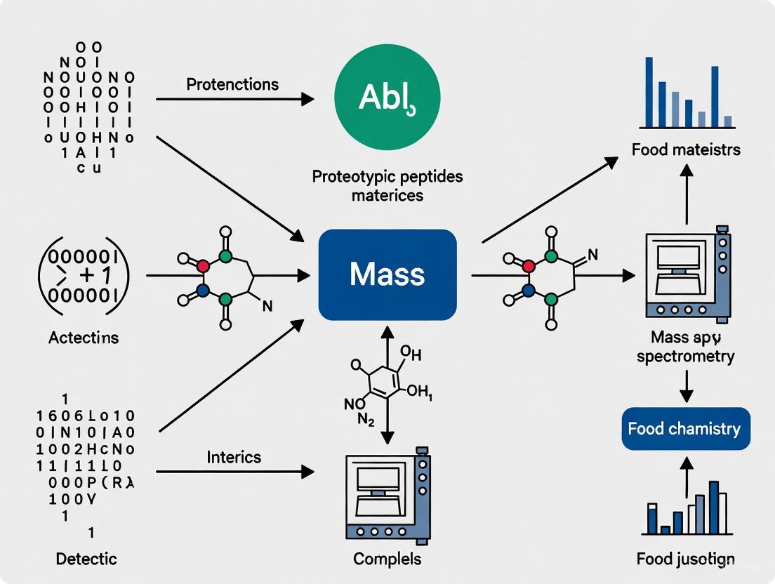
Targeted Proteomics: Leveraging Mass Spectrometry for Precise Proteotypic Peptide Detection in Complex Food Matrices
This article provides a comprehensive overview of the application of mass spectrometry (MS) for the detection and quantification of proteotypic peptides in complex food matrices, a critical methodology for food...
Targeted Proteomics: Leveraging Mass Spectrometry for Precise Proteotypic Peptide Detection in Complex Food Matrices
Abstract
This article provides a comprehensive overview of the application of mass spectrometry (MS) for the detection and quantification of proteotypic peptides in complex food matrices, a critical methodology for food safety, allergen control, and authenticity. Aimed at researchers, scientists, and drug development professionals, the content explores the foundational principles of proteotypic peptide selection, details advanced methodological workflows from sample preparation to LC-MS/MS analysis, and addresses key challenges in troubleshooting and optimization. It further examines validation strategies and compares MS performance against other detection technologies. By synthesizing current research and emerging trends, this article serves as a vital resource for implementing robust, sensitive, and accurate targeted proteomics approaches in food analysis and related biomedical fields.
The Foundation of Proteotypic Peptides: Principles and Significance in Food Analysis
Defining Proteotypic Peptides and Their Role as Unique Protein Biomarkers
In the realm of targeted proteomics, proteotypic peptides are defined as peptide sequences that uniquely represent a specific protein or protein isoform within a complex biological sample and are consistently and reliably observed in mass spectrometry (MS) experiments [1] [2]. These peptides serve as surrogate representatives for their parent proteins, enabling precise identification and quantification [3]. Their selection is crucial for developing highly specific and sensitive MS-based assays, as they must fulfill two primary criteria: uniqueness within the proteome of interest to avoid ambiguous identifications, and favorable physicochemical properties that ensure good detectability by liquid chromatography-tandem mass spectrometry (LC-MS/MS) [3].
The critical importance of proteotypic peptides lies in their ability to bridge the gap between protein discovery and clinical or industrial application. In biomarker research, they form the analytical foundation for Multiple Reaction Monitoring (MRM) or Selected Reaction Monitoring (SRM) assays, providing a highly specific method for quantifying candidate proteins in complex matrices like plasma, tissue, or food products [4] [5]. Unlike immunoassays, which rely on antibody recognition of often conformational epitopes, MS-based methods targeting proteotypic peptides identify proteins based on their fundamental amino acid sequence, reducing the risk of cross-reactivity and increasing specificity [6] [5].
Selection Criteria for Proteotypic Peptides
The selection of optimal proteotypic peptides is a meticulous process that extends beyond simple sequence uniqueness. A peptide's suitability is governed by a combination of bioinformatic predictions and empirical validation. Key selection criteria are consolidated in the table below.
Table 1: Key Criteria for Selecting Proteotypic Peptides
| Criterion | Description | Rationale |
|---|---|---|
| Sequence Uniqueness | The peptide sequence must be unique to the target protein within the background proteome. | Ensures specific monitoring of the target protein without interference from other proteins [3]. |
| MS Detectability | Exhibits consistent observation, high signal intensity, and efficient fragmentation in MS. | Provides sensitivity and robust quantification; often predicted from discovery datasets [7] [2]. |
| Absence of Problematic Residues | Should avoid methionine (oxidation) and cysteine (incomplete alkylation). | Prevents quantitative inaccuracies from variable chemical modifications [3] [7]. |
| Efficient Enzymatic Cleavage | Should have high cleavage efficiency by the protease used (e.g., trypsin). | Minimizes variability from incomplete digestion, improving quantitative accuracy [6] [3]. |
| Optimal Length | Typically between 7-25 amino acids. | Too short can be non-specific; too long may ionize or separate poorly [3]. |
| Absence of Polymorphisms/PTMs | Should not contain sites for common SNPs or post-translational modifications unless specifically targeted. | Prevents unexpected quantitative variations in clinical or population samples [3]. |
Furthermore, in specialized applications like the analysis of xenograft models (e.g., human tumors grown in mice), the selection process must ensure that the chosen peptides are unique to the species of interest (e.g., human) and are not present in the host proteome (e.g., mouse) [3]. Tools like PeptideManager have been developed to expedite this cross-species peptide selection process [3].
The following workflow diagram illustrates the logical process for selecting proteotypic peptides for a targeted MS assay.
Application in Food Allergen Detection
The detection and quantification of food allergens represents a premier example of how proteotypic peptides are applied to solve real-world analytical challenges in complex matrices. For individuals with food allergies, the accurate detection of trace amounts of allergens like milk, egg, peanut, and soy in manufactured foods is a critical safety requirement [6].
The Analytical Challenge
Food matrices are notoriously complex and are often subjected to thermal processing (e.g., baking, frying) which can denature proteins, degrade epitopes, and cause chemical modifications (e.g., Maillard reaction) [6] [8]. These processes can severely compromise the accuracy of traditional antibody-based methods like ELISA, which may fail to recognize altered protein structures [6]. Mass spectrometry, targeting stable proteotypic peptides, offers a promising alternative due to its ability to multiplex (detect multiple allergens simultaneously) and its robustness to changes in protein conformation caused by processing [6] [8].
Selection of Robust Peptide Markers
The core of the MS approach is the identification of proteotypic peptides that are stable and detectable even in processed foods. A comprehensive survey of the literature has confirmed that such peptides exist for major allergenic proteins in milk, egg, and peanut [6]. The robustness of a peptide marker is determined by its resilience to variations in the food matrix, sample preparation protocol, and MS instrumentation [6]. For instance, within the ThRAll project, a multi-allergen MS method was developed, and 16 proteotypic peptides were identified and validated for detecting milk, egg, peanut, soybean, hazelnut, and almond in incurred food materials [8].
Table 2: Exemplary Proteotypic Peptides for Food Allergen Detection
| Allergenic Food | Target Protein | Proteotypic Peptide Sequence | Key Characteristic |
|---|---|---|---|
| Peanut | Ara h 1, Ara h 3, Ara h 6 | Selected peptides from discovery | Peptides validated in incurred chocolate and bakery goods [8]. |
| Milk | Caseins, Beta-lactoglobulin | Selected peptides from discovery | Differentiates whey and casein fractions independently [6]. |
| Egg | Ovalbumin, Ovomucoid | Selected peptides from discovery | Robust to thermal processing in baked goods [8]. |
| Soy, Wheat, Tree Nuts | Various | Guidance provided via Allergen Peptide Browser | Peptide selection tools for less-studied allergens [6]. |
Experimental Workflow for Allergen Detection
The standard bottom-up proteomics workflow for allergen detection is outlined below, from sample preparation to data analysis.
Detailed Protocol: SRM-Based Allergen Quantitation in Bakery Products
The following protocol is adapted from methods used to detect allergens in incurred cookies and rusks [8].
Protein Extraction:
- Homogenize the food sample (e.g., cookie or rusk) into a fine powder.
- Extract proteins using a suitable buffer (e.g., Tris-HCl, ammonium bicarbonate) often containing additives like SDS or urea to enhance solubility and disrupt the food matrix.
- Clarify the extract by centrifugation and filtration to remove insoluble debris.
Protein Digestion:
- Reduction: Add dithiothreitol (DTT) or tris(2-carboxyethyl)phosphine (TCEP) to a final concentration of 5-10 mM and incubate at 37-60°C for 30-60 minutes to break disulfide bonds.
- Alkylation: Add iodoacetamide (IAA) to a final concentration of 15-20 mM and incubate at room temperature in the dark for 30 minutes to alkylate cysteine residues and prevent reformation of disulfide bonds.
- Proteolytic Digestion: Add sequencing-grade trypsin at an enzyme-to-protein ratio of ~1:20 to 1:50. Incubate at 37°C for 4-16 hours. The digestion is often quenched with acid (e.g., formic acid).
Peptide Clean-up:
- Desalt the resulting peptide mixture using C18 solid-phase extraction (SPE) cartridges (e.g., Waters Sep-Pak C18).
- Elute peptides in a solvent compatible with LC-MS/MS (e.g., acetonitrile with 0.1% formic acid) and dry down in a vacuum concentrator. Reconstitute in a small volume of 0.1% formic acid for MS analysis.
LC-SRM/MRM Analysis:
- Chromatography: Separate the peptides using a reverse-phase C18 nano-flow or high-performance liquid chromatography (HPLC) column with a gradient of water and acetonitrile (both with 0.1% formic acid).
- Mass Spectrometry: Analyze the eluting peptides using a triple quadrupole (QQQ) mass spectrometer.
- The first quadrupole (Q1) is set to filter the specific precursor ion (m/z) of the proteotypic peptide.
- The selected ion is fragmented in the second quadrupole (Q2) via collision-induced dissociation (CID).
- The third quadrupole (Q3) is set to filter 3-5 specific fragment ions (transitions) unique to that peptide.
- Scheduling: Transitions are monitored only during a predefined retention time window to maximize the number of data points acquired per peak and improve sensitivity.
Quantification:
- Quantify the target allergen by integrating the peak areas of the fragment ion transitions.
- Use internal standards, ideally stable isotope-labeled (SIL) versions of the proteotypic peptides, spiked into the sample at a known concentration. This corrects for losses during sample preparation and ion suppression during MS analysis, enabling absolute quantification [6] [5].
The Scientist's Toolkit: Essential Reagents and Materials
Table 3: Research Reagent Solutions for Proteotypic Peptide-Based Assays
| Item | Function | Example |
|---|---|---|
| Trypsin (Sequencing Grade) | Proteolytic enzyme that cleaves C-terminal to Lys and Arg residues, generating peptides ideal for MS analysis. | Trypsin Gold, Mass Spectrometry Grade [8]. |
| Reducing & Alkylating Reagents | Break disulfide bonds (reduction) and cap cysteine residues (alkylation) to ensure complete, reproducible digestion. | Dithiothreitol (DTT) / Tris(2-carboxyethyl)phosphine (TCEP) and Iodoacetamide (IAA) [8]. |
| Stable Isotope-Labeled (SIL) Peptides | Internal standards for absolute quantification; chemically identical to target peptides but with a mass shift. | Synthesized peptides with heavy (13C, 15N) Lys or Arg [7] [5]. |
| C18 Solid-Phase Extraction (SPE) | Desalting and purification of peptide mixtures after digestion, removing interfering salts and buffers. | Sep-Pak C18 cartridges [8]. |
| Triple Quadrupole (QQQ) Mass Spectrometer | The cornerstone instrument for targeted SRM/MRM assays, offering high sensitivity and specificity. | SCIEX QTRAP series, Agilent 6460/6490 series [6] [2]. |
Proteotypic peptides are indispensable tools in modern applied proteomics, serving as unique biomarkers that enable the precise identification and quantification of proteins in highly complex samples. Their defined selection criteria—centered on uniqueness and MS-detectability—ensure analytical specificity and robustness. This is powerfully demonstrated in the field of food safety, where MS-based methods targeting these peptides are overcoming the limitations of traditional immunoassays by providing multiplexed, specific, and reliable detection of allergens even in challenging processed food matrices like bakery products. The continued refinement of peptide selection databases, experimental protocols, and instrumentation will further solidify the role of proteotypic peptides in transitioning protein biomarker discovery from the research bench to validated clinical and industrial applications.
In the field of proteomics, particularly for applications like detecting hazardous proteins in complex food matrices, the identification and quantification of signature peptides is a fundamental task for mass spectrometry (MS). Signature peptides are unique amino acid sequences that act as surrogates for specific proteins of interest. In complex mixtures, such as alternative protein-based foods, it is often impossible to analyze intact proteins directly. Instead, proteins are digested into peptides, and specific, uniquely identifying peptides are measured to confirm the presence and quantity of the parent protein. This approach is central to targeted proteomic assays, which can confirm the presence of hazardous proteins at high sensitivity, down to the femtomole level [9]. The process relies on core principles of mass spectrometry, including the accurate mass measurement of peptides, the specific fragmentation patterns that provide sequence information, and the quantitative comparison of peptide abundances.
Core Principles of Identification and Quantification
Principle 1: Identification via Accurate Mass and Fragmentation Signature
The identification of a signature peptide is a two-step process. First, the peptide's precursor ion is identified based on its mass-to-charge ratio (m/z) with high accuracy. Second, this precursor ion is isolated and fragmented, typically by collision-induced dissociation (CID), to generate a tandem mass spectrum (MS/MS). The fragmentation process primarily breaks the peptide bonds, creating a series of b-ions (from the N-terminus) and y-ions (from the C-terminus). The pattern of these fragment ions is a unique fingerprint that can be matched against theoretical fragmentation patterns derived from a protein sequence database using search engines like Mascot or MaxQuant [10] [9]. This match confirms the amino acid sequence of the signature peptide, thereby identifying the protein from which it originated.
Principle 2: Quantification via Isotope Labeling and Peak Amplitude Comparison
Quantitative proteomic mass spectrometry involves comparing the amplitudes of peaks resulting from different isotope labeling patterns [11]. In methods like Stable Isotope Labeling by Amino Acids in Cell Culture (SILAC), samples are labeled with "light" or "heavy" isotopic forms of amino acids. When these samples are mixed and analyzed by MS, the signature peptides appear as pairs of peaks with a known mass difference. The relative intensity of these peak pairs directly reflects the relative abundance of the peptide (and thus the protein) in the original samples. For situations where full metabolic labeling is not possible, fractional isotope labeling occurs, leading to complex isotopomer distributions. Quantitative analysis of these complex distributions can be performed using a least-squares Fourier transform convolution (LS-FTC) approach to determine both the extent of labeling and the relative abundance of the peptides [11].
Principle 3: Specificity in Complex Matrices
A signature peptide must be unique to the target protein to avoid misidentification in a complex sample such as a food matrix. Mass spectrometry's high mass accuracy and resolution are critical for this discrimination. Advanced techniques like multiplexed immunoassays and mass spectrometry can simultaneously quantify specific protein fragments responsible for allergic responses by targeting their unique signature peptides. This offers high sensitivity and specificity, even in challenging samples [12]. Furthermore, the development of a comprehensive hazardous protein database as part of a data analytics pipeline is essential for reliably identifying unknown or unexpected proteins in alternative protein-based foods [9].
Quantitative Data and Performance
The following table summarizes key quantitative performance metrics for mass spectrometry-based detection and quantification of signature peptides, as demonstrated in recent applications.
Table 1: Quantitative Performance of MS-Based Signature Peptide Analysis
| Performance Metric | Demonstrated Level | Context / Technique |
|---|---|---|
| Detection Sensitivity | Femtomole (fmol) level | Targeted assay for validation of hazardous proteins [9] |
| Detection Limits | As low as 0.01 ng/mL | Multiplexed immunoassays and mass spectrometry for specific allergens [12] |
| Quantitative Analysis | Comparison of complex isotopomer distributions | Least-squares Fourier transform convolution (LS-FTC) for fractional labeling [11] |
| Labeling Efficiency | ~95% fractional atom labeling | 15N-labeling of C. elegans and Drosophila [11] |
| Labeling Efficiency | ~35% fractional residue labeling | 2H8-valine labeling in chickens [11] |
Experimental Protocols
Protocol 1: Non-Targeted Screening for Hazardous Proteins in Food
This protocol outlines an effective non-targeted workflow for screening hazardous proteins in alternative protein-based foods (APBFs) [9].
- Protein Extraction Optimization: Efficiently extract proteins from the complex APBF matrix. This is a critical step and may require optimization for different food types. The use of glass beads for homogenization can improve extraction efficiency [9].
- Protein Digestion: Digest the extracted proteins into peptides using specific enzymes. A common approach is to use a combination of Lysyl Endopeptidase (Lys-C) and sequencing-grade modified trypsin [9].
- Liquid Chromatography-Tandem Mass Spectrometry (LC-MS/MS) Analysis:
- Separate the complex peptide mixture using reversed-phase liquid chromatography.
- Analyze the eluting peptides using a tandem mass spectrometer equipped with a high-resolution mass analyzer.
- Operate the mass spectrometer in data-dependent acquisition (DDA) mode, where the most intense precursor ions are automatically selected for fragmentation to generate MS/MS spectra.
- Data Analysis:
- Process the raw MS data using software (e.g., MaxQuant [10]) to search the MS/MS spectra against a comprehensive hazardous protein database.
- Identify proteins based on the detection of their signature peptides.
Protocol 2: Quantitative Analysis using Fractional Isotope Labeling
This protocol describes a general method for quantifying peptide abundances in experiments involving fractional isotope labeling, such as pulse-labeling experiments [11].
- Pulse-Labeling Experiment: Grow cells or an organism on a medium containing a stable isotope-labeled precursor (e.g., 15N-ammonium sulfate or 13C6-isoleucine).
- Sample Preparation and Mixing: Harvest the cells and prepare the protein extract. For SILAC-style experiments, mix the labeled sample with an unlabeled control sample in a known ratio.
- LC-MS/MS Analysis: Analyze the peptide mixture via LC-MS/MS as described in Protocol 1.
- Quantitative Data Analysis using LS-FTC:
- Use Fourier transform convolution to calculate the theoretical isotope distributions for the unlabeled and fractionally labeled signature peptides. This method provides an exact calculation without approximations [11].
- Apply a non-linear least-squares fitting routine to compare the theoretical isotope distributions with the experimental mass spectrum.
- The fitting algorithm simultaneously determines the relative abundance of the peptide from the different samples and the extent of fractional isotope labeling [11].
Workflow Visualization
Diagram 1: Overall MS workflow for signature peptide analysis.
Diagram 2: Peptide identification via MS/MS fragmentation.
The Scientist's Toolkit: Research Reagent Solutions
Table 2: Essential Reagents and Materials for Signature Peptide Analysis
| Reagent / Material | Function / Application |
|---|---|
| Trypsin (Sequencing Grade) | Proteolytic enzyme for specific digestion of proteins into peptides for LC-MS/MS analysis [9]. |
| Lysyl Endopeptidase (Lys-C) | Proteolytic enzyme often used in combination with trypsin for efficient protein digestion [9]. |
| SILAC Amino Acids (e.g., 13C6-Lysine, 13C6-Arginine) | Stable isotope-labeled amino acids for metabolic labeling and quantitative comparison of protein abundance between samples [11]. |
| 15N-Ammonium Sulfate | Nitrogen source for fractional atomic labeling of cells/organisms for quantitative proteomic studies [11]. |
| Urea, Thiourea | Chaotropes for efficient protein extraction and solubilization from complex sample matrices [9]. |
| RC DC Protein Assay Kit | A detergent-compatible assay for accurate protein quantification after extraction [9]. |
| C18 Solid-Phase Extraction (SPE) Cartridges | For desalting and cleaning up peptide mixtures prior to LC-MS/MS analysis [9]. |
| High-pH Reversed-Phase Chromatography Kits | For peptide fractionation to reduce sample complexity and increase proteomic coverage [13]. |
| TMTpro Reagents | Tandem Mass Tag reagents for multiplexing, allowing simultaneous quantification of peptides from multiple samples [13]. |
The detection and quantification of specific proteins or DNA sequences in complex food matrices present significant analytical challenges. While traditional methods like Enzyme-Linked Immunosorbent Assay (ELISA) and Polymerase Chain Reaction (PCR) have been workhorse technologies for decades, they exhibit critical limitations in specificity, multiplexing capability, and robustness to food processing effects. This application note details how mass spectrometry (MS)-based proteomics, particularly through the detection of proteotypic peptides, overcomes these limitations. We present experimental protocols and data demonstrating how liquid chromatography-tandem mass spectrometry (LC-MS/MS) provides superior solutions for biomarker verification, allergen detection, and food authenticity services in complex matrices.
Traditional bioanalytical methods have served as fundamental tools for detecting proteins and nucleic acids in food products. However, their application to complex, processed food matrices reveals significant analytical challenges that can compromise result accuracy and reliability.
ELISA Limitations: The ELISA platform relies on antibody-antigen interactions, making it vulnerable to antibody cross-reactivity with non-target proteins, particularly in complex matrices containing homologous proteins [14]. Furthermore, thermal processing of foods can denature or degrade the conformational epitopes recognized by ELISA antibodies, leading to potentially false-negative results [6]. The technique also suffers from an inherent inability to multiplex effectively, requiring separate assays for different analytes and increasing sample volume requirements, analysis time, and cost [15] [6].
PCR Limitations: While PCR excels at detecting specific DNA sequences, it cannot directly quantify protein content or activity, creating a critical disconnect for applications where protein presence and quantity are clinically or regulatory relevant [6]. PCR results may also indicate the presence of a species' DNA without correlating to the actual protein concentration, especially problematic for allergenic proteins where threshold levels are based on protein content, not DNA [12].
Mass Spectrometry: A Paradigm Shift in Detection Specificity
Mass spectrometry, particularly LC-MS/MS using targeted approaches like Selected Reaction Monitoring (SRM) or Multiple Reaction Monitoring (MRM), fundamentally addresses the specificity limitations of traditional methods. Instead of relying on antibody binding or DNA amplification, MS directly detects and quantifies analytes based on their intrinsic molecular properties—mass-to-charge ratio and fragmentation patterns [16] [17].
The core innovation in MS-based protein detection involves targeting proteotypic peptides—unique peptide sequences that are specific to a parent protein and serve as reliable surrogates for its quantification [6]. This approach provides unparalleled specificity, as it can differentiate between highly homologous protein isoforms and detect specific post-translational modifications that are often invisible to immunoassays [16] [14]. MS-based methods have demonstrated detection limits as low as 0.1-5 mg kg⁻¹ (parts per million), rivaling the sensitivity of ELISA while offering significantly improved reliability in complex matrices [6].
Table 1: Comparative Analysis of Detection Technologies
| Feature | ELISA | PCR | Mass Spectrometry (LC-MS/MS) |
|---|---|---|---|
| Detection Principle | Antibody-antigen interaction [15] | DNA amplification [18] | Mass-to-charge ratio of ions [15] |
| Target Analyte | Protein (epitopes) | DNA sequences | Proteotypic peptides [6] |
| Multiplexing Capacity | Low (typically single-plex) [15] | Moderate (multiplex panels available) [18] | High (dozens to hundreds of targets) [6] [17] |
| Specificity Issues | Cross-reactivity, matrix effects [14] | Does not detect protein | High specificity via proteotypic peptides [6] |
| Effect of Food Processing | High (epitope denaturation) [6] | Low (DNA may persist) | Moderate (targets stable peptides) [6] |
| Sensitivity | 0.1-5 mg kg⁻¹ [6] | Varies by target | 0.1-5 mg kg⁻¹ (can reach femtomole) [6] [9] |
| Sample Throughput | Medium to High [15] | High | Medium (increasing with automation) [15] [17] |
Experimental Protocol: Targeted MS for Allergen Detection in Processed Foods
The following detailed protocol describes a validated workflow for multiplexed detection of allergenic proteins (e.g., from peanut, milk, and egg) in a baked goods matrix using LC-SRM/MS.
Materials and Reagents
Table 2: Research Reagent Solutions for MS-Based Allergen Detection
| Reagent / Material | Function / Application | Specifications / Notes |
|---|---|---|
| Extraction Buffer | Protein solubilization from complex food matrix | Typically contains chaotropic agents (e.g., urea), detergents, and reducing agents [9]. |
| Trypsin / Lys-C Mix | Protein digestion into peptides | Sequencing-grade enzymes for specific cleavage C-terminal to Lys and Arg [9]. |
| Stable Isotope-Labeled Peptide Standards (SIS) | Absolute quantification internal standards | Synthetic peptides identical to proteotypic peptides but with heavy isotopes (e.g., ¹³C, ¹⁵N) [6]. |
| C18 Solid-Phase Extraction (SPE) Plates | Sample clean-up and desalting | Removes interfering salts and lipids prior to LC-MS/MS analysis. |
| LC-MS/MS System | Peptide separation and detection | Nanoflow or microflow LC system coupled to triple quadrupole mass spectrometer [6] [17]. |
Step-by-Step Workflow
Step 1: Protein Extraction from Food Matrix
- Homogenize 1 g of the baked good sample with 10 mL of extraction buffer (e.g., 6 M Guanidine-HCl, 50 mM Tris-HCl, 10 mM DTT, pH 8.0) [9].
- Agitate the mixture for 2 hours at room temperature to ensure complete solubilization and reduction of disulfide bonds.
- Clarify the extract by centrifugation at 15,000 × g for 20 minutes. Transfer the supernatant containing the solubilized proteins to a new tube.
Step 2: Enzymatic Digestion into Peptides
- Determine protein concentration using a compatible assay (e.g., bicinchoninic acid assay).
- Alkylate thiol groups by adding iodoacetamide to a final concentration of 20 mM and incubating in the dark for 30 minutes.
- Dilute the sample to reduce denaturant concentration. Add trypsin/Lys-C mixture at a 1:50 (w/w) enzyme-to-protein ratio.
- Incubate at 37°C for 12-16 hours to achieve complete digestion [6] [9].
- Stop the digestion by acidifying with formic acid (final concentration 1%).
Step 3: Peptide Clean-up and Addition of Internal Standards
- Desalt the digested peptides using C18 SPE plates according to manufacturer's instructions.
- Elute peptides in a solution of 50% acetonitrile, 0.1% formic acid.
- Dry the eluents under vacuum and reconstitute in 0.1% formic acid.
- Add a known amount of stable isotope-labeled internal standard (SIS) peptides for each target proteotypic peptide to enable absolute quantification [6].
Step 4: LC-SRM/MS Analysis
- Separate peptides using a reversed-phase nanoLC system with a C18 column (75 µm × 150 mm, 2 µm particle size) and a 30-minute linear gradient of 5-35% acetonitrile in 0.1% formic acid at a flow rate of 300 nL/min.
- Introduce eluting peptides into the triple quadrupole mass spectrometer via electrospray ionization.
- Monitor 3-5 predefined precursor-product ion transitions (SRM transitions) for each proteotypic peptide and its corresponding SIS peptide. The selection of proteotypic peptides is critical and should be empirically verified for specificity and stability [6]. Example transitions for a peanut allergen (Ara h 1) peptide might be:
- Precursor ion (m/z 567.3²⁺) → Product ion (m/z 756.4)
- Precursor ion (m/z 567.3²⁺) → Product ion (m/z 863.5)
- Precursor ion (m/z 567.3²⁺) → Product ion (m/z 975.5)
Step 5: Data Analysis and Quantification
- Integrate the peak areas for both the native and SIS peptide transitions.
- Calculate the native-to-SIS peak area ratio for each target peptide.
- Interpolate the absolute amount of the native peptide (and thus the parent protein) from a calibration curve generated using synthetic peptide standards [6].
Figure 1: MS-Based Allergen Detection Workflow. The process involves protein extraction, digestion into peptides, LC separation, and highly specific MS detection.
Key Advantages of MS in Practical Applications
Unparalleled Specificity via Proteotypic Peptides
MS-based methods transcend the cross-reactivity issues of ELISA by targeting proteotypic peptides. For example, in detecting milk allergens, ELISA may struggle to distinguish between whey and casein proteins, especially when one fraction is preferentially lost during processing. In contrast, an SRM assay can independently and simultaneously quantify specific caseins (e.g., α-S1-casein) and whey proteins (e.g., β-lactoglobulin) by targeting unique peptide sequences for each, providing a more accurate and informative result [6].
Robustness to Food Processing Effects
Thermal processing can alter protein structure, masking antibody epitopes and leading to underestimation of allergen content by ELISA. MS methods that target stable, proteotypic peptides are generally more resilient to these changes. While some peptides may be modified or lost, careful selection of stable peptide targets during method development ensures reliable detection even in extensively processed foods [6].
High-Level Multiplexing for Comprehensive Analysis
The ability to monitor dozens to hundreds of peptides in a single LC-MS/MS run is a transformative advantage. A single analysis can screen for multiple allergens from different sources (e.g., peanut, milk, egg, soy), verify food authenticity by detecting species-specific peptide markers and assess the presence of protein-based toxins simultaneously [19] [9]. This multiplexing capability drastically improves analysis efficiency, reduces sample consumption, and provides a more comprehensive safety and authenticity profile compared to sequential single-plex ELISAs.
Table 3: Quantitative Performance Data of LC-MS/MS vs. ELISA
| Performance Metric | ELISA | LC-MS/MS (SRM) |
|---|---|---|
| Dynamic Range | 2-3 orders of magnitude [14] | 4-5 orders of magnitude [14] |
| Sample Volume Required | ~100 µL [15] | ~1 µL (post-digestion) [15] |
| Multiplexing Capacity | 1 protein per assay [15] | Dozens of proteins per assay [6] [17] |
| Quantitation of Specific Protein Isoforms | Limited (depends on antibody) | High (via unique peptides) [16] |
| Detection of Processed Foods | Variable; often underestimates [6] | More robust with optimized peptides [6] |
Mass spectrometry-based proteomics represents a significant technological advancement over traditional ELISA and PCR methods for the detection of proteins in complex food matrices. By directly targeting proteotypic peptides, LC-MS/MS provides superior specificity, the capacity for high-level multiplexing, and enhanced robustness to food processing effects. The experimental protocol outlined herein provides a reliable framework for researchers to implement this powerful technology for applications ranging from allergen compliance and food authenticity testing to the discovery and validation of novel protein biomarkers. As MS instrumentation and methodologies continue to evolve and become more accessible, their role in ensuring food safety and quality is poised for substantial growth.
Mass spectrometry (MS) has emerged as a powerful analytical platform for addressing critical challenges in food safety and authenticity. Its ability to detect and quantify specific protein biomarkers, known as proteotypic peptides, within complex food matrices provides researchers and food development professionals with unprecedented analytical capabilities. These peptides are uniquely representative of their parent proteins, exhibiting robust detection regardless of variations in food matrix, sample preparation protocol, and MS instrumentation [20]. The application of MS-based methods is particularly valuable for analyzing processed foods where proteins may be denatured or otherwise altered, rendering traditional antibody-based detection methods less effective [21] [22]. This application note details established protocols and key applications of MS-based detection of proteotypic peptides within the framework of food safety compliance and authenticity verification.
Key Application Areas
Allergen Detection and Quantification
Food allergen detection represents one of the most critical applications of targeted MS in food safety. With the rising prevalence of food allergies worldwide, sensitive and reliable detection of trace allergens in manufactured food is essential for consumer protection and regulatory compliance [20].
- Multiplexed Detection: Unlike enzyme-linked immunosorbent assays (ELISAs), which typically target a single allergen, MS enables simultaneous quantification of multiple allergenic proteins from different sources in a single run [21] [22]. This is particularly valuable for products manufactured in facilities handling multiple allergens.
- Robustness to Processing: MS methods target specific peptide sequences rather than conformational protein epitopes, making them more robust for detecting allergens in thermally processed foods where proteins may be denatured [21].
- Sensitivity and Standardization: Selected reaction monitoring (SRM) and multiple reaction monitoring (MRM) on triple quadrupole systems provide detection limits comparable to ELISAs (0.1–5 mg kg⁻¹) while avoiding issues of antibody cross-reactivity [21]. Ongoing work focuses on standardizing proteotypic peptide targets through resources like the Allergen Peptide Browser [20].
Food Authenticity and Fraud Detection
Food authenticity verification ensures that products match their label declarations regarding origin, composition, and processing methods. MS-based foodomics approaches provide powerful tools for detecting economically motivated adulteration.
- Geographical Origin Verification: Multi-omics strategies combining proteomics, metabolomics, and lipidomics can create unique fingerprints that authenticate the geographical origin of high-value products like olive oil, honey, and wine [23].
- Species Authentication: Proteotypic peptides enable sensitive detection of species substitution in meat and seafood products, combating fraudulent labeling practices [23]. For example, LC-MS/MS methods have been developed for detecting horse and pork in halal beef [24].
- Composition Verification: MS methods can verify premium ingredients and detect substitution with inferior alternatives, such as identifying adulteration in saffron or authenticating extra virgin olive oil [25] [23].
Safety Compliance and Regulatory Monitoring
MS technologies provide robust solutions for ensuring compliance with food safety regulations through sensitive detection of contaminants and verification of label claims.
- Regulatory Compliance: MS workflows support compliance with regulations from bodies like the FDA and European Commission regarding allergen labeling and contaminant levels [26] [22]. The US FASTER Act and EU FIC No. 1169/2011 define specific allergens requiring declaration.
- Contaminant Screening: Beyond allergens, MS platforms enable comprehensive screening for pesticides, persistent organic pollutants (POPs), and other chemical contaminants in complex food matrices [26].
- Standardized Workflows: Automated solutions like the AllergenScreener standardize sample preparation and analysis across food types, delivering consistent, reliable results for regulatory decision-making [26].
Table 1: Key Mass Spectrometry Platforms for Food Analysis Applications
| Instrument Type | Key Applications | Strengths | Example Systems |
|---|---|---|---|
| Triple Quadrupole (TQ-MS) | Targeted allergen quantification, contaminant screening | High sensitivity and specificity for SRM/MRM; Excellent quantification | SCIEX Triple Quad 4500, EVOQ LC-TQ [26] [22] |
| Q-TOF (Quadrupole Time-of-Flight) | Untargeted allergen screening, food authenticity | High resolution mass accuracy; Retrospective data analysis | impact II VIP, compact QTOF [26] |
| TIMS-TOF (Trapped Ion Mobility) | Complex authenticity profiling, contaminant ID | Additional separation dimension; High confidence compound ID | timsTOF Pro 2 [26] |
| MALDI-TOF | Food fraud detection, protein profiling | Rapid analysis; Minimal sample preparation | autoflex maX [26] |
| DART-MS | Rapid screening, quality control | Chromatography-free workflows; High throughput | EVOQ DART-TQ+ [26] |
Experimental Protocols
Sample Preparation for Allergen Detection in Processed Foods
Principle: Efficient extraction and digestion of proteins into measurable peptides is crucial for reliable allergen detection, particularly in challenging matrices like baked goods or chocolate [22].
Materials:
- Protein extraction buffer (compatible with food matrix)
- Reduction and alkylation reagents: Tris(2-carboxyethyl)phosphine (TCEP) or dithiothreitol (DTT) and iodoacetamide
- Sequencing-grade modified trypsin
- Solid-phase extraction (SPE) cartridges for clean-up (C18 or mixed-mode)
- Internal standards (stable isotope-labeled peptide standards when available)
Procedure:
- Protein Extraction: Homogenize 1 g of food sample with 10 mL of appropriate extraction buffer. The buffer composition may vary based on the food matrix (e.g., consideration of fat, carbohydrate, or polyphenol content) [22].
- Reduction and Alkylation: Denature extracted proteins with 8 M urea. Reduce with 5-10 mM TCEP or DTT at 37°C for 30-60 minutes. Alkylate with 15-20 mM iodoacetamide at room temperature for 30 minutes in the dark [5].
- Digestion: Dilute urea concentration to <1.5 M. Add trypsin at 1:20-1:50 (enzyme-to-protein ratio). Incubate at 37°C for 4-16 hours with gentle agitation [21] [5].
- Clean-up: Acidify digest with formic or trifluoroacetic acid to pH <3. Desalt using C18 SPE cartridges. Elute peptides with acetonitrile/water mixture (typically 50-80% ACN). Dry under vacuum and reconstitute in MS-compatible solvent (e.g., 0.1% formic acid) [5].
- Quality Control: Monitor digestion efficiency by SDS-PAGE or using control peptides if internal standards are available.
LC-MS/MS Analysis for Multiplexed Allergen Detection
Principle: This method uses liquid chromatography coupled to tandem mass spectrometry (LC-MS/MS) with targeted scanning (SRM/MRM) to simultaneously detect and quantify multiple allergens based on proteotypic peptides [20] [24].
Materials:
- Nano or ultra-high performance liquid chromatography (UHPLC) system
- Triple quadrupole mass spectrometer
- LC column: C18 reversed-phase (75 μm-2.1 mm ID, 1.7-3 μm particle size)
- Mobile phases: A) 0.1% formic acid in water; B) 0.1% formic acid in acetonitrile
Procedure:
- Chromatographic Separation:
MS Analysis with SRM/MRM:
- Use electrospray ionization in positive mode.
- Monitor predefined transitions (precursor ion → product ion) for proteotypic peptides of target allergens.
- Optimize instrument parameters for each transition: collision energy, declustering potential, and collision cell exit potential.
- Implement retention time scheduling to maximize the number of monitored peptides without compromising sensitivity [21].
Data Analysis:
- Integrate peak areas for all transitions for each peptide.
- Verify detection based on co-elution of multiple transitions and their relative intensity ratios matching those of standard peptides.
- Quantify using calibration curves generated with heavy isotope-labeled internal standards or matrix-matched external standards [21] [5].
Food Authenticity Verification via Non-Targeted Profiling
Principle: High-resolution mass spectrometry (HRMS) enables untargeted profiling for food authenticity assessment by detecting compositional patterns and markers indicative of origin, variety, or adulteration [23].
Materials:
- UHPLC system coupled to Q-TOF or Orbitrap mass spectrometer
- Appropriate columns for targeted compounds (e.g., C18 for metabolites, HILIC for polar compounds)
- Chemical standards for method validation
Procedure:
- Sample Preparation: Prepare samples using standardized extraction protocols suitable for the food matrix and analytes of interest (e.g., metabolites, lipids).
- LC-HRMS Analysis:
- Analyze samples using UHPLC coupled to HRMS in data-dependent acquisition (DDA) or data-independent acquisition (DIA) mode.
- Use both positive and negative electrospray ionization modes for comprehensive coverage.
- Include quality control samples (pooled quality controls) throughout the sequence [23].
- Data Processing:
- Marker Verification:
- Confirm identity of potential marker compounds using authentic standards when available.
- Validate markers across multiple samples and batches to ensure robustness.
Table 2: Performance Characteristics of MS-Based Allergen Detection Methods
| Allergen Category | Reported LOD in Food | Key Proteotypic Peptides | Noteworthy Challenges |
|---|---|---|---|
| Peanut | <10 μg/g [24] | Multiple peptides from Ara h 1, Ara h 2, Ara h 3/4 [24] | High lipid content in matrices [22] |
| Tree Nuts (Hazelnut, Almond, Walnut, etc.) | <10 μg/g for several nuts [24] | 44 identified tryptic marker peptides across 6 nut species [24] | Cross-reactivity potential in antibody-based methods avoided with MS [21] |
| Milk | Comparable to ELISA (0.1-5 mg kg⁻¹) [21] | Peptides from caseins and whey proteins [20] | Differential quantification of whey vs. casein [21] |
| Egg | Comparable to ELISA (0.1-5 mg kg⁻¹) [21] | Peptides from ovalbumin and ovomucoid [20] | Robustness to thermal processing [21] |
| Soy & Wheat | Guidance available for peptide selection [20] | Limited consensus peptides identified [20] | More research needed for standardized peptides [20] |
The Scientist's Toolkit: Essential Research Reagents and Materials
Table 3: Key Research Reagent Solutions for MS-Based Food Analysis
| Reagent/Material | Function | Application Notes |
|---|---|---|
| Sequencing-Grade Trypsin | Proteolytic digestion of proteins into measurable peptides | Selective cleavage C-terminal to lysine and arginine; most commonly used enzyme [21] [5] |
| Stable Isotope-Labeled Peptide Standards (SIL) | Absolute quantification internal standards | Chemically synthesized with heavy isotopes (13C, 15N); identical chemical properties to native peptides [5] |
| PNGase F | Glycoprotein analysis; deglycosylation enzyme | Releases N-linked glycans from glycoproteins/glycopeptides; important for glycoprotein allergen analysis [5] |
| Hydrazide Resin | Glycopeptide enrichment | Solid-phase capture of glycopeptides for enrichment; reduces sample complexity [5] |
| C18 Solid-Phase Extraction Cartridges | Sample clean-up and desalting | Removes interfering compounds and salts prior to LC-MS analysis; improves sensitivity [5] |
| UHPLC Columns (C18, 1.7-2 μm) | Chromatographic separation of peptides | Provides high-resolution separation of complex peptide mixtures; essential for multiplexed analysis [24] [5] |
| Allergen Peptide Browser Database | Proteotypic peptide selection | Online resource (AllergenPeptideBrowser.org) for verified peptide markers [20] |
Workflow Visualization
Allergen Detection Workflow
This workflow illustrates the comprehensive process for MS-based allergen detection, encompassing three critical phases: sample preparation, LC-MS/MS analysis, and data processing, culminating in multiplexed allergen reporting.
Food Authentication Strategy
This diagram outlines the integrated multi-omics approach for food authentication, demonstrating how different analytical dimensions combine through chemometric analysis to verify origin, detect adulteration, and assess label compliance.
Mass spectrometry-based detection of proteotypic peptides provides researchers and food safety professionals with powerful tools for addressing complex challenges in allergen detection, food authenticity, and regulatory compliance. The protocols and applications detailed in this document highlight the versatility, sensitivity, and specificity of MS methodologies across diverse food matrices. As MS instrumentation, analysis software, and standardized workflows continue to advance, the role of this technology in ensuring food safety and authenticity will expand, offering increasingly robust solutions for protecting consumers and maintaining integrity in the global food supply chain. The ongoing development of verified proteotypic peptide databases and standardized methods will further enhance the reliability and adoption of MS-based approaches throughout the food industry.
From Theory to Practice: Methodological Workflows for Complex Food Matrices
Mass spectrometry (MS)-based proteomics is a powerful technique for identifying and quantifying proteins in complex biological samples [27]. Within food science, its application is crucial for detecting proteotypic peptides—unique peptide sequences that reliably represent a specific parent protein—in complex food matrices [28] [29]. This process is fundamental for food authentication, allergen profiling, and safety surveillance, such as screening for hazardous proteins in alternative protein-based foods (APBFs) [29] [9]. The workflow is multi-stage, requiring careful attention at each step, from sample preparation to data interpretation, to ensure valid and reproducible results [30]. This article provides a detailed protocol for a complete MS-based proteomics workflow, framed within the context of food analysis.
Experimental Workflow
The following diagram illustrates the comprehensive workflow for a mass spectrometry-based proteomics experiment, from sample preparation through to data analysis.
Protocols and Methodologies
Sample Preparation and Protein Extraction
Efficient protein extraction from complex food matrices is a critical first step. The complexity of APBFs, for example, poses a greater challenge than traditional meat products, requiring optimized protocols [9].
Detailed Protocol for Protein Extraction from a Polyacrylamide Gel (Applicable to Pre-separated Samples) [31]:
- Materials: Polyacrylamide gel stained with an MS-compatible stain (e.g., glutaraldehyde-free silver or Coomassie), gel destain solution, 100% acetonitrile, 10 mM dithiothreitol (DTT), 55 mM iodoacetamide (freshly prepared, light-protected), gel wash solution, gel enzyme solution (e.g., trypsin), 25 mM ammonium bicarbonate, gel extraction solution, formic acid, low-binding microcentrifuge tubes.
- Procedure:
- Excise and Destain: Wearing gloves, excise the protein band/spot of interest and place it in a low-binding microcentrifuge tube. Destain with 100 µL of appropriate destain solution for 30 minutes with vigorous shaking. Remove the destain solution and wash with 400 µL of water, repeating until the gel piece is colorless [31].
- Dehydrate: Add 400 µL of 100% acetonitrile to dehydrate the gel piece for 10 minutes. Remove the supernatant and dry the gel piece in a vacuum centrifuge [31].
- Reduce and Alkylate: Add 100 µL of 10 mM DTT and incubate for 45 minutes at 55°C to reduce disulfide bonds. Remove the DTT solution, add 100 µL of 55 mM iodoacetamide, and incubate for 30 minutes at room temperature in the dark to alkylate free cysteines. Remove the iodoacetamide solution and wash the gel piece twice with 400 µL of gel wash solution for 15 minutes per wash [31].
- Dehydrate Again: Dehydrate with 100% acetonitrile as before and dry in a vacuum centrifuge [31].
For total protein extraction from complex food matrices like APBFs, optimization is key. This often involves evaluating different extraction buffers and mechanical disruption methods (e.g., using glass beads) to maximize protein yield [9].
Proteolytic Digestion
In bottom-up proteomics, proteins are enzymatically digested into peptides for MS analysis [31] [32].
Detailed Protocol for In-Gel Digestion [31]:
- Materials: Gel enzyme working solution (e.g., sequencing-grade modified trypsin at 10-20 µg/mL in 25 mM ammonium bicarbonate).
- Procedure:
- Rehydrate the dried gel piece with a sufficient volume (e.g., 10-20 µL) of the gel enzyme working solution to cover it. Incubate on ice for 1 hour.
- After rehydration, add enough 25 mM ammonium bicarbonate to cover the gel piece again to prevent drying during digestion. Incubate at 37°C for a minimum of 4 hours or overnight.
It is worth noting that digestibility is a key criterion in selecting optimal proteotypic peptides, as incomplete digestion leads to inaccurate quantification [28]. The cleavage kinetics of trypsin can vary depending on neighboring residues [28].
Peptide Cleanup and Enrichment
Prior to MS analysis, peptides require cleanup and potential enrichment to remove salts, detergents, and other interfering compounds [31].
Detailed Protocol for Peptide Desalting using Reversed-Phase Chromatography [31]:
- Materials: C18 solid-phase extraction tips or stage tips, peptide extraction solution (e.g., 5% formic acid, 50% acetonitrile), acidified solvent (e.g., 0.1% Trifluoroacetic acid - TFA).
- Procedure (using C18 tips):
- Condition the C18 material by washing with acetonitrile.
- Equilibrate the tip with an acidified solvent like 0.1% TFA.
- Bind the peptide sample by slowly passing the acidified peptide solution through the C18 material.
- Wash with 0.1% TFA to remove salts and impurities.
- Elute the purified peptides with a solution of 50-70% acetonitrile containing 0.1% formic acid.
For specific applications like phosphoproteomics, enrichment at the peptide level using affinity capture (e.g., TiO2 beads) is performed at this stage [31].
Liquid Chromatography-Mass Spectrometry (LC-MS) Analysis
Peptides are separated by liquid chromatography and analyzed by mass spectrometry.
1. Liquid Chromatography: Peptides are loaded onto a reverse-phase C18 column and separated via a gradient of increasing organic solvent (acetonitrile). This reduces sample complexity and reduces ion suppression [27] [30]. Monitoring the stability of chromatographic parameters (peak width, shape, and retention time) is critical for reproducible and quantitative results [30].
2. Mass Spectrometry: Electrospray Ionization (ESI) is the most common ion source, transferring peptides from the liquid phase to the gas phase as ions [27]. Two primary data acquisition strategies are used:
- Data-Dependent Acquisition (DDA): The mass spectrometer automatically selects the most abundant precursor ions for fragmentation (MS/MS). This is a well-proven, sensitive method [32].
- Data-Independent Acquisition (DIA): All precursor ions within a specific m/z window are fragmented simultaneously, providing highly reproducible data [32].
3. System Suitability and Calibration: To ensure validity of results, the LC-MS system must be calibrated and monitored. The mass spectrometer should be calibrated with a suitable calibration mixture that covers the full m/z measurement window and is compatible with ESI [30]. Furthermore, the stability of the chromatographic system should be checked using a simple calibration mixture of peptides before and during the analysis [30].
Data Analysis
The raw data from the mass spectrometer is processed to identify and quantify peptides and proteins. A modern approach involves workflow-based analysis for scalability and reproducibility [32].
1. Peptide Identification: Tandem mass spectra are matched against a protein sequence database using search engines (e.g., Sequest) [28] [32]. Parameters include precursor and fragment mass tolerances, and specified static (e.g., carbamidomethylation) and dynamic (e.g., oxidation) modifications [28].
2. Protein Inference and Quantification: Identified peptides are assembled into protein identifications. For quantification, label-free methods like MaxQuant [32] or isobaric labeling strategies (e.g., TMT, iTRAQ) can be used. In targeted proteomics, quantification relies on optimal proteotypic peptides [28]. The quantitative data is then aggregated to the protein level [32].
3. Downstream Statistical Analysis: Tools like MSstats perform more elaborate normalization, imputation, and statistical significance testing for differential expression [32].
4. Quality Control: Quality control metrics are gathered throughout the process, and tools like MultiQC provide summary statistics and plots to ensure the data is fit for purpose [32].
Key Data and Reagents
The following table summarizes the quantitative performance of a targeted proteomics method for Bovine Serum Albumin (BSA) in a food matrix, demonstrating key parameters for a valid assay [28].
Table 1: Quantitative Performance of a Targeted Proteomics Assay for BSA in Milk [28]
| Parameter | Value | Description |
|---|---|---|
| Linear Range | 1 - 100 ppm | The concentration range over which quantification is linear. |
| Coefficient of Determination (R²) | > 0.9990 | Indicates excellent linearity of the calibration curve. |
| Limit of Detection (LOD) in Milk | 0.78 mg/kg | The lowest concentration of BSA that can be reliably detected. |
| Optimal Proteotypic Peptide | LVNELTEFAK | The selected peptide representing BSA, chosen for its specificity, digestibility, recovery, and stability. |
A successful proteomics experiment relies on a suite of essential reagents and materials. The table below lists key components for the workflow.
Table 2: Research Reagent Solutions for Mass Spectrometry Proteomics
| Reagent/Material | Function / Purpose | Examples / Notes |
|---|---|---|
| Sequencing Grade Modified Trypsin | Proteolytic enzyme that specifically cleaves proteins at lysine and arginine residues. | Crucial for reproducible digestion [28]. |
| Dithiothreitol (DTT) | Reducing agent that breaks disulfide bonds between cysteine residues. | Used in sample preparation [31]. |
| Iodoacetamide (IAA) | Alkylating agent that modifies free cysteine sulfhydryl groups to prevent reformation of disulfides. | Must be prepared fresh and protected from light [31]. |
| C18 Solid Phase | Reversed-phase material for peptide desalting and cleanup prior to MS. | Packed in tips or columns [31]. |
| Ammonium Bicarbonate | Volatile buffer used in digestion protocols; easily removed during lyophilization. | Common buffer for in-solution and in-gel digestion [31]. |
| Mass Spec Calibrants | Solution of ions with known m/z for accurate mass calibration of the instrument. | Must be compatible with ESI and cover the required m/z range [30]. |
| LC-MS/MS System | Instrumentation for peptide separation (LC) and mass analysis/fragmentation (MS/MS). | High-resolution mass spectrometers (e.g., Orbitrap) are preferred [28] [30]. |
| Protein/Peptide Standards | Isotopically labeled internal standards for absolute quantification (e.g., AQUA peptides). | Allows for precise quantification in complex matrices [28]. |
This workflow overview provides a detailed guide for applying mass spectrometry from protein extraction to data analysis, with a specific focus on detecting proteotypic peptides in complex food matrices. The robustness of the results hinges on rigorous optimization at every stage, particularly sample preparation for challenging foods [9], and the implementation of a quality control system to ensure data validity [30]. By adhering to standardized protocols, using high-quality reagents, and employing reproducible data analysis workflows, researchers can reliably utilize MS-based proteomics for critical applications in food safety, authentication, and allergen research.
In the application of mass spectrometry for detecting proteotypic peptides in complex food matrices, the selection of optimal peptide biomarkers is a pivotal step that fundamentally determines the sensitivity, specificity, and reliability of the entire analytical method. Proteotypic peptides are uniquely representative of a specific protein within a proteome and exhibit consistent detectability by mass spectrometry [3]. Within food safety and allergen detection, this selection process becomes particularly critical, as regulatory compliance and consumer health depend on the accurate quantification of trace allergenic proteins in processed food products [33] [6]. This protocol details evidence-based selection criteria and methodologies for identifying robust proteotypic peptides, with particular emphasis on overcoming challenges posed by complex food matrices, food processing-induced modifications, and genetic variations in allergenic proteins.
Core Selection Criteria for Proteotypic Peptides
The selection of proteotypic peptides must balance multiple, sometimes competing, criteria to ensure analytical robustness. The following table synthesizes the essential parameters for evaluation:
Table 1: Comprehensive Criteria for Selecting Robust Proteotypic Peptides
| Criterion | Description | Rationale & Impact |
|---|---|---|
| Sequence Uniqueness | Peptide sequence must be unique to the target protein(s) within the relevant proteome(s) [3]. | Precludes false positives from homologous proteins; essential for specificity in complex food matrices [6]. |
| Absence of Polymorphisms & Modifications | Should lack high-frequency single nucleotide polymorphisms (SNPs), known post-translational modification (PTM) sites, and chemically modifiable residues (e.g., Cys, Met) unless specifically targeted [34] [3]. | Ensures consistent quantitation across samples and populations; avoids variability from incomplete alkylation or oxidation [3]. |
| Favorable Physicochemical Properties | Typically 7-25 amino acids; appropriate hydrophobicity for chromatography; predictable charge states [34]. | Ensures efficient ionization, separation, and detection; peptides outside these ranges may have poor MS response [34] [35]. |
| Stable & Efficient Generation | High predicted cleavage efficiency by trypsin; avoidance of missed cleavage sites and chemically unstable sequences [3]. | Maximizes yield and reproducibility of peptide generation from the protein digest, critical for quantitative accuracy [3]. |
| Robustness to Processing | Resistance to degradation or modification from thermal and chemical processing of foods [36]. | For food analysis, peptides must remain detectable and representative after cooking or other manufacturing processes [36]. |
| Optimal MS Detectability | Consistent production of high-intensity, predictable fragment ions (e.g., b- and y-ions) under collision-induced dissociation (CID) [34] [37]. | Directly impacts the sensitivity and limit of detection of the final MS assay. |
Experimental Protocol for Peptide Selection and Verification
This section provides a detailed workflow for the empirical identification and verification of proteotypic peptides, particularly for allergenic ingredients in food.
Stage 1: In Silico Selection and Prioritization
- Define the Target Proteome: Compile a list of canonical protein sequences for the allergen of interest (e.g., peanut, milk, egg) from reliable databases such as UniProt.
- In Silico Digestion: Perform a theoretical digestion of the target proteins using trypsin (cleaving C-terminal to Lys and Arg), with tools like the PeptideCutter web-tool on ExPASy [3].
- Apply Initial Filters: Filter the resulting peptide list based on length (e.g., 7-25 residues) and exclude peptides containing:
- Cysteine (to avoid variability from incomplete alkylation) and Methionine (susceptible to oxidation) [3].
- Known non-synonymous SNPs or sequence conflicts between databases.
- Potential sites for common PTMs (unless studied intentionally).
- Specificity BLAST: Perform a BLAST search of the candidate peptide sequences against the proteomes of all expected ingredients in the food matrix to ensure uniqueness [6] [3]. For xenograft models or complex matrices with multiple species, use tools like PeptideManager to select species-specific peptides [3].
- Check for Isoform Coverage: Determine if the candidate peptide is unique to a specific protein isoform or shared across all relevant isoforms, depending on the analytical goal [33] [36].
Stage 2: Empirical Validation Using Incurred Food Matrices
Theoretical selection must be followed by empirical validation in relevant matrices to account for real-world complexities [33] [36].
- Prepare Incurred Food Materials: Create model food systems by spiking the allergenic ingredient (e.g., peanut flour) into a non-allergenic base matrix (e.g., cookie dough, chocolate) at defined concentrations. Subject these materials to relevant processing (e.g., baking, extrusion) to mimic industrial conditions [33] [36].
- Protein Extraction and Digestion: Extract proteins from the incurred materials using a validated, buffered solution. Reduce (e.g., with DTT) and alkylate (e.g., with iodoacetamide) the extracted proteins. Digest the proteins into peptides using sequencing-grade trypsin under controlled conditions [6].
- Untargeted LC-HRMS Analysis: Analyze the resulting complex peptide digests using high-resolution, discovery-grade Liquid Chromatography-Mass Spectrometry (LC-MS/MS). Data-Dependent Acquisition (DDA) is typically employed to identify as many peptides as possible [36].
- Data Processing and Peptide Filtering: Process the raw HRMS data against a protein sequence database. Filter the resulting peptide identifications with high confidence. Then, cross-reference this empirical observation list with the in silico candidate list from Stage 1.
- Rank Final Candidate Peptides: Rank the empirically verified peptides based on:
- Signal Intensity and Consistency: Peptides consistently detected with high intensity across replicates and processing conditions are preferred [36].
- Robustness: Peptides that remain detectable across different food matrices and varying degrees of processing [36].
- Specificity: Confirmation that the peptide is only present in samples containing the target allergen.
The following workflow diagram summarizes this multi-stage protocol:
The Scientist's Toolkit: Key Reagents and Materials
Successful implementation of the selection protocol requires specific reagents and tools. The following table lists essential items and their functions.
Table 2: Essential Research Reagents and Materials for Peptide Selection Workflows
| Category | Item | Function / Application |
|---|---|---|
| Sample Preparation | Sequencing-grade modified trypsin | Specific enzymatic digestion of proteins C-terminal to Lys and Arg for reproducible peptide generation [6]. |
| Reduction & alkylation reagents (e.g., DTT, Iodoacetamide) | Breaks disulfide bonds and alkylates cysteine residues to prevent reformation, ensuring complete digestion [6]. | |
| Buffered protein extraction solutions | Efficient and consistent extraction of proteins from complex, often challenging, food matrices [6]. | |
| Mass Spectrometry | High-resolution mass spectrometer (e.g., Orbitrap) | Untargeted discovery analysis for high-confidence peptide identification in complex digests [36]. |
| Triple quadrupole mass spectrometer | Development and execution of highly sensitive targeted MS (SRM/MRM) assays using the finalized peptides [38] [6]. | |
| Stable isotope-labeled (SIL) synthetic peptides | Internal standards for precise and accurate quantification in targeted MS assays; corrects for matrix effects and losses [38] [6]. | |
| Bioinformatics & Databases | Protein sequence databases (e.g., UniProt, RefSeq) | Source of canonical protein sequences for in silico digestion and peptide identification [3]. |
| Peptide selection software (e.g., PeptideManager, SRMAtlas) | Facilitates and expedites the selection of proteotypic and species-specific peptides, checking for uniqueness [3] [39]. | |
| Spectral libraries (e.g., SRMAtlas, GPM) | Repository of validated peptide spectra for comparing and confirming experimental fragmentation patterns [37] [39]. |
Concluding Remarks
The rigorous, multi-stage process of proteotypic peptide selection outlined here—combining stringent in silico criteria with empirical validation in relevant, processed food matrices—is foundational to developing mass spectrometric methods for allergen detection that are both robust and reliable. Adherence to these protocols ensures that the selected peptide biomarkers are specific, sensitive, and stable, thereby enabling accurate quantification that protects allergic consumers and supports regulatory compliance. As mass spectrometry continues to evolve, the standardization of these selection criteria will be crucial for improving inter-laboratory reproducibility and advancing the field of food allergen analysis.
The accuracy of mass spectrometric analysis, particularly in the detection of proteotypic peptides within complex food matrices, is fundamentally dependent on the efficacy of the initial sample preparation. Inefficient protein extraction and purification can introduce biases, suppress ionization, and obscure low-abundance peptides, thereby compromising data quality and reliability. This application note provides a critical evaluation of several advanced protein precipitation techniques, including acetone, methanol-chloroform (M/C), and trichloroacetic acid (TCA)-acetone protocols. Framed within the context of proteomic analysis of complex biological samples, we present standardized protocols, quantitative performance data, and structured workflows to guide researchers in selecting and optimizing sample preparation methods for robust and reproducible mass spectrometry results.
Comparative Evaluation of Precipitation Methods
A comparative study of three common precipitation methods—acetone, methanol-chloroform (M/C), and TCA-acetone—applied to mammalian cell homogenates revealed significant differences in performance, which are critical for downstream mass spectrometry analysis [40].
Table 1: Quantitative Comparison of Protein Precipitation Methods [40]
| Precipitation Method | Key Procedural Variations | Protein Recovery (%) | Key Characteristics and Challenges |
|---|---|---|---|
| Acetone | With ultrasonic bath cycles | 104.18 ± 2.67 | Highest recovery; similar band profile to crude homogenate on SDS-PAGE. |
| Acetone | With NaOH addition | 103.12 ± 5.74 | High recovery; minimal protein loss. |
| Methanol-Chloroform (M/C) | With ultrasonic homogenization | 94.22 ± 4.86 | Intermediate recovery; does not adversely affect protein pattern on SDS-PAGE. |
| TCA-Acetone | Standard protocol | 77.91 ± 8.79 | Difficulties in pellet solubilization; negatively impacts recovery and band presence. |
| All Methods | -- | -- | Affected recovery of low molecular weight proteins (< 15 kDa). |
The data indicates that the acetone-based method, especially when augmented with an ultrasonic bath or NaOH, provides superior protein recovery and is recommended for proteomic workflows where capturing the full complexity of the cellular proteome is paramount [40]. The TCA-acetone protocol, while effective for some applications, presents significant challenges in solubilizing the resulting protein pellet, leading to lower recovery.
Detailed Experimental Protocols
Acetone Precipitation with Ultrasonication
This protocol is optimized for maximum protein recovery from cell homogenates [40].
- Sample Preparation: Start with a volume of cell homogenate containing 50-100 µg of protein in a microcentrifuge tube.
- Precipitation: Add four times the sample volume of ice-cold acetone. Vortex immediately to ensure thorough mixing.
- Ultrasonication: Place the tube in an ice-cooled ultrasonic bath for 3 cycles of 1 minute each, with 30-second intervals on ice.
- Incubation: Incubate the mixture at -20°C for a minimum of 1 hour to overnight for complete precipitation.
- Pelletting: Centrifuge at 15,000 × g for 15 minutes at 4°C. A visible protein pellet should form at the bottom of the tube.
- Washing: Carefully decant the supernatant without disturbing the pellet. Wash the pellet with 1 mL of ice-cold 80% acetone solution and vortex.
- Final Pelletting: Centrifuge again at 15,000 × g for 5 minutes at 4°C and carefully remove the entire wash supernatant.
- Drying and Solubilization: Air-dry the pellet for 5-10 minutes to evaporate residual acetone. Do not over-dry, as this will make resolubilization difficult. Solubilize the protein pellet in an appropriate buffer for downstream analysis.
Methanol-Chloroform (M/C) Precipitation
This method is effective for removing interfering substances and is suitable for lipid-rich samples [40].
- Sample Preparation: Use a volume of sample containing 50-100 µg of protein.
- Methanol Addition: Add 4 volumes of ice-cold methanol to the sample and vortex vigorously.
- Chloroform Addition: Add 1 volume of ice-cold chloroform, vortex thoroughly.
- Water Addition: Add 3 volumes of ultrapure water to create a phase separation. Vortex again until the mixture is cloudy.
- Centrifugation: Centrifuge at 14,000 × g for 15 minutes at room temperature. Proteins will form a fluffy white disc at the interface between the upper (aqueous) and lower (organic) phases.
- Aqueous Phase Removal: Carefully aspirate and discard the upper aqueous phase without disturbing the protein disc.
- Reprecipitation: Add 4 volumes of ice-cold methanol to the remaining lower phase and interphase. Vortex to mix.
- Final Pelletting: Centrifuge at 14,000 × g for 15 minutes at room temperature. The protein will now form a pellet at the bottom of the tube.
- Washing and Solubilization: Decant the supernatant and air-dry the pellet briefly. Solubilize the protein pellet in a suitable buffer.
TCA-Acetone Precipitation
This method is known for its stringency but can lead to lower recovery [40].
- Precipitation: Add an equal volume of ice-cold 20% (w/v) Trichloroacetic Acid (TCA) in acetone to the protein sample. Mix thoroughly.
- Incubation: Precipitate at -20°C for a minimum of 1 hour or overnight.
- Pelletting: Centrifuge at 15,000 × g for 15 minutes at 4°C.
- Washing: Wash the pellet twice with ice-cold acetone containing 0.07% (v/v) β-mercaptoethanol to remove residual TCA and salts.
- Drying: Air-dry the pellet to remove all traces of acetone.
- Solubilization: Solubilize the pellet in a strong denaturing buffer. This step often requires vigorous vortexing and may necessitate the use of ultrasonication to fully resuspend the difficult-to-solubilize pellet.
Workflow Visualization for Mass Spectrometry Analysis
The following diagram illustrates a generalized workflow for sample preparation in a bottom-up proteomics analysis, integrating the precipitation methods discussed.
The Scientist's Toolkit: Essential Reagents and Materials
Table 2: Key Research Reagent Solutions for Protein Precipitation
| Reagent / Material | Function in Sample Preparation |
|---|---|
| Acetone (HPLC grade) | Organic solvent for protein dehydration, precipitation, and washing; effectively removes water and soluble contaminants. |
| Methanol & Chloroform | Used in combination for phase separation; efficiently precipitates proteins and removes lipids and other non-polar contaminants. |
| Trichloroacetic Acid (TCA) | Strong denaturing acid that efficiently precipitates proteins; often used in an acetone mixture. |
| Ultrasonic Homogenizer / Bath | Applies ultrasonic energy to disrupt cells, fragment macromolecules, and aid in pellet resolubilization. |
| β-Mercaptoethanol / DTT | Reducing agent added to wash buffers to break disulfide bonds and prevent protein oxidation. |
| QuEChERS Kits | Ready-to-use kits for Quick, Easy, Cheap, Effective, Rugged, and Safe extraction, particularly useful for multi-residue analysis in food matrices [41]. |
| Solid-Phase Extraction (SPE) | A versatile and selective technique for purifying and concentrating analytes from complex samples, minimizing matrix effects [41]. |
The application of mass spectrometry for detecting proteotypic peptides in complex food matrices, such as spices, edible insects, and processed foods, presents significant analytical challenges. These matrices are rich in interfering compounds—including pigments, lipids, proteins, and carbohydrates—that can co-extract with target analytes, leading to ion suppression or enhancement, increased background noise, and reduced chromatographic performance. For instance, chili powder's complex matrix, rich in pigments, oils, and capsinoids, poses major challenges for accurate pesticide residue analysis by compromising sensitivity, reproducibility, and instrument longevity [42]. Similarly, edible insects contain high levels of fat and protein that complicate the extraction and analysis of pesticide residues [43]. Overcoming these matrix effects is paramount for achieving reliable quantification, particularly when employing liquid chromatography-tandem mass spectrometry (LC-MS/MS) for the precise identification and measurement of proteotypic peptides in food authentication, allergen profiling, and safety monitoring [29]. This application note details optimized protocols and strategies to mitigate these challenges, ensuring robust and reproducible results.
Optimized Sample Preparation Protocols
Effective sample preparation is the most critical step in managing complex food matrices. The following protocols are optimized to minimize matrix effects while maximizing the recovery of target peptides and analytes.
Optimized Extraction and Cleanup for Chili Powder and Spices
The following method, validated for the analysis of 135 pesticides in chili powder, can be adapted for peptide analysis from similar challenging matrices [42].
- Sample Size and Homogenization: Use a carefully optimized sample size. Smaller sizes may lack precision, while larger sizes amplify matrix effects. Pre-homogenize the sample to ensure uniformity [42].
- Extraction Solvent: Employ acetonitrile as the primary extraction solvent. It provides effective miscibility with a broad range of analytes while minimizing the co-extraction of non-polar matrix components [42] [43].
- d-SPE Cleanup: Implement a dispersive solid-phase extraction (d-SPE) cleanup. Optimize the combination of sorbents to target specific interferences [42]:
- Primary Secondary Amine (PSA): Removes organic acids and sugars.
- C18: Targets non-polar compounds like lipids and oils.
- Graphitized Carbon Black (GCB): Effective for removing pigments. Use with caution, as it can also adsorb planar molecules, potentially leading to analyte loss.
- Key Consideration: Avoid "over-cleaning" by systematically varying sorbent types and amounts to balance effective cleanup with high analyte recovery [42].
QuEChERS-Based Method for High-Fat Protein Matrices
For complex, high-fat matrices like edible insects, a modified QuEChERS (Quick, Easy, Cheap, Effective, Rugged, and Safe) method is highly effective. The protocol below, used for pesticide analysis in insects, can be modified for protein and peptide extraction [43].
- Sample Preparation: Lyophilize (freeze-dry) samples to remove water without applying heat, thus preserving the integrity of heat-labile analytes. This also allows for better control over sample weight and solvent ratios [43].
- Extraction:
- Weigh 2.5–5.0 g of homogenized, dry sample into a 50 mL centrifuge tube.
- Add 15 mL of acetonitrile and 5 mL of water. Agitate for 5 minutes.
- Add a salt mixture (e.g., 6 g MgSO₄ and 1.5 g sodium citrate) to induce phase separation. Shake vigorously.
- Centrifuge to separate the phases [43].
- Cleanup: Transfer the supernatant to a d-SPE tube containing a combination of MgSO₄, PSA, and C18. Vortex and centrifuge. The purified extract can then be concentrated and reconstituted for LC-MS analysis [43].
General Proteomics Sample Preparation for LC-MS/MS
For the specific detection of proteotypic peptides, a standard proteomics workflow is essential. The quality of this preparation directly impacts the accuracy and reproducibility of the results [44] [45].
- Sample Collection and Preservation: Immediately process samples or snap-freeze in liquid nitrogen, storing at -80°C to prevent protein degradation. Avoid repeated freeze-thaw cycles [45].
- Cell Lysis and Protein Extraction: Use reagent-based lysis (e.g., detergents like SDS, chaotropic agents like urea) combined with mechanical disruption (e.g., bead beating, sonication) to ensure complete protein extraction [44] [45].
- Protein Quantification: Accurately measure protein concentration using a colorimetric assay (e.g., BCA or Bradford assay) with a standard calibration curve [45].
- Protein Digestion:
- Denaturation and Reduction: Use urea or SDS to denature proteins, and a reducing agent like dithiothreitol (DTT) to break disulfide bonds.
- Alkylation: Alkylate cysteine residues with iodoacetamide to prevent reformation of disulfide bonds.
- Enzymatic Digestion: Digest proteins into peptides using trypsin (or other proteases like Lys-C) with an optimized enzyme-to-substrate ratio and incubation time (typically overnight at 37°C) [44] [45].
- Peptide Cleanup and Concentration: Desalt and concentrate peptides using solid-phase extraction (C18 tips or columns). A nitrogen blowdown evaporator can be used for rapid, gentle concentration of samples, increasing sensitivity for downstream LC-MS/MS analysis [45].
Table 1: Summary of Method Performance for Different Food Matrices
| Matrix | Target Analytes | Sample Preparation | Key Cleanup Sorbents | LOQ Achieved | Recovery Range |
|---|---|---|---|---|---|
| Chili Powder [42] | 135 Pesticides | Acetonitrile extraction, d-SPE | PSA, C18, GCB | 0.005 mg/kg | Satisfactory (70-120%) |
| Edible Insects [43] | 47 Pesticides | QuEChERS (ACN), d-SPE | PSA, C18, MgSO₄ | 10-15 μg/kg | 70-120% (97.87% of pesticides) |
| General Proteomics [45] | Proteins/Peptides | Lysis, Digestion, SPE | C18 for desalting | N/A | High reproducibility |
Liquid Chromatography and Mass Spectrometry Analysis
Following optimized sample preparation, the LC-MS/MS analysis requires careful method development to separate target peptides from residual matrix components effectively.
- Chromatographic Considerations: Utilize ultra-high-performance liquid chromatography (UHPLC) with sub-2-µm particle columns for superior resolution. A C18 stationary phase is standard for peptide separation. Employing a longer gradient can improve separation in complex samples but increases run time [42] [29].
- Mass Spectrometry Detection: Operate the mass spectrometer in positive electrospray ionization (ESI+) mode for most peptides. Use scheduled or parallel reaction monitoring (SRM/PRM) for targeted quantification of proteotypic peptides to ensure high sensitivity and selectivity [29].
- Mitigating Matrix Effects: Even with extensive cleanup, residual matrix effects are common.
- Matrix-Matched Calibration: Prepare calibration standards in a blank matrix extract to compensate for ion suppression/enhancement [42].
- Stable Isotope-Labeled Internal Standards (SIS): Use SIS peptides for absolute quantification. These peptides co-elute with their native counterparts but are distinguished by mass, correcting for variability in sample preparation and ionization [44].
Table 2: Research Reagent Solutions for Proteomics and Residue Analysis
| Reagent / Kit | Function | Application Note |
|---|---|---|
| Trypsin | Proteolytic enzyme for digesting proteins into peptides for MS analysis. | High specificity for cleaving at lysine and arginine residues; key for generating proteotypic peptides [45]. |
| d-SPE Sorbents (PSA, C18, GCB) | Dispersive Solid-Phase Extraction for cleanup; removes organic acids, lipids, and pigments. | Combination is crucial for managing complex matrices like chili powder and edible insects [42] [43]. |
| QuEChERS Extraction Kits | Standardized kits for quick, easy, and effective extraction of analytes from various matrices. | Ideal for high-throughput analysis; easily modified for specific matrix needs [43]. |
| Stable Isotope-Labeled Internal Standards | Synthetic peptides with heavy isotopes for absolute quantification. | Corrects for sample loss and matrix effects, ensuring accurate quantification [44]. |
| TMT (Tandem Mass Tags) | Isobaric labels for multiplexed relative quantification of proteins across samples. | Enables high-throughput profiling of multiple samples in a single MS run [44]. |
| Nitrogen Blowdown Evaporator | Gently concentrates peptide samples by evaporating solvent under a stream of nitrogen. | Increases analyte concentration and detection sensitivity prior to LC-MS/MS [45]. |
Method Validation and Quality Control
Robust method validation and stringent quality control are essential for generating reliable data that complies with regulatory guidelines.
- Validation Parameters: Validate methods by assessing linearity, precision (intra-day and inter-day), accuracy (recovery), limit of detection (LOD), and limit of quantification (LOQ). For the chili powder method, relative standard deviations (RSDs) below 15% were consistently achieved [42].
- Quality Control Measures: Incorporate quality control samples in each batch, including method blanks, spiked controls, and replicate samples. Continuously monitor recovery rates and RSDs to ensure ongoing method precision and accuracy [42] [43].
- Managing Matrix Effects: Quantify matrix effects (%ME) by comparing the analyte response in a matrix extract to its response in a pure solvent. The goal is to keep %ME within an acceptable range (e.g., -20% to +20%), indicating minimal ion suppression or enhancement [43].
Successful LC-MS/MS analysis of proteotypic peptides in complex food matrices hinges on a holistic approach that integrates optimized sample preparation, chromatographic separation, and rigorous quality control. The protocols outlined here, drawing from proven methods in pesticide residue and proteomics analysis, provide a robust framework for achieving accurate and reproducible results. By systematically addressing matrix challenges through techniques like tailored d-SPE cleanup, QuEChERS extraction, and the use of isotope-labeled standards, researchers can ensure the reliability of their data in applications ranging from food authentication and allergen detection to safety monitoring.
Experimental Workflow Diagrams
Sample Preparation Workflow for Proteomics
Matrix Cleanup and Quantification Strategy
High-Resolution Accurate-Mass (HRAM) Orbitrap mass spectrometry, combined with tandem MS/MS, has become an indispensable tool for detecting proteotypic peptides within complex food matrices. These advanced proteomic techniques enable precise identification and quantification of thousands of proteins from intricate food samples, supporting critical applications in food safety, quality control, and authenticity verification [46]. The powerful synergy of HRAM instrumentation with robust LC-MS/MS workflows allows researchers to navigate the challenges presented by complex food samples, including wide dynamic ranges of protein concentrations and interfering substances, to deliver highly specific and sensitive analyses essential for modern food research and regulatory compliance [46] [47].
Key Applications in Food Analysis
Allergen Detection and Quantification
Targeted proteomics approaches using HRAM Orbitrap technology have revolutionized allergen detection in processed foods. Multiple Reaction Monitoring (MRM) methods on triple quadrupole MS instruments enable identification and quantification of milk and egg proteins in baked goods like cookies at levels lower than 0.2 mg, surpassing the sensitivity thresholds recommended by the Voluntary Incidental Trace Allergen Labeling (VITAL) program [46]. This precision provides a reliable alternative to precautionary allergen labeling, helping to prevent cross-contamination incidents while maintaining consumer safety.
Food Authenticity and Traceability
Proteomic analysis serves as a powerful tool for food authentication through the development of characteristic protein and peptide markers. For honey, predictive models correlating amino acid profiles to aromatic compounds provide rapid multicomponent analysis of quality indicators [47]. Similarly, protein fingerprints can verify honey origin and detect adulteration, with studies revealing significant compositional differences between eucalyptus honey and other varieties [48]. In beef production, analysis of total omega-3 polyunsaturated fatty acids, micronutrients, and phytochemicals can determine the finishing diet of cattle, distinguishing grass-finished beef from conventionally fed alternatives [47].
Contaminant and Pathogen Screening
HRAM Orbitrap systems purpose-built for environmental and food safety testing, such as the Thermo Scientific Orbitrap Exploris EFOX Mass Detector, provide robust solutions for monitoring persistent contaminants like per- and polyfluoroalkyl substances (PFAS), pesticides, and other pollutants [49]. Shotgun proteomics approaches effectively characterize foodborne pathogens such as Staphylococcus aureus strains isolated from dairy products, identifying relevant phage-specific peptides that could lead to alternative treatments for mastitis beyond conventional antibiotics [46].
Bioactive Peptide Characterization
Proteomics enables detailed investigation of bioactive peptides in food products. Sensopeptidomic kinetic approaches combined with statistical tools like decision trees and random forests can identify peptide structures responsible for undesirable sensory properties, such as the bitter taste in milk protein hydrolysates [46]. This knowledge facilitates the development of improved nutritional products with enhanced palatability while maintaining the beneficial rapid release of amino acids that maximizes muscle protein anabolism.
Experimental Protocols
Sample Preparation Workflow
Protocol: Protein Extraction and Digestion for Bottom-Up Proteomics
Protein Extraction:
Protein Quantification:
- Measure protein concentration using spectrophotometric methods (e.g., Nanodrop at 280 nm) [50].
Proteolytic Digestion:
Peptide Cleanup:
LC-MS/MS Analysis
Protocol: Discovery Proteomics Using HRAM Orbitrap
Liquid Chromatography Separation:
- Use ultra-high-performance liquid chromatography (UHPLC) system with C18 reversed-phase column [51].
- Employ gradient from 4% to 22.5% mobile phase B in 3.7 minutes, then to 45% B by 5.5 minutes, and finally to 99% B for comprehensive peptide separation [50].
- Maintain flow rate at 750 nL/min for capillary columns [50].
Mass Spectrometry Data Acquisition:
- Operate Orbitrap Exploris instrument in data-dependent acquisition (DDA) mode [50].
- Set MS1 resolution to 45,000 with mass range of 375-1200 m/z [50].
- Use TopN method (e.g., Top40) with dynamic exclusion for MS2 fragmentation [50].
- Implement preaccumulation technique in bent flatapole to achieve scanning speeds up to 70 Hz [50].
- Apply HCD fragmentation with normalized collision energy of 28% [50].
Data Processing:
Targeted Analysis for Protein Quantification
Protocol: Multiple Reaction Monitoring (MRM) for Allergen Detection
Method Development:
- Select proteotypic peptides unique to target proteins (e.g., milk or egg allergens) [46].
- Optimize collision energies for each peptide precursor-fragment transition.
- Validate method specificity and sensitivity using blank matrices spiked with standards.
Data Acquisition:
- Use triple quadrupole mass spectrometer operated in MRM mode [46].
- Schedule MRM transitions based on peptide retention times.
- Maintain chromatographic resolution with UHPLC separation.
Data Analysis:
- Integrate peak areas for quantifier and qualifier transitions.
- Generate calibration curves using internal standard method.
- Apply acceptance criteria for transition ratios to confirm compound identity.
Performance Data and Technical Specifications
Table 1: Comparison of MS Instrumentation for Food Proteomics Applications
| Parameter | Orbitrap Exploris 240 | Triple Quadrupole (MRM) | Orbitrap Exploris EFOX |
|---|---|---|---|
| Mass Accuracy | <3 ppm | Not typically specified | <1 ppm |
| Resolution | Up to 240,000 | Unit resolution | High-resolution accurate mass |
| Scan Speed | Up to 22 Hz (MS2) | >300 MRM transitions/sec | Up to 70 Hz with preaccumulation [50] |
| Dynamic Range | >4,000 | >10^5 | >10^5 |
| Primary Applications | Discovery proteomics, intact protein analysis | Targeted quantification, allergen screening | PFAS, pesticide screening, retrospective analysis [49] |
| Detection Limits | Low fmol for peptides | Sub-pg for targeted compounds | ppt for environmental contaminants [49] |
Table 2: Quantitative Performance of Proteomics Methods in Food Analysis
| Application | Target Analytes | Matrix | LOD/LOQ | Throughput | Reference Technique |
|---|---|---|---|---|---|
| Allergen Detection | Milk and egg proteins | Cookies | <0.2 mg/kg (VITAL level) | Medium | Targeted proteomics (MRM) [46] |
| Bitterness Prediction | Bitter peptides | Milk protein hydrolysates | Not specified | High | LC-ESI-Q-TOF-MS/MS [46] |
| Pathogen Characterization | Bacteriophage peptides | Dairy products | Strain differentiation | Medium | LC-ESI-MS/MS [46] |
| Food Authentication | Characteristic proteins | Honey | Variety discrimination | High | UHPLC-MS/MS [47] |
The Scientist's Toolkit
Table 3: Essential Research Reagent Solutions for Food Proteomics
| Reagent/Material | Function | Application Example |
|---|---|---|
| Sequencing-grade trypsin | Proteolytic digestion of proteins into peptides | Bottom-up proteomics for protein identification [46] |
| C18 Solid-Phase Extraction Cartridges | Peptide cleanup and concentration | Sample preparation for mass spectrometry [50] |
| SDS Lysis Buffer | Protein extraction and denaturation | Efficient protein recovery from complex food matrices [50] |
| Formic Acid | Acidification of peptide solutions | Ionization enhancement for LC-MS analysis [50] |
| UHPLC Mobile Phase | Chromatographic separation | Reversed-phase separation of peptides [51] |
| Calibration Solutions | Mass accuracy calibration | Instrument performance verification [51] |
| Tandem Mass Tags | Multiplexed quantitative proteomics | Comparative analysis of multiple samples [52] |
Workflow Visualization
Figure 1: Comprehensive workflow for HRAM Orbitrap-based food proteomics analysis, from sample preparation to final applications.
Figure 2: HRAM Orbitrap instrumental workflow with preaccumulation technology for enhanced sensitivity.
This application note details a simple and reliable analytical method for the simultaneous detection of walnut and almond proteins in processed foods using liquid chromatography-tandem mass spectrometry (LC-MS/MS). The developed protocol enables specific identification and quantification of these major food allergens through the analysis of proteotypic peptides, supporting compliance with global food allergen labeling regulations. The method demonstrates sufficient recovery rates, repeatability, and reproducibility across a wide variety of processed food matrices, providing a robust solution for food safety testing laboratories.
Food allergies represent a significant public health concern, affecting millions of consumers worldwide. Regulatory frameworks including the Food Allergen Labeling and Consumer Protection Act (FALCPA) in the United States and EU Regulation No 1169/2011 mandate clear labeling of major food allergens, including tree nuts such as walnut and almond [53]. While ELISA and PCR have been traditional methods for allergen detection, they present limitations including cross-reactivity issues and inability to directly detect the causative proteins [54]. Mass spectrometry-based proteomics has emerged as a powerful alternative, enabling specific, multiplexed detection of allergenic proteins through analysis of proteotypic peptides, even in complex processed food matrices [17]. This case study validates an LC-MS/MS method for simultaneous walnut and almond detection, contributing to the broader application of mass spectrometry for detecting proteotypic peptides in complex food matrices.
Experimental Protocol
Materials and Reagents
Table 1: Essential Research Reagent Solutions
| Item | Function | Specifications |
|---|---|---|
| Acetonitrile | HPLC mobile phase | ≥99.9% purity |
| Methanol | HPLC solvent | ≥99.9% purity |
| Formic Acid | Mobile phase modifier | ≥98% purity |
| Ammonium Bicarbonate | Digestion buffer component | - |
| Trypsin (Mass Spectrometry Grade) | Protein digestion enzyme | Specific activity: >90% by BAEE |
| Dithiothreitol (DTT) | Disulfide bond reduction | - |
| Iodoacetamide | Cysteine alkylation | - |
| Cellulose Acetate Syringe Filters | Sample clarification | 5 μm, 25 mm size |
| C18 Solid-Phase Extraction Cartridges | Peptide purification | 55-105 μm particle size, 125 Å pore size |
Sample Preparation
The sample preparation follows a bottom-up proteomics approach, adapted for processed food matrices [8] [17]:
Protein Extraction:
- Homogenize 1g of processed food sample to fine powder under liquid nitrogen
- Add 10 mL of extraction buffer (50 mM Tris-HCl, 150 mM NaCl, pH 7.5)
- Vortex vigorously for 1 minute, then shake for 30 minutes at room temperature
- Centrifuge at 10,000 × g for 15 minutes at 4°C
- Collect supernatant and filter through 5 μm cellulose acetate syringe filter
Protein Digestion:
- Reduce proteins with 10 mM DTT at 56°C for 45 minutes
- Alkylate with 25 mM iodoacetamide at room temperature for 30 minutes in the dark
- Digest with trypsin (1:20 enzyme-to-protein ratio) at 37°C for 16 hours
- Stop digestion by acidification with 0.1% formic acid
- Desalt using C18 solid-phase extraction cartridges
- Concentrate samples by vacuum centrifugation and reconstitute in 0.1% formic acid for LC-MS/MS analysis
LC-MS/MS Analysis
Liquid Chromatography Conditions:
- Column: C18 reversed-phase column (2.1 × 150 mm, 1.8 μm)
- Mobile Phase A: 0.1% formic acid in water
- Mobile Phase B: 0.1% formic acid in acetonitrile
- Gradient: 5-35% B over 30 minutes
- Flow Rate: 0.2 mL/min
- Column Temperature: 40°C
- Injection Volume: 10 μL
Mass Spectrometry Parameters:
- Instrument: Triple quadrupole mass spectrometer
- Ionization: Electrospray ionization (ESI) positive mode
- Nebulizing Gas: 3 L/min
- Heating Gas: 10 L/min
- Interface Temperature: 300°C
- DL Temperature: 250°C
- Heat Block Temperature: 400°C
- Drying Gas: 10 L/min
- Detection: Multiple Reaction Monitoring (MRM)
Results and Discussion
Selection of Proteotypic Peptides
The method specificity was achieved through careful selection of proteotypic peptides that uniquely identify walnut and almond proteins while avoiding cross-reactivity with other nut species [55]. For walnut, five specific target peptides were selected from walnut 2S albumin and 7S globulin proteins. For almond, three target peptides were selected from almond 11S globulin.
Table 2: Proteotypic Peptide Markers for Walnut and Almond Detection
| Allergen Source | Protein Origin | Proteotypic Peptide Sequence | Precursor Ion (m/z) | Product Ion (m/z) |
|---|---|---|---|---|
| Walnut | 2S albumin | GEEMEEMVQSAR | 698.3 | 316.1 |
| Walnut | 7S globulin | Additional peptides not specified | - | - |
| Almond | 11S globulin | GNLDFVQPPR | 571.8 | 369.2 |
| Almond | 11S globulin | Additional peptides not specified | - | - |
Analytical Performance
The method was rigorously validated for sensitivity, linearity, and recovery using incurred samples and spiked matrices including butter cookie and chocolate ice cream [55].
Table 3: Quantitative Method Performance Characteristics
| Parameter | Walnut (GEEMEEMVQSAR) | Almond (GNLDFVQPPR) |
|---|---|---|
| Limit of Detection (LOD) | 0.22 ± 0.02 μg/g | 0.08 ± 0.02 μg/g |
| Linearity (R²) | >0.999 | >0.999 |
| Calibration Range | 0.1-50 μg/mL | 0.1-50 μg/mL |
| Recovery Rate | 90.4-101.5% | 90.4-101.5% |
The limit of detection for the walnut 2S albumin peptide GEEMEEMVQSAR was 0.22 ± 0.02 μg/g, while that for almond 11S globulin peptide GNLDFVQPPR was 0.08 ± 0.02 μg/g when extracted walnut and almond protein were spiked into challenging matrices [55]. The excellent detection limits demonstrate the method's suitability for detecting trace-level contamination of walnut and almond in processed foods.
Method Advantages in Processed Food Matrices
Food processing presents significant challenges for allergen detection due to protein structural modifications and matrix interference [8]. The selected proteotypic peptides demonstrated robustness against processing-induced modifications in baked goods and other processed foods. The LC-MS/MS method overcomes limitations of antibody-based methods which may fail to detect denatured proteins in processed foods [54]. The simultaneous detection capability provides efficiency advantages for food manufacturing quality control where multiple allergen monitoring is required.
Application in Food Safety Regulation
This LC-MS/MS method supports compliance with food allergen labeling regulations by providing accurate detection of walnut and almond proteins in processed foods [53] [55]. The sensitivity achieved meets the requirements for detecting potentially hazardous undeclared allergens, enabling food manufacturers to verify labeling accuracy and prevent cross-contamination. The method's robustness across various processed food matrices makes it particularly valuable for quality control in facilities handling multiple allergenic ingredients.
The developed LC-MS/MS method enables simple, reliable, and simultaneous detection of walnut and almond allergens in processed foods. Through targeted analysis of specific proteotypic peptides, the method achieves high sensitivity, specificity, and reproducibility while overcoming limitations of traditional detection techniques. This approach contributes significantly to the field of mass spectrometry-based allergen detection in complex food matrices and supports food safety initiatives by helping to protect consumers with food allergies.
The comprehensive analysis of wheat (Triticum aestivum) proteins is crucial for understanding its nutritional quality, functional properties for baking, and implications for human health, such as celiac disease [56]. However, the wheat proteome presents unique analytical challenges that necessitate refined mass spectrometry (MS) methods. Targeted proteomics, particularly using selected reaction monitoring (SRM), has emerged as a powerful technique for the sensitive and reliable quantification of specific proteins in complex food matrices [6] [19]. This application note details a optimized tandem digestion protocol for targeted proteomics of wheat proteins, enabling deeper characterization of the proteome, including traditionally under-represented gluten proteins and immunoreactive species. The methodologies described herein are framed within broader research applications aimed at detecting proteotypic peptides—peptides whose presence is robust across variations in matrix, preparation, and instrumentation [6].
Key Challenges in Wheat Proteomics
Wheat grain proteins are broadly categorized into gluten proteins (approximately 80%) and albumin/globulin proteins (approximately 20%) [56]. The gluten proteins, responsible for the viscoelastic properties of dough, are particularly problematic for MS analysis due to their unique amino acid composition. They are characterized by a high glutamine (32–53%) and proline (11–29%) content, while trypsin cleavage sites (lysine and arginine) are scarce, comprising only 1.0–3.8% of the total amino acids [56]. This results in long, trypsin-resistant peptides that are suboptimal for LC-MS analysis. Furthermore, the simultaneous extraction of both alcohol-soluble gliadins and insoluble glutenin polymers is technically challenging, often leading to biased representation in subsequent analyses [57].
Optimized Workflow for Wheat Protein Analysis
Protein Extraction
Efficient protein extraction is foundational to a successful proteomics workflow. Several extraction buffers were evaluated, including Urea buffer and SDS buffer. A buffer containing the acid-labile detergent RapiGest demonstrated superior performance by significantly improving the recovery of both gliadins and glutenins compared to buffers without it, including the industry standard from the Codex Alimentarius Commission [57]. RapiGest aids in disrupting protein structure and enhancing solubility, without interfering with downstream MS analysis as it is easily removed by acid hydrolysis [57].
- Recommended Buffer (Solution 3): A solution containing RapiGest, a reducing agent, and Tris buffer, with sonication at 60°C for 15 minutes [57].
Tandem Enzymatic Digestion
To overcome the limitations of trypsin, a tandem digestion strategy using multiple proteases with complementary cleavage specificities is recommended. This approach significantly increases proteome coverage.
- Enzymes: Trypsin and Glutamyl Endoproteinase (Glu-C).
- Rationale: Trypsin remains the gold standard for many proteomes. Glu-C, which hydrolyzes peptide bonds C-terminal to glutamic and aspartic acids, offers orthogonal specificity. Given the high glutamine content in gluten proteins, Glu-C provides access to otherwise inaccessible protein regions [56].
- Protocol:
- Dissolve extracted proteins in a suitable buffer (e.g., 50 mM ammonium bicarbonate).
- Reduce disulfide bonds with 10 mM dithiothreitol (DTT) at 37°C for 30 minutes.
- Alkylate cysteine residues with 20 mM iodoacetamide (IAA) at 25°C in the dark for 1 hour.
- Perform a first digestion with Glu-C (enzyme-to-substrate ratio 1:50, w/w) at 37°C for 6 hours.
- Follow with a second digestion with trypsin (enzyme-to-substrate ratio 1:20, w/w) at 37°C for 6 hours.
- Quit the reaction by adding acid (e.g., formic acid) and desalt the peptides using C18 solid-phase extraction columns prior to LC-MS/MS analysis [56].
LC-MS/MS Analysis and Data Acquisition
For targeted quantification, Selected Reaction Monitoring (SRM) on a triple quadrupole mass spectrometer is the principal technique [6]. This method involves predefined transitions specific to proteotypic peptides of interest, resulting in high sensitivity and specificity.
- LC Conditions:
- MS Conditions:
- Ionization: Electrospray ionization (ESI+) at 2.0-3.2 kV.
- Acquisition Mode: SRM/MRM with scheduled retention time windows to maximize the number of quantifiable peptides and sensitivity [6].
- Detection: Monitor 3-5 predefined precursor-product ion transitions per peptide.
The following workflow diagram illustrates the optimized protocol from extraction to data analysis:
Comparative Performance Data
The tandem digestion workflow was systematically compared against the traditional trypsin-only approach. The following tables summarize the key quantitative improvements.
Table 1: Enhancement in Protein Identification Using Tandem Digestion (Glu-C + Trypsin) vs. Trypsin Alone [56]
| Protein Category | Trypsin Alone (Number of Proteins) | Tandem Digestion (Number of Proteins) | Improvement |
|---|---|---|---|
| Total Identified Proteins | 2,218 | 2,695 | +22% |
| Gluten Proteins | 587 | 812 | +38% |
| Immunoreactive Proteins | 45 | 68 | +51% |
Table 2: Impact of Extraction Buffer and MS Acquisition Strategy on Gluten Protein Analysis [57]
| Analytical Parameter | Standard Codex Buffer | RapiGest Buffer | Improvement with RapiGest & DIA-IM-MS |
|---|---|---|---|
| Protein Identification | Baseline | - | +25% |
| Unique Peptides | Baseline | - | 10-fold increase |
| Sequence Coverage of Glutenins | Lower | Higher | +20% (e.g., for A9YSH4 LMW subunit) |
| Celiac-toxic Motifs Identified | Fewer | More | Higher for 5 out of 6 motifs searched |
The Scientist's Toolkit: Essential Research Reagents
Table 3: Key Reagents and Materials for Wheat Targeted Proteomics
| Reagent / Material | Function | Application Note |
|---|---|---|
| RapiGest SF Surfactant | Acid-labile detergent that improves protein solubility and extraction efficiency, particularly for glutenins. Removed by acid hydrolysis before MS. | Critical for unbiased extraction of both gliadin and glutenin fractions [57]. |
| Glu-C (V8 protease) | Protease that cleaves C-terminal to glutamic and aspartic acids. Provides complementary cleavage to trypsin. | Essential for accessing glutamine-rich regions of gluten proteins in tandem digestion protocols [56]. |
| Trypsin (Sequencing Grade) | Gold-standard protease that cleaves C-terminal to lysine and arginine. | Used in combination with Glu-C for comprehensive proteome coverage [56]. |
| C18 Solid-Phase Extraction (SPE) Column | Desalting and cleaning up peptide digests before LC-MS injection. | Removes salts, detergents, and other impurities that can suppress ionization [58]. |
| Proteotypic Peptide Library | Curated list of target peptides robustly detected for specific wheat proteins of interest. | Enables development of sensitive SRM assays for accurate quantification [6]. |
This application note outlines a robust and optimized protocol for targeted proteomics analysis of wheat. The combination of an efficient RapiGest-based extraction and a tandem Glu-C/trypsin digestion strategy successfully overcomes the inherent challenges of the wheat proteome. This method significantly enhances the identification and quantification of key protein classes, including glutenins and immunoreactive proteins, providing researchers with a powerful tool for applications in food safety, quality control, and nutritional science. The workflow is readily adaptable for the development of sensitive SRM assays targeting specific proteotypic peptides, aligning with the broader goals of advancing mass spectrometry for the analysis of complex food matrices.
Navigating Analytical Challenges: Troubleshooting and Method Optimization
The accurate detection and quantification of proteotypic peptides using mass spectrometry (MS) is fundamentally challenged by matrix effects, where the complex composition of a food sample interferes with analytical measurements. The food matrix is defined as the unique structure of a food, its components, and their interactions [59]. In dairy products, for instance, the dairy matrix consists of an emulsion of fat droplets suspended in an aqueous phase containing proteins, vitamins, and minerals, forming a complex structure that modulates nutrient digestion and release [59]. These matrix interactions significantly influence the efficiency of protein extraction, enzymatic digestion, peptide ionization, and ultimately, the sensitivity and accuracy of liquid chromatography-tandem mass spectrometry (LC-MS/MS) methods [6] [19].
The selection of proteotypic peptides—peptides whose presence appears robust to variations in food matrix, sample preparation protocol, and MS instrumentation—is therefore critical for reliable quantitation [6]. This document outlines standardized protocols and strategies to mitigate these effects across three challenging food categories: dairy, baked goods, and processed foods, ensuring robust method development for proteomic analysis.
Matrix-Specific Challenges and Strategies
Dairy Products
- Key Challenges: Dairy matrices are characterized by their high fat content, complex protein assemblies (casein micelles, whey proteins), and significant mineral content (e.g., calcium). These components can cause ion suppression, hinder enzymatic digestion, and co-extract with target analytes. The matrix effect varies significantly with the product type; for example, the protein network in cheese or yogurt presents a different challenge compared to liquid milk [59] [60].
- Recommended Strategies:
- Defatting: Use a two-step centrifugation protocol or petroleum ether extraction to remove fat prior to protein precipitation.
- Casein Precipitation: Employ isoelectric point precipitation (at pH 4.6) to separate caseins from whey proteins, simplifying each fraction for subsequent analysis.
- Calcium Chelation: Add EDTA to the extraction buffer to dissociate casein micelles by chelating calcium, thereby improving protein solubility and tryptic access.
Baked Goods
- Key Challenges: Baked goods present a heterogeneous matrix comprising starch, gluten, yeast, and often dairy and egg. Heat-induced modifications (e.g., Maillard reaction) can denature proteins, cross-link peptides, and generate modifications that complicate database searching and quantitation [19].
- Recommended Strategies:
- Starch Degradation: Incorporate enzymatic treatment with α-amylase to break down starch granules that can trap proteins.
- Gluten Solubilization: Use extraction buffers containing chaotropic agents (e.g., 6-8 M Urea) and reducing agents (e.g., DTT) to solubilize gluten networks effectively.
- Targeting Stable Peptides: Focus proteotypic peptide selection on sequences that are less susceptible to thermal modification during baking, as identified in discovery-mode experiments.
Processed Foods
- Key Challenges: This category encompasses a wide range of multi-ingredient products subjected to various thermal and mechanical processes. The primary challenges include the simultaneous presence of multiple protein sources (e.g., meat, soy, dairy), added stabilizers, emulsifiers, and salts, all of which contribute to severe ion suppression and unpredictable extraction efficiency [19].
- Recommended Strategies:
- Multi-Step Extraction: Implement sequential extraction protocols tailored to the solubility of different protein classes present.
- Immunoaffinity Depletion: For highly complex products, consider immunoaffinity columns to remove one or more highly abundant, non-target proteins (e.g., deprioritizing wheat protein when quantitating a minor milk allergen) [6].
- Standard Addition: Use the method of standard addition for quantification to correct for matrix-induced suppression or enhancement of the target peptide signal.
Experimental Protocols
Protocol 1: Generic Sample Preparation for LC-MS/MS Allergen Detection
This protocol is adapted for the simultaneous detection of multiple allergens (e.g., milk, egg, peanut) in a complex food [6].
- Homogenization: Pre-homogenize the entire food sample to a fine powder (for solids) or a uniform slurry (for semi-solids) using a blender or laboratory mill.
- Protein Extraction: Weigh 1.0 g of homogenized sample into a 50 mL centrifuge tube. Add 10 mL of a pre-cooled (+4°C) extraction buffer (e.g., 20 mM Tris-HCl, 1% SDS, pH 8.0). Vortex vigorously for 1 minute, then shake for 60 minutes on a horizontal shaker at room temperature.
- Centrifugation: Centrifuge at 15,000 × g for 20 minutes at 4°C. Carefully collect the supernatant. For fatty samples, a second defatting step with hexane may be necessary.
- Protein Quantification and Normalization: Determine the protein concentration of the supernatant using a compatible assay (e.g., BCA assay). Normalize all samples to a fixed protein concentration (e.g., 1 mg/mL) using the extraction buffer to minimize variability in downstream digestion.
- Reduction and Alkylation: To an aliquot containing 100 µg of protein, add DTT to a final concentration of 5 mM and incubate at 56°C for 30 minutes. Then, add iodoacetamide to a final concentration of 15 mM and incubate in the dark at room temperature for 30 minutes.
- Enzymatic Digestion: Dilute the sample with 50 mM ammonium bicarbonate to reduce the SDS concentration below 0.1%. Add sequencing-grade trypsin at a 1:20 (w/w) enzyme-to-protein ratio. Incubate at 37°C for 12-16 hours. Stop the reaction by acidifying with formic acid to a final concentration of 1%.
- Peptide Clean-up: Desalt the digested peptides using a C18 solid-phase extraction (SPE) cartridge. Elute peptides in a solution of 50% acetonitrile and 0.1% formic acid. Evaporate the solvent in a vacuum concentrator and reconstitute the peptide pellet in 2% acetonitrile / 0.1% formic acid for MS analysis.
Protocol 2: Targeted LC-SRM/MRM Analysis
This protocol describes the setup for a targeted Selected/Multiple Reaction Monitoring (SRM/MRM) assay [6] [19].
Chromatography:
- Column: C18 reversed-phase column (e.g., 2.1 mm i.d. × 150 mm, 1.9 µm particle size).
- Mobile Phase A: 0.1% Formic acid in water.
- Mobile Phase B: 0.1% Formic acid in acetonitrile.
- Gradient: 2% to 35% B over 30 minutes, followed by a wash and re-equilibration step.
- Flow Rate: 0.2 mL/min.
- Column Temperature: 40°C.
- Injection Volume: 5-10 µL.
Mass Spectrometry (Triple Quadrupole):
- Ion Source: Electrospray Ionization (ESI), positive ion mode.
- Source Parameters: Optimize capillary voltage, desolvation temperature, and gas flows for maximum signal of target peptides.
- Data Acquisition: SRM/MRM mode.
- Dwell Time: ≥ 20 ms per transition.
- Resolution: Unit resolution for both Q1 and Q3.
Scheduling: Implement scheduled SRM for assays with many transitions by specifying a retention time window (e.g., ± 2 minutes) for each peptide to maximize the number of data points acquired per peak and improve sensitivity [6].
Workflow Visualization
SRM Allergen Quantitation Workflow
Matrix Effect Mitigation Strategies
Research Reagent Solutions
Table: Essential Reagents for Proteotypic Peptide Analysis in Food Matrices
| Reagent / Material | Function / Purpose | Example Specifications / Notes |
|---|---|---|
| Sequencing-Grade Trypsin | Proteolytic enzyme that cleaves C-terminal to Lys and Arg, generating peptides of ideal length for MS analysis [6]. | Ensure high purity to minimize autolysis peptides which can cause background interference. |
| Reducing Agent (DTT) | Breaks disulfide bonds within and between proteins, denaturing the structure for improved enzymatic access. | Dithiothreitol (DTT), typically used at 5-10 mM concentration. |
| Alkylating Agent (IAA) | Modifies cysteine residues by adding a carbamidomethyl group, preventing reformation of disulfide bonds. | Iodoacetamide (IAA). Must be performed in the dark. |
| Solid-Phase Extraction (SPE) Cartridge | Desalting and purification of digested peptide mixtures prior to LC-MS/MS analysis. | C18 stationary phase is most common. |
| Chaotropic Agents | Disrupt hydrogen bonding to solubilize difficult proteins (e.g., from gluten or processed meats). | Urea (6-8 M) or Guanidine HCl. Must be diluted before trypsin addition. |
| Immunoaffinity Columns | Selective depletion of abundant, non-target proteins to reduce dynamic range and mitigate masking of low-abundance targets [6]. | Useful for complex matrices like baked goods and processed foods. |
| Stable Isotope-Labeled Peptides | Internal standards for absolute quantitation; correct for variability in digestion efficiency and ion suppression. | AQUA peptides or similar, with heavy labels (e.g., 13C, 15N). |
Data Presentation and Analysis
Performance Metrics for Allergen Quantitation
The following table summarizes typical performance characteristics achievable with a well-optimized SRM/MS method for allergen detection, based on reported literature [6].
Table: Typical SRM/MS Method Performance for Allergen Detection in Foods
| Parameter | Target Value / Range | Comments |
|---|---|---|
| Limit of Detection (LOD) | 0.1 - 5 mg allergen protein/kg food (ppm) | Sensitivity is sufficient for most clinical reactivity thresholds [6]. |
| Linear Dynamic Range | 2-3 orders of magnitude | Can be extended with the use of stable isotope-labeled internal standards. |
| Precision (Repeatability) | < 15% RSD | Relative Standard Deviation (RSD) for peak areas of replicate injections. |
| Accuracy (Recovery) | 80 - 120% | Assessed by spiking experiments into a blank or incurred matrix. |
| Number of Transitions per Peptide | 3 - 5 | Monitoring multiple fragments per precursor increases specificity [6]. |
The detection of food allergens in processed products represents a significant analytical challenge for researchers and food safety professionals. Food processing techniques, such as heating and fermentation, induce complex chemical and structural modifications to allergenic proteins, which can alter their detectability using mass spectrometry (MS) methods [61]. These modifications impact the proteins' solubility, break down existing epitopes, and can generate new ones, potentially leading to either false-negative results or an underestimation of allergen content, compromising consumer safety [62] [63]. Within this context, MS-based proteomics, particularly through the detection of proteotypic peptides, has emerged as a powerful and confirmatory technique for the unambiguous identification and quantification of multiple allergens, even in extensively processed food matrices [46] [64] [61]. This Application Note details protocols and data for reliably tracing allergenic ingredients in challenging matrices like baked goods, providing a framework for robust allergen detection within a broader research thesis on MS applications in complex foods.
Experimental Data and Impact of Processing
Quantitative Recovery of Allergens in Baked Goods
The following table summarizes the quantitative recovery data for selected peptide markers from incurred bakery products, demonstrating the impact of varying processing intensities.
Table 1: Recovery of Allergenic Peptides in Differently Processed Bakery Matrices [61]
| Allergenic Ingredient | Selected Proteotypic Peptide | Average Recovery in Cookies (180°C, 11 min) | Average Recovery in Rusks (Multiple Heating Steps) |
|---|---|---|---|
| Milk | VLVLDTDYK | >85% | ~60% |
| Egg | YLQEFLAK | >80% | ~55% |
| Peanut | DLEEAIQK | >75% | ~50% |
| Soy | VFDVELK | >80% | ~58% |
| Almond | LQGRLQDQAQLEQCR | >70% | ~45% |
| Hazelnut | DITNPNLNFIK | >75% | ~52% |
| Sesame | CQRDIVQVVGGR | >70% | ~48% |
The data reveals a clear trend: more intensive thermal processing, as encountered in rusk production, leads to a significant decrease in peptide recovery for all allergens. This reduction is attributed to the Maillard reaction, protein aggregation, and potential side-chain modifications that occur during prolonged or high-heat treatment, which can affect protein extraction and enzymatic digestion efficiency [61].
Robustness of Peptide Markers Across Platforms
A key objective in modern method development is ensuring transferability between analytical platforms. The selected proteotypic peptides were validated for the detection of traces of allergenic ingredients in two different kinds of food matrices, namely cookies and rusks [61].
Table 2: Suitability of Proteotypic Peptides for LC-MS/MS Analysis on Different Mass Spectrometry Platforms [61]
| Peptide Characteristic | Importance for Detection | Performance on Triple Quadrupole (QQQ) | Performance on High-Resolution MS (e.g., Q-TOF) |
|---|---|---|---|
| Unique Sequence | Unambiguous allergen identification | Robust for targeted MRM methods | Robust for targeted MRM and untargeted DIA methods |
| Stability to Processing | Consistent recovery in heated/fermented foods | High | High |
| Ionization Efficiency | Method sensitivity and low Limit of Detection (LOD) | High | High |
| Predictable Fragmentation | Confident identification and quantification | Excellent for SRM/MRM | Excellent; provides full-scan data for confirmation |
The robustness of these markers allows the analytical method to be implemented in laboratories with different instrumentations without compromising the reliability of the results, making it a versatile tool for the wider scientific community [61].
Detailed Experimental Protocols
Protocol 1: Sample Preparation for Allergen Detection in Bakery Products
This protocol is designed for the extraction and preparation of proteins from incurred bakery products (e.g., cookies, rusks) for subsequent LC-MS/MS analysis [61].
3.1.1 Materials and Reagents
- Incurred bakery product (e.g., cookie or rusk), homogenized to a fine powder
- Extraction Buffer: 50 mM Tris-HCl, 5 M Urea, 10 mM Dithiothreitol (DTT), pH 8.0
- Alkylating Agent: 25 mM Iodoacetamide (IAA) in the dark
- Digestion Buffer: 50 mM Ammonium Bicarbonate (AB), pH 8.0
- Protease: Trypsin, Mass Spectrometry Grade
- Solid-Phase Extraction (SPE) Cartridges: C18 desalting cartridges (e.g., Sep-Pak C18)
- Solvents: HPLC-grade water, acetonitrile (ACN), formic acid (FA)
3.1.2 Step-by-Step Procedure
- Protein Extraction: Weigh 1.0 g of homogenized sample into a 15 mL centrifuge tube. Add 10 mL of Extraction Buffer and vortex vigorously for 2 minutes. Sonicate the mixture for 5 minutes in an ice-water bath, then shake for 1 hour at room temperature on an orbital shaker.
- Clarification: Centrifuge the extract at 10,000 × g for 15 minutes at 4°C. Carefully collect the supernatant and filter it through a 5 μm cellulose acetate syringe filter.
- Protein Reduction and Alkylation: Add DTT to the filtrate to a final concentration of 10 mM and incubate at 56°C for 45 minutes. Cool to room temperature, then add IAA to a final concentration of 25 mM and incubate in the dark for 30 minutes.
- Protein Precipitation and Digestion: Precipitate proteins by adding 4 volumes of ice-cold acetone and incubating at -20°C overnight. Centrifuge at 8,000 × g for 10 minutes, discard the supernatant, and air-dry the pellet. Resuspend the protein pellet in 1 mL of Digestion Buffer. Add trypsin at a 1:50 (w/w) enzyme-to-protein ratio and incubate at 37°C for 16 hours.
- Peptide Clean-up: Acidify the digest with 1% formic acid (FA) to stop the reaction. Desalt the peptide mixture using a C18 SPE cartridge following the manufacturer's instructions. Elute peptides with 60% ACN containing 0.1% FA. Dry the eluate under a gentle stream of nitrogen and reconstitute in 100 μL of 0.1% FA in water for LC-MS/MS analysis.
Protocol 2: LC-MS/MS Analysis for Multiplex Allergen Detection
This protocol describes the liquid chromatography and mass spectrometry parameters for the simultaneous detection and quantification of multiple allergen-specific peptides.
3.2.1 LC Conditions [61]
- Column: Reversed-phase C18 column (e.g., 2.1 mm x 150 mm, 1.7 μm particle size)
- Mobile Phase A: 0.1% Formic acid in water
- Mobile Phase B: 0.1% Formic acid in acetonitrile
- Gradient:
- 0-2 min: 2% B
- 2-30 min: 2% B to 35% B
- 30-32 min: 35% B to 90% B
- 32-35 min: Hold at 90% B
- 35-36 min: 90% B to 2% B
- 36-40 min: Re-equilibrate at 2% B
- Flow Rate: 0.3 mL/min
- Column Temperature: 40°C
- Injection Volume: 5-10 μL
3.2.2 MS Conditions (for High-Resolution MS like Q-TOF) [61]
- Ionization Source: Electrospray Ionization (ESI)
- Ionization Mode: Positive
- Data Acquisition Mode: Data-Dependent Acquisition (DDA) for discovery; Targeted MS/MS (or MRM) for quantification
- Source Temperature: 150°C
- Desolvation Gas Flow: 800 L/hr
- Capillary Voltage: 3.0 kV
- Scan Range (MS1): 350-1500 m/z
- Collision Energy: Ramped based on peptide m/z and charge state
Workflow and Pathway Visualizations
Allergen Detection Workflow
The following diagram illustrates the complete end-to-end workflow for MS-based allergen detection in processed foods, from sample preparation to data analysis.
Allergen Detection Workflow
Impact of Processing on Proteins
This diagram outlines the molecular-level challenges that food processing introduces for allergen detection, highlighting the key modifications that analytical methods must overcome.
Processing-Induced Challenges
The Scientist's Toolkit: Research Reagent Solutions
Table 3: Essential Reagents and Materials for Allergen Detection Protocols
| Reagent / Material | Function / Role in Protocol | Key Considerations |
|---|---|---|
| Urea & Tris-HCl Buffer | Protein extraction and denaturation from complex food matrices. | Urea concentration must be < 6 M to avoid protein carbamylation. |
| Dithiothreitol (DTT) | Reducing agent that breaks disulfide bonds to unfold proteins. | Freshly prepared solution required for optimal activity. |
| Iodoacetamide (IAA) | Alkylating agent that caps cysteine residues, preventing reformation of disulfide bonds. | Must be prepared fresh and used protected from light. |
| Trypsin, MS Grade | Protease that specifically cleaves proteins at the C-terminal side of lysine and arginine residues. | Trypsin-to-protein ratio and incubation time are critical for complete digestion. |
| C18 Solid-Phase Extraction (SPE) Cartridges | Desalting and purification of peptide digests prior to LC-MS/MS. | Removes interfering salts and buffers that suppress ionization. |
| Ammonium Bicarbonate (AB) | Volatile buffer for digestion; easily removed by evaporation, compatible with MS. | Preferred over non-volatile buffers like phosphate. |
| Formic Acid (FA) | Acidifying agent that protonates peptides for positive-mode ESI and improves chromatographic peak shape. | High-purity "LC-MS" grade minimizes background noise. |
The protocols and data presented herein demonstrate that MS-based proteomics is a powerful and robust strategy for detecting allergens in processed foods. The selection of stable proteotypic peptides is paramount for success, as their resilience allows for reliable quantification even in extensively heated products like rusks, where recovery can be diminished but detection remains unambiguous. Integration of these detailed protocols into research on complex food matrices provides a validated path for safeguarding allergic consumers, enhancing the reliability of food labeling, and supporting the development of safer food production practices in compliance with evolving global regulations.
Within the framework of applying mass spectrometry for detecting proteotypic peptides in complex food matrices, the enzymatic digestion of proteins into peptides stands as a critical preparatory step. The accuracy of subsequent quantification hinges on the complete and reproducible release of target peptides from their parent proteins. Incomplete digestion introduces a significant source of variability and inaccuracy, particularly when using signature peptides for absolute protein quantification in food authenticity and allergen detection [6] [28].
Kinetic modeling of the proteolysis process moves digestion from an empirical, black-box procedure to a understood and controllable one. By mathematically describing the rate of peptide release, these models enable researchers to predict optimal digestion conditions and durations, ensuring that the quantification of proteotypic peptides by mass spectrometry truly reflects the original protein concentration in the sample [65] [28]. This Application Note details the implementation of such kinetic models to achieve complete peptide release, a foundational requirement for reliable data in food proteomics.
Kinetic Modeling of Peptide Release
The Peptide Release Kinetic Model
A core challenge in modeling enzymatic digestion is accounting for the fact that not all theoretically cleavable peptide bonds in a protein are immediately accessible to the enzyme. A two-step proteolysis model that considers peptide bond demasking—the process by which internal peptide bonds become exposed for enzymatic attack—provides a more accurate representation of the process [65].
The model conceptualizes proteolysis as follows:
- Demasking: A region of the polypeptide chain, initially inaccessible, undergoes a structural change (e.g., unfolding or disintegration of a protein globule) to become available to the enzyme. This step is characterized by a demasking rate constant,
k_df. - Hydrolysis: The newly exposed, enzyme-specific peptide bonds within the demasked region are hydrolyzed. Each bond
iis characterized by its own first-order hydrolysis rate constant,k_i[65].
The effective rate of hydrolysis for a given peptide bond is thus controlled by the slower of the two processes, often the demasking step. The concentration of a final peptide fragment C over time can be derived by solving a system of differential equations describing this fragmentation kinetics. For a simple trimeric block ABC with one demasking step and two hydrolyzable bonds, the concentration of the final peptide A is given by [65]:
Key Parameters in Kinetic Modeling
The development and application of a peptide release kinetic model rely on several crucial parameters, which can be determined experimentally and used to predict peptide release profiles.
Table 1: Key Parameters for Peptide Release Kinetic Models
| Parameter | Description | Experimental Determination | Impact on Digestion |
|---|---|---|---|
Demasking Rate Constant (k_df) |
Rate at which a protein region becomes accessible to the enzyme. | Monitored via spectroscopic shifts (e.g., tryptophan fluorescence) during globule degradation [65]. | Controls the initial lag phase and overall rate of hydrolysis for buried peptides. |
Hydrolysis Rate Constant (k_i) |
First-order rate constant for the cleavage of a specific peptide bond i. |
Determined by fitting peptide release data over time to the kinetic model [65] [28]. | Determines the maximum theoretical yield and release speed for a specific peptide. |
Maximum Release Ratio (C_max) |
The theoretical maximum concentration of a peptide achievable under ideal digestion conditions. | Calculated from the kinetic model or observed as the plateau in the release curve [28]. | Defines the target for complete digestion; used to calculate the percent release. |
| Degree of Hydrolysis (DH) | The fraction of peptide bonds that have been cleaved. | Calculated by measuring the concentration of free amino groups [66]. | A global indicator of digestion progress, often correlated with peptide release. |
Experimental Protocols
Protocol: Determining Peptide Release Kinetics for Model Optimization
This protocol outlines the procedure for generating data to fit and validate a peptide release kinetic model, using bovine serum albumin (BSA) as a model protein [28].
1. Materials:
- Target protein (e.g., BSA, #P02769)
- Sequencing-grade modified trypsin
- Ammonium bicarbonate buffer (e.g., 50 mM, pH 7.8)
- Reducing agent (e.g., dithiothreitol - DTT)
- Alkylating agent (e.g., iodoacetamide - IAA)
- UHPLC system coupled to a high-resolution mass spectrometer (e.g., Q-Orbitrap)
2. Method:
- Step 1: Protein Denaturation and Digestion Setup.
- Prepare a solution of the target protein (e.g., 1 mg/mL BSA) in ammonium bicarbonate buffer.
- Reduce disulfide bonds with 5 mM DTT at 56°C for 30 min.
- Alkylate with 15 mM IAA in the dark at room temperature for 30 min.
- Quench the alkylation reaction with excess DTT.
- Split the solution into multiple identical aliquots.
Step 2: Time-Course Digestion.
- Initiate digestion by adding trypsin (e.g., 1:20-1:50 enzyme-to-protein ratio) to each aliquot.
- Incubate all aliquots at 37°C with agitation.
- Stop the reaction in each aliquot at a distinct time point (e.g., 1, 5, 15, 30, 60, 120, 240, 480 minutes) by adding a strong acid (e.g., formic acid) to drop the pH below 3 or by immersing in a boiling water bath for 10 min [28].
Step 3: Peptide Quantification via Dimethylation Labeling.
- Dry down the peptide digests.
- Reconstitute in separate labeling reactions using light formaldehyde (CH₂O) for the time-course samples and heavy formaldehyde (¹³CD₂O) for a fully digested reference standard [28].
- Mix each light-labeled time-course sample with an equal amount of the heavy-labeled reference standard.
Step 4: LC-MS/MS Analysis.
- Analyze each mixture using UHPLC-MS/MS.
- For each target peptide, measure the light-to-heavy (L/H) ratio at every time point. This ratio represents the relative amount of peptide released at that specific time [28].
3. Data Analysis and Model Fitting:
- Plot the L/H ratio (representing concentration) for each peptide against digestion time.
- Fit the resulting curve to the kinetic model equation (e.g.,
[Peptide] = C_max * (1 - exp(-k_obs * t))or a more complex demasking model). - Iterate until the fit converges and residues are minimized (e.g., <20%) [28].
- From the model, extract the kinetic parameters
k_obs(observed rate constant) andC_max(maximum release) for each peptide.
Workflow Visualization: From Protein to Kinetic Model
The following diagram illustrates the integrated workflow for optimizing enzymatic digestion through kinetic modeling, culminating in the selection of ideal proteotypic peptides for mass spectrometry.
The Scientist's Toolkit
The successful implementation of this kinetic approach requires specific reagents and tools. The following table details essential solutions for this field.
Table 2: Research Reagent Solutions for Digestion Optimization
| Item | Function & Application | Justification |
|---|---|---|
| Sequencing-Grade Modified Trypsin | Serine protease that specifically cleaves C-terminal to Lys and Arg. The workhorse enzyme for bottom-up proteomics. | "Sequencing grade modified trypsin" is specified for its high purity and specificity, minimizing side reactions and ensuring reproducible cleavage [28]. |
| Ammonium Bicarbonate Buffer | A volatile buffer (50 mM, pH ~7.8-8.0) used to maintain optimal pH for tryptic activity during digestion. | Its volatility prevents interference with subsequent LC-MS analysis, making it the standard buffer for proteomic sample preparation [67]. |
| Stable Isotope-Labeled Peptides (AQUA) | Synthetic peptides with heavy isotopes (e.g., ¹³C, ¹⁵N) used as internal standards for absolute quantification. | The AQUA method allows for precise quantification of specific peptides, independent of digestion yield, and is a core component of targeted proteomics [28]. |
| Dimethylation Labeling Reagents | Formaldehyde (CH₂O) and isotopic formaldehyde (¹³CD₂O) for stable isotope labeling of primary amines post-digestion. | Provides a cost-effective and efficient method for multiplexed relative quantification of many peptides simultaneously, crucial for generating kinetic release data [28]. |
| UHPLC-Q-Orbitrap Mass Spectrometer | High-resolution mass spectrometry system for accurate mass measurement and quantification of peptides. | Essential for distinguishing and quantifying light- and heavy-dimethylated peptide pairs with high mass accuracy and sensitivity [28]. |
Integrating kinetic models of peptide release into the sample preparation workflow for mass spectrometry is a powerful strategy to enhance the accuracy and reliability of protein quantification in complex food matrices. By moving beyond fixed-time digestion protocols and explicitly accounting for factors like peptide bond demasking, researchers can make informed decisions to achieve complete digestion. The protocols and models described herein provide a roadmap for selecting proteotypic peptides with favorable release kinetics, thereby strengthening the foundation of food analysis related to allergens, authenticity, and safety. This rigorous approach ensures that the quantification of proteotypic peptides truly reflects the abundance of the parent protein, delivering more dependable data for regulatory compliance and scientific research.
Mass spectrometry (MS) has emerged as a powerful confirmatory method for the unequivocal identification of proteotypic peptides and low-abundance biomarkers within complex food matrices [61]. The analysis of alternative protein-based foods (APBFs) presents particular challenges due to the complexity of the matrix, which can impair protein extraction and final detection [9]. The sensitivity and specificity of liquid chromatography-tandem mass spectrometry (LC-MS/MS) workflows are fundamentally dependent on rigorous instrument tuning and strategic parameter selection. This protocol details a comprehensive methodology for optimizing MS parameters to achieve femtomole-level sensitivity required for detecting hazardous proteins and allergens in processed foods, supporting critical food safety surveillance [9].
Experimental Protocols
Sample Preparation and Protein Extraction
Efficient protein extraction from complex food matrices represents a critical first step in the analytical pipeline. The following optimized protocol ensures high protein recovery while maintaining compatibility with downstream LC-MS/MS analysis [9].
Materials:
- APBF samples (soybean, rice, mycoprotein, egg white, wheat, yeast, potato, pea, faba bean)
- Lysis buffer (e.g., containing SDS or urea)
- Reducing agent (Dithiothreitol, DTT)
- Alkylating agent (Iodoacetamide, IAA)
- Digestive enzymes (Trypsin, Lysyl Endopeptidase/Lys-C)
- Desalting cartridges (e.g., Sep-Pak C18)
- RC DC Protein Assay Kit
Procedure:
- Homogenization: Mechanically disrupt food matrices using glass beads in a bead beater to ensure complete cell lysis and protein liberation.
- Protein Extraction: Incubate samples with optimized lysis buffer. The extraction protocol should be largely optimized to facilitate not only the detection of harmful proteins but also the protein profiling of the complex food matrix [9].
- Protein Quantification: Determine protein concentration using the RC DC protein assay following manufacturer's instructions.
- Reduction and Alkylation: Add DTT to a final concentration of 5mM and incubate at 56°C for 30 minutes. Subsequently, add IAA to 15mM and incubate in darkness at room temperature for 30 minutes.
- Enzymatic Digestion: Perform protein digestion using trypsin or Lys-C at an enzyme-to-substrate ratio of 1:50 (w/w). Incubate at 37°C for 12-16 hours.
- Peptide Clean-up: Desalt digested peptides using C18 solid-phase extraction cartridges. Elute peptides with 50-70% acetonitrile containing 0.1% formic acid.
- Lyophilization: Concentrate peptide samples by vacuum centrifugation and reconstitute in 0.1% formic acid for LC-MS/MS analysis.
Liquid Chromatography Optimization
Proper LC method configuration is essential for effective peptide separation prior to mass spectrometric analysis.
Gradient Optimization:
- Use a nonlinear acetonitrile gradient ranging from 5% to 35% over 60-120 minutes
- Maintain a constant flow rate of 300 nL/min for nano-flow systems
- Column temperature should be stabilized at 40-50°C
Column Selection:
- Analytical column: C18 reversed-phase capillary column (75μm ID × 25cm length, 2μm particle size)
- Trap column: C18 reversed-phase (5μm particle size) for sample loading and desalting
Mass Spectrometer Tuning and Calibration
Regular instrument calibration ensures optimal sensitivity and mass accuracy. The following procedure should be performed weekly or when switching between ionization modes.
Calibration Solution Preparation:
- ESI-L Low Concentration Tuning Mix (e.g., from Thermo Scientific)
- Prepare according to manufacturer's specifications in 50% acetonitrile with 0.1% formic acid
Tuning Procedure:
- Ion Source Optimization:
- Infuse calibration solution at 500 nL/min using a syringe pump
- Adjust source voltage (1.5-2.5 kV for nano-ESI) for stable spray
- Optimize capillary temperature (275-300°C) and S-lens RF level
- Fine-tune ion transfer tube position for maximum signal intensity
- Mass Analyzer Calibration:
- For Orbitrap systems: calibrate mass analyzer using recommended ions
- Verify mass accuracy (< 3 ppm error) across the entire mass range (300-2000 m/z)
- For quadrupole mass analyzers: optimize resolution settings and collision energies
Data-Dependent Acquisition (DDA) Parameter Optimization
Maximize peptide identifications by optimizing DDA parameters for complex food samples.
Full MS Scan Parameters:
- Resolution: 120,000 at 200 m/z (for Orbitrap systems)
- Scan range: 350-1600 m/z
- AGC target: 4e5
- Maximum injection time: 100 ms
MS/MS Parameters:
- Resolution: 15,000 at 200 m/z
- Isolation window: 1.4-2.0 m/z
- Normalized collision energy: 28-32% for HCD fragmentation
- AGC target: 1e5
- Dynamic exclusion: 30 seconds
Parallel Reaction Monitoring (PRM) Method Development
For targeted analysis, PRM provides high sensitivity and specificity for validated peptide markers [9].
PRM Optimization Steps:
- Peptide Selection: Identify proteotypic peptides from discovery experiments
- Scheduling Windows: Define retention time windows (±2 minutes around expected elution time)
- Resolution Settings: Set to 30,000-60,000 for improved selectivity
- Collision Energy Optimization: Determine optimal CE for each precursor (typically 25-35% for doubly charged peptides)
- Injection Time Optimization: Adjust maximum injection time (100-250 ms) based on peptide abundance
Table 1: Key MS Instrument Parameters for Enhanced Sensitivity in Food Proteomics
| Parameter Category | Specific Parameter | Recommended Setting | Impact on Sensitivity/Specificity |
|---|---|---|---|
| Ion Source | Spray Voltage | 1.8-2.4 kV (nano-ESI) | Higher voltage improves ionization efficiency |
| Capillary Temperature | 275-300°C | Aids desolvation without degrading peptides | |
| S-Lens RF Level | 50-70% | Optimizes ion transfer to mass analyzer | |
| Mass Analysis | MS1 Resolution | 120,000 | Higher resolution improves specificity but reduces scan speed |
| MS2 Resolution | 15,000-30,000 | Balances identification confidence and speed | |
| AGC Target | 4e5 (MS1), 1e5 (MS2) | Prevents overfilling while maintaining signal | |
| Fragmentation | Collision Energy | 28-32% (HCD) | Optimized fragmentation for peptide identification |
| Isolation Window | 1.4-2.0 m/z | Narrower windows reduce co-isolation | |
| Chromatography | Gradient Length | 60-120 min | Longer gradients improve peptide separation |
| Column Temperature | 40-50°C | Higher temperature improves peak shape |
The Scientist's Toolkit: Essential Research Reagents and Materials
Table 2: Key Research Reagent Solutions for MS-Based Food Proteomics
| Reagent/Material | Function/Application | Specifications/Alternatives |
|---|---|---|
| Trypsin (Mass Spectrometry Grade) | Proteolytic digestion of proteins; cleaves C-terminal to Lys and Arg | Promega, Sequencing grade; Alternatively: Lys-C for complementary digestion |
| Lysyl Endopeptidase (Lys-C) | Proteolytic enzyme cleaving C-terminal to Lysine | Fujifilm Wako Pure Chemical Corporation; Used alone or in combination with trypsin |
| Sep-Pak C18 Cartridges | Solid-phase extraction for peptide desalting and cleanup | Waters Spa, 55-105 µm particle size, 125 Å pore size, 50 mg/1cc |
| PD-10 Desalting Cartridges | Size-exclusion based desalting of protein extracts | Cytiva, GE Healthcare Life Sciences |
| RC DC Protein Assay Kit | Colorimetric quantification of protein concentration | Sigma-Aldrich; Compatible with reducing agents and detergents |
| Formic Acid | Mobile phase additive for LC-MS; improves ionization | ≥98% purity, LC-MS grade; Typically used at 0.1% in water and acetonitrile |
| Acetonitrile (HPLC grade) | Organic component of LC mobile phase | ≥99.9% purity, low UV absorbance |
| Ammonium Bicarbonate | Digestion buffer component; maintains optimal pH | 50-100 mM concentration, pH ~8.0 |
Workflow Visualization
Diagram 1: MS-Based Proteomics Workflow for Complex Food Matrices
Diagram 2: Key Parameter Tuning for Enhanced MS Sensitivity
Concluding Remarks
The meticulous optimization of instrument parameters detailed in this application note provides a robust framework for achieving high sensitivity and specificity in mass spectrometry-based detection of proteotypic peptides in complex food matrices. By systematically addressing each component of the workflow—from sample preparation through data acquisition—researchers can develop highly sensitive assays capable of detecting hazardous proteins at femtomole levels, thereby significantly contributing to food safety surveillance [9]. The presented protocols and parameters establish a foundation for reliable biomarker discovery and validation in challenging food matrices, with particular relevance for the growing alternative protein sector where comprehensive safety assessment remains critical [9].
The application of mass spectrometry (MS) for detecting proteotypic peptides in complex food matrices represents a significant advancement in foodomics, a discipline defined by the integration of advanced omics technologies to evaluate complex biological systems [68]. The foodome—the pool of all compounds present in a food sample and/or in a biological system interacting with it—presents a formidable analytical challenge due to its sheer complexity and dynamic nature [68]. In this context, proteotypic peptides serve as unique molecular signatures that unequivocally identify specific proteins within food matrices, enabling applications ranging from authenticity verification and allergen detection to safety control and quality assessment [68]. However, the reliable detection of these peptides is hampered by two interconnected hurdles: the management of complex spectral data and the implementation of robust, automated computational workflows. These challenges are particularly pronounced in foodomics, where researchers must contend with a large dynamic range of protein concentrations, diverse biochemical properties, and intricate food processing effects that further complicate spectral interpretation [68]. Overcoming these limitations requires not only advanced instrumentation but also sophisticated computational strategies that can transform raw spectral data into biologically meaningful insights.
Analytical Challenges in Complex Food Matrices
Complex food matrices, including processed foods, cereals, fruits, vegetables, and animal-based products, present unique analytical challenges for mass spectrometry-based detection of proteotypic peptides. The fundamental obstacle lies in the simultaneous presence of multiple confounding factors that obfuscate the target peptide signal and compromise detection accuracy.
Chemical Complexity and Dynamic Range
Food matrices contain an immense diversity of compounds that interfere with proteotypic peptide detection. These include:
- High-abundance proteins (e.g., storage proteins in cereals, muscle proteins in meat) that dominate the spectral space and mask lower-abundance targets
- Lipids and carbohydrates that cause ion suppression during MS analysis
- Processing-induced modifications (e.g., Maillard reaction products, oxidation products) that generate unexpected spectral features
- Natural variation in food composition due to species, variety, geographical origin, and agricultural practices
The dynamic range of protein concentrations in food often exceeds the analytical capabilities of conventional LC-MS systems, making detection of low-abundance proteotypic peptides particularly challenging. For instance, in allergen detection, the target protein may be present at concentrations several orders of magnitude lower than the dominant matrix proteins [68].
Matrix-Induced Modifications and Interferences
Food processing and storage introduce chemical modifications that alter peptide mass and behavior, creating discrepancies between experimental spectra and database references. These include:
- Chemical modifications: Non-enzymatic glycosylation, oxidation, and deamidation induced by thermal processing or storage conditions
- Enzymatic modifications: Endogenous enzyme activity that continues post-harvest or post-mortem, generating truncated or modified peptide forms
- Adduct formation: Complexation with matrix components (e.g., metals, polyphenols) that affects ionization efficiency and mass accuracy
Mass spectrometry imaging (MSI) has emerged as a powerful technique to visualize the spatial distributions of compounds in complex food samples, providing insights into the localization of target peptides and potential interferences [69]. However, limited data are currently available for MSI applications in food authenticity and safety, presenting a significant research gap [69].
Experimental Protocols for Proteotypic Peptide Detection
Sample Preparation Methodology
Robust sample preparation is critical for successful detection of proteotypic peptides in complex food matrices. The following protocol has been optimized for diverse food types:
Materials and Reagents:
- Lysis Buffer: 8 M Urea, 2 M Thiourea, 50 mM Tris-HCl (pH 8.0)
- Reduction Buffer: 10 mM Dithiothreitol (DTT) in 50 mM Ammonium Bicarbonate
- Alkylation Buffer: 50 mM Iodoacetamide (IAA) in 50 mM Ammonium Bicarbonate
- Digestion Buffer: 50 mM Ammonium Bicarbonate with sequencing-grade Trypsin/Lys-C mix
- Solid-Phase Extraction: C18 cartridges for desalting
Procedure:
- Homogenization: Cryogrind 100 mg of food sample under liquid nitrogen to a fine powder.
- Protein Extraction: Add 1 mL lysis buffer per 100 mg sample, vortex vigorously, and sonicate on ice (3 cycles of 10 s pulse, 20 s rest). Centrifuge at 14,000 × g for 15 min at 4°C. Collect supernatant.
- Protein Quantification: Determine protein concentration using Bradford assay with BSA standards.
- Reduction: Add DTT to final concentration of 10 mM, incubate at 56°C for 30 min.
- Alkylation: Add IAA to final concentration of 50 mM, incubate in darkness at room temperature for 20 min.
- Digestion: Dilute sample 1:4 with digestion buffer. Add trypsin/Lys-C at 1:50 (enzyme:protein ratio). Incubate at 37°C for 16 h with gentle agitation.
- Peptide Cleanup: Acidify with 1% trifluoroacetic acid (TFA). Desalt using C18 cartridges according to manufacturer's instructions. Elute with 60% acetonitrile/0.1% TFA. Lyophilize and store at -80°C.
Critical Steps:
- Maintain temperature control during extraction to prevent artifactual modifications
- Include appropriate controls for processing contaminants (e.g., keratin)
- Optimize digestion time for specific food matrices (may require 4-18 h)
LC-MS/MS Data Acquisition Parameters
The following instrument methods have been validated for proteotypic peptide detection in complex food matrices:
Table 1: Liquid Chromatography Parameters for Peptide Separation
| Parameter | Setting | Notes |
|---|---|---|
| Column | C18, 2.1 mm × 150 mm, 1.9 μm | Small particle size for high resolution |
| Mobile Phase A | 0.1% Formic acid in water | LC-MS grade solvents essential |
| Mobile Phase B | 0.1% Formic acid in acetonitrile | LC-MS grade solvents essential |
| Flow Rate | 0.3 mL/min | Optimized for ESI source |
| Gradient | 5-35% B in 60 min | Shallower gradients improve separation |
| Column Temperature | 45°C | Reduces backpressure, improves efficiency |
| Injection Volume | 5-10 μL | Dependent on sample concentration |
Table 2: Mass Spectrometry Acquisition Parameters
| Parameter | Setting | Rationale |
|---|---|---|
| Spray Voltage | 2.2 kV (positive) | Optimized for peptide ionization |
| Capillary Temperature | 320°C | Efficient desolvation |
| Sheath Gas Flow | 12 arb | Auxiliary gas for stabilization |
| Auxiliary Gas Flow | 5 arb | Additional desolvation assistance |
| S-Lens RF Level | 65% | Optimal ion transmission |
| MS1 Resolution | 70,000 | High resolution for accurate mass |
| Scan Range | 375-1500 m/z | Optimal for tryptic peptides |
| AGC Target | 3e6 | Optimal ion accumulation |
| Maximum IT | 100 ms | Balance depth and cycle time |
| MS2 Resolution | 17,500 | High-speed fragmentation |
| Isolation Window | 2.0 m/z | Precise precursor selection |
| HCD Energy | 28% | Optimal for peptide fragmentation |
| AGC Target | 1e5 | Sufficient fragment ion signal |
| Dynamic Exclusion | 30 s | Prevent repeated sequencing |
Data Processing Workflow
The transformation of raw spectral data into confident peptide identifications requires a multi-step computational workflow:
Diagram 1: Data Processing Workflow for Proteotypic Peptide Detection
Workflow Automation and Computational Strategies
Implementing Reproducible Data Analysis Workflows
The principles of reproducible data analysis workflows provide a framework for managing complex spectral data in foodomics research. The Explore, Refine, and Produce (ERP) workflow offers a systematic approach to moving from raw data to coherent research questions and insightful contributions [70].
Explore Phase: In this initial phase, researchers "meet" their data through initial processing and interrogation. For proteotypic peptide detection, this includes:
- Data Gut Checks: Quick, broad testing to assess data quality before deep analysis, including checking data dimensions after processing, assessing missing values, and verifying value ranges to ensure they make biological sense [70]
- Initial Visualization: Basic spectral quality metrics and precursor intensity distributions
- Hypothesis Generation: Preliminary database searches to identify potential proteotypic peptides
Refine Phase: Iterative development and testing of analytical methods:
- Defensive Programming: Implementing strategies to guard against failures or bugs in code, including the use of tests and assertions to validate data processing steps [70]
- Modular Code Development: Creating separate, testable functions for specific tasks (e.g., peak detection, retention time alignment, intensity normalization)
- Parameter Optimization: Systematic testing of search parameters and filtering criteria
Produce Phase: Finalization of methods and generation of research outputs:
- Version Control: Managing changes to code or documentation while maintaining a record of changes over time [70]
- Provenance Tracking: Documenting the complete history of data transformations and analytical decisions
- Research Products: Generating not only traditional publications but also reusable code, curated datasets, and workflow documentation
FAIR Principles for Computational Workflows
Implementing FAIR (Findable, Accessible, Interoperable, Reusable) principles maximizes the value of computational workflows as research assets and facilitates their adoption by the wider research community [71].
Table 3: FAIR Implementation for Spectral Analysis Workflows
| FAIR Principle | Implementation for Proteotypic Peptide Detection | Tools and Standards |
|---|---|---|
| Findable | Workflow assigned persistent identifier; Rich metadata describing purpose and requirements | DOI through Zenodo; WorkflowHub registry; Structured metadata (Bioschemas) |
| Accessible | Retrievable via standardized protocols; Metadata accessible even when workflow unavailable | HTTPS downloads; GitHub/GitLab repositories; Long-term preservation |
| Interoperable | Formal language for knowledge representation; Standard vocabularies | Common Workflow Language (CWL); EDAM ontologies; Standard file formats (mzML, mzIdentML) |
| Reusable | Clear license and provenance; Domain-relevant community standards | MIT or Apache 2.0 license; Git version history; Community guidelines (Proteomics Standards Initiative) |
Workflow Management Systems for Complex Analyses
For complex proteomics workflows with multiple interconnected steps, workflow Management Systems (WMS) provide essential capabilities for automation and reproducibility [71].
When to Implement WMS:
- Your workflow has more than 5-10 interconnected processing steps
- You need to process multiple datasets with the same pipeline
- You require parallel processing or cluster computing for large datasets
- You collaborate with multiple researchers or institutions
- You need detailed provenance tracking for publication
- Your workflow takes more than a few hours to complete
Recommended WMS Platforms:
- Nextflow: Domain-specific language (DSL) optimized for computational pipelines; built-in support for containers and cloud execution
- Snakemake: Python-based workflow definition; excellent integration with statistical analysis steps
- Galaxy: Web-based platform with graphical interface; lower barrier to entry for non-programmers
- Common Workflow Language (CWL): Standardized, platform-agnostic workflow descriptions
Data Analysis Strategies for Complex Spectra
Advanced Spectral Processing Algorithms
The management of complex spectra requires sophisticated algorithms specifically designed to address food matrix challenges:
Spectral Library Searching:
- Targeted Analysis: Using experimentally verified spectra of proteotypic peptides as references for identification
- Spectral Clustering: Grouping similar spectra across samples to identify conserved proteotypic patterns
- Transfer Learning: Leveraging spectral libraries from related food matrices to improve identification rates in novel samples
De Novo Sequencing:
- Deep Learning Approaches: Neural networks trained on fragmentation patterns to sequence novel peptides without database dependence
- Spectral Graph Theory: Representing spectra as graphs where paths correspond to potential peptide sequences
- Hybrid Methods: Combining database searching with de novo sequencing to maximize identifications
Statistical Validation and Quality Control
Robust statistical methods are essential for distinguishing true proteotypic peptides from false identifications:
False Discovery Rate (FDR) Control:
- Target-Decoy Approach: Searching against reverse or randomized databases to estimate FDR
- PeptideProphet/ProteinProphet: Bayesian frameworks for probability-based validation
- q-value Estimation: Storey-Tibshirani method for multiple testing correction
Quality Metrics for Proteotypic Peptide Detection:
- Signal-to-Noise Ratio: Minimum threshold of 10:1 for reliable quantification
- Retention Time Consistency: Coefficient of variation < 2% across technical replicates
- Mass Accuracy: < 5 ppm error for high-confidence identifications
- Fragmentation Quality: High coverage of y- and b-ion series with minimal neutral losses
Research Reagent Solutions
Table 4: Essential Materials for Proteotypic Peptide Detection in Food Matrices
| Reagent/Material | Function | Specifications |
|---|---|---|
| Sequencing-grade Trypsin | Protein digestion | Specific cleavage at K/R residues; Modified to prevent autolysis |
| C18 Solid-Phase Extraction Cartridges | Peptide desalting and cleanup | 50 mg bed weight; Compatible with wide polarity range |
| Urea and Thiourea | Protein denaturation and solubilization | Ultra-pure grade (< 0.1 ppm heavy metals) |
| Iodoacetamide | Cysteine alkylation | Freshly prepared in dark to prevent degradation |
| Trifluoroacetic Acid (TFA) | Ion-pairing agent for LC separation | LC-MS grade to minimize background signals |
| Ammonium Bicarbonate | Digestion buffer component | Molecular biology grade; pH stabilized |
| Mass Spec Standards | Retention time calibration | Mixture of synthetic peptides across hydrophobicity range |
| Internal Standard Peptides | Quantification control | Stable isotope-labeled versions of target proteotypic peptides |
The detection of proteotypic peptides in complex food matrices represents both a significant challenge and opportunity in foodomics research. Effectively managing complex spectra requires integrated strategies spanning sample preparation, instrumental analysis, and computational processing. The implementation of automated, FAIR-compliant workflows ensures that analytical methods remain reproducible, transferable, and scalable across different food systems and laboratories. As mass spectrometry imaging and other spatial omics technologies continue to advance [69], they will provide essential molecular context that enhances our understanding of food matrix effects on proteotypic peptide detection. By addressing the dual hurdles of spectral complexity and workflow management detailed in this protocol, researchers can unlock the full potential of mass spectrometry for food safety, authenticity, and quality applications, ultimately contributing to improved human health and consumer confidence in the food supply chain.
Ensuring Accuracy: Method Validation, Comparison, and Regulatory Compliance
The application of mass spectrometry (MS) for the detection of proteotypic peptides serves as a powerful tool for authenticating food origin, verifying label claims, and detecting adulterants in complex food matrices [19]. The reliability of these quantitative analyses, however, is critically dependent on the rigorous establishment of method robustness, which is demonstrated through the validation of key analytical performance characteristics. This document provides detailed application notes and protocols for determining the Limit of Detection (LOD), Limit of Quantification (LOQ), linearity, and recovery within the context of a targeted MS workflow for proteotypic peptides in complex food samples. The guidance is structured to assist researchers, scientists, and drug development professionals in developing robust, reliable, and regulatory-compliant analytical methods.
Theoretical Foundations and Key Definitions
A clear understanding of fundamental concepts is a prerequisite for proper method implementation.
- Proteotypic Peptides: These are peptides whose amino acid sequences are unique to a specific protein from a particular species or food commodity. Their presence is robust to variations in food matrix, sample preparation protocol, and MS instrumentation, making them ideal biomarkers for targeted MS analysis [6].
- Limit of Detection (LOD): The LOD is the lowest quantity or concentration of a component that can be reliably distinguished from a blank sample. Modern definitions, such as that from the International Organization for Standardization (ISO), incorporate statistical probabilities for both false positives (α, Type I error) and false negatives (β, Type II error) [72].
- Limit of Quantification (LOQ): The LOQ is the lowest concentration at which the analyte can not only be reliably detected but also quantified with acceptable precision and accuracy (typically defined by a predetermined imprecision and bias) [73].
- Linearity: This refers to the ability of the method to obtain test results that are directly proportional to the concentration of the analyte within a given range. It is crucial to distinguish this from the calibration function, which is the relationship between the instrumental signal response and the concentration [74].
- Recovery: Recovery assesses the efficiency of the sample preparation process, representing the percentage of an analyte that is successfully extracted from a complex matrix. It is a critical parameter for confirming that the method accurately measures the true analyte concentration present in the original sample [75].
Critical Experimental Parameters and Protocols
Determination of Limit of Detection (LOD) and Limit of Quantification (LOQ)
The LOD and LOQ can be established through several approaches, each with its own application context.
Protocol 1: LOD/LOQ from Replicate Measurements of a Low-Concentration Sample
This method is based on the standard deviation of the response and is widely applicable [72].
- Procedure:
- Take a test sample (ideally a real but spiked matrix) where the concentration of the proteotypic peptide is low, near the expected detection limit.
- Analyze a minimum of 10 portions of this sample following the complete analytical procedure under specified precision conditions (e.g., repeatability).
- Convert the instrument responses (e.g., peak areas) to concentrations using the analytical calibration curve.
- Calculate the standard deviation (SD) of the measured concentrations from the replicates.
- Calculation:
- LOD is calculated as 3.3 × SD.
- LOQ is calculated as 10 × SD. These multipliers assume a risk level (α and β) of 0.05 and a constant standard deviation near the detection limit [72].
Protocol 2: LOD/LOQ via Signal-to-Noise Ratio
This approach is common in chromatographic analyses, including LC-MS [72].
- Procedure:
- Analyze standard solutions of the proteotypic peptide at decreasing concentrations.
- For each chromatogram, measure the height of the analyte peak (H) and the maximum amplitude of the background noise (h) in a region close to the analyte's retention time.
- Calculation:
- The LOD is typically defined as the concentration that yields a signal-to-noise ratio (S/N) of 3:1.
- The LOQ is typically defined as the concentration that yields a S/N of 10:1.
Table 1: Summary of LOD and LOQ Determination Methods
| Method | Basis of Calculation | Typical Application | Key Formula(s) |
|---|---|---|---|
| Standard Deviation | Statistical variability of replicate measurements | General purpose; required for formal method validation | LOD = 3.3 × SDLOQ = 10 × SD |
| Signal-to-Noise | Chromatographic peak response versus baseline noise | LC-MS/MS methods using peak height for quantification | LOD: S/N ≥ 3LOQ: S/N ≥ 10 |
Establishing Linearity and the Calibration Curve
A robust calibration model is the foundation of accurate quantification. The following protocol outlines best practices for its establishment [74].
Protocol 3: Construction and Assessment of the Calibration Curve
- Calibrator Preparation:
- Use matrix-matched calibrators wherever possible to minimize bias from matrix effects. The calibration matrix should be as commutable as possible with the patient or test sample matrix.
- For endogenous analytes, a "proxy" blank matrix (e.g., stripped serum or synthetic matrix) may be required.
- A minimum of six non-zero calibrators is recommended, along with a blank (zero) standard.
- Space calibrators appropriately to cover the entire expected concentration range, including the low end near the LOQ and the high end of the expected range.
- Internal Standards:
- Employ a stable isotope-labeled (SIL) internal standard for each target proteotypic peptide. The SIL-IS perfectly mimics the analyte and compensates for matrix effects and losses during sample preparation [74].
- Regression Modeling and Weighting:
- Plot the measured signal response (typically analyte-to-internal-standard peak area ratio) against the known concentration.
- Investigate the heteroscedasticity (non-constant variance) of the data. If the standard deviation of the response increases with concentration, apply an appropriate weighting factor (e.g., 1/x or 1/x²) during regression to ensure accuracy across the range.
- Use statistical parameters, not just the correlation coefficient (r), to assess linearity. The residuals plot is more informative for diagnosing lack-of-fit.
The workflow for establishing a linear and reliable calibration is summarized in the diagram below.
Assessing Recovery and Matrix Effects
Matrix effects—the suppression or enhancement of analyte ionization by co-eluting compounds—are a major challenge in LC-MS/MS. The following protocol provides a standardized way to measure them and determine recovery [75].
Protocol 4: Determination of Matrix Effects and Analyte Recovery
- Sample Set Preparation: Prepare three distinct sets of samples, all taken to the same final solvent composition.
- Set A (Solvent Standard): The proteotypic peptide and SIL-IS spiked into pure mobile phase or solvent.
- Set B (Post-Extraction Spiked): A blank matrix sample taken through the entire extraction process. After extraction, spike the proteotypic peptide and SIL-IS into the final extract.
- Set C (Pre-Extraction Spiked): A blank matrix sample spiked with the proteotypic peptide and SIL-IS before the extraction process, then taken through the entire procedure.
- Data Acquisition: Analyze all sets within a single analytical run.
- Calculation:
- Matrix Effect (ME): Compare the response of the post-extraction spike (Set B) to the solvent standard (Set A).
ME (%) = [(Peak Area Set B - Peak Area Set A) / Peak Area Set A] × 100A value less than 0 indicates suppression; greater than 0 indicates enhancement. Best practice recommends action (e.g., improved cleanup, chromatography, or IS correction) if effects exceed ±20% [75]. - Recovery (RE): Compare the response of the pre-extraction spike (Set C) to the post-extraction spike (Set B). This measures the efficiency of the extraction process.
RE (%) = (Peak Area Set C / Peak Area Set B) × 100 - Process Efficiency (PE): The overall efficiency, accounting for both extraction recovery and matrix effects.
PE (%) = (Peak Area Set C / Peak Area Set A) × 100
- Matrix Effect (ME): Compare the response of the post-extraction spike (Set B) to the solvent standard (Set A).
The logical relationship and experimental design for this assessment is illustrated below.
The Scientist's Toolkit: Research Reagent Solutions
Successful implementation of these protocols requires specific, high-quality materials. The following table details essential research reagents and their functions.
Table 2: Key Research Reagents and Materials for Targeted Peptide Quantitation
| Item | Function/Description | Critical Notes |
|---|---|---|
| Stable Isotope-Labeled (SIL) Peptide Standards | Synthesized proteotypic peptides containing heavy isotopes (e.g., ¹³C, ¹⁵N). Used as internal standards (IS). | Essential for correcting for matrix effects and variable sample preparation efficiency. Must be identical in sequence to the target proteotypic peptide [74]. |
| Trypsin (Sequencing Grade) | Protease that cleaves proteins C-terminal to arginine and lysine. Used to generate peptides from proteins in a sample. | The most common enzyme for generating peptides for MS analysis due to the predictable peptide lengths it produces [6]. |
| Matrix-Matched Calibrators | Calibration standards prepared in a blank matrix that is representative of the sample being tested. | Mitigates bias from matrix differences between calibrators and test samples. The blank matrix should be verified for commutability [74]. |
| Quality Control (QC) Materials | Samples with known concentrations of the target analytes, representing low, medium, and high levels within the calibration range. | Used to monitor the performance and stability of the analytical method in each batch. Not used for calibration. |
| Solid-Phase Extraction (SPE) Cartridges | Devices used for sample clean-up and concentration. | Helps remove interfering matrix components, thereby reducing matrix effects and improving sensitivity. |
The rigorous establishment of LOD, LOQ, linearity, and recovery is non-negotiable for generating reliable quantitative data from mass spectrometric detection of proteotypic peptides in complex food matrices. By adhering to the detailed protocols and best practices outlined in this document—particularly the use of stable isotope-labeled internal standards, matrix-matched calibration, and comprehensive assessment of matrix effects—researchers can develop robust, sensitive, and precise methods. These validated methods are crucial for upholding the integrity of food authentication, ensuring regulatory compliance, and protecting public health.
The detection and quantification of protein allergens in food matrices represent a significant analytical challenge, crucial for protecting consumers with food allergies. Mass spectrometry (MS) has emerged as a confirmatory technique to unequivocally identify multiple allergens, thereby increasing the level of protection for allergic consumers [61]. Within this field, a critical practical challenge is the transfer of analytical methods from low-resolution (e.g., triple quadrupole) to high-resolution (e.g., Orbitrap, FTICR) MS platforms. Such transfers are often undertaken to leverage the increased specificity, accuracy, and mass measurement precision of high-resolution systems [76]. This process, however, is not straightforward. Food processing introduces additional complexities, causing chemical and structural modifications to proteins or creating matrix interferences that can impair the detection of selected peptide markers [61]. This application note details a standardized protocol for the successful transfer of a multi-allergen LC-MS/MS method from a low-resolution to a high-resolution MS platform, framed within the context of detecting proteotypic peptides for allergenic ingredients in baked goods.
Theoretical Background and Key Concepts
The Role of Proteotypic Peptides in Allergen Detection
Food allergen quantitation using MS is accomplished by selecting and measuring proteotypic peptides—peptides whose presence is robust to variations in food matrix, sample preparation protocol, and MS instrumentation [6]. These peptides act as surrogate markers for the parent allergenic protein. The selection of stable and unique proteotypic peptides is therefore the foundation of any reliable MS-based allergen method [6]. The robustness of these peptides is critical when transferring methods across platforms, as they must be detectable and quantifiable regardless of the instrument's mass analyzer technology.
Comparative Advantages of MS Platforms
- Low-Resolution MS/MS Platforms (e.g., Triple Quadrupoles): These instruments are the workhorses for targeted quantitation using methods like Selected Reaction Monitoring (SRM) or Multiple Reaction Monitoring (MRM). They offer high sensitivity and rapid cycle times, ideal for monitoring many transitions in a single method [6]. Peptide identity is confirmed by the co-elution of multiple precursor-fragment ion transitions at a consistent ratio.
- High-Resolution MS Platforms (e.g., Orbitrap, FTICR): These instruments provide accurate mass measurements of the precursor and fragment ions. This adds a powerful additional dimension of specificity, as the measured mass of a peptide can be distinguished from isobaric interferences with high confidence [76]. This is particularly beneficial in complex food matrices like baked goods.
Database Search Strategies with High Mass Accuracy
The acquisition of high-accuracy precursor masses can be leveraged in two primary ways during database searches, which is a key consideration in method transfer [76]:
- Narrow-Window Searching: The database search is constrained to a very narrow mass tolerance window (e.g., 5-10 ppm) around the measured precursor mass. This reduces the number of candidate peptides considered, speeding up search times but potentially missing correct matches if the mass calibration drifts.
- Wide-Tolerance Searching with Post-Search Filtering: The database search is performed with a wide mass tolerance (e.g., ± 3 Da), and the accurate mass is used as a post-search filter to discard incorrect matches. This strategy has been shown to maximize the number of correct peptide identifications, though it does not reduce database search times [76]. For method validation, this approach can be more comprehensive in verifying the identity of proteotypic peptides.
Experimental Protocol: Method Transfer and Validation
The following diagram illustrates the comprehensive workflow for transferring and validating a multi-allergen MS method from a low-resolution to a high-resolution platform.
Detailed Methodology
Sample Preparation and Protein Extraction
This protocol uses cookies and rusks as model baked good matrices, incurred with allergenic ingredients at two concentration levels (e.g., 24 and 48 µg of total allergenic food protein per gram of food) alongside an allergen-free control [61].
- Protein Extraction: Homogenize the food sample. Extract proteins using a suitable buffer (e.g., 50 mM Ammonium bicarbonate, pH 8, containing 0.1% Rapigest) [61] [76].
- Protein Quantification: Determine protein concentration using a colorimetric assay such as BCA.
- Reduction and Alkylation: Treat the protein extract with 5 mM DTT for 30 minutes at 60°C to reduce disulfide bonds. Subsequently, alkylate free sulfhydryl groups with 15 mM iodoacetamide for 30 minutes at room temperature in the dark [76].
- Enzymatic Digestion: Digest 50 µg of protein with trypsin (1:100 enzyme-to-protein ratio) for 1 hour at 37°C [61] [76]. Quench the digestion by acidifying with formic acid or HCl.
- Clean-up: Desalt the resulting peptide mixture using C18 solid-phase extraction cartridges (e.g., Sep-Pak C18) [61]. Dry down the eluate and reconstitute in a mobile phase compatible with LC-MS (e.g., 0.1% formic acid in water).
Liquid Chromatography Conditions
- Column: A homemade or commercially available reversed-phase column (e.g., 40 cm fused silica capillary packed with Jupiter Proteo C12 resin, 75 µm inner diameter) [76].
- Mobile Phase: Buffer A: 5% acetonitrile, 0.1% formic acid; Buffer B: 80% acetonitrile, 0.1% formic acid.
- Gradient: Use a linear gradient from 5% B to 32% B over 200 minutes, at a flow rate of approximately 200 nL/min [76].
- Temperature: Ambient.
Mass Spectrometry Method Transfer and Data Acquisition
The core of the transfer involves adapting the detection parameters from the low-res to the high-res platform.
- Low-Res MS Method (Source): The original method is defined on a triple quadrupole instrument. It consists of SRM/MRM transitions for proteotypic peptides from each allergen (e.g., milk, egg, peanut, soy, almond, hazelnut, sesame). Each transition includes a precursor ion (Q1) and a characteristic product ion (Q3), monitored within a specific retention time window [6].
- High-Res MS Method (Transferred):
- Full Scan MS: Acquire a survey scan in the high-resolution mass analyzer (e.g., Orbitrap) at a resolution of ≥50,000 (at m/z 400) over the m/z range 400–1400.
- Targeted MS/MS (tMS² or PRM): For each proteotypic peptide, trigger data-dependent or scheduled data-independent MS/MS acquisition on the precursor ions. Fragment the precursors in the HCD cell and analyze the fragments in the Orbitrap at a resolution of ≥15,000.
- Key Parameter Adjustment: The mass tolerance for precursor isolation and fragment detection will be significantly tighter on the high-res instrument (e.g., 5-10 ppm) compared to the low-res instrument (e.g., 0.5-1.0 Da).
Data Analysis and Peptide Validation
- Database Searching: Process the raw files using software (e.g., Bullseye [76]) to assign high-accuracy monoisotopic masses to MS/MS spectra. Search the resulting peak lists against a protein sequence database using a search algorithm (e.g., SEQUEST).
- Search Strategy: Employ a wide-tolerance search (e.g., ± 3 Da) with post-search filtering using a narrow mass window (e.g., 5 ppm) to maximize peptide identifications [76].
- Search Parameters: Use trypsin as the enzyme, static modification of carbamidomethylation for cysteine, and variable modifications as needed (e.g., oxidation of methionine).
- Peptide Marker Validation: A proteotypic peptide is considered successfully transferred and valid if it meets all following criteria in the high-resolution data:
- It is consistently identified in all replicates of the incurred samples.
- Its measured precursor mass matches the theoretical mass within 5-10 ppm.
- It displays a consistent retention time profile.
- Its fragment ions provide sufficient coverage and intensity for reliable quantification.
Results and Performance Metrics
Quantitative Performance of the Transferred Method
The following table summarizes typical performance metrics that should be evaluated for the transferred high-resolution method in the target matrices (e.g., cookies and rusks) and compared, where possible, to the original low-resolution method.
Table 1: Performance Metrics for a Transferred High-Resolution MS Allergen Method
| Allergenic Ingredient | Proteotypic Peptide | LOD (µgTAFP/gF) | LOQ (µgTAFP/gF) | Linear Range (µgTAFP/gF) | Recovery (%) in Cookies | Recovery (%) in Rusks | Precision (RSD%) |
|---|---|---|---|---|---|---|---|
| Milk | VLVLDTDYK | <5 | 10 | 10-1000 | 85-95 | 75-90 | <10 |
| Egg | GGLEPINFQTAADQAR | <5 | 10 | 10-1000 | 88-98 | 78-92 | <10 |
| Peanut | DLEEQESVQR | <5 | 10 | 10-1000 | 82-95 | 70-88 | <12 |
| Soy | VTSGNVAGSLK | <5 | 10 | 10-1000 | 80-92 | 72-85 | <12 |
| Almond | LLSGNPQQQQR | <5 | 10 | 10-1000 | 84-96 | 76-89 | <10 |
| Hazelnut | LSNGEATR | <5 | 10 | 10-1000 | 83-94 | 74-87 | <11 |
| Sesame | SCGGLFNPQLCR | <5 | 10 | 10-1000 | 81-93 | 71-84 | <12 |
Note: The values in this table are illustrative examples based on research findings [61]. Actual values must be determined experimentally during validation. LOD: Limit of Detection; LOQ: Limit of Quantification; RSD: Relative Standard Deviation; µgTAFP/gF: microgram of Total Allergenic Food Protein per gram of Food.
Decision Framework for Analytical Transfers
The process of transferring a method and interpreting the validation data involves critical decision points. The following flowchart guides the user through this process.
The Scientist's Toolkit: Essential Research Reagents and Materials
Table 2: Key Reagents and Materials for MS-Based Allergen Method Development and Transfer
| Item | Function / Purpose | Example / Specification |
|---|---|---|
| Trypsin (Mass Spectrometry Grade) | Enzymatic digestion of proteins into peptides for LC-MS/MS analysis. Selective cleavage C-terminal to Lys and Arg. | Trypsin Gold, Mass Spectrometry Grade [61] |
| Reducing Agent | Breaks disulfide bonds in proteins to unfold the structure for complete digestion. | Dithiothreitol (DTT) [76] |
| Alkylating Agent | Modifies and caps free cysteine residues to prevent reformation of disulfide bonds. | Iodoacetamide (IAA) [76] |
| Ammonium Bicarbonate (AB) | Common buffering agent used to maintain optimal pH during protein extraction and digestion. | ≥99.0% purity [61] |
| Rapigest SF Surfactant | Acid-labile surfactant that aids in protein solubilization and extraction without interfering with MS analysis. | Waters Corp. [76] |
| Formic Acid (FA) | Ion-pairing agent and acidifier for mobile phases in LC-MS; improves peptide ionization. | ≥98% purity for LC-MS [61] |
| LC-MS Solvents | High-purity solvents for mobile phase preparation to minimize background noise and ion suppression. | Acetonitrile & Methanol (HPLC grade ≥99.9%) [61] |
| Solid-Phase Extraction (SPE) Cartridges | Desalting and clean-up of peptide digests prior to LC-MS analysis to remove interfering salts and buffers. | C18, 55–105 µm, 125 Å pore size (e.g., Sep-Pak C18) [61] |
| Syringe Filters | Clarification of protein extracts to remove particulate matter that could clog LC systems. | Cellulose acetate, 5 µm pore size [61] |
| Heavy Isotope-Labeled Peptides | Internal standards for absolute quantification; identical chemistry but distinct mass. | Synthetic peptides with (13C, 15N) labels [77] |
Discussion
The successful transfer of a multi-allergen MS method from a low-resolution to a high-resolution platform, as demonstrated in complex matrices like cookies and rusks, confirms that appropriately selected proteotypic peptides are robust markers [61]. The primary advantage gained is the increased specificity from high-accuracy mass measurements, which reduces the risk of false positives from matrix interferences [76]. However, the transfer process also reveals challenges, most notably the impact of extensive food processing. Harsher processing conditions, such as those used in rusk production (involving fermentation and double baking), can lead to a noticeable decrease in allergen recovery compared to simpler matrices like cookies [61]. This underscores that peptide selection must consider not only the target allergen and matrix but also the specific food processing techniques employed.
Furthermore, the choice of data analysis strategy is paramount. Utilizing a wide-tolerance database search followed by stringent mass accuracy filtering, as opposed to a narrow-window search from the outset, has been shown to maximize peptide identifications—a critical factor when validating that all intended markers have successfully transferred [76]. This approach ensures a more comprehensive validation of the method's transferred specificity.
Within food safety and biomedical research, the accurate detection of target molecules in complex matrices is a cornerstone of analytical science. The selection of an appropriate analytical technique is critical, as it directly impacts the reliability, specificity, and utility of the data generated. This application note provides a detailed comparative analysis of three foundational technologies—Mass Spectrometry (MS), Immunoassays, and PCR (Polymerase Chain Reaction). Framed within the context of detecting proteotypic peptides in complex food matrices for allergen research, this document delineates the operational principles, relative advantages, and limitations of each method. It further presents structured experimental protocols and data to guide researchers, scientists, and drug development professionals in selecting the optimal platform for their specific application needs, with a particular emphasis on the growing role of MS-based proteomics in clinical and food safety laboratories [17].
Performance Comparison at a Glance
The following tables summarize the key characteristics and performance metrics of the three techniques, highlighting their suitability for different types of analytes and applications.
Table 1: Core Characteristics and Applicability
| Feature | Mass Spectrometry (MS) | Immunoassays | PCR |
|---|---|---|---|
| Primary Analyte | Proteins, peptides, small molecules [6] | Proteins, peptides (via epitopes) | Nucleic Acids (DNA, RNA) [78] |
| Multiplexing Capacity | High (dozens of targets simultaneously) [6] | Low (typically single-plex) [6] | Moderate (usually 2-5 targets per reaction) |
| Specificity | High (based on molecular mass and fragmentation pattern) | High (but susceptible to cross-reactivity) [6] | Very High (sequence-specific primer binding) |
| Sensitivity | High (sub-ppm levels achievable) [6] | High (sub-ppm levels achievable) [6] | Exceptional (few copies per reaction) [78] |
| Throughput | Moderate to High | High | High |
| Susceptibility to Matrix Effects | Moderate (can be mitigated with sample clean-up) | High (especially in processed foods) [6] | Low to Moderate (inhibitors can be an issue) |
| Robustness to Food Processing | High (targets stable peptide sequences) [61] | Low (epitopes can be denatured) [6] | Not applicable for protein targets |
Table 2: Quantitative Performance in Food Allergen Detection
| Metric | Mass Spectrometry (Selected Reaction Monitoring) [6] [55] | Immunoassay (ELISA) [6] |
|---|---|---|
| Limit of Detection (LOD) | 0.1 - 5.0 mg/kg (ppm) [6] | 0.1 - 5.0 mg/kg (ppm) [6] |
| Dynamic Range | > 3 orders of magnitude [6] | ~2 orders of magnitude |
| Quantitation | Absolute, using stable isotope-labeled internal standards [6] | Relative, against a protein standard curve |
| Impact of Processing | Lower; proteotypic peptides are stable [61] | Significant; thermal processing denatures conformational epitopes [6] |
| Example: Walnut LOD | 0.22 μg/g for peptide GEEMEEMVQSAR [55] | Varies by kit and processing conditions |
| Example: Almond LOD | 0.08 μg/g for peptide GNLDFVQPPR [55] | Varies by kit and processing conditions |
Detailed Experimental Protocols
Protocol: Multi-Allergen Detection in Processed Foods using LC-MS/MS
This protocol is designed for the simultaneous detection and quantitation of multiple allergenic proteins (e.g., from milk, egg, peanut, soy, almond, hazelnut) in baked goods like cookies and rusks, which involve thermal processing [61].
1. Sample Preparation and Protein Extraction
- Weigh and Homogenize: Accurately weigh 1 g of the finely ground food sample (e.g., cookie or rusk).
- Extract Proteins: Add 10 mL of a suitable extraction buffer (e.g., 20 mM Tris-HCl buffer, pH 8.0, containing 1% SDS) to the sample. Vortex vigorously and incubate with shaking for 1 hour at 60°C to maximize protein solubilization, especially from complex, processed matrices [9].
- Clarify Extract: Centrifuge the mixture at 10,000 × g for 15 minutes at 4°C. Collect the supernatant containing the extracted proteins.
- Protein Quantification and Normalization: Determine the protein concentration of the supernatant using a colorimetric assay (e.g., Bicinchoninic Acid assay). Normalize the protein concentration across all samples to ensure consistent analysis.
2. Protein Digestion into Peptides
- Reduction and Alkylation: To an aliquot of the extracted protein (e.g., 100 μg), add dithiothreitol (DTT) to a final concentration of 5 mM and incubate at 56°C for 30 minutes to reduce disulfide bonds. Then, add iodoacetamide (IAA) to a final concentration of 15 mM and incubate in the dark at room temperature for 30 minutes to alkylate the cysteine residues [61].
- Trypsin Digestion: Add sequencing-grade trypsin at a 1:50 (enzyme-to-protein) ratio. Incubate the mixture at 37°C for 4-16 hours to enzymatically cleave proteins into peptides.
- Reaction Quenching: Acidify the digest with 1% formic acid to stop the enzymatic reaction.
3. Peptide Clean-up
- Desalt the peptide mixture using a C18 solid-phase extraction (SPE) cartridge or a StageTip [61]. Elute peptides with an organic solvent like 50-80% acetonitrile in 0.1% formic acid. Concentrate the eluate in a vacuum centrifuge and reconstitute in 0.1% formic acid for MS analysis.
4. LC-MS/MS Analysis with SRM/MRM
- Liquid Chromatography (LC): Inject the reconstituted peptides onto a reversed-phase C18 LC column. Separate peptides using a nano-flow or micro-flow LC system with a gradient of water and acetonitrile, both containing 0.1% formic acid.
- Mass Spectrometry (MS): Use a triple quadrupole (QQQ) mass spectrometer operated in Selected/Multiple Reaction Monitoring (SRM/MRM) mode [6] [17].
- Data Acquisition: For each target proteotypic peptide (e.g., GEEMEEMVQSAR for walnut; GNLDFVQPPR for almond [55]), predefined the specific precursor ion (m/z) and its most abundant fragment ions (product ions). Monitor these transitions within a specific retention time window. Include stable isotope-labeled versions of each target peptide as internal standards for precise quantification [6].
5. Data Analysis and Quantification
- Use software (e.g., Skyline) to integrate the peak areas for the target peptide transitions.
- Generate a calibration curve using the internal standards to calculate the absolute amount of each allergenic protein in the original food sample [6].
Protocol: SARS-CoV-2 Detection using RT-PCR and Comparison with MS
This protocol outlines the gold-standard RT-PCR method for viral RNA detection and highlights key comparison points with a mass spectrometry-based protein detection method [78].
1. Sample Collection and RNA Extraction
- Sample Collection: Collect nasopharyngeal swabs or saliva from patients using appropriate kits [78] [79].
- RNA Extraction: Extract total nucleic acids (including viral RNA) from the sample using a commercial extraction kit (e.g., column-based or magnetic bead-based methods). Elute the purified RNA in nuclease-free water.
2. One-Step Reverse Transcription-PCR (RT-PCR)
- Reaction Setup: Prepare a one-step RT-PCR master mix containing: reverse transcriptase, DNA polymerase, dNTPs, buffer, magnesium chloride, and sequence-specific primers and probes targeting conserved regions of the SARS-CoV-2 genome (e.g., ORF1ab and N genes) [78].
- Amplification: Combine the master mix with the extracted RNA template in a real-time PCR instrument. The thermocycling conditions typically are:
- Reverse Transcription: 50°C for 15 minutes.
- Initial Denaturation: 95°C for 2 minutes.
- Amplification (40-45 cycles): Denature at 95°C for 15 seconds, then anneal/extend at 60°C for 30-60 seconds. Fluorescence is measured at the end of each annealing/extension step.
- Analysis: The cycle threshold (Ct) value, representing the cycle number at which the fluorescence exceeds a background threshold, is determined for each sample. A sample is considered positive if the Ct value is below a validated cut-off (e.g., ≤38) [78].
3. Comparative MS-Based Method (SISCAPA-LC-MS)
- Principle: This method detects viral proteins (e.g., Nucleocapsid protein) rather than RNA, which may indicate active infection [79].
- Workflow: Proteins are extracted from saliva, digested with trypsin into peptides, and then specific SARS-CoV-2 peptides are enriched using anti-peptide antibodies (Stable Isotope Standards and Capture by Anti-Peptide Antibodies - SISCAPA). The enriched peptides are then quantified using LC-MS/MS [79].
- Key Comparison: A study showed that while RT-PCR had higher sensitivity, the SISCAPA-LC-MS method showed a strong correlation with RT-PCR at high viral RNA loads (low Ct values <20) and offers high specificity and throughput scalability [79].
Workflow Visualization
The following diagrams illustrate the core logical and experimental pathways for the key techniques discussed.
Technique Selection Logic
Targeted MS Proteomics Workflow
The Scientist's Toolkit: Essential Research Reagents
Successful implementation of the described MS-based proteomics protocol requires a set of core reagents and materials. The following table details these essential components.
Table 3: Key Research Reagent Solutions for MS-Based Allergen Detection
| Reagent / Material | Function and Critical Notes |
|---|---|
| Extraction Buffer (e.g., Tris-SDS) | Solubilizes proteins from complex, processed food matrices. SDS is crucial for denaturing proteins and ensuring efficient extraction from baked goods [9]. |
| Trypsin, Sequencing Grade | Proteolytic enzyme that specifically cleaves proteins C-terminal to lysine and arginine, generating predictable peptides for MS analysis [6] [61]. |
| Reducing & Alkylating Agents (DTT, IAA) | DTT reduces disulfide bonds, unfolding proteins. IAA alkylates cysteine thiols to prevent reformation, ensuring complete digestion [61]. |
| Stable Isotope Labeled (SIL) Peptides | Internal standards chemically identical to target peptides but with a heavier mass. Essential for precise absolute quantification by correcting for sample loss and ion suppression [6]. |
| C18 Solid-Phase Extraction Tips | For desalting and concentrating the peptide digest prior to LC-MS/MS analysis, removing contaminants that interfere with ionization [61]. |
| LC Columns (C18, nano-flow) | Provides high-resolution separation of complex peptide mixtures, reducing ion suppression and improving sensitivity [17]. |
| Triple Quadrupole Mass Spectrometer | The instrument platform of choice for SRM/MRM assays, offering high sensitivity, speed, and robustness for targeted quantitation [6] [17]. |
The analysis of proteotypic peptides in complex food matrices, essential for applications ranging from food authenticity to allergen detection, must navigate a stringent global regulatory landscape. In the United States, the Food and Drug Administration (FDA) provides oversight, while in the European Union, the European Food Safety Authority (EFSA) sets the standards for food safety assessment. Furthermore, laboratory methodologies must comply with specific quality standards, underscoring the need for robust, validated analytical techniques. Mass spectrometry has emerged as a premier technology in this field, offering the superior reproducibility, high sensitivity, and specificity required to meet these regulatory demands for the accurate quantification of peptide and protein markers [80]. This document outlines detailed application notes and experimental protocols designed to align mass spectrometry-based workflows with the current requirements of the FDA, EFSA, and other regulatory bodies, ensuring data integrity and regulatory compliance.
Current Regulatory Guidelines and Data Requirements
Navigating the requirements of regulatory agencies is fundamental to gaining approval for new methods and products. The key principles emphasized across major agencies include thorough characterization, rigorous stability testing, and comprehensive method validation.
FDA and ICH Guidelines for Bioanalytical Method Validation
For bioanalytical methods, including those for peptide and protein quantification, the FDA and the International Council for Harmonisation (ICH) have established clear guidelines. The ICH Q2(R2) guideline provides the standard for the validation of analytical procedures, detailing key parameters that must be assessed [81]. These parameters, summarized in Table 1, ensure that a method is fit for its intended purpose. According to ICH M10, the validation of bioanalytical methods for biologics requires specific adaptations, with parameters like selectivity and matrix effects being critically evaluated to ensure accurate quantification in complex biological and food matrices [81]. The entire process of method development and validation is further guided by the enhanced approach described in ICH Q14, which promotes a more systematic and science-based framework [81].
Table 1: Key Validation Parameters for Bioanalytical Methods (Based on ICH Q2(R2) and ICH M10)
| Validation Parameter | Description | Typical Regulatory Requirement |
|---|---|---|
| Selectivity/Specificity | Ability to assess the analyte unequivocically in the presence of other components | No interference from blank matrix ≥ 20% of LLOQ response |
| Accuracy | Closeness of agreement between the measured value and the accepted true value | Within ±15% bias (±20% at LLOQ) |
| Precision | Closeness of agreement between a series of measurements | RSD ≤15% (≤20% at LLOQ) |
| Linearity | Ability of the method to obtain results proportional to analyte concentration | Specific linear model with r² >0.99 |
| Range | Interval between the upper and lower concentration for which linearity, accuracy, and precision are established | Confirmed from LLOQ to ULOQ |
| Limit of Detection (LOD) | Lowest concentration that can be detected | Signal-to-Noise ratio ≥ 3:1 |
| Limit of Quantification (LOQ) | Lowest concentration that can be quantified with acceptable accuracy and precision | Signal-to-Noise ratio ≥ 10:1; Accuracy & Precision within ±20% |
| Matrix Effects | Impact of sample matrix on the ionization efficiency of the analyte | Internal Standard normalized matrix factor RSD ≤15% |
EFSA Requirements for Novel Foods and Allergenicity
When developing novel foods or assessing allergenicity, EFSA's guidance documents are critical. The updated guidance on novel food applications, applicable from February 2025, mandates a thorough characterization of the protein and peptide profile for novel foods that are high in protein [82]. Furthermore, Section 10 of the technical dossier specifically addresses allergenicity data, categorizing foods into four types and defining different testing requirements for each [82]. EFSA has also highlighted the need for advanced omics technologies and improved allergenicity risk assessment as key research and innovation needs, pointing to the growing role of advanced mass spectrometry techniques in regulatory science [83].
For laboratories, adherence to the Laboratory Accreditation for Analysis of Foods (LAAF) rule ensures that analytical data is generated under a standardized quality system. While the search results do not explicitly detail LAAF rules, the principles of GMP and method validation described in FDA and ICH documents form the foundation of such accredited laboratory work [81].
Experimental Protocol: A Targeted MS Workflow for Meat Authentication
The following protocol details a targeted mass spectrometry-based method for the quantification of species-specific peptides in meat products, demonstrating a workflow designed to meet regulatory standards for food authenticity [80].
Materials and Reagents
Table 2: Research Reagent Solutions for Sample Preparation
| Item | Function / Description |
|---|---|
| Tris-HCl Buffer (0.05 M, pH 8.0) | Extraction and digestion buffer for protein solubilization |
| Urea (7 M) / Thiourea (2 M) | Denaturing agents for efficient protein extraction |
| Dithiothreitol (DTT, 0.1 M) | Reducing agent to break disulfide bonds |
| Iodoacetamide (IAA, 0.1 M) | Alkylating agent to cap cysteine residues |
| Trypsin (Sequencing Grade) | Protease for enzymatic digestion of proteins into peptides |
| C18 Solid-Phase Extraction (SPE) Column | Purification and desalting of peptide digests |
| Formic Acid (FA, 0.1%) | Mobile phase additive for LC-MS to improve ionization |
| Acetonitrile (ACN), LC-MS Grade | Organic mobile phase for LC-MS separation |
Sample Preparation and Digestion
- Protein Extraction: Homogenize 2 g of the meat sample in 20 mL of pre-cooled extraction solution (0.05 M Tris-HCl, 7 M urea, 2 M thiourea, pH 8.0) in an ice-water bath. Centrifuge the homogenate at 12,000 × g for 20 minutes at 4°C [80].
- Reduction and Alkylation: Pipette 200 µL of the supernatant. Add 30 µL of 0.1 M DTT solution and incubate at 56°C for 60 minutes. After cooling, add 30 µL of 0.1 M IAA solution and incubate in the dark at room temperature for 30 minutes [80].
- Enzymatic Digestion: Dilute the mixture with 1.8 mL of 25 mM Tris-HCl buffer (pH 8.0). Add 60 µL of 1.0 mg/mL trypsin solution and incubate at 37°C overnight [80].
- Digestion Termination and Purification: Stop the reaction by adding 15 µL of formic acid. Purify the peptide digest using a C18 SPE column activated with methanol and equilibrated with 0.5% acetic acid. After loading the sample and washing, elute peptides with 2 mL of ACN/0.5% acetic acid (60:40, v/v). Filter the eluate through a 0.22 µm membrane prior to LC-MS analysis [80].
LC-MS/MS Data Acquisition for Quantification
- Liquid Chromatography: Use a UPLC system with a C18 column (e.g., Hypersil GOLD, 2.1 mm × 150 mm, 1.9 µm). Employ a gradient of mobile phase A (0.1% formic acid in water) and B (0.1% formic acid in acetonitrile). A representative gradient is: 0.0–0.2 min, 97–90% A; 0.2–16.0 min, 90–60% A [80].
- Mass Spectrometry: Acquire data on a high-resolution mass spectrometer (e.g., Q Exactive HF-X). For quantification, use a Parallel Reaction Monitoring (PRM) method. Key MS parameters include:
- Resolution: 60,000 (MS1) and 15,000 (MS2)
- AGC target: 3e6 for MS1, 1e5 for MS2
- Maximum injection time: 100 ms
- Isolation window: 1.2-2.0 m/z
- Normalized collision energy: 25-30 eV
Data Analysis and Validation
- Peptide Identification and Specificity: Process the raw data using proteomics software (e.g., MaxQuant, Skyline) to identify peptides. Confirm the specificity of candidate peptides by searching against a protein database for the target and non-target species.
- Quantification: For absolute quantification, use stable isotope-labeled (AQUA) peptides as internal standards [84]. Construct a calibration curve with known concentrations of the AQUA peptides spiked into a blank matrix. Calculate the concentration of the endogenous target peptide based on the ratio of light (endogenous) to heavy (AQUA) peptide peak areas.
- Method Validation: Validate the method according to the parameters in Table 1. Assess accuracy and precision via spike-recovery experiments. The described methodology has been shown to achieve recoveries of 78–128% with RSD <12% [80].
Workflow Visualization
The following diagram illustrates the logical workflow for developing and validating a regulatory-compliant targeted MS method.
Regulatory Considerations for Specific Applications
Allergen Quantification
Targeted proteomics using Multiple Reaction Monitoring (MRM) with isotope-labeled internal standards (AQUA peptides) offers absolute quantitation of food allergens, capable of reaching levels as low as 10 ppb in a multiplexed fashion [84]. This is crucial for protecting public health as allergen levels can vary dynamically in fresh and processed foods due to factors like genetic differences, agricultural practices, and post-harvest treatments [84]. Regulatory compliance requires methods to be sensitive enough to detect allergens below established threshold doses and to account for this variability.
Stability and Quality Control
For any therapeutic peptide or protein-based product, regulatory bodies like the FDA and ICH mandate rigorous stability testing under various conditions (e.g., temperature, humidity, light) to establish shelf life and recommended storage conditions [81]. These stress stability studies help identify degradation products and pathways. ICH Q1A(R2) provides detailed instructions for stability testing, including recommended storage conditions for long-term, intermediate, and accelerated studies, which are essential for defining the quality control parameters of the final product [81].
Table 3: Example Stability Study Conditions for Biopharmaceuticals (based on ICH Q1A(R2))
| Study Type | Minimum Storage Time | Storage Conditions |
|---|---|---|
| Long-term | 12 months | 5°C ± 3°C |
| Intermediate | 6 months | 25°C ± 2°C / 60% RH ± 5% RH |
| Accelerated | 6 months | 40°C ± 2°C / 75% RH ± 5% RH |
Adherence to the regulatory standards set by the FDA, EFSA, and LAAF is non-negotiable for the application of mass spectrometry in detecting proteotypic peptides. The protocols and guidelines outlined here provide a framework for developing robust, validated, and regulatory-compliant analytical methods. By integrating rigorous method validation, controlled sample preparation, advanced LC-MS/MS techniques, and thorough stability testing, researchers can generate the high-quality, reliable data required to ensure food authenticity, assess product safety, and protect public health. As regulatory science evolves, a proactive approach to understanding and implementing new guidance will be essential for continued compliance and innovation.
Mass spectrometry (MS)-based proteomics has emerged as a powerful confirmatory method for detecting and quantifying multiple food allergens, offering a high level of protection for allergic consumers. This application note details the experimental protocol and results from a study validating a multi-allergen liquid chromatography-tandem mass spectrometry (LC-MS/MS) method for the simultaneous detection of seven allergenic ingredients (egg, milk, soy, almond, hazelnut, peanuts, and sesame) in two complex baked goods—cookies and rusks. The method was successfully transferred to a high-resolution mass spectrometry (HRMS) platform, demonstrating that selected proteotypic peptide markers are robust enough to trace culprit ingredients despite the extensive technological and thermal processing involved in bakery production. The method's sensitivity, in terms of Limit of Detection (LOD) and Limit of Quantification (LOQ), was established, and the impact of processing conditions on allergen detection was rigorously investigated [85] [8].
For individuals with food allergies, strict avoidance is the primary management strategy, making accurate food labeling a critical public health tool. EU Regulation (EU) No 1169/2011 mandates the labeling of 14 major allergen classes. However, the risk of accidental cross-contamination during manufacturing remains a significant concern, leading to increased use of Precautionary Allergen Labeling (PAL). The reliability of PAL is hampered by a lack of standardized rules, and its presence or absence does not always accurately reflect the allergen status of a product [8].
Food processing, particularly the thermal and mechanical treatments used in bakery production, presents a substantial challenge for allergen detection. These processes can induce chemical and structural modifications to proteins, potentially masking epitopes recognized by antibody-based methods like ELISA (Enzyme-Linked Immunosorbent Assay) [86]. Mass spectrometry, especially a bottom-up proteomics approach, overcomes this limitation by targeting stable proteotypic peptides, which are robust to such processing effects [85] [87]. This document outlines the application and validation of a multi-target MS method, originally devised within the ThrAll project, for the sensitive and accurate control of multiple allergens in challenging bakery matrices [8].
Experimental Protocol
Production of Incurred Bakery Products at Pilot-Scale
To mimic real-world scenarios, allergen-free, laboratory-scale batches of cookies and rusks were produced in a pilot plant and incurred with seven allergenic ingredients at two defined concentration levels: 24 and 48 μg of Total Allergenic Food Protein per gram of Food (μgTAFP/gF) [8].
- Cookie Production: Ingredients (powdered sugar, sunflower seed oil, water, baking soda, ammonium bicarbonate, and flour) were mixed in stages through creaming and dough formation. The formed cookies (≈10 g each) were baked at 180°C for 11 minutes [8].
- Rusk Production: This product was selected as a model for a highly processed food. Its production involves more intensive technological phases, including a fermentation step, followed by baking and a secondary drying/toasting process, resulting in a low-moisture final product [85] [8].
Sample Preparation and Protein Extraction
The well-established bottom-up proteomics protocol was followed [8]:
- Protein Extraction: Proteins were extracted from the homogenized cookie and rusk samples (allergen-free and incurred at two levels) using an appropriate extraction buffer.
- Protein Digestion: The extracted proteins were subjected to in-solution digestion using trypsin (Gold, Mass Spectrometry Grade) as the proteolytic enzyme. The digestion protocol included steps for protein denaturation, reduction (e.g., with dithiothreitol, DTT), and alkylation (e.g., with iodoacetamide) to ensure complete and reproducible digestion into peptides [8] [87].
- Peptide Clean-up: The resulting peptide mixtures were purified and desalted using C18 solid-phase extraction (SPE) cartridges (e.g., Sep-Pak C18) to remove interfering compounds and salts before LC-MS/MS analysis [8].
Liquid Chromatography and High-Resolution Mass Spectrometry Analysis
- Chromatography: The purified peptides were separated using ultra-high-performance liquid chromatography (UHPLC).
- Mass Spectrometry: The multi-target method, initially developed on a low-resolution MS/MS platform, was transferred to a high-resolution mass spectrometer (HRMS). Analyses were performed on this platform to confirm the robustness of the pre-identified proteotypic peptide markers across different instrumentation [85] [8].
- Quantification: The method leveraged isotopically labeled synthetic peptides as internal standards for precise and accurate quantification, correcting for sample preparation and ionization variability [87].
Data Analysis
The sensitivity of the method was calculated by determining the Limit of Detection (LOD) and Limit of Quantification (LOQ) for each allergen in both matrices. Furthermore, the overall recovery was estimated to evaluate the effect of different processing conditions on the detection of the peptide markers [85] [8].
The workflow for the analytical method is summarized below:
Results and Data Analysis
Method Performance and Sensitivity
The validated LC-MS/MS method demonstrated high sensitivity and reliability for detecting multiple allergens in both cookie and rusk matrices. The method's performance, characterized by its LOD and LOQ, was suitable for identifying trace-level contaminations that pose a risk to allergic consumers. The use of isotopically labeled peptides allowed for recovery rates within an acceptable range (70-113%), as established in similar studies [87].
Table 1: Key Advantages of MS-Based Allergen Detection over ELISA
| Feature | Mass Spectrometry (MS) | Enzyme-Linked Immunosorbent Assay (ELISA) |
|---|---|---|
| Multiplexing | Simultaneous detection of multiple allergens [86] [22] | Typically single-analyte; multiple tests needed [86] |
| Target | Detects proteotypic peptides [86] | Detects protein epitopes [86] |
| Effect of Processing | Robust to thermal processing; targets stable peptide sequences [85] [22] | Susceptible to epitope denaturation/degradation by heat [86] |
| Specificity | High; based on mass-to-charge ratio, avoids cross-reactivity [22] | Can yield false positives due to antibody cross-reactivity [86] |
| Quantification | Absolute quantification via isotopic labels [87] | Relative to a protein standard; can vary between kits [86] |
Impact of Food Processing
The study confirmed that food processing impacts allergen detection. However, the careful selection of proteotypic peptides that are relatively thermostable ensured robust detection even in extensively processed products like rusks. The structural modifications that can occur on the protein moiety during baking did not impair the final detection of these validated markers, underscoring the advantage of MS for analyzing processed foods [85] [8] [87].
The Scientist's Toolkit: Essential Research Reagents and Materials
The following table details the key reagents and instruments critical for implementing this multi-allergen MS method.
Table 2: Key Research Reagents and Instrumentation for Multi-Allergen Analysis
| Item | Function/Application | Example/Specification |
|---|---|---|
| Trypsin (MS Grade) | Proteolytic enzyme for specific protein digestion into peptides [8] | Trypsin Gold, Mass Spectrometry Grade [8] |
| Isotopically Labeled Peptides | Internal standards for precise quantification [87] | Synthesized peptides (e.g., AQUA peptides) [87] |
| C18 Solid-Phase Extraction (SPE) Cartridges | Desalting and purification of peptide mixtures before MS analysis [8] | Sep-Pak C18, 55–105 µm, 125 Å [8] |
| UHPLC System | High-resolution separation of complex peptide digests | Systems capable of nano-flow or micro-flow gradients |
| High-Resolution Mass Spectrometer | Accurate mass detection and quantification of target peptides | Triple Quadrupole (QqQ) or HRMS platforms (e.g., Orbitrap) [85] [22] |
| Ammonium Bicarbonate (AB) | Buffer for protein extraction and digestion [8] | CAS: 1066-33-7 [8] |
| Dithiothreitol (DTT) | Reducing agent for breaking protein disulfide bonds [8] | CAS: 3483-12-3 [8] |
| Iodoacetamide | Alkylating agent for cysteine side chain capping [8] | CAS: 144-48-9 [8] |
This application note validates a robust and sensitive high-resolution mass spectrometry-based method for the simultaneous detection and quantification of seven major food allergens in complex baked goods. The study successfully demonstrates that carefully selected proteotypic peptide markers remain detectable even after intensive thermal and mechanical processing, such as that required for producing cookies and rusks. The method provides a powerful analytical tool for food manufacturers and control laboratories, enabling improved allergen management, more accurate risk assessment for cross-contamination, and ultimately, supporting better protection for consumers with food allergies. The protocol outlined herein, including the use of incurred materials and isotopic dilution, serves as a benchmark for future method development and standardization in food allergen analysis.
Conclusion
The application of mass spectrometry for detecting proteotypic peptides has unequivocally transformed food safety and analysis, providing unparalleled specificity, sensitivity, and multiplexing capability for complex matrices. The synthesis of insights from foundational principles, optimized methodologies, rigorous troubleshooting, and comprehensive validation confirms MS as a confirmatory and often superior technique, especially for processed foods where traditional methods fail. Future directions point toward increased automation, AI-enhanced data analysis for predictive allergenicity, and the integration of high-resolution accurate mass (HRAM) platforms as standard tools. These advancements, driven by a growing market and stringent regulations, will not only enhance food safety but also pave the way for applications in personalized nutrition and clinical diagnostics, bridging the gap between food science and biomedical research.
References
- 1. Proteotypic Peptides and Their Applications [https://pubmed.ncbi.nlm.nih.gov/27975286/]
- 2. Delineating protein biomarkers for gastric cancers [https://www.sciencedirect.com/science/article/abs/pii/S1874391924001945]
- 3. PeptideManager: a peptide selection tool for targeted ... [https://www.frontiersin.org/journals/genetics/articles/10.3389/fgene.2014.00305/full]
- 4. Mass spectrometry based biomarker discovery, verification ... [https://www.sciencedirect.com/science/article/pii/S1574789114000544]
- 5. Mass Spectrometric Identification of Proteotypic Peptides from ... [https://clinicalproteomicsjournal.biomedcentral.com/articles/10.1007/s12014-008-9011-2]
- 6. Food allergen detection by mass spectrometry: the role of ... [https://pmc.ncbi.nlm.nih.gov/articles/PMC5516885/]
- 7. An extra dimension in protein tagging by quantifying ... [https://www.nature.com/articles/srep27220]
- 8. Mass Spectrometry-Based Method for Multiple Allergens ... [https://www.mdpi.com/2304-8158/14/5/726]
- 9. Mass spectrometry-based proteomics pipeline for ... [https://www.sciencedirect.com/science/article/abs/pii/S0308814625026469]
- 10. Mass Spectrometry – Protocols - Chemistry [https://chemistry.gsu.edu/mass-spectrometry-protocols/]
- 11. Quantitative Analysis of Isotope Distributions In Proteomic ... [https://pmc.ncbi.nlm.nih.gov/articles/PMC2502059/]
- 12. Innovations shaping the future of food allergen testing [https://allergenbureau.net/innovations-shaping-the-future-of-food-allergen-testing/]
- 13. Mass Spectrometry Whitepapers and Application Notes [https://www.thermofisher.com/us/en/home/global/forms/life-science/mass-spec-whitepaper-app-notes.html]
- 14. Immunoassays and Mass Spectrometry for Determination ... [https://pmc.ncbi.nlm.nih.gov/articles/PMC11046482/]
- 15. Comparing Mass Spectrometry, ELISA, and Olink for ... [https://blog.genewiz.com/comparing-mass-spectrometry-elisa-and-olink-for-proteomics]
- 16. The advantages and the limitations of LC-MS/MS and Elisa ... [https://www.biotrial.com/bioanalysis-lc-ms-ms-vs-elisa/]
- 17. Mass spectrometry-based proteomics as an emerging tool in ... [https://clinicalproteomicsjournal.biomedcentral.com/articles/10.1186/s12014-023-09424-x]
- 18. PCR–Electrospray Ionization Mass Spectrometry [https://pmc.ncbi.nlm.nih.gov/articles/PMC7106027/]
- 19. Mass spectrometry-based protein and peptide profiling for ... [https://www.sciencedirect.com/science/article/abs/pii/S030881462101462X]
- 20. allergen detection by Food : the role of systems... mass spectrometry [https://pubmed.ncbi.nlm.nih.gov/28725476/]
- 21. allergen detection by Food : the role of systems... mass spectrometry [https://www.nature.com/articles/npjsba201622?error=cookies_not_supported]
- 22. Food allergens testing [https://sciex.com/applications/food-and-beverage-testing/quality-assurance-and-quality-control/food-allergens-testing]
- 23. Foodomics as a Tool for Evaluating Food Authenticity and ... [https://pmc.ncbi.nlm.nih.gov/articles/PMC11719641/]
- 24. Comprehensive peptide marker identification for the detection of... [https://link.springer.com/article/10.1007/s00216-016-9384-4]
- 25. Food Authenticity Workshop Hamburg 2025 [https://www.bruker.com/pt/landingpages/bams/food-authenticity-workshop.html]
- 26. Food & Beverage Analysis with Mass Spectrometry [https://www.bruker.com/en/landingpages/bams/markets/food-beverage.html]
- 27. Quantitative mass spectrometry: an overview - PMC [https://pmc.ncbi.nlm.nih.gov/articles/PMC5031646/]
- 28. Peptide Selection for Accurate Targeted Protein Quantification ... [https://pmc.ncbi.nlm.nih.gov/articles/PMC7057324/]
- 29. Proteins and peptides: proteomics approaches for food ... [https://www.sciencedirect.com/science/article/pii/S221479932400050X]
- 30. A Standardized Protocol for Assuring the Validity ... [https://pmc.ncbi.nlm.nih.gov/articles/PMC10093877/]
- 31. Preparation of Proteins and Peptides for Mass ... [https://pmc.ncbi.nlm.nih.gov/articles/PMC2905857/]
- 32. Quantitative mass spectrometry data analysis [https://docs.quantms.org/en/latest/introduction.html]
- 33. Critical review on proteotypic peptide marker tracing for six allergenic ingredients in incurred foods by mass spectrometry - PubMed [https://pubmed.ncbi.nlm.nih.gov/31955787/]
- 34. Selecting Optimal Peptides for Targeted Proteomic ... [https://pmc.ncbi.nlm.nih.gov/articles/PMC5614897/]
- 35. Quantitative Proteomics | Thermo Fisher Scientific - US [https://www.thermofisher.com/us/en/home/life-science/protein-biology/protein-biology-learning-center/protein-biology-resource-library/pierce-protein-methods/quantitative-proteomics.html]
- 36. High-resolution mass spectrometry-based selection of peanut peptide biomarkers considering food processing and market type variation - PubMed [https://pubmed.ncbi.nlm.nih.gov/31476548/]
- 37. Critical Evaluation of Product Ion Selection and Spectral ... [https://clinicalproteomicsjournal.biomedcentral.com/articles/10.1007/s12014-009-9023-6]
- 38. Quantitative analysis of peptides and proteins in biomedicine ... [https://pmc.ncbi.nlm.nih.gov/articles/PMC3943160/]
- 39. Human SRMAtlas: A Resource of Targeted Assays to ... [https://www.sciencedirect.com/science/article/pii/S0092867416308492]
- 40. Comparison of protein precipitation methods for sample ... [https://www.sciencedirect.com/science/article/pii/S0717345820300452]
- 41. Advanced Sample Preparation Techniques for Multi ... [https://www.sciltp.com/journals/fm/articles/2510001759]
- 42. Overcoming Matrix Challenges in Pesticide Residue ... [https://www.chromatographyonline.com/view/tackling-the-heat-overcoming-matrix-challenges-in-pesticide-residue-analysis-of-chili-powder-using-lc-ms-ms]
- 43. Optimization and Validation of a QuEChERS-Based ... [https://www.mdpi.com/1420-3049/30/11/2293]
- 44. Proteomics Sample Preparation Information [https://www.thermofisher.com/us/en/home/clinical/clinical-translational-research/clinical-translational-research-learning-center/translational-research-information/proteomics-research-information/proteomics-sample-preparation-information.html]
- 45. Proteomics Sample Preparation [https://www.organomation.com/proteomics-sample-preparation]
- 46. Proteomics and Food Analysis: Principles, Techniques, ... [https://pmc.ncbi.nlm.nih.gov/articles/PMC8620185/]
- 47. Chromatography and Mass Spectrometry: Evolving ... [https://www.mdpi.com/2304-8158/14/15/2694]
- 48. Applications of proteomics - Food science [https://www.nautilus.bio/blog/applications-of-proteomics-food-science/]
- 49. Thermo Fisher Scientific Launches Industry-First Orbitrap ... [https://www.biospace.com/press-releases/thermo-fisher-scientific-launches-industry-first-orbitrap-mass-detector-for-environmental-and-food-safety-testing]
- 50. Enhancing Tandem MS Sensitivity and Peptide ... [https://pmc.ncbi.nlm.nih.gov/articles/PMC12322952/]
- 51. Food Proteomics Research and Extension Mass Spectrometry ... [https://portal.nifa.usda.gov/web/crisprojectpages/1029392-food-proteomics-research-and-extension-mass-spectrometry-platform.html]
- 52. Advances in protein complex analysis using mass ... [https://pmc.ncbi.nlm.nih.gov/articles/PMC1665575/]
- 53. Food Allergies [https://www.fda.gov/food/nutrition-food-labeling-and-critical-foods/food-allergies]
- 54. Allergens in Food: Analytical LC-MS/MS Method for the ... [https://pmc.ncbi.nlm.nih.gov/articles/PMC12428185/]
- 55. Development of a simple and reliable LC-MS/MS method ... [https://www.sciencedirect.com/science/article/pii/S2665927123000126]
- 56. Accelerating wheat (Triticum aestivum) proteomics using a ... [https://www.sciencedirect.com/science/article/abs/pii/S0733521025000311]
- 57. A Label-free, Data-independent Approach to Defining the ... [https://www.waters.com/nextgen/us/en/library/application-notes/2016/label-free-data-independent-approach-to-defining-the-gluten-proteome.html?srsltid=AfmBOoqdVJRjeaVzl-r_JAZx3n1L2Jix2R9n4RyjOekXdhkdw3YXaDFA]
- 58. Proteomic profiling reveals important regulators of ... [https://pmc.ncbi.nlm.nih.gov/articles/PMC12220436/]
- 59. The Dairy Matrix: Its Importance, Definition, and Current ... [https://pmc.ncbi.nlm.nih.gov/articles/PMC11397276/]
- 60. What is the Dairy Matrix and Why It Matters [https://www.usdairy.com/news-articles/what-is-the-dairy-matrix-and-why-it-matters]
- 61. Mass Spectrometry-Based Method for Multiple Allergens ... [https://pmc.ncbi.nlm.nih.gov/articles/PMC11899636/]
- 62. AllergenScreener [https://www.bruker.com/en/products-and-solutions/mass-spectrometry/ms-solutions/allergenscreener.html]
- 63. Proteomics in Food Science: Safety, Quality & Processing [https://www.creative-proteomics.com/resource/proteomics-food-science-advancement.htm]
- 64. Mass spectrometry-based proteomics as an emerging tool in ... [https://pmc.ncbi.nlm.nih.gov/articles/PMC10464495/]
- 65. Modeling of the Peptide Release during Proteolysis of β- ... [https://www.mdpi.com/1422-0067/24/15/11929]
- 66. Enhancing Digestibility and Intestinal Peptide Release of ... [https://pmc.ncbi.nlm.nih.gov/articles/PMC11678449/]
- 67. Ultrafast enzymatic digestion of proteins by microdroplet ... [https://www.nature.com/articles/s41467-020-14877-x]
- 68. Foodomics: Analytical Opportunities and Challenges - PMC [https://pmc.ncbi.nlm.nih.gov/articles/PMC8756396/]
- 69. Mass spectrometry imaging in food safety and authenticity [https://www.sciencedirect.com/science/article/pii/S0924224424004795]
- 70. Principles for data analysis workflows - PMC - PubMed Central [https://pmc.ncbi.nlm.nih.gov/articles/PMC7971542/]
- 71. Computational Workflows - Data Management for Research [https://guides.library.cmu.edu/researchdatamanagement/FAIR_compworkflows]
- 72. The Limit of Detection | LCGC International [https://www.chromatographyonline.com/view/limit-detection]
- 73. Rate of Advancement of Detection Limits in Mass Spectrometry [https://pmc.ncbi.nlm.nih.gov/articles/PMC10209656/]
- 74. Calibration Practices in Clinical Mass Spectrometry [https://pmc.ncbi.nlm.nih.gov/articles/PMC9467832/]
- 75. Determining Matrix Effects in Complex Food Samples [https://www.waters.com/blog/understanding-sample-complexity-determining-matrix-effects-in-complex-food-samples/?srsltid=AfmBOoq6Fc1ozsAZuUWAB8UBDEHSjU8-SSy3O40_rjhtf1xRD6GCUebg]
- 76. Comparison of Database Search Strategies for High ... [https://pmc.ncbi.nlm.nih.gov/articles/PMC2818881/]
- 77. Analytical Validation Considerations of Multiplex Mass- ... [https://pmc.ncbi.nlm.nih.gov/articles/PMC4261948/]
- 78. Superiority of MALDI-TOF Mass Spectrometry over Real ... [https://pmc.ncbi.nlm.nih.gov/articles/PMC8145549/]
- 79. A high throughput immuno-affinity mass spectrometry ... [https://www.degruyterbrill.com/document/doi/10.1515/cclm-2023-0243/html?lang=en&srsltid=AfmBOopIRY9nDMcp_PfhZf7oU7MPrvz4OheBuIXZrR25SDNa6-pMTSTN]
- 80. Visualization-Based Rapid Screening and Quantitative ... [https://www.mdpi.com/2304-8158/14/17/3048]
- 81. Regulatory Guidelines for the Analysis of Therapeutic ... [https://pmc.ncbi.nlm.nih.gov/articles/PMC11806371/]
- 82. Novel Food Guidance: Latest Updates [https://www.chemsafe-consulting.com/en/2024/12/10/novel-food-guidance-latest-updates/]
- 83. Advancing EFSA's regulatory science: Updated research and ... [https://www.efsa.europa.eu/en/efsajournal/pub/e220401]
- 84. Targeted Proteomics: Current Status and Future ... [https://pmc.ncbi.nlm.nih.gov/articles/PMC5082697/]
- 85. Mass Spectrometry-Based Method for Multiple Allergens ... [https://pubmed.ncbi.nlm.nih.gov/40077429/]
- 86. Food allergen detection by mass spectrometry: the role of ... [https://www.nature.com/articles/npjsba201622]
- 87. Quantification of allergenic plant traces in baked products ... [https://www.sciencedirect.com/science/article/abs/pii/S0023643816304674]