Sample Dilution Strategies for Matrix Effects Reduction: A Comprehensive Guide for Bioanalytical Research
Matrix effects present significant challenges in quantitative LC-MS analysis, potentially compromising accuracy, precision, and sensitivity in biomedical research and drug development.
Sample Dilution Strategies for Matrix Effects Reduction: A Comprehensive Guide for Bioanalytical Research
Abstract
Matrix effects present significant challenges in quantitative LC-MS analysis, potentially compromising accuracy, precision, and sensitivity in biomedical research and drug development. This comprehensive review explores sample dilution as a practical and effective strategy to mitigate matrix effects across diverse analytical contexts. Drawing from recent advancements in chromatographic and mass spectrometric techniques, we examine the fundamental mechanisms of matrix effects, methodological considerations for dilution optimization, troubleshooting approaches for complex matrices, and validation frameworks for comparative assessment. By synthesizing evidence from pesticide residue analysis, metabolomics, pharmaceutical bioanalysis, and clinical applications, this article provides researchers with evidence-based protocols to enhance analytical reliability while addressing common pitfalls in dilution-based approaches.
Understanding Matrix Effects: Mechanisms, Impacts, and the Scientific Basis for Dilution
Matrix effects are a critical challenge in liquid chromatography-electrospray ionization-mass spectrometry (LC-ESI-MS), defined as the alteration of ionization efficiency for target analytes due to co-eluting compounds from the sample matrix [1] [2]. These effects manifest primarily as ion suppression (signal decrease) or less commonly as ion enhancement (signal increase), significantly impacting assay accuracy, precision, and sensitivity [2] [3]. In ESI-MS, the mechanisms behind matrix effects include competition for charge and droplet surface, changes in droplet viscosity and surface tension, and ion pairing with pre-formed analyte ions [4]. Understanding and addressing matrix effects is particularly crucial within sample dilution research, where dilution serves as a primary strategy to minimize these interferences while balancing sensitivity requirements [4] [5].
Quantitative Data on Matrix Effects
The extent and impact of matrix effects vary significantly across different analytical contexts. The tables below summarize key quantitative findings from recent studies.
Table 1: Documented Magnitude of Matrix Effects Across Sample Types
| Sample Matrix | Observed Matrix Effect | Key Findings | Source |
|---|---|---|---|
| Urban Runoff Water | Median suppression: 0–67% (at REF 50) | "Dirty" samples after dry periods showed >50% suppression at REF 50, while "clean" samples had <30% suppression even at REF 100. | [5] |
| Atmospheric Aerosols | Average ME: 109.5 ± 6.1% (Range: 89.9–158.2%) | Both suppression and enhancement observed; 2,6-dimethyl-4-nitrophenol showed strong enhancement (158.2%) due to suspected isobaric interference. | [3] |
| Plasma Metabolomics | Ion suppression: 1% to >90% | Extent varied with LC system (IC, HILIC, RPLC) and ion source cleanliness. | [6] |
| Derivatized Amino Acids | Concentration-dependent | FMOC-derivatives caused significant signal suppression for other FMOC-derivatives; DEEMM derivatives were least affected by sample matrix. | [4] |
Table 2: Effectiveness of Mitigation and Correction Strategies
| Strategy | Performance and Key Metrics | Source |
|---|---|---|
| Sample Dilution | Logarithmic relationship with ME; small dilutions have limited impact. Required REF 50 to keep suppression <50% in "dirty" urban runoff. | [4] [5] |
| Post-Column Infusion of Standards (PCIS) | 89% (17/19) agreement in PCIS selection between artificial and biological matrix methods; improved ME for most affected analytes. | [1] |
| IROA TruQuant Workflow | Corrected ion suppression from 1% to >97%; enabled linear signal increase with sample input even in concentrated samples. | [6] |
| Individual Sample-Matched IS (IS-MIS) | Achieved <20% RSD for 80% of features, outperforming pooled sample correction (70% of features). | [5] |
Experimental Protocols for Assessing Matrix Effects
Post-Column Infusion of Standards (PCIS)
The PCIS technique provides a real-time chromatographic profile of matrix effects [1] [4].
- Objective: To visually monitor and correct for matrix effects throughout the chromatographic run.
- Procedure:
- Infusion Setup: Connect a syringe pump containing a standard solution of the analyte(s) of interest to a T-union located between the LC column outlet and the ESI source.
- Chromatographic Analysis: Inject the extracted sample matrix (without the analyte) onto the LC column. Begin the separation method.
- Post-Column Mixing: As the LC effluent passes through the T-union, continuously infuse the standard solution at a low, constant flow rate (e.g., 10 µL/min), mixing it with the column eluent just before ionization [7].
- Data Acquisition: Monitor the MS signal for the infused standard over the entire chromatographic run time.
- Data Interpretation: A stable signal indicates no matrix effect. A dip or peak in the baseline signal corresponds to ion suppression or enhancement, respectively, at that specific retention time from co-eluting matrix components [4].
- Application for Correction: This method can be used to select optimal PCIS compounds for matrix effect compensation in untargeted analyses, with one study showing 89% agreement between PCIS selected using artificial versus biological matrix effects [1].
Systematic Assessment of ME, Recovery, and Process Efficiency
This multi-faceted approach, based on pre- and post-extraction spiking, is essential for comprehensive bioanalytical method validation [2].
- Objective: To simultaneously quantify the absolute and relative impacts of the matrix effect, extraction recovery, and total process efficiency.
- Procedure:
- Sample Set Preparation: Prepare three sets of samples across multiple matrix lots (at least 5-6) and two concentration levels.
- Set 1 (Neat Solvent): Spike standards and internal standard (IS) into a neat solvent (e.g., mobile phase) to represent the 100% response baseline.
- Set 2 (Post-Extraction Spiked): Spike standards and IS into extracted, analyte-free matrix. This set assesses the absolute matrix effect.
- Set 3 (Pre-Extraction Spiked): Spike standards into the matrix before extraction and add IS after extraction. This set reflects the total process efficiency.
- LC-ESI-MS/MS Analysis: Analyze all samples and record the peak areas for the analytes and IS.
- Sample Set Preparation: Prepare three sets of samples across multiple matrix lots (at least 5-6) and two concentration levels.
- Calculations:
- Absolute Matrix Effect (ME%):
(Mean Peak Area Set 2 / Mean Peak Area Set 1) * 100 - Extraction Recovery (RE%):
(Mean Peak Area Set 3 / Mean Peak Area Set 2) * 100 - Process Efficiency (PE%):
(Mean Peak Area Set 3 / Mean Peak Area Set 1) * 100 - IS-Normalized Matrix Factor (MF):
(Analyte Peak Area Set 2 / Analyte Peak Area Set 1) / (IS Peak Area Set 2 / IS Peak Area Set 1)
- Absolute Matrix Effect (ME%):
- Interpretation: This integrated experiment identifies whether inaccuracies originate from the ionization process (ME%), the sample preparation (RE%), or both (PE%). IS-normalized MF values with CV < 15% are typically acceptable, indicating the IS successfully compensates for variability [2].
Ion Suppression Correction via the IROA TruQuant Workflow
This protocol uses a stable isotope-labeled internal standard (IROA-IS) library to measure and correct for ion suppression in non-targeted metabolomics [6].
- Objective: To accurately correct for ion suppression across all detected metabolites in a sample.
- Procedure:
- Standard Preparation: Create an IROA Internal Standard (IROA-IS) with a 95% ¹³C label and a Long-Term Reference Standard (IROA-LTRS) as a 1:1 mixture of 95% ¹³C and 5% ¹³C equivalent standards.
- Sample Preparation: Spike a constant amount of the IROA-IS into all experimental samples and the IROA-LTRS into quality control samples.
- LC-ESI-MS Analysis: Run the samples on the LC-MS system. The IROA standards generate a unique, formula-specific isotopolog ladder for each metabolite.
- Data Processing with ClusterFinder: Use specialized software to identify true metabolites based on their characteristic IROA isotopic pattern.
- Suppression Calculation and Correction: The software applies a dedicated algorithm (Eq. 1 in the source) to calculate and correct for ion suppression. The core principle is that the ¹³C-labeled IROA-IS and the endogenous ¹²C metabolites experience the same degree of ion suppression. The loss of the ¹³C signal in each sample is used to correct the corresponding ¹²C signal, restoring accurate relative quantitation [6].
Workflow Visualization
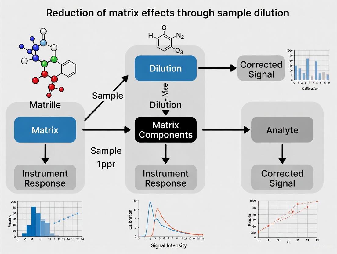
The following diagram illustrates the logical relationship and workflow for the key experimental protocols described in this note, highlighting how they can be integrated into a comprehensive strategy for defining and mitigating matrix effects.
The Scientist's Toolkit: Key Research Reagents & Materials
Table 3: Essential Reagents and Materials for Matrix Effect Research
| Item | Function/Application in Matrix Effect Research |
|---|---|
| Stable Isotope-Labeled (SIL) Internal Standards | Correct for variability in ionization efficiency and ion suppression; essential for calculating IS-normalized Matrix Factors [1] [2] [6]. |
| IROA Internal Standard (IROA-IS) Library | A comprehensive ¹³C-labeled standard library enabling ion suppression measurement and correction for a wide range of metabolites in non-targeted studies [6]. |
| Post-Column Infusion T-union | Allows for the mixing of a continuously infused standard with the LC effluent just prior to the ESI source, enabling real-time visualization of matrix effects [1] [4]. |
| Theta Emitters (Dual-Channel Nano-ESI Emitters) | Permit the introduction of sample and a compensating solution (e.g., ammonium acetate with additives) from separate channels, helping to generate droplets depleted of non-volatile salts and reduce adduction [8]. |
| Derivatization Reagents (e.g., DEEMM, FMOC-Cl) | Modify analyte properties to improve chromatography and ionization; choice of reagent (e.g., DEEMM being less affected by matrix) can inherently reduce matrix effects [4]. |
Matrix effects represent a significant challenge in bioanalysis, particularly when using electrospray ionization mass spectrometry (ESI-MS) for pharmacokinetic screening in drug development. These effects can severely compromise data accuracy, leading to incorrect rejection of potential drug candidates. The fundamental mechanisms underpinning these effects are competition for charge and droplet surface effects in the electrospray process [9] [10]. Within the broader context of reducing matrix effects through sample dilution research, understanding these core mechanisms is essential for developing effective analytical protocols. This application note details the underlying principles, provides experimental validation data, and outlines standardized protocols for identifying and mitigating these effects to ensure reliable bioanalytical results.
Theoretical Background
Electrospray Ionization (ESI) and Matrix Effects
Electrospray Ionization operates by generating a fine mist of charged droplets at the MS interface. The formation of gas-phase ions from these droplets is vulnerable to interference from co-eluting compounds present in the biological matrix [10]. Ion suppression occurs when these interfering species reduce the ionization efficiency of the target analyte, leading to diminished signal intensity and inaccurate quantification.
Core Mechanisms of Ion Suppression
Competition for Charge: In the electrospray droplet, the available charge (protons or other ions) is finite. Surface-active compounds or those with higher proton affinity can outcompete analyte molecules for this limited charge. This competition is particularly pronounced with formulation excipients like Cremophor EL (CrEL), which contains numerous polyethyleneglycol (PEG) oligomers that effectively compete for available protons [10]. The presence of such agents can lead to a significant, and often variable, reduction in the analyte signal.
Droplet Surface Effects: The physical properties of the electrospray droplet itself are critical for efficient ion release. Interfering matrix components can alter the surface tension, viscosity, or evaporation rate of the droplet. Compounds like CrEL are highly surface-active and can preferentially occupy the droplet surface, thereby forming a barrier that impedes the "ion evaporation" process through which analyte ions are released into the gas phase [10]. This phenomenon directly impacts the sensitivity and robustness of the LC-MS/MS method.
Table 1: Fundamental Mechanisms of Ion Suppression in ESI-MS
| Mechanism | Primary Cause | Impact on Analysis |
|---|---|---|
| Competition for Charge | Co-eluting compounds with high proton affinity or surface activity deplete the available charge in the ESI droplet. | Reduced analyte signal intensity; non-linear response; inaccurate quantification. |
| Droplet Surface Effects | Matrix components alter droplet physics (surface tension, viscosity), hindering the efficient release of analyte ions. | Lowered sensitivity and poor method robustness; signal instability. |
Experimental Validation & Data
A study investigating CrEL, a common dosing vehicle, clearly demonstrates these mechanisms. CrEL causes significant ion suppression for a wide range of analytes, with plasma concentrations of 0.50-1.0 mg/mL causing a 2 to 10-fold suppression in signal [10]. This effect is most severe in the initial sampling points after intravenous or oral administration, where excipient concentration is highest.
The contained-ESI process, which controls droplet exposure time to acid vapor, has been shown to mitigate these effects. This method generates fine initial droplets with a high proton abundance, which together work to eliminate competition for charge and space during ion formation. This approach can yield an improvement of at least one order of magnitude in detection limits, sensitivity, and accuracy when compared to conventional electrospray [9].
Table 2: Quantitative Impact of Cremophor EL (CrEL) on Ion Suppression
| Parameter | Finding | Experimental Context |
|---|---|---|
| CrEL Concentration Causing Suppression | 0.50 - 1.0 mg/mL in plasma | Observed in initial sampling points post IV/oral dosing in rats [10]. |
| Magnitude of Signal Suppression | 2 to 10-fold reduction | Impact observed on a panel of diverse analytes (e.g., atenolol, propranolol, warfarin) [10]. |
| Improvement with Contained-ESI | >1 order of magnitude | Enhancement in detection limits, sensitivity, and accuracy compared to standard ESI [9]. |
Detailed Experimental Protocols
Protocol 1: Assessing Matrix Effects Caused by Formulation Excipients
Objective: To identify and quantify the ion suppression effect of a formulation excipient (e.g., Cremophor EL) on target analytes.
Materials:
- Test Analytes: A panel of compounds with diverse physicochemical properties (e.g., atenolol, propranolol, warfarin) [10].
- Excipient: Cremophor EL (CrEL).
- Biological Matrix: Blank rat plasma.
- Equipment: LC-MS/MS system with ESI source; 96-well polypropylene plates.
Procedure:
- Preparation of Solutions:
- Prepare a master stock of CrEL (e.g., 50 mg/mL) in DMSO.
- Prepare working standard solutions of target analytes.
Sample Preparation:
- Spike working standard solutions into blank rat plasma to create calibration standards and quality control (QC) samples.
- For the suppression test, prepare samples in duplicate: one set in pure matrix and one set in matrix containing a high concentration of CrEL (e.g., equivalent to 1 mg/mL in plasma).
- Extract samples using a suitable method (e.g., protein precipitation with acetonitrile). Vortex mix for 10 minutes and centrifuge at 3350 g for 10 minutes at 4°C [10].
- Dilute the supernatant with water for LC-MS/MS analysis.
LC-MS/MS Analysis:
- Inject aliquots (e.g., 5 μL) using a generic gradient LC method.
- Use a C18 column (e.g., 50 x 4.6 mm, 3.5 μm) with a mobile phase of 0.1% formic acid in water and acetonitrile with 20% tetrahydrofuran.
- Monitor analyte signals using Multiple Reaction Monitoring (MRM).
Data Analysis:
- Compare the peak areas of analytes in the presence and absence of CrEL.
- Calculate the Matrix Effect (ME) as follows: ME (%) = (Peak Area in Presence of CrEL / Peak Area in Absence of CrEL) x 100%
- A value significantly less than 100% indicates ion suppression.
Protocol 2: Mitigation of Matrix Effects via Alternative Ionization
Objective: To eliminate ion suppression by switching from Electrospray Ionisation (ESI) to Atmospheric Pressure Chemical Ionisation (APCI).
Materials:
- The same materials as in Protocol 1.
- LC-MS/MS system equipped with both ESI and APCI sources.
Procedure:
- Sample Preparation:
- Prepare calibration and QC samples containing the analyte and the suppressing excipient (CrEL) as described in Protocol 1.
LC-MS/MS Analysis with APCI:
- Use the same chromatographic conditions as in Protocol 1.
- Switch the ion source from ESI to APCI.
- Optimize APCI source parameters (e.g., vaporizer temperature, corona needle current) for the target analytes.
- Analyze the samples using the MRM method.
Data Analysis:
- Compare the chromatographic signals and calculated concentrations from the APCI method with those obtained from the ESI method.
- The APCI mode is often found to be completely free of the suppression effects caused by CrEL, which are severe in ESI mode [10].
Protocol 3: Mitigation via Sample Preparation - Liquid-Liquid Extraction (LLE)
Objective: To remove the ion-suppressing agent (CrEL) from the sample prior to LC-MS/MS analysis.
Materials:
- Extraction Solvents: tert-Butyl methyl ether (TBME) or hexane.
- Other materials as listed in Protocol 1.
Procedure:
- LLE Procedure:
- Aliquot 50 μL of plasma sample into a 96-well plate.
- Add 200-300 μL of organic extraction solvent (TBME or hexane).
- Seal the plate and vortex mix vigorously for 10-15 minutes.
- Centrifuge the plate at 3350 g for 10 minutes to achieve phase separation.
- Transfer the organic (upper) layer to a new plate.
- Evaporate the organic solvent under a gentle stream of nitrogen at 40°C.
- Reconstitute the dried extract with a compatible mobile phase for LC-MS/MS analysis.
LC-MS/MS Analysis:
- Analyze the reconstituted extracts using the LC-MS/MS conditions from Protocol 1 (ESI mode).
Data Analysis:
- Compare the matrix effect and analyte recovery between the LLE-prepared samples and those prepared via protein precipitation. LLE with TBME or hexane has been shown to effectively eliminate CrEL-based ion suppression in ESI mode [10].
The Scientist's Toolkit: Research Reagent Solutions
Table 3: Essential Materials for Investigating Charge Competition and Droplet Effects
| Item | Function / Role in Research |
|---|---|
| Cremophor EL (CrEL) | A model formulation excipient used as a probe to study ion suppression mechanisms due to its high surface activity and abundance of PEG oligomers [10]. |
| Diverse Analytic Panel | A set of reference compounds with varying logP, pKa, and chemical structures to test the universality of suppression effects and mitigation strategies [10]. |
| LC-MS/MS with ESI/APCI | The core analytical platform. The ability to switch between ESI (prone to suppression) and APCI (more robust) is key for comparative studies [10]. |
| Tert-Butyl Methyl Ether | An organic solvent for Liquid-Liquid Extraction (LLE), effective at removing CrEL from plasma samples, thereby mitigating matrix effects in ESI [10]. |
| Polypropylene Glycol | Used as an internal standard for the specific quantification of CrEL (PEG oligomers) in plasma to understand its pharmacokinetic profile [10]. |
Signaling Pathways and Workflow Diagrams
Diagram 1: Ion Suppression Mechanism
Diagram 2: Matrix Effect Mitigation
Impact on Analytical Parameters: Accuracy, Precision, and Sensitivity
Matrix effects represent a significant challenge in analytical chemistry, particularly in techniques like liquid chromatography-tandem mass spectrometry (LC-MS/MS). They are defined as the combined effect of all components of the sample other than the analyte on the measurement of the quantity [11]. These effects arise when matrix components co-elute with the analyte, altering its ionization efficiency in the mass spectrometer source, leading to either ion suppression or enhancement [12] [13] [14]. The presence of matrix effects can severely compromise key analytical parameters, including accuracy, precision, and sensitivity, resulting in erroneous data, reduced method robustness, and potential failures in method validation [13] [14]. This application note details protocols for assessing matrix effects and demonstrates how strategic sample dilution can mitigate their impact, thereby improving the reliability of analytical methods.
Assessing Matrix Effects: Foundational Protocols
Before implementing mitigation strategies, it is crucial to qualitatively and quantitatively assess the presence and extent of matrix effects. The following established protocols are essential for this characterization.
Protocol 1: Qualitative Assessment via Post-Column Infusion
This method provides a visual map of ion suppression or enhancement regions throughout the chromatographic run [13] [14].
- Principle: A solution of the analyte is infused post-column into the mobile phase while a blank matrix extract is injected onto the LC column. This allows for the continuous monitoring of the analyte signal, with any dips or rises indicating regions of matrix effect [13] [14].
- Procedure:
- Set up the LC-MS/MS system with a T-connector between the column outlet and the MS ion source.
- Connect a syringe pump containing a neat solution of the analyte (or a stable isotope-labeled internal standard) and initiate a constant infusion at a low flow rate (e.g., 10-20 µL/min).
- Inject a processed blank matrix sample (e.g., extracted plasma, urine, or a sample-specific matrix) onto the LC column and start the chromatographic method.
- Monitor the ion chromatogram of the analyte for any significant signal disruption (suppression or enhancement) against a stable baseline.
- Data Interpretation: A stable signal indicates minimal matrix effect. Signal suppression appears as a negative peak, while enhancement appears as a positive peak, identifying the retention time windows most affected by the sample matrix [14].
Protocol 2: Quantitative Assessment via Post-Extraction Spiking
This quantitative method, often considered a "golden standard," calculates a Matrix Factor (MF) to measure the degree of matrix effect [13] [14].
- Principle: The response of an analyte spiked into a blank matrix extract is compared to its response in a neat solution at the same concentration [14].
- Procedure:
- Prepare a set of calibration standards in a neat solvent (e.g., mobile phase).
- Process multiple lots of blank matrix (at least six different sources are recommended) through the entire sample preparation procedure [14].
- After extraction, spike the analyte at a known concentration (e.g., Low and High QC levels) into the cleaned-up blank matrix extracts.
- Analyze both the neat standards and the post-extraction spiked samples.
- Data Interpretation: Calculate the Matrix Factor (MF) and the IS-normalized MF using the formulas below. An absolute MF <1 indicates ion suppression, >1 indicates enhancement, and ≈1 indicates no effect. The IS-normalized MF should be close to 1, demonstrating that the internal standard effectively compensates for the matrix effect [14].
- Absolute MF = Peak Area (analyte in spiked matrix extract) / Peak Area (analyte in neat solution)
- IS-normalized MF = MF (analyte) / MF (internal standard)
Table 1: Quantitative Evaluation of Matrix Effects via Post-Extraction Spiking
| Parameter | Acceptance Criteria | Interpretation |
|---|---|---|
| Absolute Matrix Factor (MF) | Ideally 0.75 - 1.25 [14] | Indicates the absolute signal suppression/enhancement. Values outside this range suggest significant matrix effects. |
| IS-normalized MF | Close to 1.0 [14] | Indicates how well the internal standard compensates for the matrix effect. Critical for method robustness. |
The Dilution Strategy: A Practical Protocol for Mitigation
Sample dilution is a straightforward and effective strategy to reduce the concentration of interfering matrix components, thereby minimizing their impact on ionization [15].
- Principle: Diluting the sample with solvent decreases the absolute amount of matrix components entering the mass spectrometer, reducing their capacity to cause ion suppression or enhancement, without proportionally affecting the analyte signal if sensitivity allows [15] [16].
- Procedure:
- Perform an initial analysis to estimate the concentration of the analyte in the sample.
- Based on the estimated concentration and the required sensitivity, select an appropriate dilution factor. A dilution factor of 15 has been shown to eliminate most matrix effects in analyses of pesticides in fruits and vegetables [15].
- Perform a serial dilution for high precision: Dilute the sample stepwise (e.g., 1:5, then 1:3 from the first dilution to achieve a final 1:15 dilution) to ensure mixing homogeneity and accuracy [16].
- Analyze the diluted sample. The calibration standards and quality controls must be diluted in the same manner to maintain consistency.
- Data Interpretation: Compare the accuracy and precision of the diluted samples against undiluted ones. A successful dilution will bring the IS-normalized MF closer to 1 and improve the accuracy of QC samples.
Table 2: Impact of Dilution on Analytical Parameters in Different Matrices
| Matrix | Analyte | Dilution Factor | Impact on Accuracy & Precision | Impact on Sensitivity | Citation |
|---|---|---|---|---|---|
| Fruits & Vegetables | 53 Pesticides | 15 | Reduced signal suppression, enabling quantification with solvent standards [15] | Reduced, but sufficient with modern sensitive instruments [15] | [15] |
| Skin Moisturizers | Primary Aliphatic Amines | Not Specified | Improved accuracy via reduced matrix effects [17] | High sensitivity maintained via vortex-assisted liquid-liquid microextraction for preconcentration [17] | [17] |
| Plasma (General) | Drugs/Metabolites | Variable (e.g., 2-10 fold) | Pre-dilution of study samples mitigates anticipated matrix effects from dosing vehicles [14] | May require evaluation; can be offset by pre-concentration or high sensitivity instruments [18] [14] | [14] |
The following workflow diagram outlines the decision-making process for assessing and mitigating matrix effects, positioning dilution as a key strategy.
The Scientist's Toolkit: Essential Reagents and Materials
Successful implementation of dilution and other mitigation strategies requires specific reagents and materials.
Table 3: Key Research Reagent Solutions for Mitigating Matrix Effects
| Reagent / Material | Function / Explanation | Application Note |
|---|---|---|
| Stable Isotope-Labeled Internal Standard (SIL-IS) | The gold standard for compensating for matrix effects; co-elutes with the analyte and experiences nearly identical ionization suppression/enhancement, normalizing the signal [13] [14]. | Crucial for bioanalysis. Its use is recommended even when dilution is applied to ensure accuracy [14]. |
| Selective Adsorbents (e.g., Zirconia-coated silica, MAA@Fe₃O₄) | Used in clean-up to selectively remove phospholipids or other specific matrix interferents without retaining the target analytes, thereby reducing the matrix load [17] [19]. | Effective in procedures like dispersive micro-solid phase extraction (DµSPE) for complex matrices like skin moisturizers [17]. |
| Matrix-Matched Calibration Standards | Calibrators prepared in a blank matrix that matches the sample, compensating for consistent matrix effects by mirroring the sample's composition [13] [16]. | Requires a source of blank matrix. Used when complete removal of matrix effects is not feasible. |
| Appropriate Diluents (e.g., Mobile Phase, Buffer) | The solvent used for dilution. It should be compatible with the LC-MS system and not cause precipitation or instability of the analyte [15] [16]. | Using the initial mobile phase as a diluent is a common and safe practice to avoid chromatographic issues. |
Matrix effects pose a direct threat to the accuracy, precision, and sensitivity of analytical methods. A systematic approach involving rigorous assessment through post-column infusion and post-extraction spiking is fundamental. When matrix effects are identified, strategic sample dilution emerges as a highly effective and practical protocol for mitigation. As demonstrated in various studies, a sufficient dilution factor can significantly reduce ion suppression, enabling accurate quantification. The dilution strategy is most effective when integrated with other best practices, such as the use of stable isotope-labeled internal standards and selective sample clean-up, ultimately leading to the development of robust and reliable analytical methods for drug development and beyond.
In quantitative bioanalysis, the presence of interfering compounds in a sample matrix can significantly compromise the accuracy, precision, and sensitivity of analytical results. These matrix effects occur when co-eluting compounds alter the ionization efficiency of the target analyte, leading to either ion suppression or enhancement. Dilution represents a fundamental sample preparation strategy to mitigate these effects. The theoretical foundation is straightforward: by reducing the concentration of all components in the sample, the absolute amount of interfering compounds introduced into the analytical system is decreased to a level where their impact on the analyte of interest becomes negligible. This approach is particularly valuable in liquid chromatography-tandem mass spectrometry (LC-MS/MS) bioanalysis, where matrix effects are a major concern affecting data reliability. When a sample is diluted, the proportional relationship between the analyte and the interferent may remain, but their absolute concentrations fall below a threshold where interference occurs, thereby improving the fidelity of the quantitative measurement.
Theoretical Principles and Mechanisms
Fundamental Relationship Between Dilution and Concentration Reduction
The core principle of dilution is that it uniformly reduces the concentration of all solutes present in a solution. The dilution factor (DF) is calculated as the ratio of the final volume to the initial volume: DF = Vfinal / Vinitial. Consequently, the concentration of any compound after dilution is its original concentration divided by the dilution factor. For interfering compounds, this reduction in concentration diminishes their capacity to cause ion suppression or enhancement in the mass spectrometer source. The effectiveness of dilution hinges on the premise that the analyte possesses sufficient detection sensitivity to withstand the dilution process while the interferents do not significantly affect the ionization process at their new, lower concentrations. This makes dilution a practical and efficient first-line strategy for managing matrix effects, especially when the exact identity of the interfering substances is unknown.
The Concept of Parallelism in Dilution
A critical validation step when employing dilution is assessing parallelism. A dilution experiment, often referred to as a parallelism study, judges whether diluted samples lie parallel to the calibration curve [20]. This confirms that the analyte, when corrected for the dilution factor, provides the same result regardless of the extent of dilution. Non-parallelism indicates that the dilution does not correctly compensate for matrix effects, potentially due to issues like differing antibody affinities in immunoassays or the presence of an interferent whose effect is not linearly reduced by dilution [20]. Samples containing high-affinity antibodies may show over-recovery on dilution, while those with low-affinity antibodies show under-recovery [20]. Therefore, demonstrating parallelism is essential to confirm that dilution is a valid approach for a given analyte-matrix combination.
Quantitative Assessment of Dilution Efficacy
The success of a dilution protocol in reducing matrix effects can be systematically evaluated by calculating key parameters. The following table summarizes the formulas and acceptance criteria for these metrics.
Table 1: Key Parameters for Assessing Dilution Efficacy
| Parameter | Calculation Formula | Purpose | Interpretation |
|---|---|---|---|
| Dilution Factor (DF) | ( DF = \frac{V{final}}{V{initial}} ) | To determine the factor by which the sample has been diluted. | A higher DF leads to greater reduction of interferents but requires higher analyte sensitivity. |
| Matrix Effect (ME) | ( ME (\%) = \left( \frac{Peak Area{Post-extraction Spiked Matrix}}{Peak Area{Neat Solvent}} - 1 \right) \times 100\% ) [2] | To quantify ion suppression/enhancement. | A value of 0% indicates no matrix effect. Negative values indicate suppression; positive values indicate enhancement. |
| Process Efficiency (PE) | Derived from pre- and post-extraction spiked samples [2] | To measure the combined effect of recovery and matrix effect on the overall method. | Reflects the total impact of the sample preparation and analysis process on the measured signal. |
| Recovery (R) | ( Recovery (\%) = \frac{2 \times Concentration{after PEG}}{Concentration{before PEG}} \times 100\% ) [21] | To measure the fraction of analyte regained after a preparation step. | High recovery indicates minimal analyte loss during dilution or other clean-up procedures. |
Empirical data from validation studies provides concrete evidence of dilution's utility. For instance, in a study investigating unexplained elevations of the tumor marker CA 19-9, a polyethylene glycol (PEG) precipitation method was used to detect interference. The recovery rate after PEG treatment was a critical indicator, with a cutoff below 37.9% providing an area under the curve (AUC) of 0.993 for identifying interference, showing high sensitivity and specificity [21]. Furthermore, Matuszewski et al. established methodologies that integrate the assessment of matrix effect, recovery, and process efficiency into a single experiment, allowing for a comprehensive understanding of how dilution influences the entire analytical process [2].
Experimental Protocols for Dilution and Matrix Effect Evaluation
Protocol 1: Standard Dilution for Matrix Effect Reduction
This protocol outlines a simple dilution method suitable for samples with low protein matrix content, such as urine or cerebrospinal fluid (CSF).
Principle: Reducing matrix component concentrations via dilution with a compatible solvent to minimize ionization interference in LC-MS/MS.
Materials & Reagents:
- Internal Standard (IS) Solution: A stable isotope-labeled analog of the analyte is ideal.
- Diluent: LC-MS grade water, mobile phase B, or a buffered solution compatible with the analytical system.
- Analytical Instrumentation: LC-MS/MS system.
- Labware: Precision micropipettes, polypropylene microcentrifuge tubes.
Procedure:
- Aliquot Sample: Pipette a measured volume (e.g., 50 µL) of the sample into a clean microcentrifuge tube.
- Add Internal Standard: Add a fixed volume of the IS working solution to the sample. Vortex to mix.
- Dilute Sample: Add the appropriate volume of diluent to achieve the desired dilution factor (e.g., 1:2, 1:10). The total volume should be within the operational range of the tube and the analytical instrument.
- Mix and Centrifuge: Vortex the mixture thoroughly for 30-60 seconds. Centrifuge at high speed (e.g., 10,000-14,000 × g) for 5-10 minutes to pellet any particulates.
- Analysis: Transfer the supernatant to an LC vial and inject into the LC-MS/MS system.
Notes: The simplicity of this protocol is its main advantage, but it is limited by the assay's sensitivity and cannot concentrate the analyte [20]. The required dilution factor should be determined experimentally during method validation.
Protocol 2: Integrated Assessment of Matrix Effect, Recovery, and Process Efficiency
This protocol, based on the approaches of Matuszewski et al., allows for the simultaneous evaluation of how dilution and sample clean-up impact method performance [2].
Principle: Comparing analyte response in different sample sets (neat solvent, post-extraction spiked matrix, and pre-extraction spiked matrix) to deconvolute the contributions of the matrix and recovery.
Materials & Reagents:
- At least 6 independent lots of blank matrix.
- Analyte standard solution.
- Internal standard solution.
- Appropriate solvents and reagents for sample preparation (e.g., protein precipitation reagents, solid-phase extraction cartridges).
Procedure:
- Prepare Sample Sets:
- Set 1 (Neat Solution): Spike analyte and IS into a neat solution of mobile phase. This set represents the ideal signal response without matrix.
- Set 2 (Post-extraction Spiked): Spike analyte and IS into a blank matrix extract that has already undergone the sample preparation procedure. This set assesses the matrix effect (ME).
- Set 3 (Pre-extraction Spiked): Spike analyte and IS into the blank matrix before performing the sample preparation. This set assesses the overall process efficiency (PE), which includes both extraction recovery and the matrix effect.
- Perform Sample Preparation: Process all sets according to the established method (e.g., protein precipitation, dilution).
- LC-MS/MS Analysis: Analyze all samples and record the peak areas for the analyte and IS.
- Data Calculation:
- Matrix Effect (ME): Compare the peak response of Set 2 to Set 1. ( ME = (Peak Area{Set 2} / Peak Area{Set 1}) \times 100\% ).
- Recovery (R): Calculate by comparing the peak response of Set 3 to Set 2. ( R = (Peak Area{Set 3} / Peak Area{Set 2}) \times 100\% ).
- Process Efficiency (PE): Calculate by comparing the peak response of Set 3 to Set 1. ( PE = (Peak Area{Set 3} / Peak Area{Set 1}) \times 100\% ). It should also equal ( (ME \times R) / 100 ).
Notes: This integrated approach is crucial for a comprehensive understanding of the factors influencing analyte quantification and for validating that dilution effectively controls for matrix effects [2].
Workflow Visualization
Diagram 1: Dilution Workflow for Matrix Effect Reduction. This diagram outlines the decision-making process for implementing and validating sample dilution to mitigate matrix effects in LC-MS/MS analysis.
The Scientist's Toolkit: Essential Research Reagents and Materials
Table 2: Key Reagent Solutions for Dilution Studies
| Reagent/Material | Function/Purpose | Example Specifications |
|---|---|---|
| Stable Isotope-Labeled Internal Standard (SIL-IS) | Corrects for variability in sample preparation and ionization; the gold standard for compensating matrix effects [22]. | Creatinine-d3 for a creatinine assay; purity >95%. |
| LC-MS Grade Solvents | Used as diluents; high purity minimizes background noise and prevents introduction of new interferents. | Water, methanol, acetonitrile; low UV absorbance, low particle count. |
| Polyethylene Glycol (PEG) 6000 | Used in precipitation protocols to remove high molecular weight interferents like proteins and macro-complexes [21]. | 25% w/v solution in appropriate buffer. |
| Blank Matrix | Essential for method development and validation to prepare calibration standards and quality controls. | Human plasma, urine, or cerebrospinal fluid from multiple donors. |
| Heterophile Antibody Blocking Reagent (HBR) | Added to samples to neutralize heterophile antibodies, a common source of immunoassay interference [21]. | Commercially available blocking tubes or solutions. |
| Formic Acid / Ammonium Formate | Common mobile phase additives for LC-MS to improve chromatographic separation and ionization efficiency. | LC-MS grade, 0.1% formic acid, 2-10 mM ammonium formate. |
Dilution remains a cornerstone technique for reducing the concentration of interfering compounds and mitigating matrix effects in quantitative bioanalysis. Its theoretical basis is rooted in the fundamental principles of solution chemistry, which dictate that a reduction in absolute concentration can render interferents insignificant without proportionally affecting a sensitive analyte's detectability. The successful application of dilution requires rigorous validation, including the demonstration of parallelism and a systematic assessment of its impact on matrix effect, recovery, and overall process efficiency. When implemented within a well-designed analytical method and supported by appropriate internal standards, dilution is a powerful, simple, and cost-effective strategy to enhance data accuracy and reliability, thereby supporting robust drug development and clinical research.
Identifying Matrix-Prone Analytes and Sample Types
Matrix effects are a critical challenge in quantitative bioanalysis, particularly in liquid chromatography-mass spectrometry (LC-MS), where they can severely compromise accuracy, precision, and sensitivity [22] [13]. These effects occur when compounds co-eluting with the analyte interfere with the ionization process in the mass spectrometer, causing ion suppression or enhancement [22] [13]. Within the broader context of research on reducing matrix effects through sample dilution, the first essential step is the systematic identification of analytes and sample types most susceptible to these interference phenomena. This application note provides detailed protocols and data for characterizing matrix-prone analytes and matrices, establishing a foundational framework for developing effective dilution-based mitigation strategies.
Defining Matrix-Prone Characteristics
Analyte Properties Conferring Susceptibility
Certain inherent physicochemical properties significantly increase an analyte's vulnerability to matrix effects. Understanding these properties allows researchers to predict and preemptively address potential interference issues.
Table 1: Analyte Properties Associated with Increased Matrix Effect Risk
| Property | Risk Level | Mechanistic Rationale | Example Analytes |
|---|---|---|---|
| High Polarity | High | Competes with polar matrix components for ionization in ESI source [13] | Metabolites, inorganic salts |
| Surface Activity | High | Affects droplet formation and charge transfer in ESI; can be suppressed by other surface-active compounds [15] | Phospholipids, certain pharmaceuticals |
| Low Volatility | Medium | Can be affected by less-volatile matrix compounds that impact droplet evaporation efficiency [22] | Large molecules, some polymers |
| Basicity | Medium | Susceptible to interference from other basic compounds that may deprotonate and neutralize analyte ions [22] | Basic pharmaceuticals, amines |
Electrospray ionization (ESI) is particularly prone to matrix effects compared to atmospheric pressure chemical ionization (APCI), as ionization occurs in the liquid phase where matrix components can directly interfere with the analyte's ability to form stable ions and transfer to the gas phase [13]. The relative similarity in polarity between an analyte and its matrix composition also increases susceptibility, as this similarity makes selective extraction more challenging, leaving more co-eluting interferences [13].
High-Risk Sample Matrices
The complexity and composition of the sample matrix itself are major determinants of matrix effect severity. Biological matrices contain numerous components that can co-elute with analytes and interfere with ionization.
Table 2: High-Risk Sample Matrices and Their Problematic Components
| Matrix Type | Key Interfering Components | Primary Concerns | Common Applications |
|---|---|---|---|
| Plasma/Serum | Phospholipids, proteins, amino acids, lipids [13] [23] | High concentration of phospholipids causing ion suppression; protein binding [23] | Drug monitoring, bioanalysis [24] [23] |
| Urine | Inorganic salts, urea, metabolic derivatives [13] | High salt content; variable composition between individuals [13] | Metabolite studies, clinical chemistry [22] [23] |
| Whole Blood | Phospholipids, proteins, cellular components [23] | Additional complexity from hemolysis and cell lysis products [23] | Forensic analysis, whole blood studies |
| Tissue Homogenates | Phospholipids, fats, cellular debris [25] | Complex mixture with high concentration of interfering compounds [26] | Drug distribution studies, biomarker research |
| Food & Beverages | Fats, proteins, carbohydrates, additives [25] | Highly variable and complex composition; natural pigments [25] | Pesticide residue analysis, contaminant testing [15] |
| Environmental Water | Humic acids, dissolved organic matter, salts [24] | Natural organic matter that can suppress ionization [24] | Pesticide analysis, environmental monitoring [15] |
Notably, matrix effects can vary significantly between individual matrix sources. For instance, plasma from healthy volunteers may present a different interference profile compared to plasma from terminally ill patients with different genetics and diets [24]. This highlights the importance of testing matrix effects using blank matrices from multiple relevant sources (recommended: at least six) during method validation [24] [13].
Experimental Protocols for Identifying Matrix Effects
Post-Column Infusion for Qualitative Assessment
The post-column infusion method provides a qualitative assessment of matrix effects throughout the chromatographic run, identifying regions of ion suppression or enhancement [22] [13].
Protocol:
- System Setup: Connect a syringe pump containing the analyte standard solution to a T-piece between the HPLC column outlet and the MS ion source [13].
- Infusion Conditions: Infuse the analyte standard at a constant rate (typical flow rates: 5-20 μL/min) to establish a stable baseline signal [13].
- Sample Injection: Inject a blank matrix extract (e.g., processed plasma, urine, or tissue homogenate) onto the chromatographic system using the intended analytical method [13] [22].
- Data Analysis: Monitor the signal of the infused analyte. A decrease in signal indicates ion suppression; an increase indicates ion enhancement [13]. Record the retention time zones where these effects occur.
Interpretation: This method provides a "matrix effect fingerprint" of the chromatographic run, highlighting regions where analytes would be most susceptible to matrix effects. It is particularly valuable during method development for optimizing chromatographic separation to position analyte peaks in regions with minimal interference [22] [13].
Post-Extraction Spike Method for Quantitative Assessment
The post-extraction spike method provides a quantitative measure of matrix effects for specific analytes by comparing their response in neat solution versus matrix [13] [22].
Protocol:
- Prepare Solutions:
- Solution A (Neat Standard): Prepare the analyte at a known concentration in a suitable solvent (typically the reconstitution solvent or mobile phase) [13].
- Solution B (Spiked Matrix): Take a blank matrix extract (processed without analyte), and spike with the same concentration of analyte as Solution A [13] [22].
- Analysis: Analyze both solutions using the intended LC-MS/MS method, ensuring identical chromatographic and detection conditions [13].
- Calculation: Calculate the matrix effect (ME) using the formula: ME (%) = (Peak Area of Solution B / Peak Area of Solution A) × 100 [13]
- Interpretation:
- ME ≈ 100%: No significant matrix effect
- ME < 100%: Ion suppression
- ME > 100%: Ion enhancement [13]
Table 3: Matrix Effect Classification Based on ME Percentage
| ME Percentage | Effect Category | Recommended Action |
|---|---|---|
| 85-115% | Minimal | Generally acceptable for bioanalytical methods [13] |
| 70-85% or 115-130% | Moderate | Consider mitigation strategies; may require stable isotope internal standard [13] |
| <70% or >130% | Severe | Requires significant method modification; dilution may be effective [15] |
This quantitative approach is essential for validating methods according to regulatory guidelines, which often require demonstrating that matrix effects do not compromise assay accuracy [24] [23].
Slope Ratio Analysis for Concentration-Dependent Assessment
Slope ratio analysis extends the post-extraction spike method across a concentration range to evaluate how matrix effects may vary at different analyte levels [13].
Protocol:
- Calibration Curves: Prepare two calibration curves in parallel:
- Analysis: Analyze both calibration sets using the intended LC-MS/MS method.
- Calculation: Calculate the slope ratio (SR): SR = Slope of Matrix-Matched Calibration Curve / Slope of Solvent-Based Calibration Curve [13]
- Interpretation: An SR significantly different from 1.0 indicates consistent matrix effects across the concentration range, with values <1 indicating suppression and >1 indicating enhancement.
The Scientist's Toolkit: Essential Research Reagents and Materials
Table 4: Key Research Reagent Solutions for Matrix Effect Evaluation
| Item | Function/Application | Considerations |
|---|---|---|
| Blank Matrix | Assessing background interference and preparing matrix-matched standards [24] | Source from at least six different lots; match to study population as closely as possible [24] |
| Stable Isotope-Labeled Internal Standards (SIL-IS) | Compensating for matrix effects by normalizing analyte response [22] [13] | Ideal but often expensive; may not be available for all analytes [22] [13] |
| Structural Analog Internal Standards | Alternative to SIL-IS when isotopes unavailable [22] | Must have similar physicochemical properties and co-elute with analyte [22] |
| Phospholipid Removal Plates | Selective removal of phospholipids from biological samples [23] | Effective for reducing major source of matrix effects in plasma/serum [23] |
| Solid Phase Extraction (SPE) Cartridges | Sample clean-up and concentration; reduces matrix components [25] [27] | Various chemistries available; select based on analyte properties [27] |
| Protein Precipitation Plates | Rapid protein removal from biological fluids [27] [23] | Simple but may not remove phospholipids effectively [23] |
| Appropriate Solvents | Sample reconstitution, dilution, and mobile phase preparation [25] | Must be compatible with both sample matrix and LC-MS system [25] |
Systematic identification of matrix-prone analytes and sample types is a critical prerequisite for developing effective dilution-based strategies to mitigate matrix effects. By employing the protocols outlined in this application note—post-column infusion, post-extraction spike, and slope ratio analysis—researchers can accurately characterize matrix effects and make informed decisions on appropriate dilution factors. The data generated through these methods provides a scientific foundation for optimizing sample preparation and chromatographic conditions, ultimately leading to more robust and reliable bioanalytical methods. Within the broader thesis context of reducing matrix effects through dilution, this characterization work enables the rational application of dilution protocols tailored to specific analyte-matrix combinations, balancing the need to reduce interferences with maintaining adequate analytical sensitivity.
Practical Dilution Methods: Protocols, Optimization, and Workflow Integration
Matrix effects pose a significant challenge in analytical methods, particularly in liquid chromatography-tandem mass spectrometry (LC-MS/MS), where co-eluting compounds can suppress or enhance analyte ionization, leading to inaccurate quantification. Sample dilution is a straightforward and effective strategy to reduce matrix effects by decreasing the concentration of interfering compounds in the sample. However, this approach must carefully balance matrix reduction with the preservation of analytical sensitivity. This application note provides a detailed protocol for determining optimal dilution factors, supported by experimental data and workflows tailored for researchers and drug development professionals.
Key Concepts and Quantitative Data
The Dilution-Sensitivity Trade-Off
Dilution reduces matrix effects but concurrently decreases analyte concentration, potentially impacting detection limits. The optimal dilution factor minimizes matrix interference while maintaining analyte concentrations above the instrument’s limit of quantification (LOQ).
Experimental Evidence on Dilution Efficacy
Studies evaluating matrix effects for pesticides in complex matrices (e.g., orange, tomato, and leek) demonstrated that dilution significantly reduces signal suppression. The data below summarize the relationship between dilution factors and matrix effect reduction:
Table 1: Impact of Dilution on Matrix Effects in LC-MS/MS Analysis
| Matrix | Dilution Factor | Matrix Effect Reduction | Notes |
|---|---|---|---|
| Orange | 1:15 | Significant reduction | Enabled use of solvent-based standards for most pesticides [15] |
| Tomato | 1:15 | Significant reduction | Similar efficacy as in orange matrix [15] |
| Leek | 1:15 | Moderate to significant reduction | Persistent matrix effects for some pesticides required additional measures [15] |
A dilution factor of 1:15 was found to eliminate most matrix effects, allowing quantification with solvent-based standards in many cases [15]. For analytes where dilution alone was insufficient, stable isotope-labeled internal standards provided an effective alternative for accurate quantification.
Experimental Protocols
Protocol 1: Determining Optimal Dilution Factor
Objective: Identify the dilution factor that minimizes matrix effects without compromising sensitivity.
Materials:
- Stock standard solution of the analyte
- Blank matrix (e.g., plasma, plant extract)
- Appropriate diluent (e.g., methanol, acetonitrile, or buffer)
- LC-MS/MS system
Steps:
- Prepare Matrix-Matched Standards: Spike the analyte into the blank matrix at a concentration within the linear range of the instrument.
- Perform Serial Dilutions: Dilute the spiked matrix extracts at factors of 1:5, 1:10, 1:15, and 1:20 using the diluent.
- Analyze Samples: Inject each diluted sample into the LC-MS/MS system and record the peak areas.
- Compare with Solvent Standards: Analyze solvent-based standards at equivalent concentrations.
- Calculate Matrix Effects (ME): [ ME\% = \left(\frac{\text{Peak area in matrix}}{\text{Peak area in solvent}} - 1\right) \times 100 ] A value of ±20% indicates minimal matrix effects [15] [13].
- Select Optimal Dilution Factor: Choose the lowest dilution factor that achieves ME within ±20% and maintains the analyte signal above the LOQ.
Protocol 2: Serial Dilution for High-Dilution Factors
Objective: Achieve high dilution factors accurately, especially when working with limited sample volumes or high precision requirements.
Materials:
- Precision pipettes
- Diluent (e.g., acetonitrile for LC-MS compatibility)
- Multi-well plates or microcentrifuge tubes
Steps:
- Calculate Dilution Scheme: For a final dilution factor of 1:1000, use intermediary dilutions (e.g., 1:10 followed by 1:100) to improve accuracy [28] [29].
- First Dilution (1:10): Combine 10 µL of sample with 90 µL of diluent. Mix thoroughly.
- Second Dilution (1:100): Combine 10 µL of the 1:10 dilution with 990 µL of diluent. Mix thoroughly.
- Validate Dilution: Confirm the final concentration using calibration standards.
Formulas:
- Dilution Factor (DF): [ DF = \frac{\text{Final Volume}}{\text{Solute Volume}} ]
- Serial Dilution Calculations: [ \text{Move Volume} = \frac{\text{Final Volume}}{DF - 1} ] [ \text{Diluent Volume} = \text{Final Volume} - \text{Move Volume} ] [29]
Visual Workflows and Diagrams
Workflow for Dilution Factor Optimization
The following diagram outlines the decision-making process for balancing matrix effects and sensitivity:
Serial Dilution Protocol Workflow
This diagram illustrates the stepwise procedure for performing serial dilutions:
Research Reagent Solutions and Materials
Table 2: Essential Materials for Dilution-Based Matrix Effect Reduction
| Item | Function | Example Applications |
|---|---|---|
| Stable Isotope-Labeled Internal Standards | Compensates for residual matrix effects after dilution; improves accuracy [15] [13] | LC-MS/MS quantification of problematic pesticides or metabolites |
| Solid Phase Extraction (SPE) Cartridges | Pre-concentrates analytes and removes interfering matrix components before dilution [18] [13] | Environmental and bioanalytical sample preparation |
| LC-MS/MS Compatible Solvents | Act as diluents; ensure chemical compatibility and minimal background interference [15] [18] | Sample dilution in HPLC, GC, and MS protocols |
| Precision Pipettes and Automated Liquid Handlers | Enable accurate serial dilutions, reducing human error [28] [29] | High-throughput dilution for calibration curves |
Discussion and Best Practices
- Assess Matrix Effects Early: Incorporate matrix effect evaluation during method development rather than validation to improve robustness [13]. Techniques like post-column infusion provide qualitative insights, while post-extraction spike methods offer quantitative data [13].
- Prioritize Dilution for Simplicity: Dilution is cost-effective and straightforward, particularly for methods with adequate sensitivity. For example, a 1:15 dilution factor is a practical starting point for many vegetable matrices [15].
- Combine Strategies for Challenging Matrices: For persistent matrix effects, combine dilution with stable isotope-labeled internal standards or advanced clean-up techniques (e.g., SPE) [15] [13].
- Validate Dilution Integrity: Ensure dilution steps do not introduce contamination or analyte loss. Use quality control samples to verify precision and accuracy at each dilution level.
Determining the optimal dilution factor is critical for mitigating matrix effects without sacrificing sensitivity. Experimental data support a dilution factor of 1:15 as effective for many applications, though matrix-specific validation is essential. By integrating the protocols, workflows, and reagent solutions outlined here, researchers can enhance the reliability of quantitative analyses in drug development and other fields.
Matrix effects, the suppression or enhancement of analyte signal by co-eluting compounds from a sample matrix, represent a significant challenge in mass spectrometry, compromising data accuracy and reproducibility in fields from clinical diagnostics to environmental monitoring [30] [5]. Automated dilution addresses this by systematically reducing matrix component concentration, thereby minimizing their interference with ionization efficiency [31] [15]. This application note details the integration of Automated Micro-Dilution and Injection (AMDI) systems and online auto-injection platforms as robust, reproducible strategies for matrix effect mitigation within sample preparation workflows. These approaches are particularly vital for high-throughput laboratories analyzing complex biological and environmental samples, where manual dilution is a bottleneck prone to human error [32] [33].
Quantitative Data on Dilution for Matrix Effect Reduction
The relationship between dilution factor and the reduction of matrix effects has been quantitatively demonstrated across various analytical techniques and sample types. The following table summarizes key experimental findings from recent research.
Table 1: Summary of Quantitative Data on Dilution for Matrix Effect Reduction
| Analytical Technique | Sample Matrix | Key Analytic(s) | Observation | Minimum DF for Negligible ME | Citation |
|---|---|---|---|---|---|
| SERS | Fish Feed | Malachite Green | Linear correlation between ME and logarithm of DF; MEs weaken with increasing DF. | DF > 249 | [31] |
| SERS | Fish Meat | Malachite Green | MEs increased with matrix complexity; MEs become negligible at high DF. | DF > 374 | [31] |
| LC-ESI-MS/MS | Orange, Tomato, Leek | 53 Pesticides | Dilution reduced signal suppression in most cases. | DF = 15 (for most matrix effects) | [15] |
| UHPSFC-MS | Plasma | 8 Vitamin E forms | Sample preparation combined with appropriate calibration model is crucial; dilution is a key strategy. | Not Specified | [30] |
| LC-ESI-MS (Urban Runoff) | Urban Runoff Water | Pesticides, Pharmaceuticals | High variability in signal suppression (0–67% median); "dirty" samples required higher dilution (REF 50). | Sample-Dependent (REF 50-100) | [5] |
These studies confirm that while the specific dilution factor required is matrix- and analyte-dependent, the general principle holds: strategic dilution is a simple yet powerful tool for mitigating matrix effects [31] [15] [5].
Experimental Protocols
Protocol 1: Establishing a Minimum Dilution Factor for SERS Analysis
This protocol is adapted from a 2025 study investigating the detection of malachite green in complex aquaculture-related matrices using Surface-Enhanced Raman Spectroscopy (SERS) [31].
1. Objective: To determine the minimum dilution factor (DF) required to negate matrix effects in SERS analysis for a specific analyte-matrix combination.
2. Materials:
- SERS Substrate: Highly sensitive Cu(OH)₂-Ag/CN-CDots substrate.
- Analyte: Malachite green (MG) standard.
- Matrices: Aquaculture water, fish feed, fish meat.
- Solvents: Appropriate extraction solvents (e.g., acetonitrile).
- Equipment: Raman spectrometer, centrifuge, vortex mixer, pipettes, volumetric flasks.
3. Procedure:
- Step 1: Sample Extraction.
- Homogenize solid matrices (fish feed, fish meat).
- Extract MG from each matrix type using a validated method (e.g., solvent extraction with acetonitrile). Centrifuge to obtain a clear supernatant.
- Step 2: Dilution Series Preparation.
- Prepare a series of diluted extracts from the original sample extract. For example, create dilutions with DFs of 10, 50, 100, 200, 300, 400, and 500 using an appropriate solvent.
- Spike each diluted extract with a known, constant concentration of MG analyte standard.
- Step 3: SERS Measurement.
- Apply a fixed volume of each diluted and spiked extract to the SERS substrate.
- Acquire Raman spectra for each sample under consistent instrumental parameters (laser power, integration time).
- Step 4: Data Analysis and Calculation.
- Measure the peak intensity or area for the characteristic MG band in each spectrum.
- Calculate the apparent recovery (%) at each DF:
(Measured Concentration / Spiked Concentration) * 100. - A recovery of 100% indicates no matrix effect. Plot recovery (%) against the logarithm of the DF (Log(DF)).
- Perform linear regression. The minimum DF for negligible ME is the point where the recovery confidence interval consistently contains 100%.
Protocol 2: Online SPE-LC/MS with Integrated Automated Dilution for PFAS Analysis
This protocol outlines an automated workflow for the analysis of per- and polyfluoroalkyl substances (PFAS) in complex seafood matrices, leveraging the PAL RTC autosampler for dilution and clean-up [33].
1. Objective: To perform automated calibration, sample dilution, clean-up, and analysis of PFAS in complex samples to minimize matrix effects and analyst exposure.
2. Materials:
- Automated System: PAL RTC autosampler configured for LC/MS, integrated with an Agilent triple quadrupole LC/MS system.
- Consumables: μSPE cartridges (e.g., C18 or graphitized carbon for PFAS), microplates, QuEChERS extraction salts.
- Standards: Native PFAS analytical standards (e.g., 73 compounds), internal standards.
3. Procedure:
- Step 1: System Configuration.
- Program the PAL method composer software to sequence the following steps: calibration standard preparation, sample weighing, QuEChERS extraction, extract dilution, μSPE clean-up, and injection.
- Step 2: Automated Calibration and Sample Preparation.
- The system automatically serially dilutes a PFAS stock standard to prepare a multi-point calibration curve in solvent.
- For seafood samples, the system dispenses a homogenized sample into a vial, adds solvent (e.g., acetonitrile) for QuEChERS extraction, and shakes.
- Step 3: Automated Dilution and Micro-Solid Phase Extraction (μSPE).
- An aliquot of the raw extract is automatically diluted with water or a weak solvent to achieve a predetermined DF (e.g., 1:5 or 1:10). This step reduces the organic solvent content and matrix load, preconditioning the sample for μSPE.
- The diluted extract is passed through a μSPE cartridge. The PAL system controls the load, wash, and elution steps. PFAS analytes are retained on the cartridge while many matrix interferents are washed away.
- The analytes are eluted with a strong solvent in a small, predefined volume, achieving clean-up and concentration.
- Step 4: LC/MS Analysis and Data Processing.
- The purified eluate is automatically injected into the LC/TQ-MS for separation and MRM quantification.
- Quantify against the solvent-based calibration curve prepared in Step 2, demonstrating the effectiveness of the automated dilution/clean-up in eliminating matrix effects.
Workflow and Strategy Diagrams
Automated Dilution and Analysis Workflow
The following diagram illustrates the logical flow of an automated sample preparation and dilution protocol, as implemented in systems like the PAL platform [33] [34].
Diagram 1: Automated Dilution and Analysis Workflow.
Matrix Effect Reduction Strategy Selection
This diagram outlines a decision pathway for selecting the appropriate matrix effect reduction strategy based on sample complexity and analytical requirements [15] [30] [35].
Diagram 2: Matrix Effect Reduction Strategy Selection.
The Scientist's Toolkit: Research Reagent Solutions
The following table details key reagents, materials, and instrumentation essential for implementing the automated dilution protocols described in this note.
Table 2: Essential Research Reagents and Materials for Automated Dilution Workflows
| Item Name | Function / Application | Specific Example / Note |
|---|---|---|
| Mercaptoacetic acid-modified magnetic adsorbent (MAA@Fe3O4) | Dispersive µSPE adsorbent for selective matrix interference removal without adsorbing target analytes like primary amines. | Effective for cleaning complex matrices like skin moisturizers prior to analysis; reusable for up to 5 cycles [17]. |
| µSPE Cartridges | Miniaturized solid-phase extraction for high-throughput, automated sample clean-up with reduced solvent consumption. | Used in PAL System for automated clean-up in applications like pesticide or PFAS analysis [33]. |
| Butyl Chloroformate (BCF) | Derivatization agent for primary aliphatic amines. Converts polar amines into less polar, volatile carbamate derivatives suitable for GC analysis. | Enables simultaneous derivatization and extraction in VALLME, improving chromatographic behavior [17]. |
| QuEChERS Kits | Quick, Easy, Cheap, Effective, Rugged, and Safe method for extracting analytes from complex solid/semi-solid matrices. | Can be automated on platforms like the PAL System for food safety (pesticides) and environmental analysis [33]. |
| PAL RTC Autosampler | Robotic automated sample preparation and injection system. Integrates liquid handling, dilution, SPE, and thermal mixing. | Enables end-to-end automation from sample weighing to injection for LC/MS and GC/MS [33] [34]. |
| GERSTEL MultiPurpose Sampler (MPS) | Automated sampler for GC or LC that can be configured to perform liquid handling, LLE, SPE, and derivatization. | Automates complex sample prep steps, standardizing processes and improving reproducibility [36]. |
Automated dilution systems represent a paradigm shift in managing matrix effects, moving beyond manual, variable methods to standardized, reliable workflows. The integration of AMDI and online auto-injection platforms, as exemplified by the PAL and GERSTEL MPS systems, enables precise dilution, robust clean-up, and seamless integration with analytical instrumentation [32] [33] [36]. This approach not only enhances data quality and reproducibility but also increases laboratory efficiency and frees skilled personnel for higher-value tasks. As the field advances, the synergy of automation with green chemistry principles, modular design, and AI-driven optimization will further solidify automated dilution as a cornerstone of high-quality analytical science [32].
Matrix effects (MEs) represent a significant challenge in the quantitative analysis of analytes in complex biological and environmental samples using techniques like liquid chromatography-tandem mass spectrometry (LC-MS/MS) and gas chromatography-mass spectrometry (GC-MS). MEs are defined as the unintended impact of co-eluting matrix components on the ionization efficiency and detection of the target analyte, leading to signal suppression or enhancement [37]. The complexity of the matrix—whether plasma, urine, tissues, or agricultural commodities—introduces numerous compounds that can co-extract and co-elute with the analyte, thereby compromising the reliability, accuracy, and precision of the quantitative results [38] [39]. Understanding and mitigating MEs is therefore a critical component of robust analytical method development.
The core of this application note is framed within a broader research thesis investigating sample dilution as a primary strategy for reducing matrix effects. Dilution reduces the concentration of interfering matrix components, thereby minimizing their impact on ionization without necessarily compromising analyte detectability, given the high sensitivity of modern mass spectrometers. This document provides detailed, matrix-specific protocols for assessing and correcting MEs, with a focus on practical application for researchers and scientists in drug development and environmental monitoring.
Universal Workflow for Matrix Effect Assessment
The following diagram illustrates the generalized decision-making workflow for assessing and mitigating matrix effects across different sample matrices, which is detailed in the subsequent matrix-specific protocols.
Matrix-Specific Protocols and Experimental Methodologies
Protocol for Human Plasma and Serum
1. Experimental Protocol for ME Evaluation in Plasma: The following protocol is adapted from research on TRAM-34 analysis in rat plasma [39].
Sample Preparation (Protein Precipitation):
- Piper 50 µL of plasma into a microcentrifuge tube.
- Add 150 µL of an ice-cold precipitating solvent (e.g., Acetonitrile or Methanol, containing the internal standard).
- Vortex vigorously for 1-2 minutes.
- Centrifuge at 14,000 × g for 10 minutes at 4°C.
- Transfer the supernatant to a new vial. A dilution step with water or mobile phase can be incorporated here to further reduce MEs.
ME Assessment via Post-Extraction Addition:
- Prepare two sets of samples in quintuplicate (n=5) [37].
- Set A (Solvent Standard): Spike the analyte at a known concentration (e.g., near the lower limit of quantification) into the pure reconstitution solvent.
- Set B (Matrix Standard): Spike the same concentration of analyte into the supernatant of an extracted blank plasma sample (from which the analyte was never present).
- Analyze both sets under identical LC-MS/MS conditions.
Data Analysis:
- Calculate the Matrix Effect (ME) for each analyte using the peak areas (A and B) [37]:
- Equation 1:
ME (%) = [(B - A) / A] × 100
- Equation 1:
- A negative value indicates signal suppression; a positive value indicates enhancement. An absolute ME value greater than 20% is typically considered significant and requires mitigation [37].
- Calculate the Matrix Effect (ME) for each analyte using the peak areas (A and B) [37]:
Visualization of Phospholipid Interference:
- Phospholipids are a major source of MEs in plasma [39]. Use "in-source multiple-reaction monitoring" (IS-MRM) to monitor characteristic transitions for glycerophosphocholines (PCs), such as m/z 184 → 184, and for lysophosphatidylcholines (LPCs), such as m/z 104 → 104.
- This "visualized MEs" approach helps identify the chromatographic region where phospholipids elute, allowing for method adjustments (e.g., changing mobile phase or gradient) to shift the analyte's retention time away from this suppression zone [39].
2. Key Research Reagent Solutions for Plasma:
| Reagent / Solution | Function & Rationale |
|---|---|
| Acetonitrile (LC-MS Grade) | Protein precipitation solvent; effectively denatures and removes proteins, a major source of phospholipids. |
| Isotopically Labeled Internal Standards | Corrects for variability in sample preparation and ionization suppression/enhancement; ideal standard co-elutes with analyte [38] [5]. |
| Formic Acid (FA) | Mobile phase additive (0.1%) to improve protonation and chromatographic peak shape in ESI+ mode. |
| Glycerophosphocholine Standards | Used to map the elution profile of phospholipids via IS-MRM for method development and troubleshooting [39]. |
Protocol for Human Urine
1. Experimental Protocol for ME Evaluation in Urine: This protocol is based on the assessment of amino acids in human urine via GC-MS [38].
Sample Preparation (Solid-Phase Extraction - SPE):
- Adjust the pH of the urine sample to 6.5 using formic acid.
- Filter through 0.7 µm glass fiber filters.
- Perform multilayer solid-phase extraction (ML-SPE) using a combination of sorbents (e.g., Oasis HLB and Isolute ENV+).
- Elute analytes with methanol and preconcentrate the eluent by evaporation under a gentle stream of nitrogen at 40°C.
ME Assessment using Isotopologs:
- A novel approach for GC-MS involves using stable isotope-labeled analogs (isotopologs) of the target analytes.
- Spike a known amount of the deuterated standard into the sample before derivatization and extraction.
- The Matrix Effect is quantified by comparing the specific peak area of the isotopolog in the matrix to its peak area in a pure solvent standard. A deviation indicates the presence of MEs [38].
Dilution as a Mitigation Strategy:
- Urine samples have highly variable ionic strength and organic carbon content. A direct injection of a preconcentrated sample can lead to severe MEs.
- After SPE and reconstitution, a dilution series should be prepared (e.g., 1:2, 1:5, 1:10) and the MEs re-evaluated. The optimal dilution factor is the one that brings the ME below 20% while maintaining the analyte signal above the limit of quantification.
Protocol for Complex Agricultural Commodities
1. Experimental Protocol for ME Evaluation in Food Samples: This protocol is derived from the analysis of pesticides in complex food matrices like egg and soybean [37].
Sample Preparation (QuEChERS):
- For a representative commodity (e.g., soybean, egg), homogenize the sample.
- Extract using a QuEChERS (Quick, Easy, Cheap, Effective, Rugged, and Safe) protocol, which involves extraction with acetonitrile followed by a dispersive-SPE clean-up step to remove fatty acids and other interferences.
Comprehensive ME and Recovery Assessment:
- This requires three sets of samples, all prepared in the same solvent composition [37]:
- Set A: Solvent standard.
- Set B: Blank matrix extract, spiked with analyte after extraction (for ME calculation).
- Set C: Blank matrix, spiked with analyte before extraction (for recovery calculation).
- Analyze all sets and calculate:
- Matrix Effect (ME):
ME (%) = [(B - A) / A] × 100 - Analyte Recovery (RE):
RE (%) = [C / B] × 100[37]
- Matrix Effect (ME):
- This dual assessment distinguishes between the loss of analyte during extraction (poor recovery) and the suppression/enhancement during ionization (ME).
- This requires three sets of samples, all prepared in the same solvent composition [37]:
2. Key Research Reagent Solutions for Agricultural Commodities:
| Reagent / Solution | Function & Rationale |
|---|---|
| QuEChERS Extraction Kits | Standardized kits for efficient extraction and clean-up of diverse agrochemicals from various food matrices. |
| Relative Enrichment Factor (REF) | A measure of sample preconcentration. Diluting the sample reduces the REF, which is a key strategy for managing MEs in dirty samples [5]. |
| Individual Sample-Matched IS (IS-MIS) | A novel strategy where an internal standard is matched to an analyte based on its behavior in that specific individual sample, outperforming methods that use a pooled sample for matching [5]. |
Comparative Data and Mitigation Strategies
The table below summarizes quantitative data on matrix effects and the efficacy of dilution as a mitigation strategy, synthesized from the cited research.
Table 1: Summary of Matrix Effects and Dilution Efficacy Across Sample Types
| Sample Matrix | Analyte Class | Observed Matrix Effect | Recommended Mitigation Strategy | Outcome Post-Mitigation |
|---|---|---|---|---|
| Urban Runoff ("Dirty") [5] | Mixed Pesticides, Pharmaceuticals | Median suppression 0-67% at REF 50 | Dilution to a lower REF (e.g., < REF 50) | Suppression reduced to < 50% |
| Urban Runoff ("Clean") [5] | Mixed Pesticides, Pharmaceuticals | Suppression < 30% at REF 100 | Minimal dilution required (e.g., REF 100) | Suppression remains acceptable (< 30%) |
| Soybean [37] | Picolinafen | 40% Signal Enhancement | Use of isotope-labeled internal standard; sample dilution | Accurate quantification achieved |
| Raw Egg [37] | Fipronil | 30% Signal Suppression | Use of isotope-labeled internal standard; sample dilution | Accurate quantification achieved |
| Rat Plasma [39] | TRAM-34 (Drug) | Significant suppression at low conc. | Mobile phase optimization; SPE clean-up; dilution | ME reduced; precise & accurate method |
The following diagram synthesizes the logical pathway for selecting the optimal strategy to overcome matrix effects, positioning sample dilution as a primary research focus.
The protocols detailed herein confirm that matrix effects are a pervasive challenge whose severity is highly dependent on the specific sample matrix and its history (e.g., "dirty" vs. "clean" runoff) [5]. While several mitigation strategies exist, including improved sample clean-up and chromatographic separation, sample dilution stands out as a universally applicable, cost-effective, and robust first-line approach. The research data clearly shows that diluting a sample to a lower Relative Enrichment Factor (REF) directly reduces the concentration of interfering matrix components, thereby attenuating signal suppression or enhancement [5] [37].
For methods requiring high precision, dilution should be combined with effective internal standardization. The emerging Individual Sample-Matched Internal Standard (IS-MIS) strategy offers a significant advantage for heterogeneous sample sets, ensuring accurate correction by accounting for sample-specific effects [5]. By adopting these matrix-specific protocols and systematically investigating dilution factors, researchers can develop more reliable quantitative methods, ultimately enhancing the quality of data in drug development and environmental monitoring.
Matrix effects pose a significant challenge in analytical chemistry, particularly in liquid chromatography electrospray mass spectrometry (LC/ESI/MS), where co-eluting compounds can suppress or enhance analyte ionization, ultimately degrading analytical accuracy [40]. While simple sample dilution is known to reduce these effects, its efficiency is often limited by analyte detection limits and can vary substantially between samples [41] [40].
The extrapolative dilution method provides a sophisticated solution to this limitation. This hybrid approach combines dilution with mathematical extrapolation to estimate analyte concentrations at infinite dilution—theoretically a matrix-free environment [41] [40]. First proposed for atomic spectrometry in 1990 [42] and later adapted for LC/ESI/MS [40], this method enables researchers to obtain accurate concentration measurements even in complex matrices where traditional methods fail.
Theoretical Foundation
The Principle of Extrapolation to Infinite Dilution
The extrapolative dilution method operates on a fundamental principle: matrix effects diminish with increasing dilution but may not be completely eliminated within practical detection limits [40]. By performing consecutive dilutions of the sample and measuring the apparent analyte concentration at each dilution factor, a relationship emerges between calculated concentration and dilution. Extrapolating this relationship to infinite dilution (zero concentration of matrix components) provides an estimate of the true analyte concentration unaffected by matrix interferences [40] [42].
This approach addresses a critical limitation of simple dilution methods, where the dilution factor needed to eliminate matrix effects may push analyte concentrations below detection limits [41]. The extrapolative method leverages data from multiple dilution levels where measurements are reliable, using mathematical extrapolation to estimate the concentration that would be measured if matrix effects were completely eliminated.
Mathematical Framework
The mathematical foundation involves plotting the calculated analyte concentration against the dilution factor for each successive dilution. Three distinct patterns may emerge from this graphical representation [40]:
- No matrix effect: A horizontal line indicates the absence of significant matrix effects across dilution levels.
- Eliminable matrix effect: A curve that plateaus at higher dilutions shows that matrix effects can be eliminated through sufficient dilution.
- Persistent matrix effect: A continuous curve where no plateau is reached within practical dilution limits requires extrapolation to infinite dilution.
The extrapolation is typically performed using linear or non-linear regression models fitted to the measured data points. The y-intercept of this regression (where dilution factor approaches zero) represents the estimated true concentration in the absence of matrix effects [40] [42].
Experimental Protocols
Protocol for LC/ESI/MS Analysis of Pesticides
The following protocol is adapted from the seminal work by Kruve et al. (2009) on pesticide analysis in complex matrices [40] [43]:
Reagents and Equipment
- Analytical instrument: LC/ESI/MS system with capability for selected reaction monitoring
- Chromatography column: Appropriate C18 column for pesticide separation
- Mobile phase: Acetonitrile (J.T. Baker) and methanol (J.T. Baker) in water
- Pesticide standards: Methomyl, thiabendazole, aldicarb, imazalil, methiocarb (Dr. Ehrenstorfer GmbH)
- Matrices: Tomato, cucumber, apple, rye, garlic samples
- Extraction solvents: Acetone, acetonitrile, or other appropriate solvents
Sample Preparation Procedure
- Extraction: Homogenize sample matrix and extract pesticides using appropriate extraction protocol for each matrix type.
- Initial dilution: Prepare initial sample extract at a concentration suitable for LC/ESI/MS analysis.
- Dilution series: From the initial extract, prepare consecutive dilutions covering a range of dilution factors (e.g., 1:2, 1:5, 1:10, 1:20, 1:30) until approaching the quantitation limit of the method.
- Standard solutions: Prepare pesticide standard solutions in pure solvent for comparison.
LC/ESI/MS Analysis Parameters
- Flow rate: 0.8 mL/min
- Injection volume: Optimize based on sensitivity requirements and matrix effects
- Ionization mode: Electrospray ionization (ESI)
- Detection: Selected reaction monitoring (SRM)
- Chromatographic conditions: Optimize gradient elution program for separation of target pesticides
Data Analysis and Extrapolation
- Calculate apparent pesticide concentration in each dilution using solvent-based calibration.
- Plot calculated concentration against dilution factor.
- Fit appropriate regression model to the data points.
- Extrapolate to dilution factor = 0 to obtain matrix-effect-free concentration estimate.
- Compare extrapolated results with known spiked concentrations for validation.
Protocol for Plasma Volume Estimation Using Indicator Dilution
While not strictly extrapolative dilution, the indocyanine green (ICG) dilution method for plasma volume estimation employs similar back-extrapolation principles and illustrates the broader application of dilution-extrapolation methodologies in physiological measurements [44]:
Materials and Equipment
- Indicator: Indocyanine green (ICG)
- Administration: Intravenous injection system
- Sampling: Blood collection system for frequent sampling
- Analysis: Spectrophotometer for ICG concentration measurement
Procedure
- ICG administration: Inject known dose of ICG intravenously.
- Blood sampling: Collect frequent blood samples during the first 5 minutes post-injection.
- ICG measurement: Determine plasma ICG concentration in each sample.
- Back-extrapolation: Fit mono-exponential decay to measurements from 2-5 minutes and extrapolate back to estimate theoretical initial concentration (ICG₀).
- Plasma volume calculation: Calculate plasma volume using the formula: Plasma volume (L) = Dose of ICG administered (mg) / ICG₀ (mg/L)
Advanced Modeling
For improved accuracy, newer methods apply physiologically-based mathematical models of ICG kinetics that better represent the initial distribution phase, reducing underestimation common with traditional back-extrapolation [44].
Applications and Validation Data
Quantitative Performance in Pesticide Analysis
The extrapolative dilution method has been rigorously validated for pesticide analysis in complex matrices. Kruve et al. (2009) demonstrated its effectiveness across five pesticides in five different food matrices at two concentration levels (0.5 and 5.0 mg kg⁻¹) [40] [43].
Table 1: Performance of Extrapolative Dilution for Pesticide Analysis
| Pesticide | Matrix | Spiked Conc. (mg kg⁻¹) | Measured Conc. (mg kg⁻¹) | Recovery (%) |
|---|---|---|---|---|
| Methomyl | Tomato | 0.5 | 0.49 | 98.0 |
| Thiabendazole | Cucumber | 5.0 | 4.95 | 99.0 |
| Aldicarb | Apple | 0.5 | 0.48 | 96.0 |
| Imazalil | Rye | 5.0 | 5.10 | 102.0 |
| Methiocarb | Garlic | 0.5 | 0.51 | 102.0 |
The method demonstrated excellent agreement between analyzed and spiked concentrations across all 50 analyte-matrix combinations tested, with recovery rates typically within 95-105% [40]. This performance was notably superior to simple dilution approaches, where approximately 22% of results (11 out of 50) were deemed unacceptable due to residual matrix effects [40] [43].
Comparative Method Performance
A comprehensive comparison of methods for addressing matrix effects in LC/ESI/MS analysis demonstrated the superior performance of extrapolative dilution [35]. When evaluated for pesticide determination in challenging matrices such as onion and garlic, the method provided results statistically indistinguishable from true values and achieved the highest accuracy among all evaluated approaches [35].
Table 2: Comparison of Methods for Overcoming Matrix Effects
| Method | Principle | Advantages | Limitations |
|---|---|---|---|
| Extrapolative Dilution | Multiple dilutions with extrapolation to infinite dilution | High accuracy; works for various matrices; provides true values | Time-consuming; requires multiple measurements |
| Matrix-Matched Calibration | Calibration standards prepared in matrix-free or similar matrix | Simple implementation; widely used | Requires similar matrix; may not account for all interferences |
| Standard Addition | Standards added directly to the sample | Accounts for matrix effects; good accuracy | Time-consuming; requires multiple sample preparations |
| Isotope Dilution | Stable isotope-labeled analog as internal standard | High precision; compensates for losses | Expensive standards; may not be available for all analytes |
| Post-Column Infusion | Continuous infusion of standard post-column | Identifies regions of matrix effect | Does not quantify matrix effect; primarily diagnostic |
The exceptional performance of extrapolative dilution stems from its hybrid nature—it both reduces matrix effects through physical dilution and corrects for residual effects through mathematical extrapolation [35].
Implementation Workflows
Decision Framework for Method Selection
The extrapolative dilution method is particularly valuable in specific scenarios. The following workflow illustrates the decision process for implementing this technique:
Detailed Procedural Workflow
For researchers implementing the technique, the following comprehensive workflow ensures proper execution:
The Scientist's Toolkit
Essential Research Reagent Solutions
Successful implementation of the extrapolative dilution method requires specific reagents and materials:
Table 3: Essential Research Reagents and Materials
| Item | Function/Application | Example Specifications |
|---|---|---|
| LC/ESI/MS System | Separation and detection of analytes; equipped with electrospray ionization source | Capable of selected reaction monitoring (SRM) |
| Analytical Standards | Preparation of calibration solutions; purity reference for quantitation | Certified reference materials (CRMs) from reputable suppliers |
| Extraction Solvents | Sample preparation; extraction of analytes from various matrices | HPLC-grade acetone, acetonitrile, methanol |
| Matrix Samples | Representative matrices for method development and validation | Tomato, cucumber, apple, rye, garlic, etc. |
| Micro dilution Plates | Preparation of serial dilutions; compatible with autosamplers | 96-well plates with 200 μL capacity |
| Mobile Phase Additives | Modification of chromatographic separation; influence on ionization efficiency | LC/MS-grade formic acid, ammonium acetate |
Practical Implementation Guidelines
For optimal results, consider these practical recommendations:
- Dilution Range: Ensure dilution series covers a sufficiently wide range, typically from minimal dilution to near the method's quantitation limit.
- Replication: Perform replicate measurements at each dilution level to account for analytical variability.
- Model Selection: Choose appropriate regression models based on the observed concentration-dilution relationship.
- Quality Control: Include quality control samples to monitor method performance throughout the analysis.
- Matrix Characterization: Understand the major matrix components that may contribute to ionization effects.
The extrapolative dilution method represents a powerful hybrid approach for overcoming matrix effects in analytical chemistry. By combining physical dilution with mathematical extrapolation, it enables accurate determination of analyte concentrations in complex matrices where traditional methods fail. The technique has been rigorously validated for pesticide analysis in various food matrices and shows superior performance compared to other methods for addressing matrix effects.
While more time-consuming than simple dilution approaches, extrapolative dilution provides unparalleled accuracy in situations where precise quantification is essential, such as regulatory analysis near maximum residue limits or method development/validation studies. As analytical challenges continue to grow with increasingly complex samples, this method offers a robust solution for obtaining reliable quantitative data in the presence of significant matrix interferences.
Matrix effects (MEs) pose a significant challenge in bioanalytical chemistry, particularly in liquid chromatography–mass spectrometry (LC-MS) applications, where co-eluting compounds can cause signal suppression or enhancement, ultimately compromising quantitative accuracy. [30] [5] Sample preparation is a critical front-line strategy for mitigating these effects by removing interfering compounds from the sample matrix. Among the most effective techniques are Solid-Phase Extraction (SPE), Liquid-Liquid Extraction (LLE), and Protein Precipitation (PP), each offering distinct mechanisms for sample clean-up and analyte enrichment. [45] [46] [47] This application note details the integration of these three core sample preparation protocols within a broader research thesis focused on reducing matrix effects through sample dilution. The content is structured to provide drug development professionals and researchers with actionable, detailed methodologies and quantitative comparisons to enhance the reliability of their analytical results.
Theoretical Background: Matrix Effects and the Role of Sample Preparation
Matrix effects primarily stem from endogenous compounds (e.g., salts, lipids, metabolites) or exogenous compounds (e.g., polymers, anticoagulants) that co-elute with target analytes during chromatographic analysis. [30] In techniques using electrospray ionization (ESI), this often results in signal suppression, though enhancement can also occur. [30] [5] The fundamental mechanism involves competition between analytes and interfering compounds for available charge and access to the droplet surface during the ionization process. [30]
Sample preparation and dilution are two key strategies for managing matrix effects. [5] While sample preparation aims to remove interfering compounds physically, dilution reduces their concentration relative to the analyte, thereby diminishing their influence. [31] A recent study investigating malachite green detection via Surface-Enhanced Raman Spectroscopy (SERS) established a linear correlation between matrix effects and the logarithm of the dilution factor (DF), determining that MEs became negligible at DFs exceeding 249 for fish feed and 374 for fish meat. [31] This underscores dilution's power as a simple, effective strategy. The sample preparation techniques detailed herein serve to complement dilution by providing a robust initial clean-up, allowing for higher, more practical dilution factors without sacrificing necessary analytical sensitivity.
Solid-Phase Extraction (SPE)
Principles and Applications
SPE is a sample preparation technique that uses a solid sorbent packed in cartridges or well-plates to selectively retain analytes or interfering compounds from a liquid sample. [46] [48] It operates on the same principles as liquid chromatography and is widely used to remove interfering compounds, reduce sample complexity, and concentrate analytes, thereby extending chromatography column life and improving detection sensitivity. [46] SPE is particularly valuable in pharmaceutical, environmental, forensics, and food safety applications. [46]
The two primary strategies in SPE are:
- Bind and Elute: The analyte is retained on the sorbent while matrix components pass through. The analyte is subsequently washed and eluted with a strong solvent. [48]
- Interferent Removal/Trapping: The analyte passes through while interferents are retained on the sorbent. [48]
SPE is noted for using significantly smaller solvent volumes than traditional Liquid-Liquid Extraction (LLE). [46]
Detailed SPE Protocol
The following protocol is adapted for processing plasma samples to analyze small molecules, such as pharmaceuticals or xenobiotics, with the goal of minimizing matrix effects. [46]
Pre-Treatment:
- Dilute the plasma sample with an equal volume of water or a suitable buffer (e.g., phosphate buffer) to optimize sample composition for retention. [46]
- Ensure the sample is at the appropriate pH for maximum analyte retention. For reversed-phase SPE of acidic or basic compounds, adjusting the pH can ensure the analyte is in its neutral form.
- Centrifuge the sample at high speed (e.g., 10,000 × g for 10 minutes) to remove any particulates. [46]
Conditioning:
- Pass 1–2 column volumes of methanol (or another solvent stronger than the sample matrix) through the SPE sorbent at a flow rate of approximately 1 mL/min. [46]
- Pass 1–2 column volumes of water or a buffer matching the sample matrix's solvent strength and pH. Do not allow the sorbent to dry before sample application. [46]
Sample Loading:
- Load the pre-treated sample onto the conditioned SPE cartridge. A typical flow rate for this step is 1 mL/minute to ensure consistent and quantitative retention. [46]
Washing:
- Wash the sorbent with 1–2 column volumes of a solvent strong enough to remove weakly retained interferences but weak enough to leave the analytes bound. A common wash is 5–20% methanol or acetonitrile in water or buffer. [46]
Elution:
- Elute the retained analytes using two small aliquots (e.g., 0.5–1 mL each) of a strong solvent, such as pure methanol or acetonitrile, or a mixture containing a volatile acid like formic acid. Using two small aliquots increases elution efficiency compared to one large volume. [46]
- Collect the eluate for direct analysis or concentrate it via evaporation and reconstitution in a mobile-phase-compatible solvent. [46]
SPE and Matrix Effect Considerations
The choice of SPE sorbent phase is critical for effectively removing phospholipids, a major source of ion suppression in plasma analysis. [30] A study comparing sample preparation methods for vitamin E analysis in plasma found that SPE used in "interferent removal" mode was the least affected by matrix effects. [30] The format selection (cartridge vs. 96-well plate) depends on throughput needs; 96-well plates are ideal for processing many small-volume samples simultaneously in a high-throughput setting. [46]
Liquid-Liquid Extraction (LLE)
Principles and Applications
LLE separates compounds based on their relative solubilities in two immiscible liquids, typically an organic solvent and an aqueous phase. [49] [50] Non-polar (hydrophobic) compounds tend to partition into the organic phase, while polar (hydrophilic) compounds remain in the aqueous phase. [50] LLE is a cornerstone technique in clinical research (e.g., for therapeutic drug monitoring from urine and plasma), pharmaceutical purification, and food safety analysis (e.g., for pesticide residue extraction). [50] Its advantages include simplicity, low material costs, high selectivity, and effective cleanup of complex matrices. [50] A micro-scale version, Liquid-Liquid Microextraction (LLME), and Supported Liquid Extraction (SLE) have been developed to reduce solvent consumption and improve reproducibility. [50]
Detailed LLE Protocol
This protocol describes the extraction of a neutral organic compound from a biological fluid such as plasma or urine using a separatory funnel. [49] [50]
Sample Pre-Treatment:
- Transfer the aqueous sample (e.g., urine or plasma diluted 1:1 with water or buffer) to a separatory funnel. Ensure the stopcock is closed. [46] [49]
- Adjust the pH if extracting ionizable compounds. For acidic analytes, acidify the sample; for basic analytes, make it basic to suppress ionization and promote partitioning into the organic phase. [49]
Extraction:
- Add a volume of immiscible organic solvent (e.g., ethyl acetate or methyl tert-butyl ether) roughly equal to the sample volume. [50]
- Seal the funnel with a stopper, invert it, and immediately vent the pressure by opening the stopcock (pointed away from yourself and others). [49]
- Shake the funnel vigorously for 1–2 minutes, venting periodically. [49]
- Place the funnel in a ring stand and allow the phases to separate completely. [49]
Phase Separation:
- Remove the stopper and drain the lower (aqueous) layer from the stopcock. [49]
- Collect the upper (organic) layer through the top of the funnel. [49]
- Repeat the extraction 2–3 more times on the aqueous phase with fresh organic solvent and combine the organic extracts. [49]
Post-Processing:
- Dry the combined organic extract over a drying agent, such as anhydrous sodium sulfate. [49]
- Filter the dried organic solution and evaporate it to dryness under a gentle stream of nitrogen or using a vacuum concentrator. [50]
- Reconstitute the residue in an appropriate solvent for analysis. [50]
LLE and Matrix Effect Considerations
The efficiency of LLE is measured by the distribution ratio (D) or partition coefficient (K~d~), which are influenced by temperature, solute concentration, and pH. [50] In the context of matrix effect reduction, a study on vitamin E analysis in plasma found that while Supported Liquid Extraction (SLE, based on LLE principles) resulted in the highest recoveries, it could still be susceptible to matrix effects, the extent of which depended on the data processing model used. [30] The selectivity of LLE can be powerfully harnessed to remove specific classes of phospholipids and salts that contribute to matrix effects.
Protein Precipitation
Principles and Applications
Protein precipitation (PP) is a rapid, straightforward technique for removing high-abundance proteins from biological fluids like plasma or serum. [47] It works by altering the solvent environment to reduce protein solubility, causing them to aggregate and precipitate out of solution. [47] The primary mechanisms include:
- Salting Out: Adding high concentrations of salts (e.g., ammonium sulfate) to disrupt the hydration shell around proteins. [47]
- Organic Solvent Addition: Adding water-miscible organic solvents (e.g., acetonitrile or methanol) to reduce the dielectric constant of the solvent and destabilize proteins. [47]
- Isoelectric Precipitation: Adjusting the pH to the protein's isoelectric point (pI), where its net charge is zero and solubility is minimal. [47]
While simple, traditional PP can be ineffective for concentrating analytes and may not remove all matrix interferences. An advanced form, Differential Protein Precipitation (DPPT), has been developed for challenging analytes like siRNA. This method uses an optimized concentration of organic solvent (e.g., 55% acetonitrile) to precipitate large, high-abundance plasma proteins while leaving the target analytes (e.g., GalNAc-siRNA conjugates) in the supernatant. [51]
Detailed Protein Precipitation Protocol
This protocol describes a general PP method for plasma and an optimized DPPT for siRNA molecules. [51] [47]
General Protein Precipitation for Plasma/Serum:
- Add 3 volumes of ice-cold organic solvent (typically acetonitrile or methanol) to 1 volume of plasma/serum in a microcentrifuge tube. [47]
- Vortex the mixture vigorously for 1–2 minutes to ensure complete mixing.
- Centrifuge the sample at >10,000 × g for 10 minutes to pellet the precipitated proteins.
- Carefully collect the supernatant for direct analysis, or dry and reconstitute it in a compatible solvent.
Differential Protein Precipitation for siRNA (DPPT): [51]
- Spike the siRNA standard into rat plasma (e.g., 58 μL of plasma).
- Add a calculated volume of acetonitrile to achieve a final concentration of 55% ACN (v/v). The optimized percentage is critical for precipitating proteins while keeping siRNA in solution.
- Centrifuge the mixture at 1,500 × g for 5 minutes.
- Transfer the supernatant to a new tube and dry it under a gentle stream of nitrogen gas.
- Reconstitute the dried extract in RNase-free water for LC-MS/MS analysis.
Protein Precipitation and Matrix Effect Considerations
While PP is fast, it often provides the least selective clean-up, potentially leaving many interfering compounds in the supernatant and leading to significant matrix effects. [30] The DPPT method, however, demonstrates that optimization can yield excellent results for specific analyte classes, achieving low ng/mL sensitivity for siRNA in plasma. [51] The success of PP in mitigating matrix effects is highly dependent on the nature of the sample and the analyte.
Comparative Data and Workflow Integration
Quantitative Comparison of Techniques
The table below summarizes key performance metrics for SPE, LLE, and Protein Precipitation, highlighting their utility in reducing matrix effects.
Table 1: Quantitative Comparison of Sample Preparation Techniques for Matrix Effect Reduction
| Parameter | Solid-Phase Extraction (SPE) | Liquid-Liquid Extraction (LLE) | Protein Precipitation (PP) |
|---|---|---|---|
| Typical Recovery | High (often >90%), but analyte-dependent [51] | High, can be optimized via pH and solvent choice [50] | High for proteins; analyte recovery in supernatant may vary [47] |
| Matrix Effect Reduction Efficiency | High when optimized; shown to be least affected in "interferent removal" mode for vitamin E [30] | High selectivity possible; SLE (a supported format) showed high recovery for vitamin E [30] | Can be low due to non-selective cleanup; advanced forms like DPPT are more effective [30] [51] |
| Best Suited For | High-throughput processing (96-well plates); broad-range clean-up [45] [46] | Separating ionic/neutral compounds; cost-effective batch processing [50] | Rapid deproteination; differential precipitation of specific analyte classes (e.g., siRNA) [51] |
| Dilution Factor (DF) Compatibility | Enables high DF by pre-concentrating analytes | Compatible with post-extraction dilution | Filtrate/supernatant often requires high DF to reduce MEs [31] |
| Relative Cost | Moderate to High (sorbent cost) [51] | Low (solvent cost) [46] | Very Low [47] |
The Scientist's Toolkit: Essential Research Reagent Solutions
Table 2: Key Reagents and Materials for Sample Preparation Protocols
| Item | Function/Application |
|---|---|
| Oasis HLB SPE Cartridges/Plates | A reversed-phase, hydrophilic-lipophilic balanced sorbent for extracting a wide range of analytes from biological matrices. [5] |
| Ammonium Sulfate | A high-efficiency salt for "salting out" and precipitating proteins based on the Hofmeister series. [47] |
| Acetonitrile (LC-MS Grade) | A versatile organic solvent for protein precipitation and as a component in SPE and LC mobile phases. [51] [5] |
| Acidified Solvents (e.g., with Formic Acid) | Used in LLE and SPE to protonate basic analytes or as an elution strength modifier. [5] |
| Stable Isotopically Labelled Internal Standards (SIL-IS) | Crucial for compensating for matrix effects and losses during sample preparation; added at the start of the procedure. [30] |
| Supelclean ENVI-Carb | A graphitized carbon black sorbent used in multilayer SPE for the clean-up of complex environmental water samples. [5] |
Integrated Workflow for Matrix Effect Mitigation
The following workflow diagram illustrates how SPE, LLE, and Protein Precipitation can be integrated with dilution within an analytical method development strategy.
Workflow for Matrix Effect Mitigation
SPE, LLE, and Protein Precipitation are powerful and complementary techniques for sample clean-up, each with distinct advantages in reducing matrix effects. SPE offers selectivity and high-throughput potential, LLE provides excellent selectivity for compounds with different solubilities, and Protein Precipitation delivers speed and simplicity, with advanced forms like DPPT enabling analysis of challenging biomolecules. The integration of these sample preparation methods with a strategic post-preparation dilution protocol forms a robust defense against matrix effects, significantly enhancing the accuracy and reliability of quantitative bioanalysis in drug development and clinical research.
Advanced Challenges and Optimization Strategies for Complex Matrices
Addressing Non-Linear Effects and Concentration-Dependent Matrix Impacts
Matrix effects represent a significant challenge in quantitative bioanalysis, particularly when using sophisticated detection techniques like liquid chromatography-mass spectrometry (LC-MS). These effects occur when components in the sample matrix, other than the analyte itself, alter the detector response for the target analyte, leading to ion suppression or enhancement [13] [52]. The conventional definition of the sample matrix is "the portion of the sample that is not the analyte" [52]. When these matrix components co-elute with the analyte of interest, they can significantly interfere with accurate quantification, resulting in either suppressed or enhanced signals compared to pure standard solutions [53].
The situation becomes considerably more complex with concentration-dependent matrix impacts and non-linear dilution effects (NLD). NLD describes the phenomenon where measured analyte concentrations deviate substantially from expected values when samples are analyzed at different dilution factors [54]. This non-linearity poses particular challenges for assays requiring quantification of analytes across wide concentration ranges, as conventional approaches often necessitate sample splitting and differential dilutions that are vulnerable to these effects [54]. Understanding and addressing these phenomena is crucial for developing robust analytical methods, especially in regulated environments such as pharmaceutical development and clinical diagnostics where accurate quantification is paramount.
Understanding Matrix Effects and Non-Linearity
Matrix effects primarily arise from competitive processes during analyte ionization, especially in techniques using atmospheric pressure ionization (API) interfaces such as electrospray ionization (ESI) and atmospheric pressure chemical ionization (APCI) [13]. In ESI, where ionization occurs in the liquid phase, matrix components can interfere with the transfer of charged analyte to the gas phase, leading to ion suppression. In APCI, where the analyte is transferred as a neutral molecule and ionized in the gas phase, matrix effects are generally less pronounced but can still occur [13].
The fundamental problem lies in the matrix's ability to either enhance or suppress the detector response to the presence of the analyte [52]. This effect is particularly problematic in mass spectrometric detection, where "analytes compete with matrix components for available charge during the desolvation process, leading to enhanced or suppressed ionization of the analyte" [52]. The extent of matrix effects is highly variable and unpredictable, depending on specific interactions between the analyte and interfering co-eluting compounds [13].
Dilutional Non-Linearity (NLD)
Dilutional non-linearity presents additional complications for quantitative analysis. NLD occurs when the measured concentration of an analyte deviates significantly from expected values at different dilution factors [54]. This phenomenon is especially problematic when analyzing samples with analyte concentrations spanning multiple orders of magnitude, as there is "currently no single assay that can quantify both low- and high-abundance proteins simultaneously from a single sample" without encountering non-linearity issues [54].
The hook effect represents a specific manifestation of non-linearity in immunoassays, where "the concentration of analyte begins to exceed the amount of antibody" available, causing the dose-response curve to plateau and potentially develop a negative slope with further concentration increases [55]. This effect seriously compromises accurate quantification of the true analyte concentration in a sample matrix.
Table 1: Common Phenomena Leading to Matrix Effects and Non-Linearity Across Detection Techniques
| Detection Technique | Phenomenon | Impact on Quantitation |
|---|---|---|
| Mass Spectrometry (MS) | Ionization suppression/enhancement | Altered ion response due to competition for available charge |
| Fluorescence Detection | Fluorescence quenching | Reduced quantum yield leading to signal suppression |
| UV/Vis Absorbance Detection | Solvatochromism | Changes in absorptivity based on solvent environment |
| Evaporative Light Scattering (ELSD) & Charged Aerosol Detection (CAD) | Effects on aerosol formation | Altered aerosol formation process affecting detection |
Experimental Evaluation Protocols
Post-Column Infusion Method
The post-column infusion method, initially described by Bonfiglio et al., provides a qualitative assessment of matrix effects throughout the chromatographic run [13]. This technique enables identification of specific retention time zones susceptible to ion enhancement or suppression.
Protocol Steps:
- Setup: Connect a T-piece between the LC column outlet and the MS inlet. Prepare a solution of the analyte standard in a compatible solvent at a concentration within the analytical range being investigated.
- Infusion: Continuously infuse the analyte standard solution post-column at a constant flow rate while injecting a blank sample extract through the LC system.
- Chromatographic Conditions: Use the intended chromatographic method, including mobile phase composition and gradient profile.
- Data Acquisition: Monitor the analyte signal throughout the chromatographic run. A stable signal indicates minimal matrix effects, while signal depression or enhancement at specific retention times indicates regions of matrix interference.
- Interpretation: Identify retention time windows affected by matrix components and note the severity of suppression or enhancement.
This method is particularly valuable during method development as it "permits the identification of the retention time zones in a chromatographic plot most likely to experience phenomena of ion enhancement or suppression" [13]. If a blank matrix is unavailable, the post-column infusion can be performed using a labeled internal standard instead of the analyte standard [13].
Post-Extraction Spike Method
The post-extraction spike method, pioneered by Matuszewski et al., provides quantitative assessment of matrix effects by comparing analyte response in different matrices [13].
Protocol Steps:
- Sample Preparation: Prepare three sets of samples:
- Set A: Pure standard solutions in mobile phase or reconstitution solvent at multiple concentration levels.
- Set B: Blank matrix samples extracted and then spiked with analyte standards at the same concentration levels as Set A.
- Set C: Samples spiked with analyte before extraction to assess extraction efficiency.
- Analysis: Analyze all samples using the developed LC-MS method.
- Calculation: Calculate matrix effect (ME) using the formula: ME (%) = (Peak area of post-spiked sample / Peak area of standard solution) × 100
- Interpretation: ME values around 100% indicate no matrix effect, values <100% indicate ion suppression, and values >100% indicate ion enhancement. The extraction efficiency can be calculated by comparing Set C with Set B.
This approach provides "a quantitative assessment of matrix effect" and is particularly useful during method validation [13]. The European Medicines Agency recommends that the matrix effect should be quantified by calculating the internal standard-normalized matrix factor [56].
Linearity-of-Dilution Experiments
Linearity-of-dilution experiments are essential for identifying and addressing dilutional non-linearity, especially when analyte concentrations exceed the assay's upper limit of quantification.
Protocol Steps:
- Sample Selection: Identify samples with analyte concentrations above the upper limit of quantification or suspected of matrix effects.
- Serial Dilution: Prepare a series of factored dilutions within the analytical range of the assay using an approved assay diluent. Common dilution factors include 2-fold, 5-fold, and 10-fold serial dilutions.
- Analysis: Analyze all diluted samples using the validated method.
- Data Analysis: Calculate the observed concentration for each dilution and compare it to the expected concentration (accounting for the dilution factor).
- Recovery Calculation: Calculate the percent recovery for each dilution using: Recovery (%) = (Observed concentration / Expected concentration) × 100
- Interpretation: Samples demonstrating dilutional linearity should show recoveries within 80-120% of expected values across the dilution series [55]. The minimal required dilution (MRD) is the lowest dilution factor that yields acceptable recovery.
This protocol helps establish that "a sample with a spiked concentration above the upper limit of quantification can be diluted to a concentration within the working, standard curve range and still produce an accurate and reliable result" [55].
Quantitative Assessment and Data Interpretation
Matrix Effect Calculation and Interpretation
Accurate quantification of matrix effects is essential for method validation. The matrix effect can be calculated using several approaches:
Matrix Factor (MF) Calculation: MF = Peak response in presence of matrix / Peak response in pure solution MF < 1 indicates ion suppression; MF > 1 indicates ion enhancement
IS-Normalized Matrix Factor: MF_IS = Matrix factor (analyte) / Matrix factor (internal standard) This normalized approach provides more reliable assessment of matrix effects.
Classification of Matrix Effects:
- |ME| ≤ 20%: Negligible matrix effects
- 20% < |ME| ≤ 50%: Medium matrix effects
- |ME| > 50%: Strong matrix effects [56]
Table 2: Quantitative Assessment Methods for Matrix Effects
| Method | Type of Information | Calculation Approach | Acceptance Criteria |
|---|---|---|---|
| Post-Extraction Spike | Quantitative | ME (%) = (B/A) × 100Where A=standard solution, B=post-spiked matrix | 85-115% recovery |
| Slope Ratio Analysis | Semi-quantitative | Ratio of calibration curve slopes in matrix vs. pure solvent | Ratio close to 1.0 |
| Matrix Factor | Quantitative | MF = Response in matrix / Response in solvent | 0.85-1.15 |
| IS-Normalized MF | Quantitative | MF_IS = MF(analyte) / MF(IS) | 0.85-1.15 |
Statistical Approaches for Non-Linearity Assessment
Non-linear regression models can be employed to characterize and compensate for dilutional non-linearity. The Bayesian non-linear model for serial dilutions provides a robust framework for addressing these challenges [57]. This model uses a scaled and shifted logistic curve defined by:
E[y | x, β] = g(x, β) = β₁ + β₂ / (1 + (x/β₃)^(-β₄))
Where:
- β₁: Color intensity when concentration is 0
- β₂: Increase to saturation
- β₃: Inflection point of the curve
- β₄: Rate of saturation [57]
This model incorporates measurement error variance that accounts for potential non-linearity:
τ(α, σy, g(x, β), A) = (g(x,β)/A)^(2α) × σy²
Where α (between 0 and 1) allows variance to be higher for larger measurement values, and A is a constant that improves interpretability of σ_y [57].
Mitigation Strategies and Solutions
Sample Preparation Techniques
Sample preparation represents the first line of defense against matrix effects. The selectivity of sample clean-up directly correlates with matrix effect reduction.
Solid Phase Extraction (SPE): SPE provides superior matrix removal compared to simple protein precipitation. As demonstrated in one study, Strata-X PRO polymeric sorbent achieved a ten-fold reduction in phospholipid interference compared to protein precipitation alone [53]. When developing SPE methods for multianalyte determination, hierarchical cluster analysis (HCA) can classify compounds based on their SPE behavior, allowing selection of representative analytes to streamline method development [58].
Selective Extraction Techniques: Recent advances in selective extraction include molecular imprinted technology (MIP), which offers "new opportunities in terms of selective extraction, high recovery percentage and low matrix effects" [13]. Although not yet commercially widespread, MIP shows promise for future applications.
Phospholipid Removal: Specific products like HybridSPE-Phospholipid Ultra Cartridges target phospholipids, the primary contributors to matrix effects in biological samples, providing specialized clean-up for demanding applications.
Chromatographic and Mass Spectrometric Approaches
Chromatographic Separation: Optimizing chromatographic conditions represents another key strategy for mitigating matrix effects. This includes:
- Extending run times to separate analytes from matrix components
- Modifying mobile phase composition to improve separation
- Using alternative column chemistries to change selectivity
- Implementing divert valve switching to direct matrix-rich regions to waste [13] [52]
Ion Source Selection: APCI sources generally exhibit less pronounced matrix effects compared to ESI because "ionization occurs in the gas phase and most mechanisms causing ion suppression in ESI in the liquid phase are not present in APCI" [13]. When developing methods for compounds susceptible to matrix effects, evaluating both ionization sources is recommended.
Calibration Strategies
Internal Standardization: The internal standard method represents "a very potent way to mitigate matrix effects on quantitation" [52]. Stable isotope-labeled internal standards (SIL-IS) are particularly effective because they exhibit nearly identical chemical behavior to the analytes but can be distinguished mass spectrometrically. The calibration curve is constructed using the ratio of analyte to internal standard response versus the ratio of their concentrations.
Matrix-Matched Calibration: When blank matrix is available, preparing calibration standards in the same matrix as samples can compensate for matrix effects. This approach requires "demonstrating similar MS response of the analyte in both original and surrogate matrix" when analyzing endogenous compounds [13].
Standard Addition Method: For particularly challenging matrices, the standard addition method can be employed by spiking known amounts of analyte into the sample. This approach directly addresses matrix effects but requires additional sample processing and analysis.
Table 3: Research Reagent Solutions for Matrix Effect Mitigation
| Reagent/Category | Function/Application | Key Characteristics |
|---|---|---|
| Stable Isotope-Labeled Internal Standards | Compensation for matrix effects during quantitation | Nearly identical chemical behavior to analytes with mass distinction |
| Strata-X PRO Polymeric Sorbent | Enhanced matrix removal in SPE | Specifically designed for phospholipid removal |
| Molecular Imprinted Polymers | Selective extraction of target analytes | High selectivity, recovery percentage, and low matrix effects |
| HybridSPE-Phospholipid Ultra Cartridges | Targeted phospholipid removal | Specialized clean-up for challenging biological samples |
| Tunable Proximity Assay (EVROS) | Overcoming dilutional non-linearity | Wide dynamic range spanning multiple orders of magnitude |
Advanced Solutions for Dilutional Non-Linearity
Tunable Proximity Assays
The EVROS ( tunable proximity assay) technology represents a groundbreaking approach to overcoming dilutional non-linearity. This method utilizes "paired oligonucleotide-tagged affinity reagent detection of target analytes" where simultaneous binding to the same target molecule enables DNA strand ligation [54]. The key innovation lies in two independent tuning strategies:
- Probe Loading Strategy: Ensures similar signals are produced for all targets regardless of abundance
- Epitope Depletion Strategy: Shifts the binding curve of detection reagents to match the physiological concentration range of the target [54]
This technology demonstrates "the power of EVROS over the Luminex approach in solid phase proximity ligation assay format to simultaneously quantify four different proteins with physiological concentrations ranging from low femtomolar to high nanomolar – a dynamic range spanning seven orders of magnitude in a single 5 µL sample of undiluted serum" [54].
Non-Linear Modeling Approaches
Advanced statistical modeling provides another avenue for addressing non-linearity. The proportion model with beta ratio (BR) calibration offers a method to "approximately quantify the nonlinearity in the dilution design" [59]. This approach enables prediction of true fold-change values without non-linearity interference, particularly for large concentration ranges.
The Bayesian non-linear model implementation with Stan probabilistic programming language facilitates robust curve fitting for serial dilution data, enabling estimation of unknown concentrations despite non-linear effects [57].
Integrated Workflow for Matrix Effect Management
Based on comprehensive evaluation of the literature, we propose the following integrated workflow for addressing non-linear effects and concentration-dependent matrix impacts:
- Early Assessment: Implement post-column infusion during method development to identify potential matrix effect zones in the chromatogram [13].
- Sample Preparation Optimization: Select appropriate sample clean-up techniques based on matrix complexity and required sensitivity. SPE with polymeric sorbents generally provides superior matrix removal compared to protein precipitation [53].
- Chromatographic Method Development: Optimize separation to elute analytes away from matrix interference regions identified in post-column infusion experiments.
- Internal Standard Selection: Incorporate stable isotope-labeled internal standards early in method development to compensate for residual matrix effects [52].
- Linearity-of-Dilution Assessment: Conduct systematic dilution experiments to identify the minimal required dilution (MRD) that eliminates non-linear effects [55].
- Quantitative Matrix Effect Evaluation: Perform post-extraction spike experiments at multiple concentration levels across the calibration range to quantify matrix effects [13].
- Advanced Solutions Implementation: For methods requiring wide dynamic range, consider innovative approaches like EVROS technology to overcome fundamental limitations of conventional assays [54].
- Method Validation: Incorporate matrix effect assessment as an integral component of method validation, following regulatory guidelines when applicable [56].
This comprehensive approach ensures development of robust analytical methods capable of producing accurate and reliable results despite challenging matrix environments and concentration-dependent effects.
In the analysis of complex samples using Liquid Chromatography-Mass Spectrometry (LC-MS), sample dilution serves as a primary strategy to mitigate the matrix effect, a phenomenon where co-eluting compounds interfere with the ionization of target analytes, compromising accuracy, reproducibility, and sensitivity [22]. However, this straightforward approach creates a critical analytical conflict: as dilution reduces matrix interference, it simultaneously reduces the analyte concentration, potentially pushing it below the method's limit of detection (LoD) [60]. This application note examines this fundamental trade-off within the context of pesticide residue and metabolomics analysis, providing structured protocols and data to guide researchers in developing robust, sensitive, and accurate quantitative methods.
Theoretical Framework: The Dilution-Sensitivity Balance
Understanding Matrix Effects
Matrix effects occur when compounds co-eluting with the analyte suppress or enhance its ionization in the mass spectrometer's electrospray source [22]. These effects are particularly pronounced in complex matrices like biological fluids (plasma, urine) and agricultural commodities. The consequences include:
- Inaccurate Quantification: Suppression or enhancement of the analyte signal leads to incorrect concentration measurements.
- Poor Reproducibility: Variable matrix effects between samples impair precision.
- Reduced Analytical Sensitivity: Signal suppression can elevate the practical LoD [1] [22].
Defining Sensitivity Thresholds
To understand the impact of dilution, it is crucial to define key sensitivity parameters:
- Limit of Blank (LoB): The highest apparent analyte concentration expected to be found when replicates of a blank sample (containing no analyte) are tested. It is calculated as:
LoB = mean_blank + 1.645(SD_blank)[60]. - Limit of Detection (LoD): The lowest analyte concentration that can be reliably distinguished from the LoB. Its calculation incorporates the variability of a low-concentration sample:
LoD = LoB + 1.645(SD_low concentration sample)[60]. - Limit of Quantitation (LoQ): The lowest concentration at which the analyte can be quantified with acceptable precision and bias, representing a concentration at or above the LoD [60].
When a sample is diluted, the concentration of the analyte of interest is reduced. If the post-dilution concentration falls near or below the method's LoD, detection becomes unreliable or impossible, thus resolving the matrix effect at the cost of losing detectability.
Experimental Protocols
Protocol 1: Determining the Maximum Practical Dilution
This protocol outlines a procedure to establish the highest dilution factor that can be applied before analyte concentration falls below the LoD.
Materials Needed:
- Stock solution of the analyte of known concentration
- Blank matrix (free of the analyte but otherwise matching the sample matrix)
- Diluent (appropriate solvent, e.g., mobile phase or buffer)
- Pipettes and clean tips
- Microcentrifuge or dilution tubes
- LC-MS/MS system
Procedure:
- Prepare a Matrix-Matched Standard: Spike the analyte into the blank matrix at a concentration well above the expected LoD.
- Perform Serial Dilutions: Create a dilution series of the spiked matrix with the diluent. For example, prepare 1:2, 1:5, 1:10, 1:20, 1:50, and 1:100 dilution factors in triplicate [28] [61].
- LC-MS/MS Analysis: Analyze each dilution level in the series.
- Data Analysis:
- Plot the measured analyte concentration (or peak area) against the dilution factor.
- Identify the dilution factor at which the signal becomes irreproducible or the measured concentration falls below the experimentally determined LoD for the method. This is the Maximum Practical Dilution.
Protocol 2: Automated Matrix Dilution Injection (AMDI) for LC-MS/MS
The AMDI method leverages the autosampler's capabilities to perform online dilution, automating matrix-matched calibration and mitigating manual errors [62].
Materials Needed:
- Standard solutions of target analytes
- Blank matrix extracts
- LC-MS/MS system with an autosampler capable of programmable online dilution
Procedure:
- System Setup: Configure the autosampler's injection program to automatically draw a defined volume of the sample and a specified volume of diluent (e.g., a blank mobile phase or solvent) into the injection syringe, mixing them before injection [62].
- Calibration Curve Preparation: The autosampler program is set to inject the same standard solution at multiple, progressively higher dilution levels, effectively constructing a matrix-matched calibration curve from a single standard vial [62].
- Sample Analysis: Unknown samples are injected using the same automated dilution protocol.
- Validation: The method was validated for 71 pesticides in four agricultural commodities, demonstrating accuracy of 70-120% and precision with relative standard deviations (RSD) below 10% for most compounds [62].
The workflow below illustrates the core automated process.
Protocol 3: Assessing Matrix Effects via Post-Column Infusion
This protocol qualitatively assesses matrix effects to guide dilution strategy development [22].
Materials Needed:
- Standard solution of the analyte
- Blank sample extract
- LC-MS/MS system with a post-column infusion tee and a secondary infusion pump
Procedure:
- Infusion Setup: Connect a syringe containing a standard solution of the analyte to a secondary pump. Use a post-column tee to mix the column effluent with the infused standard solution before it enters the mass spectrometer.
- Chromatographic Run: Inject the blank sample extract onto the LC column while continuously infusing the standard.
- Data Acquisition: Monitor the signal of the infused analyte in MRM mode throughout the chromatographic run.
- Interpretation: A stable signal indicates no matrix effects. A dip or rise in the baseline indicates ionization suppression or enhancement, respectively, at that retention time. The goal of dilution is to minimize these signal perturbations.
Data Presentation and Analysis
Impact of Dilution on Sensitivity and Matrix Effects
The following table summarizes quantitative data from studies investigating the balance between dilution and detection.
Table 1: Impact of Dilution on Analytical Parameters in Different Studies
| Study Focus | Matrix | Key Analytical Technique | Optimal Dilution Finding | Impact on Matrix Effect | Impact on Sensitivity/LoD |
|---|---|---|---|---|---|
| Automated Dilution [62] | Agricultural commodities | UHPLC-MS/MS | Automated online dilution | Superior linearity vs. conventional method | Accuracy maintained at 70-120% for most of 71 pesticides |
| Matrix Effect Correction [1] | Plasma, Urine, Feces | LC-ESI-MS (Untargeted) | Use of Post-Column Infusion of Standards (PCIS) | 89% agreement in PCIS selection for effective correction | Improved data accuracy for affected features |
| General Strategy [22] | Human Urine | HPLC-MS/MS | Sample dilution or low volume injection | Feasible only when assay sensitivity is very high | Potential for analyte concentration to fall below LoD |
The Scientist's Toolkit: Essential Research Reagents and Materials
Table 2: Key Reagents and Materials for Dilution and Matrix Effect Studies
| Item | Function/Description | Application Example |
|---|---|---|
| Stable Isotope-Labeled Internal Standards (SIL-IS) | Co-eluting internal standard that experiences identical matrix effects as the analyte, enabling signal correction [22]. | Correcting for ionization suppression in quantitative bioanalysis. |
| Structural Analog Internal Standards | A chemically similar compound used as a more affordable alternative to SIL-IS, though potentially less accurate [22]. | Method development when SIL-IS are unavailable or too expensive. |
| Blank Matrix | A sample of the biological or environmental material that is verified to be free of the target analyte(s). | Preparing matrix-matched calibration standards for post-extraction spike experiments [22]. |
| Post-Column Infusion Tee | A hardware component that allows the mixing of column effluent with an externally infused standard solution. | Qualitatively mapping matrix effects across the chromatographic run time [22]. |
| Programmable Autosampler | An autosampler capable of performing automatic dilution steps within its syringe before injection. | Automating the preparation of calibration curves and sample dilution (AMDI method) [62]. |
Integrated Workflow for Mitigating Matrix Effects Without Losing Detection
The following diagram integrates the core concepts and protocols into a single decision-making workflow for analysts.
Navigating the compromise between dilution and detection is a central challenge in modern LC-MS analysis. A methodical approach that involves accurately determining the LoD and LoQ, empirically testing the maximum practical dilution, and leveraging advanced strategies like automated dilution and effective internal standardization is crucial for success. The protocols and data presented herein provide a framework for researchers to optimize their methods, ensuring that the reduction of matrix effects does not come at the unacceptable cost of losing the ability to detect and quantify critical analytes.
In analytical chemistry, the fundamental physicochemical properties of an analyte, particularly its polarity, dictate every stage of method development, from sample preparation to instrumental analysis [63]. Accurately identifying and quantifying a diverse range of chemical species requires a deep understanding of the distinct behaviors of polar and non-polar compounds. Polarity describes the distribution of electrical charge across a molecule. Polar compounds have a separation of charge, with distinct positive and negative poles, while non-polar compounds have a more balanced charge distribution [63]. This single property profoundly influences solubility, retention in chromatographic systems, and ionization efficiency, especially in mass spectrometry.
This application note details the practical considerations for handling polar and non-polar analytes, with a specific focus on strategies to mitigate matrix effects (MEs)—a major challenge in quantitative analysis. MEs occur when co-eluting components from the sample matrix alter the ionization efficiency of the target analyte, leading to signal suppression or enhancement and compromising data accuracy [13]. By integrating analyte-specific preparation and analysis protocols, researchers can achieve more reliable and reproducible results.
Fundamental Properties and Analytical Challenges
The core distinction between polar and non-polar analytes gives rise to specific analytical challenges, which are summarized in the table below.
Table 1: Key Characteristics and Primary Analytical Challenges of Polar and Non-Polar Compounds
| Aspect | Polar Compounds | Non-Polar Compounds |
|---|---|---|
| Chemical Definition | Uneven charge distribution (presence of dipoles) [63] | Even charge distribution [63] |
| Common Functional Groups | -OH, -COOH, -NH₂, ionic moieties [63] | Long hydrocarbon chains, aromatic rings |
| Solubility | High in polar solvents (e.g., water, methanol) | High in non-polar solvents (e.g., hexane, chloroform) |
| Primary Challenge in RP-LC | Poor retention on conventional C18 columns [64] | Strong retention, requiring high organic solvent for elution |
| Typical Matrix Effects | Often severe in ESI-MS due to competition for charge [13] | Can be influenced by co-extracted non-polar interferences |
| Common Ionization Mode (ESI) | Positive mode for basic compounds; Negative mode for acidic compounds | Less prone to ionization suppression, but still susceptible |
A critical parameter for quantifying polarity is the logD (distribution coefficient) at pH 7.4, which describes the partitioning of a molecule between organic and aqueous phases at a physiologically relevant pH. A compound with a logD > 0 is considered non-polar to moderately polar, while a logD < 0 is classified as polar to very polar [65]. Research shows that reversed-phase liquid chromatography (RP-LC), the most common chromatographic technique, covers approximately 90% of compounds with logD > 0, but its coverage drops significantly for very polar compounds (logD < 0) [65]. This inherent limitation of RP-LC for polar analytes is a major source of knowledge gaps in environmental and bioanalytical screening [65].
Chromatographic Techniques for Comprehensive Coverage
No single chromatographic method can universally cover the entire polarity spectrum. A systematic study comparing 12 methods across four platforms demonstrated that while 125 of 127 environmentally relevant compounds were detected by at least one platform, none provided complete coverage alone [65]. The choice of technique must therefore be analyte-specific.
Reversed-Phase Liquid Chromatography (RP-LC)
- Typical Setup: Non-polar stationary phase (e.g., C18) with a polar mobile phase (e.g., water/acetonitrile mixture) [64].
- Best For: Non-polar to moderately polar analytes (logD > 0). It is the "gold standard" for this domain due to its robustness and ease of use [65].
- Challenges with Polar Analytes: Inadequate retention, potential for "dewetting" of the stationary phase in highly aqueous mobile phases, and often requires ion-pairing agents which can contaminate the MS system [64].
- Solutions: Specialized column chemistries can enhance performance. T3 columns, with a lower C18 ligand density, improve polar analyte retention and reduce dewetting. Core-shell technology columns provide higher efficiency separations [66] [64].
Hydrophilic Interaction Liquid Chromatography (HILIC)
- Typical Setup: Polar stationary phase (e.g., zwitterionic) with a mobile phase rich in organic solvent (typically >80% acetonitrile) and a small amount of aqueous buffer [64].
- Best For: Highly polar and hydrophilic compounds that are poorly retained by RP-LC. It is ideal for sugars, metabolites, amino acids, and polar pesticides [64].
- Advantages: Provides excellent retention for polar analytes, often yields greater sensitivity in electrospray ionization-MS due to the organic-rich mobile phase, and is fully MS-compatible [65] [64].
- Considerations: Requires longer column equilibration times, and the sample diluent must be compatible with the high organic starting conditions to maintain peak shape [64].
Mixed-Mode Chromatography
- Typical Setup: Stationary phases that combine multiple retention mechanisms, most commonly reversed-phase and ion-exchange [64].
- Best For: Polar ionic compounds, such as acids and bases, and samples containing a mixture of polar and non-polar analytes.
- Advantages: Offers greater flexibility in method development by allowing control over ionic and hydrophobic interactions via pH, ionic strength, and organic solvent content. Eliminates the need for ion-pairing reagents [64].
- Solutions: Advanced mixed-mode columns (e.g., those with MaxPeak High Performance Surfaces) mitigate issues like nonspecific adsorption and improve batch-to-batch reproducibility [64].
Supercritical Fluid Chromatography (SFC)
- Typical Setup: Uses supercritical CO₂ as the primary mobile phase, often with organic modifiers.
- Best For: A broad range of polarities. Studies show it covers about 70% of compounds with logD > 0 and up to 60% of very polar analytes (logD < 0), serving as a strong complementary technique to RP-LC [65].
- Advantages: Very narrow peak widths (~2.5 seconds), leading to high efficiency, and low environmental impact [65].
Table 2: Platform Coverage Based on Analyte Polarity (logD at pH 7.4)
| Chromatographic Platform | Coverage of Compounds (logD > 0) | Coverage of Very Polar Compounds (logD < 0) |
|---|---|---|
| Reversed-Phase LC (RP-LC) | ~90% | Low (Coverage drops significantly) [65] |
| Hydrophilic Interaction LC (HILIC) | <30% | Up to ~60% [65] |
| Supercritical Fluid Chromatography (SFC) | ~70% | Up to ~60% [65] |
| Ion Chromatography (IC) | <30% | Good for charged species (performance depends on ionization mode) [65] |
| Combination (RP-LC + SFC or HILIC) | ~94% | ~94% [65] |
The data strongly advocates for a multi-platform approach for comprehensive non-targeted screening. Combining RP-LC with a complementary technique like HILIC or SFC can increase overall chemical space coverage to approximately 94% [65].
Sample Preparation and Matrix Effect Mitigation
Sample preparation is a critical line of defense against matrix effects. The goal is to isolate the analyte from matrix components that co-elute and interfere with ionization.
Sample Preparation Techniques
- Protein Precipitation with Phospholipid Removal (PLR): A simple technique that goes beyond traditional protein precipitation by incorporating a sorbent to remove phospholipids—a major class of compounds responsible for ion suppression in ESI-MS [66]. This provides a cleaner extract than protein precipitation alone with minimal additional method development.
- Solid Phase Extraction (SPE): Offers superior sample clean-up. Mixed-mode SPE sorbents that utilize both reversed-phase and ion-exchange mechanisms are particularly powerful as they allow for selective retention and washing of analytes based on both hydrophobicity and charge, effectively removing more interferences [66].
- Microelution SPE: A modern evolution of SPE that uses much smaller sorbent bed masses and volumes. It is ideal for limited sample volumes, reduces organic solvent consumption, and often eliminates the need for evaporation and reconstitution, streamlining the workflow [66].
- Pressurized Liquid Extraction (PLE): An efficient technique for solid samples, such as sediments. Using an optimal dispersant like diatomaceous earth and successive extraction with methanol and a methanol-water mixture can achieve high recoveries for a wide range of trace organic contaminants [67].
Evaluating Matrix Effects
It is crucial to evaluate MEs during method development, not just validation [13]. Two primary methods are used:
- Post-Column Infusion: Provides a qualitative assessment. A standard analyte is infused post-column into the MS while a blank matrix extract is injected. Signal suppression or enhancement across the chromatogram reveals "hot spots" of matrix interference [13].
- Post-Extraction Spiking: Provides a quantitative assessment. The response of an analyte spiked into a blank matrix extract is compared to its response in a pure solvent at the same concentration. The ratio of the responses quantifies the matrix effect [13].
Strategies to Overcome Matrix Effects
The choice of strategy often depends on the required sensitivity and the availability of a blank matrix [13].
- When Sensitivity is Crucial: The focus should be on minimizing ME.
- Improve Chromatographic Separation: Optimize the gradient to separate the analyte from matrix interferences.
- Enhance Sample Clean-up: Implement more selective extraction techniques like mixed-mode SPE or PLR.
- Adjust MS Parameters: Using a divert valve to direct the initial and late eluting solvent to waste can reduce source contamination [13].
- When a Blank Matrix is Available: The focus can be on compensating for ME.
- Stable Isotope-Labeled Internal Standards (SIL-IS): This is the gold standard. The SIL-IS experiences nearly identical MEs as the analyte, allowing for perfect compensation [13] [67].
- Matrix-Matched Calibration: Preparing calibration standards in the blank matrix to mimic the sample's composition [13].
The following workflow diagram illustrates the decision-making process for handling matrix effects:
Detailed Experimental Protocols
Protocol: Post-Column Infusion for Qualitative ME Assessment
Purpose: To identify regions of ion suppression/enhancement in a chromatographic run [13].
Materials & Reagents:
- LC-MS system with a post-column T-piece.
- Syringe pump for infusion.
- Analytical column and mobile phases.
- Blank matrix extract (e.g., processed sample without analyte).
- Standard solution of the target analyte.
Procedure:
- Connect the syringe pump containing the analyte standard solution to the post-column T-piece.
- Start a constant infusion of the standard at a defined flow rate.
- Inject the blank matrix extract onto the LC column and start the chromatographic method with MS detection.
- The MS signal will reflect the infused standard's baseline. Any deviation (dip or peak) in this baseline indicates where co-eluting matrix components from the blank extract are causing ion suppression or enhancement.
- Analyze the chromatogram to identify retention time zones affected by MEs. The goal of method optimization is then to shift the analyte's retention time away from these problematic zones.
Protocol: Mixed-Mode Solid Phase Extraction for Polar Ionic Analytes
Purpose: To selectively extract and clean up polar ionic analytes from complex matrices, reducing MEs.
Materials & Reagents:
- Mixed-mode SPE cartridges (e.g., reversed-phase/strong cation exchange for bases or reversed-phase/strong anion exchange for acids).
- SPE vacuum manifold.
- Solvents: methanol, water, ammonium hydroxide, formic acid, ammonium acetate, or formate buffers.
Procedure:
- Conditioning: Sequentially pass methanol and then water or a weak buffer through the sorbent bed. Do not let the bed run dry.
- Loading: Acidify or basify the sample to ensure the ionic analytes are in their charged state. Load the sample onto the conditioned cartridge.
- Washing:
- Wash 1 (Aqueous): Use a buffered aqueous solution to remove water-soluble interferences (e.g., salts, polar neutrals) without eluting the charged analytes.
- Wash 2 (Organic): Use a moderate percentage of organic solvent (e.g., 20-40% methanol) to remove non-polar interferences that are retained only by reversed-phase mechanisms.
- Elution: Pass an elution solvent that neutralizes the analyte's charge and disrupts the ionic interaction. For basic analytes, this is typically an organic solvent (e.g., methanol) containing 2-5% ammonium hydroxide. For acids, an organic solvent with 2-5% formic acid is used.
- Analysis: Collect the eluate, evaporate if necessary, reconstitute in a mobile phase-compatible solvent, and analyze by LC-MS.
The Scientist's Toolkit: Research Reagent Solutions
Table 3: Essential Materials for Handling Polar and Non-Polar Analytes
| Item | Function/Application |
|---|---|
| Mixed-Mode SPE Sorbents | Selective retention of analytes via ionic and hydrophobic interactions; crucial for cleaning up polar ionic compounds and reducing MEs [66]. |
| Phospholipid Removal Plates (e.g., Phree) | Removes proteins and phospholipids from biological samples in a single step, significantly reducing a major source of ion suppression in ESI [66]. |
| HILIC Columns (e.g., BEH Amide, Z-HILIC) | Provides strong retention and separation for highly polar compounds that are unretained in RP-LC [65] [64]. |
| Specialized RP-LC Columns (e.g., T3, Biphenyl) | T3 columns enhance retention of polar analytes under aqueous conditions; Biphenyl/Phenyl-Hexyl columns offer complementary selectivity for aromatic compounds [66] [64]. |
| Stable Isotope-Labeled Internal Standards | The most effective method for compensating for matrix effects, as they co-elute with the analyte and behave identically during ionization [13] [67]. |
This application note provides a systematic framework for identifying, evaluating, and mitigating reagent-induced matrix effects in analytical methods that incorporate chemical derivatization. Within the broader context of research on reducing matrix effects through sample dilution, we present optimized protocols that integrate dilution strategies with selective derivatization to maintain method sensitivity while significantly improving analytical accuracy and precision. The strategies outlined are particularly relevant for LC-MS and GC-MS analyses in complex matrices, enabling researchers to develop more robust quantification methods for pharmaceutical development.
Chemical derivatization is an indispensable technique in analytical chemistry, employed to enhance detectability, improve chromatographic behavior, and facilitate the analysis of compounds lacking inherent detection properties [68] [69]. However, the derivatization process itself can introduce a specific category of matrix effects termed "reagent-induced matrix effects," where excess reagents, reaction byproducts, or solvent impurities interfere with analyte detection and quantification [22] [13].
These effects are particularly problematic in mass spectrometry, where co-eluting compounds can cause severe ionization suppression or enhancement, detrimentally affecting accuracy, reproducibility, and sensitivity [22] [13]. Within a research framework focused on dilution-mediated matrix reduction, this note provides practical strategies for managing derivatization-specific interference while maintaining the sensitivity gains achieved through derivatization.
Understanding Reagent-Induced Matrix Effects
Mechanisms of Interference
Reagent-induced matrix effects primarily manifest through three mechanisms:
- Ionization Competition: Excess derivatization reagents and their byproducts compete with the target analytes for ionization capacity in the MS source, leading to signal suppression [22] [13].
- Chromatographic Co-elution: Reagent-related compounds with similar chemical properties may co-elute with analytes, especially when the derivatizing agent is present in large molar excess [22].
- Surface Activity Alteration: Some derivatization reagents can alter the surface tension of electrospray droplets, reducing the efficiency of charged droplet conversion to gas-phase ions [22] [13].
The complexity increases because matrix effects are highly dependent on the specific analyte, the ionization technique (ESI being more prone than APCI), the source design of the mass spectrometer, and the sample matrix itself [15] [13].
Detection and Assessment Methods
Several established methods can detect and quantify the extent of matrix effects, each providing complementary information.
Table 1: Methods for Detecting and Assessing Matrix Effects
| Method | Principle | Assessment Type | Key Advantages | Key Limitations |
|---|---|---|---|---|
| Post-Column Infusion [22] [13] | Continuous infusion of analyte during LC-MS analysis of a blank, derivatized sample extract. | Qualitative | Identifies regions of ionization suppression/enhancement throughout the chromatogram. | Does not provide quantitative data; requires additional hardware. |
| Post-Extraction Spike [22] [13] | Comparison of analyte response in neat solution vs. response spiked into a blank, derivatized matrix extract. | Quantitative | Provides a quantitative measure (e.g., Matrix Factor) for the analyte at a specific concentration. | Requires a blank matrix; may not reflect effects across the calibration range. |
| Slope Ratio Analysis [13] | Comparison of the calibration curve slope in the matrix versus the slope in neat solvent. | Semi-Quantitative | Evaluates matrix effects over the entire calibration range. | Does not isolate ionization efficiency from extraction recovery. |
The following workflow diagram outlines the strategic decision process for managing these effects based on the required sensitivity of the analytical method, a crucial consideration when dilution is a primary mitigation tool.
Figure 1: Strategic Decision Workflow for Managing Matrix Effects (ME). Based on the required sensitivity, the path diverges to either minimize or compensate for matrix effects [13].
The Scientist's Toolkit: Research Reagent Solutions
The selection of appropriate reagents and materials is critical for successfully implementing derivatization protocols while controlling for matrix effects.
Table 2: Essential Research Reagents and Materials for Derivatization Protocols
| Reagent/Material | Function/Application | Key Considerations |
|---|---|---|
| Stable Isotope-Labeled Internal Standards (SIL-IS) [22] [13] | Gold standard for compensating matrix effects; co-elutes with analyte but distinguished by mass. | Expensive; not always commercially available. Essential for high-quality quantitative bioanalysis. |
| Structural Analog Internal Standards [22] | A co-eluting compound with similar structure and properties to the analyte as a cheaper alternative to SIL-IS. | Must demonstrate similar matrix effect and recovery to the analyte; may not be as effective as SIL-IS. |
| Acyl Chlorides (e.g., Benzoyl Chloride) [69] | Derivatization of hydroxyl groups in compounds like triterpenoids to introduce chromophores/fluorophores. | Requires anhydrous conditions (e.g., pyridine solvent); used in excess. Byproducts can cause matrix effects. |
| Silylation Reagents (e.g., BSTFA) [70] | Replace active hydrogens in -OH, -COOH, -NH groups to increase volatility and reduce polarity for GC analysis. | Highly moisture-sensitive; reaction byproducts are volatile and can cause ghost peaks or source contamination. |
| Solid-Phase Extraction (SPE) Cartridges [13] | Clean-up step post-derivatization to remove excess reagents, byproducts, and matrix interferents. | Select sorbent chemistry based on the properties of the derivative, not the underivatized analyte. |
| LC-MS Compatible Solvents & Filters | Sample preparation and reconstitution post-derivatization. | Use high-purity solvents to avoid background interference; use low-binding PTFE filters (e.g., 0.22 µm) [22]. |
Protocols for Managing Derivatization Interference
Protocol 1: Post-Derivatization Dilution to Mitigate Matrix Effects
This protocol leverages sample dilution as a primary strategy to reduce matrix effects originating from derivatization reagents, based on research demonstrating its effectiveness [15].
Application: Universal approach for multi-analyte methods where sensitivity is not the limiting factor. Principle: Diluting the final extract reduces the concentration of interfering reagents and matrix components more than the analyte signal (if detector sensitivity permits), thereby decreasing ionization suppression/enhancement [15].
Materials:
- Derivatized sample extract
- HPLC-grade dilution solvent (e.g., Acetonitrile/Water mixture matching initial mobile phase)
- Volumetric flasks or autosampler vials
- Positive displacement pipettes
Procedure:
- Complete Derivatization: Perform the derivatization reaction as optimized (e.g., with appropriate molar excess of reagent, time, and temperature).
- Initial Preparation: Prepare the sample for injection according to the standard protocol (e.g., reconstitute in a specific volume of solvent).
- Serial Dilution: Create a series of diluted extracts (e.g., 1:2, 1:5, 1:10, 1:15) from the initial prepared sample using the HPLC-grade dilution solvent.
- Analysis and Evaluation: Analyze all diluted samples and a solvent standard.
- Calculate Matrix Effect (ME): For each dilution, calculate the matrix factor (MF) using the post-extraction spike method [13]:
MF = (Peak Area of Analyte in Spiked Matrix Extract) / (Peak Area of Analyte in Neat Solution)An MF of 1 indicates no matrix effect, <1 indicates suppression, and >1 indicates enhancement. - Determine Optimal Dilution Factor (ODF): Identify the dilution factor where the MF stabilizes closest to 1.0 (typically within 0.85-1.15) for the majority of analytes [15]. A dilution factor of 15 has been shown to be sufficient for most pesticides in food matrices [15].
Validation:
- Construct calibration curves in both solvent and matrix at the ODF.
- Compare slopes; the difference should be <15% to indicate successful mitigation.
- Ensure precision (RSD) and accuracy (%Bias) at the LLOQ meet validation criteria (e.g., ±20%).
Protocol 2: Standard Addition for Endogenous Analytes or When Blank Matrix is Unavailable
This protocol is ideal for situations where a blank matrix is unavailable (e.g., for endogenous compounds) and uses standard addition to compensate for matrix effects without requiring a SIL-IS [22].
Application: Quantification of endogenous compounds or when a blank matrix is inaccessible. Principle: The analyte is quantified by adding known amounts of the standard to aliquots of the sample. The method inherently corrects for the matrix effect because the standard and analyte experience the same interference [22].
Materials:
- Sample of interest (e.g., biological fluid)
- Primary stock solution of the analyte standard
- Identical materials from Protocol 1
Procedure:
- Sample Aliquoting: Divide the derivatized sample extract into at least four equal aliquots.
- Standard Spiking: Spike all but one aliquot with increasing, known volumes of the analyte standard solution. Leave one aliquot unspiked. Add an equivalent volume of pure solvent to the unspiked aliquot to maintain constant volume.
- Dilution and Analysis: Dilute all aliquots to the same final volume with solvent. Analyze all samples.
- Data Analysis: Plot the measured instrumental response (peak area) against the concentration of the standard added to each aliquot.
- Extrapolation and Calculation: Perform linear regression on the data points. The absolute value of the x-intercept of the line corresponds to the original concentration of the analyte in the sample.
Validation:
- The coefficient of determination (R²) of the standard addition curve should be >0.99.
- Precision can be assessed by repeating the standard addition on different days or with different sample preparations.
Protocol 3: Integrated Clean-up and Dilution for Derivatized Triterpenoids
This specific protocol combines chemical derivatization, solid-phase clean-up, and dilution to analyze triterpenoids by HPLC-UV/FLD/MS, addressing their lack of chromophores and low ionization efficiency [69].
Materials:
- Triterpenoid standard or sample extract
- Benzoyl chloride (or other acyl chloride) in pyridine [69]
- Solid-Phase Extraction (SPE) system with C18 or suitable sorbent cartridges
- Elution solvents (e.g., methanol, acetonitrile)
- HPLC vials
Procedure:
- Derivatization: a. Add a 10-fold molar excess of benzoyl chloride in anhydrous pyridine to the dried triterpenoid sample. b. Heat the mixture at 80°C for 2 hours to ensure complete derivatization of all hydroxyl groups [69]. c. Cool the reaction mixture to room temperature.
- Solid-Phase Clean-up: a. Condition the SPE cartridge with methanol followed by water or a weak solvent. b. Dilute the derivatization reaction mixture with an aqueous buffer (e.g., 1-5% pyridine in water, pH ~7) and load onto the cartridge. c. Wash with a water/organic solvent mixture (e.g., 20% methanol) to remove polar reagents and reaction byproducts (e.g., benzoic acid). d. Elute the derivatized triterpenoids with a strong solvent (e.g., 100% acetonitrile).
- Post-Clean-up Dilution: a. Evaporate the eluent under a gentle stream of nitrogen. b. Reconstitute the dried sample in a small volume of HPLC mobile phase. c. Perform a dilution series (as in Protocol 1) to determine the optimal dilution factor that minimizes matrix effects while maintaining adequate signal for the benzoylated derivative.
- HPLC-UV/FLD/MS Analysis: Analyze the diluted, cleaned-up sample.
Validation:
- Confirm the complete removal of excess benzoyl chloride and its hydrolysis products by analyzing a reagent blank.
- Compare the signal-to-noise ratio of the derivatized triterpenoid before and after clean-up/dilution; a significant improvement indicates reduced interference.
The following diagram illustrates the logical progression of this integrated protocol, highlighting the key stages where interference is addressed.
Figure 2: Integrated Clean-up and Dilution Workflow. This multi-stage approach sequentially addresses different sources of interference to ensure reliable analysis of derivatized compounds [15] [69].
Managing reagent-induced matrix effects is not merely an optional optimization step but a fundamental requirement for developing robust, reproducible, and accurate analytical methods involving chemical derivatization. The strategies outlined—particularly the systematic integration of post-derivatization dilution with selective clean-up and appropriate calibration techniques—provide a actionable roadmap for scientists. By adopting these protocols, researchers in drug development can effectively mitigate a significant source of analytical variability, ensuring that the sensitivity gains from derivatization are not negated by compromised data quality. This approach aligns with the broader objective of employing sample dilution as a primary, efficient strategy for matrix normalization in complex analytical workflows.
Constant Serum Concentration Approach for Biological Assays
Matrix effects, defined as the alteration of analyte ionization efficiency by co-eluting compounds from the sample matrix, represent a significant challenge in the bioanalysis of biologics and cell-based assays [2] [22]. These effects can cause severe ion suppression or enhancement, detrimentally affecting assay accuracy, reproducibility, and sensitivity [22]. The Constant Serum Concentration Approach provides a systematic methodology to mitigate these interferences by maintaining a consistent, diluted serum concentration across calibration standards, quality controls, and study samples, thereby reducing the variability introduced by diverse sample matrices.
This protocol is situated within a broader research thesis investigating sample dilution as a primary strategy for matrix effect reduction. The approach is grounded on the principle that dilution decreases the concentration of interfering compounds below a threshold where they significantly impact ionization efficiency, without proportionally reducing the analyte signal, especially when aided by sensitive detection techniques [31] [22]. The following sections detail the application notes and step-by-step protocols for implementing this strategy in bioanalytical workflows.
Key Principles and Theoretical Foundation
The Constant Serum Concentration Approach functions on several interconnected principles. First, the composition of biological matrices like serum or plasma varies significantly between individuals (e.g., in lipid, salt, and metabolite content) [30] [2]. This variability causes differential matrix effects when undiluted samples are analyzed. By diluting all samples to a uniform serum concentration, the compositional variability is minimized, leading to a more consistent and predictable matrix background [2].
Second, matrix effects demonstrate a strong concentration dependence [30]. Research on compounds including vitamin E forms in plasma has confirmed that matrix effects can vary significantly across concentration levels, even within a single order of magnitude [30]. A controlled dilution brings the concentration of most interfering substances into a range where their collective effect is stabilized and can be effectively compensated by a stable isotopically labelled internal standard (SIL-IS).
Finally, the relationship between the Dilution Factor (DF) and matrix effects is often logarithmic. A study on SERS detection of malachite green established a linear correlation between the observed matrix effect and the logarithm of the dilution factor [31]. This relationship allows for the predictive calculation of the minimum dilution required to render matrix effects negligible for a given assay and matrix type [31].
Experimental Protocols
Protocol 1: Determination of Minimum Required Dilution (MRD)
Objective: To establish the minimum dilution factor that adequately minimizes matrix effects for a specific analyte in a target serum matrix, ensuring accuracy and precision meet validation criteria.
Materials:
- Test Matrix: Pooled human serum from at least 6 individual donors [2].
- Analyte Stock Solution: High-purity reference standard.
- Internal Standard (IS) Solution: Preferably a Stable Isotopically Labelled Internal Standard (SIL-IS).
- Diluent: Appropriate buffer (e.g., phosphate-buffered saline) or surrogate matrix.
- Equipment: LC-MS/MS system, calibrated pipettes, volumetric flasks/tubes.
Procedure:
- Prepare Post-Extraction Spiked Samples:
- Begin with multiple aliquots of pooled blank serum.
- Perform a sample preparation procedure (e.g., protein precipitation) that effectively removes the analyte.
- Spike a fixed, medium-level concentration of the analyte and SIL-IS into the resulting supernatant (post-extraction).
Create Dilution Series:
Prepare Neat Solvent Standards:
- Prepare a set of standards in neat solvent (e.g., mobile phase) at the same nominal analyte concentration as the spiked serum samples.
Analysis and Calculation:
- Analyze all diluted samples and neat solvent standards using the LC-MS/MS method.
- For each dilution factor, calculate the absolute matrix effect (ME) using the post-extraction addition approach [30] [2]:
- ME (%) = (As / Ar) × 100
- Where
A_sis the peak area of the analyte in the post-extraction spiked matrix, andA_ris the peak area of the analyte in the neat solvent.
- Calculate the IS-normalized Matrix Factor (MF) [2]:
- MF = (As / A{is,s}) / (Ar / A{is,r})
- Where
A_{is,s}andA_{is,r}are the peak areas of the IS in the matrix and neat solvent, respectively.
Interpretation:
Table 1: Example Data from MRD Determination for a Hypothetical Biologic Therapeutic
| Dilution Factor (DF) | Absolute ME (%) | IS-Normalized MF | CV of MF (n=6 lots, %) |
|---|---|---|---|
| 2 (No dilution) | 45 (Suppression) | 0.72 | 22.5 |
| 5 | 62 (Suppression) | 0.89 | 18.1 |
| 10 | 85 (Suppression) | 0.97 | 12.3 |
| 20 | 92 (Suppression) | 1.02 | 8.5 |
| 50 | 96 (Suppression) | 1.05 | 6.1 |
| 100 | 98 (Suppression) | 1.01 | 4.9 |
Based on this data, a DF of 10 would be selected as the MRD, as it is the lowest dilution meeting the precision and accuracy criteria.
Protocol 2: Implementation of Constant Serum Concentration in Quantitative Workflow
Objective: To apply the determined MRD for the accurate quantification of a biologic drug in study samples using a constant serum concentration.
Materials:
- Calibrators: Drug substance in a surrogate matrix (e.g., stripped serum) or the same buffer used for dilution.
- Quality Controls (QCs): Prepared in pooled serum at low, medium, and high concentrations.
- Study Samples: Serum samples from pharmacokinetic/toxicokinetic studies.
- Internal Standard Working Solution.
- Diluent.
Procedure:
- Define the Constant Serum Concentration: The chosen MRD defines the constant serum concentration. For an MRD of 10, the constant serum concentration in all processed samples is 10%.
Prepare Calibration Standards:
- Prepare calibrators in surrogate matrix or buffer at the required concentrations.
- Dilute an aliquot of each calibrator with an appropriate volume of blank diluent (or a small, fixed amount of blank serum if using buffer) to achieve the final constant serum concentration (e.g., 10%). The final volume after dilution must be consistent.
Prepare Quality Controls and Study Samples:
- Dilute all QCs and unknown study samples with diluent to achieve the same constant serum concentration as the calibrators (e.g., 10%).
Sample Processing:
- Add a fixed volume of Internal Standard working solution to all samples (calibrators, QCs, and study samples).
- Proceed with the established sample preparation procedure (e.g., solid-phase extraction, protein precipitation) [30].
Data Acquisition and Analysis:
- Analyze the processed samples by LC-MS/MS.
- Construct the calibration curve using the processed calibrators. The chosen regression model significantly impacts results; models with logarithmic transformation or 1/x² weighting can provide better fits and lower errors [30].
- Use the calibration curve to interpolate the concentrations of QCs and study samples. Apply the MRD factor to back-calculate the original concentration in the undiluted study sample.
Data Analysis and Regulatory Considerations
Assessment of Method Performance
A systematic assessment is critical for method validation. The experiment outlined in Protocol 1 provides data for three key parameters, which should be evaluated according to international guidelines [2]:
- Matrix Effect (ME): Assess the absolute and IS-normalized ME. The CV of the IS-normalized ME across at least 6 matrix lots should be <15% to demonstrate the consistency of the constant serum approach [2].
- Reccovery (RE): Calculated by comparing the peak areas of samples spiked with the analyte before extraction to those spiked after extraction. Recovery should be consistent and precise.
- Process Efficiency (PE): Reflects the combined impact of ME and RE (PE = ME × RE). A high process efficiency indicates that the overall method is effective despite the sample preparation and ionization steps.
Table 2: Key Parameters for Systematic Assessment of Matrix Effects, Recovery, and Process Efficiency
| Parameter | Calculation Method | Acceptance Criterion | Purpose |
|---|---|---|---|
| Absolute Matrix Effect (%) | (Apost / Aneat) × 100 | N/A (For information) | Quantifies the extent of ion suppression/enhancement. |
| IS-Normalized Matrix Factor | (Apost / Ais,post) / (Aneat / Ais,neat) | CV < 15% across matrix lots [2] | Assesses the effectiveness of the IS in compensating for ME variability. |
| Recovery (%) | (Apre / Apost) × 100 | Consistent and precise | Measures the efficiency of the sample preparation/extraction. |
| Process Efficiency (%) | (Apre / Aneat) × 100 | As high and consistent as possible | Evaluates the overall efficiency of the entire method. |
Abbreviations: A_post: Peak area in post-extraction spiked matrix; A_neat: Peak area in neat solution; A_is: Internal standard peak area; A_pre: Peak area in pre-extraction spiked matrix.
Compliance with Regulatory Guidelines
Adherence to regulatory guidelines is mandatory. The ICH M10 guideline on bioanalytical method validation requires the evaluation of matrix effects using at least 6 individual matrix lots at two concentration levels (low and high) [30] [2]. The constant serum concentration approach directly facilitates this requirement by standardizing the matrix background. Furthermore, for assays with wide calibration ranges, it is crucial to evaluate matrix effects at multiple concentration levels, as they can be concentration-dependent [30]. The use of a stable isotopically labelled internal standard (SIL-IS) remains the gold standard for compensating for residual matrix effects, as it co-elutes with the analyte and experiences nearly identical ionization conditions [30] [22].
The Scientist's Toolkit
Table 3: Essential Research Reagent Solutions for Constant Serum Concentration Assays
| Reagent / Solution | Function / Purpose | Key Considerations |
|---|---|---|
| Pooled Human Serum | Serves as a quality control matrix and for MRD determination. | Source from at least 6 individual donors to assess inter-individual variability [2]. |
| Stable Isotopically Labelled Internal Standard (SIL-IS) | Compensates for variability in sample preparation and ionization efficiency. | Ideally, elutes at the same retention time as the analyte for identical matrix effect compensation [30] [22]. |
| Surrogate Matrix / Diluent | Used for preparation of calibration standards. | Can be stripped serum, buffer, or artificial plasma. Must be demonstrated to behave similarly to the authentic matrix after dilution. |
| Mobile Phase Additives | Enable chromatographic separation in LC-MS. | Use high-purity, LC-MS grade solvents and additives (e.g., formic acid, ammonium formate) to minimize background noise and source contamination [30] [2]. |
| Solid-Phase Extraction (SPE) Cartridges | Clean up samples and remove phospholipids, a major cause of matrix effects. | "Interferent removal" mode SPE has been shown to be less affected by matrix effects for certain analytes like vitamin E [30]. |
Validation Frameworks and Comparative Assessment of Dilution Efficacy
In quantitative bioanalysis, sample dilution is a fundamental sample preparation technique employed to bring analyte concentrations within the dynamic range of an analytical method, particularly in liquid chromatography-tandem mass spectrometry (LC-MS/MS) [18]. Dilution is also a critical strategy for reducing matrix effects (MEs), which are the alterations in analyte ionization efficiency caused by co-eluting components from the sample matrix, leading to ion suppression or enhancement [13] [22]. These effects can severely compromise the accuracy, reproducibility, and sensitivity of an assay [13]. However, the dilution process itself must be rigorously validated to ensure that it does not introduce analytical uncertainty. This application note details the protocols and acceptance criteria for validating the key parameters of linearity, accuracy, and precision specifically in the context of post-dilution analysis, framed within a thesis investigating the reduction of matrix effects through sample dilution. The guidance is aligned with principles from the US Food and Drug Administration (FDA) Bioanalytical Method Validation guidance [71].
Core Validation Parameters Post-Dilution
When a sample is diluted, the validation must demonstrate that the dilution step is accurate, precise, and does not alter the analyte's response in a non-linear fashion. The following parameters are paramount.
Linearity and Dilution Integrity
Dilution integrity, or linearity, confirms that a sample can be diluted with the intended matrix (e.g., blank plasma) without affecting the quantitative result [71]. The diluted samples should demonstrate a linear response proportional to the dilution factor.
- Objective: To demonstrate that samples above the upper limit of quantification (ULOQ) can be diluted to fall within the calibration curve range with maintained accuracy and precision.
- Experimental Protocol:
- Prepare a stock solution of the analyte at a concentration significantly above the ULOQ.
- Spike this high-concentration stock into the appropriate blank matrix to create a sample above the ULOQ.
- Perform a serial dilution of this sample using the blank matrix as the diluent to achieve concentrations within the calibrated range [28] [61]. Common dilution factors are 2-fold, 10-fold, or a series thereof [28].
- Analyze these diluted samples against a freshly prepared calibration curve.
- Acceptance Criteria: The accuracy and precision of the calculated original concentration of the diluted samples should be within ±15% (or ±20% at the LLOQ) [71].
Table 1: Example Data for Dilution Integrity Assessment
| Nominal Pre-Dilution Concentration (ng/mL) | Dilution Factor | Theoretical Post-Dilution Concentration (ng/mL) | Measured Concentration (ng/mL) | Accuracy (% Nominal) | Precision (% RSD) |
|---|---|---|---|---|---|
| 10000 | 10 | 1000 | 1025 | 102.5% | 3.5% |
| 10000 | 100 | 100 | 97.5 | 97.5% | 4.1% |
| 50000 | 500 | 100 | 104.0 | 104.0% | 5.8% |
Accuracy
Accuracy measures the closeness of the measured value to the true value of the analyte after the dilution process [71].
- Objective: To determine the bias introduced by the dilution procedure.
- Experimental Protocol:
- Prepare quality control (QC) samples at a concentration above the ULOQ.
- Dilute these QC samples with blank matrix to a concentration within the calibration range (e.g., at the low, mid, and high QC levels).
- Process and analyze the diluted QCs alongside a calibration curve.
- Calculate the accuracy by comparing the measured original concentration (accounting for the dilution factor) to the nominal original concentration.
- Acceptance Criteria: Accuracy should be within ±15% of the nominal value for all dilution levels tested [71].
Precision
Precision, measured as the relative standard deviation (RSD%), evaluates the reproducibility of the dilution process and subsequent analysis [71].
- Objective: To assess the variability associated with the dilution step.
- Experimental Protocol:
- Prepare at least five replicates of QC samples at a high concentration requiring dilution.
- Dilute each replicate identically to bring them within the working range.
- Analyze all replicates in a single batch (for repeatability) or across different batches/analysts/days (for intermediate precision).
- Calculate the RSD% for the calculated original concentrations of the replicates.
- Acceptance Criteria: The precision (RSD%) should not exceed 15% for all dilution levels tested [71].
Table 2: Summary of Validation Parameters and Protocols Post-Dilution
| Parameter | Objective | Key Steps in Protocol | Acceptance Criteria |
|---|---|---|---|
| Linearity | Verify proportional analyte response after dilution. | Prepare samples >ULOQ; perform serial dilution with blank matrix [61]; analyze against a calibration curve. | Accuracy and precision within ±15% (±20% LLOQ). |
| Accuracy | Measure bias from the true value introduced by dilution. | Dilute high-concentration QCs to within range; calculate original concentration vs. nominal. | Mean accuracy within ±15% of nominal value. |
| Precision | Assess reproducibility (repeatability) of the entire dilution procedure. | Analyze multiple replicates of diluted high-concentration QCs; calculate RSD% of the results. | RSD% ≤ 15% for all dilution factors. |
The Scientist's Toolkit: Essential Materials for Dilution Studies
Table 3: Key Research Reagent Solutions and Materials
| Item | Function / Explanation |
|---|---|
| Blank Matrix | The biological fluid (e.g., plasma, urine) from which the analyte is absent. It is used as the diluent to maintain matrix consistency and for preparing calibration standards [71]. |
| Stable Isotope-Labeled Internal Standard (SIL-IS) | The gold standard for compensating for matrix effects and volume inaccuracies during dilution. It is a chemically identical version of the analyte with a different mass [13] [22]. |
| Analyte Stock Solution | A concentrated solution of the analyte of known concentration, used to prepare spiked samples for validation [71]. |
| Appropriate Diluent (Buffer/MeOH) | A solvent compatible with the matrix and analyte (e.g., methanol, buffer solution) used for reconstitution or as a solvent in serial dilutions [61]. |
| Calibrated Pipettes & Tips | Essential for accurate and precise liquid handling during serial dilution steps to prevent the accumulation of errors [61]. |
Experimental Workflow for Validating Post-Dilution Parameters
The following workflow diagrams the process of validating linearity, accuracy, and precision in the context of a dilution study designed to mitigate matrix effects.
Workflow for Validating Dilution as a Strategy
Assessing Matrix Effects Post-Dilution
A critical part of the thesis context is to quantitatively demonstrate that dilution reduces matrix effects. The following workflow integrates this assessment.
The Matrix Effect (ME%) is calculated using the post-extraction addition method [13] [22]: ME% = (B / A) × 100%, where A is the peak area of the analyte in neat solution, and B is the peak area of the analyte spiked into a blank matrix extract. An ME% of 100% indicates no matrix effect, <100% indicates suppression, and >100% indicates enhancement. A successful dilution strategy will show ME% values approaching 100% and RSD% values within acceptance criteria (e.g., <15%) as the dilution factor increases, proving that dilution minimizes the impact of the matrix [22].
Within a research framework focused on reducing matrix effects, the validation of linearity, accuracy, and precision post-dilution is not merely a regulatory formality but a fundamental scientific requirement. A rigorously validated dilution protocol ensures that the observed reduction in analyte concentration is a true reflection of the dilution process and not an artifact caused by unresolved matrix interferences or procedural inaccuracies. By adhering to the detailed protocols and acceptance criteria outlined in this application note, researchers can confidently employ sample dilution as a robust and reliable strategy to mitigate matrix effects, thereby enhancing the quality and reliability of data generated in drug development and other bioanalytical applications.
Matrix effects represent a significant challenge in quantitative liquid chromatography-mass spectrometry (LC-MS), detrimentally affecting the accuracy, reproducibility, and sensitivity of analyses in drug development and bioanalytical research [72]. These effects occur when compounds co-eluting with the analyte interfere with the ionization process in the MS detector, causing ionization suppression or, less commonly, enhancement [72]. The mechanisms, while not fully understood, may involve competition for charge, changes in droplet formation efficiency, or alterations in solution viscosity and surface tension [72].
This application note provides a comparative analysis of sample dilution against other established strategies for mitigating matrix effects. Dilution is a fundamental physical approach that reduces the concentration of interfering compounds, while alternative methods—such as advanced sample clean-up, sophisticated internal standard calibration, and instrumental automation—seek to remove or correct for these interferences without sacrificing analytical sensitivity [72] [73]. We frame this discussion within the context of a broader thesis on reducing matrix effects, providing detailed protocols and data to guide researchers in selecting and implementing the most appropriate mitigation strategy for their specific applications.
Understanding and Assessing Matrix Effects
The Challenge of Complex Matrices
Matrix effects are not uniform and can vary dramatically based on sample origin and history. For instance, in environmental analysis, urban runoff samples collected after prolonged dry periods ("dirty" samples) can exhibit severe signal suppression (median suppression of 0–67% at a 50x relative enrichment factor), whereas samples from wet periods ("clean" samples) show much lower suppression (below 30% even at REF 100) [5]. This variability complicates the development of robust analytical methods, as a one-size-fits-all approach to mitigation is often ineffective.
Detection Methods
Before mitigation strategies can be applied, matrix effects must be properly detected and quantified. Two primary methods are commonly employed:
- Post-column Infusion: This method involves infusing a constant flow of analyte into the HPLC eluent while injecting a blank matrix extract. Variations in the signal response indicate regions of ionization suppression or enhancement in the chromatogram [72]. While excellent for qualitative assessment, this method is time-consuming and requires additional hardware [72].
- Post-extraction Spike: This quantitative method compares the signal response of an analyte in neat mobile phase with the response of an equivalent amount of analyte spiked into a blank matrix extract post-extraction. The difference in response determines the extent of the matrix effect [72]. A significant limitation is the requirement for an analyte-free matrix, which is unavailable for endogenous analytes [72].
Mitigation Strategy 1: Sample Dilution
Principle and Application
Sample dilution is a straightforward physical approach to reduce matrix effects by simply decreasing the concentration of interfering compounds in the injected sample [72]. The underlying principle follows a logarithmic relationship between matrix effects and matrix concentration, meaning that small dilutions may not significantly impact matrix effects, while substantial dilution can dramatically reduce them, provided the instrument sensitivity is sufficient [4].
Detailed Protocol: Automated Matrix Dilution Injection (AMDI)
A modern implementation of the dilution approach is the Automated Matrix Dilution Injection (AMDI) method, which leverages the autosampler's built-in dilution functionality [62].
- Objective: To prepare matrix-corrected dilutions and quantify analytes without manual manipulation for the LC-MS/MS analysis of multiple pesticide residues.
- Materials:
- Instrumentation: Liquid chromatography system (HPLC or UHPLC) coupled with tandem mass spectrometry (MS/MS) and an autosampler capable of automated dilution.
- Samples: Matrix extracts from agricultural commodities.
- Standards: Target analyte standards (e.g., a mix of 71 pesticides for validation).
- Procedure:
- Sample Preparation: Prepare matrix extracts using a validated method (e.g., QuEChERS).
- AMDI Programming: Configure the autosampler's injection program to perform online serial dilutions of the matrix extract directly in the injection vial using an appropriate diluent (e.g., a buffer or organic solvent).
- Calibration: The autosampler creates a calibration curve by injecting these automated dilutions.
- LC-MS/MS Analysis:
- Chromatography: Separate analytes using a reversed-phase C18 column. Employ a gradient elution with mobile phase A (e.g., water with 0.1% formic acid) and B (e.g., acetonitrile with 0.1% formic acid).
- Mass Spectrometry: Operate the MS in multiple reaction monitoring (MRM) mode with electrospray ionization (ESI).
- Data Analysis: Quantify analytes based on the automatically generated matrix-matched calibration curve.
- Validation: The AMDI method was validated by assessing linearity, accuracy, and precision. In one study, it exhibited superior linearity in UHPLC analyses compared to conventional methods, with most pesticides showing accuracies of 70–120% and relative standard deviations (RSD) below 10% [62].
Mitigation Strategy 2: Advanced Sample Clean-up
Principle and Application
Advanced sample clean-up techniques aim to physically remove matrix interferents before the sample is introduced into the LC-MS system. These methods offer a more targeted solution compared to generic dilution.
Detailed Protocol: Dispersive Micro Solid-Phase Extraction (D-μSPE) with Magnetic Adsorbent
This protocol uses a functionalized magnetic adsorbent to remove matrix components while leaving the analytes of interest in solution [17].
- Objective: To eliminate matrix effects for the accurate analysis of primary aliphatic amines (PAAs) in complex skin moisturizer samples.
- Materials:
- Adsorbent: Mercaptoacetic acid-modified magnetic adsorbent (MAA@Fe3O4).
- Samples: Skin moisturizers.
- Chemicals: EDTA, NaOH, HCl for pH adjustment.
- Equipment: Vortex mixer, magnetic rack, GC-FID or LC-MS system for analysis.
- Procedure:
- Sample Pre-treatment: Weigh 5 mL of the sample and add 10 mg of disodium EDTA to chelate cations and prevent precipitation in alkaline media.
- pH Adjustment: Adjust the pH of the sample to an optimal value (e.g., 10) using NaOH or HCl.
- D-μSPE Clean-up:
- Add a predetermined amount of MAA@Fe3O4 adsorbent (e.g., 20 mg) to the sample.
- Vortex the mixture vigorously for a set time (e.g., 2 minutes) to ensure efficient contact between the adsorbent and the matrix.
- Place the sample vial on a magnetic rack to separate the magnetic adsorbent (now bound with matrix interferents) from the supernatant.
- Analysis: The supernatant, now with reduced matrix effect, is subjected to further derivatization and analysis (e.g., Vortex-Assisted Liquid-Liquid Microextraction followed by GC-FID) [17].
- Performance: This method demonstrated high matrix removal efficiency, with unadsorbed analyte percentages of 92–97% and excellent precision (RSD 1.4–2.7%) [17].
Mitigation Strategy 3: Internal Standardization
Principle and Application
Internal standardization corrects for matrix effects by adding a reference compound that experiences the same ionization suppression/enhancement as the analyte. The most effective internal standards are stable isotope-labeled versions of the analytes (SIL-IS), which have nearly identical chemical and chromatographic properties [72]. When SIL-IS are unavailable or too expensive, alternative strategies can be employed.
Detailed Protocol: Individual Sample-Matched Internal Standard (IS-MIS)
This protocol is designed for non-target screening where traditional internal standard matching fails due to high sample heterogeneity [5].
- Objective: To correct for residual matrix effects and instrumental drift in the analysis of highly variable urban runoff samples.
- Materials:
- Instrumentation: LC system coupled to a high-resolution mass spectrometer (e.g., qTOF).
- Standards: A mix of isotopically labeled internal standards covering a wide range of polarities.
- Procedure:
- Sample Preparation: Enrich samples via solid-phase extraction and evaporate to a desired relative enrichment factor (REF).
- Multi-REF Analysis: Analyze each individual sample at multiple REFs (e.g., three different dilutions) as part of the same analytical sequence.
- Data Acquisition: Acquire data in a data-independent (DIA) or data-dependent (DDA) mode.
- IS-MIS Normalization: Process the data using software that matches features (potential compounds) across the different REF injections for the same sample. The internal standard that shows the most similar response across the dilutions for a given feature is selected as its "individual sample-matched" standard.
- Performance: The IS-MIS strategy consistently outperformed methods using a pooled sample for matching, achieving a relative standard deviation (RSD) of <20% for 80% of all detected features, compared to only 70% with the conventional method [5]. This accuracy comes at the cost of a 59% increase in analysis runs for the most cost-effective strategy [5].
Comparative Data Analysis
The following tables summarize the key performance metrics and characteristics of the different matrix effect mitigation strategies discussed.
Table 1: Quantitative Performance of Mitigation Strategies
| Mitigation Strategy | Reported Accuracy | Reported Precision (RSD) | Key Analytes / Matrix |
|---|---|---|---|
| Automated Dilution (AMDI) [62] | 70% - 120% | < 10% | 71 Pesticides / Agricultural Commodities |
| D-μSPE with Magnetic Adsorbent [17] | - | 1.4% - 2.7% | Primary Aliphatic Amines / Skin Moisturizers |
| IS-MIS Normalization [5] | - | < 20% (for 80% of features) | Non-Target Compounds / Urban Runoff |
Table 2: Strategic Comparison of Mitigation Approaches
| Mitigation Strategy | Relative Cost | Throughput | Ease of Implementation | Best Suited For |
|---|---|---|---|---|
| Sample Dilution | Low | High | Easy | High-sensitivity methods; initial go-to strategy |
| Advanced Clean-up (e.g., D-μSPE, HybridSPE) [73] [17] | Low to Medium | Medium | Moderate | Targeted removal of specific interferents (e.g., phospholipids) |
| Stable Isotope IS [72] | High | High | Easy (if available) | Gold standard for targeted quantitation |
| Individual Sample-Matched IS [5] | Medium (runtime cost) | Low | Complex | Highly variable samples; non-target screening |
The Scientist's Toolkit: Essential Research Reagents & Materials
Table 3: Key Reagents and Materials for Matrix Effect Mitigation
| Item Name | Function / Application | Key Characteristics |
|---|---|---|
| HybridSPE-Phospholipid [73] | Selective depletion of phospholipids from plasma/serum. | Zirconia-coated silica particles that bind phospholipids via Lewis acid/base interaction. |
| MAA@Fe3O4 Adsorbent [17] | Dispersive micro-SPE clean-up for complex matrices. | Magnetic, functionalized with mercaptoacetic acid to bind matrix interferents. |
| Stable Isotope-Labeled Internal Standards (SIL-IS) [72] | Optimal internal standardization for targeted quantitation. | Isotopic labels (e.g., ^2H, ^13C) ensure nearly identical chemical behavior to the analyte. |
| Biocompatible SPME (bioSPME) Fibers [73] | Targeted analyte isolation with minimal matrix co-extraction. | C18-modified silica in a biocompatible binder that excludes large biomolecules. |
Workflow and Decision Pathway
The following diagram illustrates the logical workflow for selecting an appropriate matrix effect mitigation strategy based on the analytical problem and available resources.
The comparative analysis presented herein demonstrates that while sample dilution remains a viable and straightforward first-line defense against matrix effects, a range of powerful alternative strategies exists. The choice of the optimal strategy is context-dependent. Simple dilution is effective when sensitivity is not a limiting factor. For targeted quantitation where the highest accuracy is required, stable isotope-labeled internal standards are unparalleled. In cases of high sample heterogeneity, such as in environmental or non-target screening, the Individual Sample-Matched IS (IS-MIS) method provides superior correction. Finally, for challenges dominated by a known class of interferents, such as phospholipids in plasma, targeted clean-up techniques like HybridSPE or functionalized D-μSPE offer a robust solution. A thorough understanding of these options empowers scientists to significantly improve the reliability and accuracy of their LC-MS analyses in drug development and beyond.
In liquid chromatography–mass spectrometry (LC–MS) analysis, matrix effects (MEs) pose a significant challenge by causing ion suppression or enhancement, which detrimentally impacts the accuracy, reproducibility, and sensitivity of quantitative measurements [22]. These effects are caused by co-eluting compounds from the sample that interfere with the ionization process in the mass spectrometer. MEs are particularly problematic in the analysis of complex and variable sample matrices, such as urban runoff, where the chemical composition can change dramatically based on factors like rainfall frequency and the length of dry periods between events [5]. Traditional methods for correcting MEs, such as using a pooled sample for internal standard (IS) matching, often fall short for these heterogeneous samples.
The Individual Sample-Matched Internal Standard (IS-MIS) approach is a novel normalization strategy designed to overcome these limitations. By matching features and internal standards through the analysis of each individual sample at multiple relative enrichment factors (REFs) within the same analytical sequence, the IS-MIS method effectively corrects for sample-specific matrix effects and instrumental drift. This protocol details the application of the IS-MIS method within the context of a research thesis focused on reducing matrix effects, providing a robust and cost-effective solution for large-scale environmental monitoring programs [5].
Key Principles of the IS-MIS Approach
Core Concept and Advantages
The IS-MIS strategy fundamentally shifts how internal standards are matched to analytes. Instead of relying on a single, pooled sample to determine internal standard assignment for all samples, it performs this matching on a per-sample basis. This is achieved by analyzing each individual sample at three different relative enrichment factors, which creates a data set that allows for the optimal pairing of an internal standard to each feature based on its real behavior within that specific sample matrix.
This method consistently outperforms established ME correction strategies. In a direct comparison, the IS-MIS method achieved a <20% Relative Standard Deviation (RSD) for 80% of features analyzed. In contrast, internal standard matching with a pooled sample resulted in only 70% of features meeting this same reliability threshold [5]. Although the IS-MIS approach requires additional analysis time—approximately 59% more runs for the most cost-effective strategy—the significant improvement in accuracy and reliability makes it a viable choice for demanding applications [5]. Furthermore, the data generated across multiple REFs provides valuable, direct measurements of peak reliability, which can be used to identify and remove "false" peaks during data preprocessing [5].
Comparison of ME Correction Methods
Table 1: Comparison of Matrix Effect Correction Methods for LC-MS Analysis.
| Method | Key Principle | Advantages | Limitations |
|---|---|---|---|
| Individual Sample-Matched IS (IS-MIS) | Matches internal standards to features by analyzing each sample at multiple dilutions (REFs). | Corrects for sample-specific MEs and instrumental drift; highest accuracy for heterogeneous samples [5]. | Increased analytical time (59% more runs) [5]. |
| Stable Isotope-Labeled IS (SIL-IS) | Uses chemically identical, isotopically labeled analogs of the analyte as internal standard. | Ideal correction; compensates for both MEs and losses in sample preparation [22]. | Expensive; not always commercially available [22]. |
| Pooled Sample IS (B-MIS) | Uses replicate injections of a pooled sample to select the best internal standard for all samples. | More robust than random IS assignment; reduces RSD [5]. | Less accurate for highly heterogeneous samples; can introduce bias [5]. |
| Standard Addition | Analyzes the sample spiked with known increments of the analyte. | Does not require a blank matrix; good for endogenous compounds [22]. | Labor-intensive; not suitable for a large number of samples. |
| Sample Dilution | Reduces the concentration of matrix components by diluting the sample. | Simple and effective if sensitivity permits [5] [22]. | Not a correction method; only a reduction strategy; may compromise detection limits. |
Experimental Protocols for IS-MIS Correction
Reagent and Solution Preparation
Table 2: Key Research Reagent Solutions for IS-MIS Protocol.
| Item | Specification / Composition | Function / Purpose |
|---|---|---|
| Internal Standard Mix (ISMix) | 23 isotopically labeled compounds covering a wide polarity range (0.04–1.9 mg/L) [5]. | Serves as the pool of internal standards for matching against analyte features. |
| Standard Mix (StdMix) | 104 runoff-relevant pesticides, pharmaceuticals, rubber, and industrial compounds (5–250 μg/L) in methanol [5]. | Used for quantification and method performance evaluation. |
| Solid-Phase Extraction (SPE) Sorbents | Multilayer SPE with 250 mg Supelclean ENVI-Carb, 550 mg 1:1 Oasis HLB, and Isolute ENV+ [5]. | Pre-concentrates samples and performs a preliminary clean-up to remove some matrix components. |
| LC-MS Grade Solvents | Methanol, water, acetonitrile with 0.1% formic acid [5]. | Ensures minimal background interference and optimal LC-MS performance. |
| Dilution Series | Sample prepared at three different Relative Enrichment Factors (REFs), e.g., REF 50, REF 100, REF 500 [5]. | Enables the matching of internal standards to features based on their behavior across different concentrations. |
Sample Preparation and Enrichment Workflow
- Sample Collection and Pre-treatment: Collect urban runoff samples in clean containers. For composite samples, combine subsamples taken at different time points. Measure standard parameters like turbidity, pH, and total organic carbon (TOC). Adjust the sample pH to 6.5 using formic acid and filter through 0.7 μm glass fiber filters [5].
- Solid-Phase Extraction (SPE): Process the filtered samples using a multilayer SPE method. Elute analytes with 11 mL of methanol [5].
- Pre-concentration and Dilution Series: Evaporate the eluent to a final volume of 2 mL using a nitrogen evaporator at 40°C, achieving a high REF (e.g., REF 500). This is your stock extract. From this stock, prepare at least three different dilutions for each sample (e.g., REF 50, REF 100) to be injected into the LC-MS system. The ISMix should be added to these dilutions at a consistent concentration [5].
LC-MS Instrumental Analysis
- Liquid Chromatography:
- Column: BEH C18 column (100 × 2.1 mm, 1.7 μm) [5].
- Gradient: Use a gradient elution starting at 1% B, held for 1 min, increased to 30% B after 3 min, further to 99% B at 16 min, and maintained until 21 min before re-equilibration [5].
- Mobile Phase: A) Water with 0.1% formic acid; B) Acetonitrile with 0.1% formic acid [5].
- Flow Rate: 0.3 mL/min [5].
- Injection Volume: 2 μL (in triplicate) [5].
- Mass Spectrometry:
Data Processing and IS-MIS Normalization
- Feature Detection and Extraction: Process the raw data using software like MSDial for feature detection, extraction, and filtering. Set appropriate mass tolerances (e.g., 0.01 Da for MS1) [5].
- Peak Integration: Integrate peaks for target analytes and internal standards using software (e.g., TargetLynx), manually inspecting peak areas to ensure accuracy [5].
- IS-MIS Normalization: For each individual sample, use the data from the three analyzed REFs to match each detected feature with the internal standard that shows the most similar behavior across the dilution series. This matched internal standard is then used to normalize the feature's signal, correcting for matrix effects and instrumental variance specific to that sample [5].
Key Data and Performance Metrics
Quantitative Performance of ME Correction Strategies
The following table summarizes key quantitative findings from a study that implemented the IS-MIS approach on urban runoff samples, demonstrating its superior performance.
Table 3: Quantitative Performance of IS-MIS vs. Other Methods.
| Performance Metric | IS-MIS Method | Pooled Sample IS (B-MIS) | Context / Conditions |
|---|---|---|---|
| Features with <20% RSD | 80% of features [5] | 70% of features [5] | Evaluation of precision. |
| Median Signal Suppression | 0–67% [5] | Not Applicable | Observed in 21 urban runoff samples at REF 50. |
| Increase in Analysis | ~59% more runs [5] | Baseline | Cost for the most cost-effective IS-MIS strategy. |
| Required REF for "Dirty" Samples | Below REF 50 [5] | Not Specified | To avoid >50% suppression after dry periods. |
| Signal Suppression for "Clean" Samples | Below 30% [5] | Not Specified | Even at a high REF of 100. |
Troubleshooting and Optimization Guidelines
Table 4: Troubleshooting Common Issues in IS-MIS Protocol.
| Problem | Potential Cause | Suggested Solution |
|---|---|---|
| High RSD after IS-MIS | Poor peak integration or insufficient chromatographic separation. | Manually inspect and optimize integration parameters; improve LC gradient [5]. |
| Weak or noisy signals | Over-dilution or loss of analyte during SPE. | Re-assess the REF series; check SPE recovery with a standard mix [5]. |
| Inconsistent IS matching | High sample heterogeneity or co-elution of IS and analytes. | Ensure REFs are appropriately spaced; optimize chromatography to separate IS from interferents. |
| Signal saturation at high REF | Analyte concentration is too high for the detector's dynamic range. | Include a higher dilution factor (e.g., REF 200) in the analysis series. |
The IS-MIS correction method represents a significant advancement in the accurate quantification of analytes in complex and variable matrices by LC-MS. By moving beyond pooled sample normalization to a sample-specific internal standard matching process, it directly addresses the critical challenge of variable matrix effects. When integrated into a research framework focused on optimizing sample dilution strategies, the IS-MIS protocol provides a powerful, data-rich approach for achieving highly reliable and precise results in environmental monitoring, pharmaceutical development, and other fields where analytical accuracy is paramount.
Matrix effects pose a significant challenge in quantitative bioanalysis, particularly when using liquid chromatography with mass spectrometry (LC-MS). This phenomenon occurs when components co-eluting with the analyte of interest alter ionization efficiency, leading to signal suppression or enhancement that compromises analytical accuracy [14]. The Matuszewski post-extraction addition method, first described in 2003, has emerged as the gold standard for quantitatively assessing these effects during method development and validation [14] [30]. Within the broader context of research on reducing matrix effects through sample dilution, this protocol provides a systematic framework for evaluating whether dilution effectively mitigates matrix-related inaccuracies. For researchers in drug development, implementing this robust assessment strategy is essential for ensuring the reliability of analytical methods supporting preclinical and clinical studies [14] [2].
Principle of the Method
The fundamental principle of Matuszewski's approach involves comparing analyte response in a clean solution versus response in a processed sample matrix to quantitatively determine the extent of ionization suppression or enhancement [14] [30]. This method calculates a Matrix Factor (MF) by analyzing samples spiked with the analyte after the extraction process, thereby isolating the impact of the matrix on ionization efficiency from extraction recovery [2].
The method specifically evaluates:
- Absolute Matrix Effect: The direct impact of matrix components on analyte ionization
- IS-Normalized Matrix Effect: The ability of an internal standard to compensate for matrix effects
- Concentration Dependence: Whether matrix effects vary across the analytical range [30]
Table: Quantitative Framework for Matrix Effect Assessment Using Matuszewski's Method
| Parameter | Calculation Formula | Interpretation | Acceptance Criteria |
|---|---|---|---|
| Absolute Matrix Factor (MF) | MF = (Peak Area in Post-spiked Matrix Extract) / (Peak Area in Neat Solution) |
MF < 1: Ion suppressionMF > 1: Ion enhancement | Ideally 0.75-1.25 [14] |
| IS-Normalized MF | IS-normalized MF = (MF of Analyte) / (MF of IS) |
Value ≈ 1: Effective compensation by IS | Close to 1.0 [14] |
| Matrix Effect (%) | %ME = (MF - 1) × 100% |
Negative %: SuppressionPositive %: Enhancement | Consistent across levels [30] |
Experimental Protocol
Materials and Equipment
Table: Essential Research Reagents and Materials
| Item/Category | Specification | Function/Purpose |
|---|---|---|
| Blank Matrix | At least 6 individual lots from unique biological sources [14] [2] | Represents natural biological variability |
| Analyte Standards | Certified reference materials at minimum 3 concentration levels (low, medium, high) [30] | Assessment across analytical range |
| Stable Isotope-Labeled Internal Standard (SIL-IS) | 13C-, 15N-labeled analogues of target analyte [14] | Optimal compensation for matrix effects |
| Sample Preparation Materials | Solid-phase extraction plates, supported liquid extraction devices, or protein precipitation plates [30] | Matrix component removal |
| LC-MS/MS System | Liquid chromatography system coupled to tandem mass spectrometer with electrospray ionization [14] | Analytical separation and detection |
Sample Preparation Workflow
The following workflow outlines the critical steps for implementing Matuszewski's post-extraction addition method:
Detailed Procedural Steps
Sample Set Preparation (as per Matuszewski's Original Design)
Source Blank Matrix: Obtain at least six individual lots of blank matrix from different biological sources. For plasma/serum, include lots with varied lipid content (lipemic) and hemolyzed samples when possible [14] [2].
Process Blank Matrix: Subject each matrix lot to the intended sample preparation procedure (e.g., protein precipitation, solid-phase extraction, supported liquid extraction) without adding any analyte or internal standard [30].
Prepare Three Sample Sets:
- Set A (Neat Solution): Prepare analyte and internal standard in mobile phase or pure solvent at low, medium, and high concentrations (minimum of three levels) in triplicate. This set represents the reference standard without matrix [2].
- Set B (Post-extraction Spiked): Spike the processed blank matrix from Step 2 with analyte and internal standard at the same concentration levels as Set A, in triplicate. This set isolates the matrix effect [14] [30].
- Set C (Pre-extraction Spiked): Spike the native (unprocessed) blank matrix with analyte and internal standard before sample preparation, then process through the entire method. This set evaluates process efficiency [2].
LC-MS/MS Analysis
Analyze Samples: Inject all samples from Sets A, B, and C in a randomized sequence to avoid bias from instrument drift [30].
Data Collection: Record peak areas for both the analyte and internal standard for all samples. Ensure chromatographic quality with consistent retention times and stable peak shapes [14].
Data Analysis and Interpretation
Matrix Effect Calculations
Calculate the Matrix Factor (MF) for each concentration level and each matrix lot using the formula:
Absolute MF = Peak AreaSet B (post-extraction spiked) / Peak AreaSet A (neat solution)
IS-Normalized MF = MFAnalyte / MFInternal Standard
Process Efficiency = Peak AreaSet C (pre-extraction spiked) / Peak AreaSet A (neat solution)
Recovery = Process Efficiency / Absolute MF [2]
Interpretation Guidelines
- Acceptable Matrix Effect: Absolute MF values between 0.75-1.25 with CV ≤15% across different matrix lots [14]
- Effective IS Compensation: IS-normalized MF close to 1.0 with minimal variability [14]
- Concentration Independence: Consistent MF values across low, medium, and high concentration levels indicates no concentration-dependent matrix effects [30]
Application to Sample Dilution Research
When evaluating sample dilution as a strategy for mitigating matrix effects:
Design Dilution Series: Prepare samples at multiple dilution factors (e.g., 2-fold, 5-fold, 10-fold) and apply Matuszewski's method at each dilution level [74].
Assess Improvement: Determine the dilution factor at which MF values approach 1.0 and variability between matrix lots meets acceptance criteria [22].
Verify Sensitivity: Confirm that diluted samples maintain adequate sensitivity for quantification, as dilution reduces analyte concentration [74].
Table: Matrix Effect Assessment in Dilution Context
| Dilution Factor | Absolute MF (Mean ± CV) | IS-Normalized MF (Mean ± CV) | Interpretation |
|---|---|---|---|
| No Dilution | 0.45 ± 25% | 1.15 ± 18% | Severe suppression, high variability |
| 2-fold | 0.68 ± 16% | 1.08 ± 12% | Moderate suppression, moderate variability |
| 5-fold | 0.89 ± 9% | 0.98 ± 7% | Acceptable, minimal variability |
| 10-fold | 0.94 ± 5% | 1.02 ± 4% | Optimal, consistent performance |
Troubleshooting and Method Optimization
Addressing Unacceptable Matrix Effects
If matrix effects fall outside acceptance criteria:
Modify Sample Preparation: Implement more selective cleanup techniques such as solid-phase extraction (SPE) or supported liquid extraction (SLE) to better remove phospholipids and other interfering compounds [30].
Chromatographic Optimization: Adjust LC conditions to shift analyte retention away from regions of ion suppression/enhancement identified by post-column infusion experiments [14] [52].
Alternative Ionization: Switch from electrospray ionization (ESI) to atmospheric-pressure chemical ionization (APCI), which is generally less susceptible to matrix effects [14].
Increase Dilution Factor: Apply higher dilution factors if sensitivity permits, as this reduces concentration of interfering matrix components [74].
Regulatory Considerations
The International Council for Harmonisation (ICH) M10 guideline recommends matrix effect assessment using a minimum of six matrix lots at low and high QC concentrations, with accuracy within ±15% and precision ≤15% for each individual matrix source [14] [2]. Matuszewski's method directly supports these requirements by providing the rigorous quantitative assessment needed for regulated bioanalysis.
Matrix effects represent a significant challenge in quantitative bioanalysis, particularly in liquid chromatography coupled to mass spectrometry (LC-MS), where co-eluting compounds can cause ion suppression or enhancement, detrimentally affecting accuracy, reproducibility, and sensitivity [22] [2]. The selection of instrument platforms, analytical techniques, and sample matrices directly influences the magnitude of these effects and the success of their mitigation. Within the broader context of reducing matrix effects through sample dilution research, this application note provides a structured comparison of success rates across different analytical scenarios, detailed protocols for key experiments, and visualization of optimal workflows to guide researchers in drug development and bioanalysis.
Quantitative Success Rates Across Platforms and Matrices
The effectiveness of strategies to overcome matrix effects is highly dependent on the specific instrument platform, sample matrix, and the applied sample preparation technique. The following tables consolidate quantitative data from multiple studies to enable direct comparison.
Table 1: Success Rates of Sample Preparation Techniques in Reducing Matrix Effects in LC-MS/MS Bioanalysis (Plasma/Serum Matrix)
| Sample Preparation Technique | Typical Matrix Effect Reduction | Key Advantages | Key Limitations |
|---|---|---|---|
| Protein Precipitation (PPT) | Variable; high phospholipid content often remains [19] | Simplicity, minimal sample loss, inexpensive, easily automated [19] | Significant ion suppression from phospholipids; cannot concentrate analytes [19] |
| Liquid-Liquid Extraction (LLE) | Effective removal of phospholipids with pH control [19] | Good selectivity, effective with optimized solvents [19] | Can be time-consuming; may require hazardous solvents [19] |
| Solid-Phase Extraction (SPE) | High; polymeric mixed-mode phases are particularly effective [19] | Selective preconcentration (10–100-fold), effective phospholipid removal [19] | More complex and costly than PPT; requires method development [19] |
| Supported Liquid Extraction (SLE) | High matrix effect reduction; results in high recoveries [30] | High recovery compared to other techniques [30] | Method development required for optimal performance [30] |
| Salting-out Assisted LLE (SALLE) | Broader application range but higher matrix effect vs. LLE [19] | Covers lipophilic to hydrophilic molecules; good recovery [19] | Extracts contain more endogenous compounds [19] |
Table 2: Platform Comparison and Dilution Efficacy for Different Matrices
| Analytical Platform / Matrix | Optimal Dilution Factor | Resulting Matrix Effect | Key Findings / Success Rate |
|---|---|---|---|
| LC-ESI-MS/MS (Fruits/Vegetables) | 15-fold | Elimination of most matrix effects [15] | Enabled quantification with solvent-based standards for most pesticides [15] |
| SFC-MS (Plasma - Vitamin E) | N/A (Sample Prep Dependent) | Wide variation (+92% to -72%) [30] | Matrix effect highly dependent on calibration model; logarithmic transformation provided best fit [30] |
| UHPSFC-MS (Plasma) | N/A | Ion suppression and enhancement observed [30] | Phospholipids, a major cause of effects, are well-separated in SFC vs. LC [30] |
| LC-ESI-MS/MS (Urban Runoff) | 50x REF (Dirty samples) 100x REF (Clean samples) | <50% suppression (Dirty) <30% suppression (Clean) [5] | "Dirty" samples after dry periods required greater dilution; IS-MIS correction strategy was most effective [5] |
| Immunoassays (ELISA/MSD) | Optimized via pre-validation | Minimized interference [75] | Dilution reduces interferents; optimal factor balances LLOQ and interference reduction [75] |
Detailed Experimental Protocols
Protocol 1: Systematic Assessment of Matrix Effect, Recovery, and Process Efficiency in LC-MS/MS
This integrated protocol, based on Matuszewski's approach, evaluates critical validation parameters in a single experiment for bioanalytical methods, crucial for assays with limited sample volume (e.g., cerebrospinal fluid) [2].
Materials:
- Analytical Platform: LC-ESI-MS/MS system.
- Chemicals: Analyte standards, stable isotopically labelled internal standards (SIL-IS), LC-MS grade solvents (methanol, acetonitrile, water, formic acid, ammonium formate).
- Biological Matrix: At least 6 different lots of the target matrix (e.g., human plasma, cerebrospinal fluid).
Method:
- Solution Preparation: Prepare intermediate and working standard (WS(STD)), internal standard (WS(IS)), and mixed (Sol) solutions in mobile phase.
- Sample Set Preparation: For each of the six matrix lots, prepare three sets in triplicate at low and high concentrations [2]:
- Set 1 (Neat Solution): Spiked with WS(STD) and WS(IS) in mobile phase. Represents the standard response.
- Set 2 (Post-Extraction Spiked): Blank matrix taken through the entire sample preparation workflow. The resulting extract is then spiked with WS(STD) and WS(IS). Used to calculate the Matrix Effect.
- Set 3 (Pre-Extraction Spiked): Blank matrix spiked with WS(STD) and WS(IS) before being taken through the entire sample preparation workflow. Used to calculate Recovery and Process Efficiency.
- LC-MS/MS Analysis: Analyze all sample sets using the validated chromatographic and mass spectrometric method.
- Data Calculation: For each matrix lot and concentration, calculate [2]:
- Matrix Effect (ME):
(Mean Peak Area of Set 2 / Mean Peak Area of Set 1) * 100 - Recovery (RE):
(Mean Peak Area of Set 3 / Mean Peak Area of Set 2) * 100 - Process Efficiency (PE):
(Mean Peak Area of Set 3 / Mean Peak Area of Set 1) * 100or(ME * RE) / 100 - IS-Normalized Values: Calculate ME, RE, and PE using analyte-to-internal standard peak area ratios to assess SIL-IS compensation.
- Matrix Effect (ME):
Evaluation: The precision (CV%) of the ME, RE, and PE across the six matrix lots is calculated. A CV < 15% is generally acceptable, indicating consistent performance regardless of the individual matrix composition [2].
Protocol 2: Overcoming Matrix Effects via the Dilution Approach in LC-ESI-MS/MS
This protocol is designed for multiresidue analysis in complex food matrices like fruits and vegetables, where dilution is a simple and effective strategy to reduce matrix load [15].
Materials:
- Analytical Platform: LC-ESI-MS/MS system.
- Chemicals: Pesticide analytical standards (>98% purity), acetonitrile, methanol, formic acid.
- Samples: Representative matrices (e.g., orange, tomato, leek).
Method:
- Sample Preparation: Homogenize the sample. Extract using a validated multiresidue method (e.g., QuEChERS).
- Dilution Series: Prepare a series of dilutions from the initial extract using a mobile phase-compatible solvent (e.g., acetonitrile, water). Test factors such as 1.5, 3, 5, 10, and 15-fold dilution [15].
- Calibration Curves: Prepare calibration curves in pure solvent (solvent-based standards) and in the blank matrix extract (matrix-matched standards) for comparison.
- LC-ESI-MS/MS Analysis: Analyze all diluted samples and calibration standards.
- Matrix Effect Calculation: For each pesticide and dilution level, quantify the matrix effect by comparing the slope of the matrix-matched calibration curve to the solvent-based standard curve:
%ME = [(Slope_matrix / Slope_solvent) - 1] * 100[15]. A value of 0% indicates no matrix effect.
Evaluation: Identify the dilution factor at which the %ME falls within an acceptable range (e.g., -20% to +20%) for the majority of analytes. A dilution factor of 15 was found to be sufficient to eliminate most matrix effects in pesticide analysis, allowing for quantification with solvent-based standards [15].
Workflow and Strategy Visualization
Systematic ME Assessment Workflow
The following diagram illustrates the integrated experimental workflow for the simultaneous assessment of matrix effect, recovery, and process efficiency as described in Protocol 3.1.
Matrix Effect Mitigation Decision Pathway
This diagram outlines a logical decision-making pathway for selecting the appropriate strategy to overcome matrix effects, positioning dilution as a primary investigative approach.
The Scientist's Toolkit: Essential Reagent Solutions
This table details key reagents and materials essential for implementing the protocols and strategies discussed for mitigating matrix effects.
Table 3: Key Research Reagent Solutions for Matrix Effect Mitigation
| Reagent / Material | Function / Purpose | Application Context |
|---|---|---|
| Stable Isotope-Labelled Internal Standards (SIL-IS) | Gold standard for compensating matrix effects; co-elutes with analyte, correcting for ionization variability [22] [2]. | Quantitative LC-MS/MS and SFC-MS bioanalysis when commercially available and cost-effective [30] [2]. |
| Stable Isotope-Labelled Analogues (for NTS) | Internal standard mix for non-target screening; corrects for ME and instrumental drift across retention times [5]. | Suspect and Non-Target Screening (NTS) in environmental analysis (e.g., urban runoff) [5]. |
| Phospholipid-Specific SPE Sorbents | Selectively removes phospholipids, a major source of ion suppression in plasma/serum [19]. | Sample preparation for biological fluids prior to LC-MS/MS. |
| Mixed-Mode SPE Sorbents | Combines reversed-phase and ion-exchange mechanisms for superior selective cleanup of complex matrices [19]. | Sample preparation for multiresidue analysis or when dealing with problematic matrices. |
| Heterophilic Antibody Blockers | Neutralizes interfering antibodies that cause false signals in immunoassays [75]. | Plate-based immunoassays (ELISA, MSD). |
| Matrix-Matched Calibrators | Calibration standards prepared in a blank matrix to mimic sample background, improving quantitation accuracy [75]. | Used when a blank matrix is available and SIL-IS are not viable. |
Conclusion
Sample dilution remains a fundamentally sound, cost-effective, and widely applicable strategy for mitigating matrix effects in bioanalytical applications. The evidence demonstrates that appropriate dilution factors can eliminate up to 70-80% of matrix effects in complex samples, with optimal factors ranging from 15-fold for agricultural commodities to 374-fold for challenging matrices like fish meat in SERS analysis. Successful implementation requires careful consideration of analyte sensitivity, matrix complexity, and the concentration-dependence of matrix effects. While dilution serves as a powerful standalone approach, its efficacy is enhanced when integrated with complementary strategies such as stable isotope-labeled internal standards, improved sample clean-up, and advanced data processing models. Future directions should focus on developing standardized dilution protocols for specific biomedical applications, automated dilution systems for high-throughput environments, and computational models predicting optimal dilution factors based on sample composition. As analytical technologies advance toward greater sensitivity, the practical utility of dilution strategies will continue to expand, offering researchers a reliable tool to ensure data quality in drug development and clinical research.
References
- 1. Matrix Effects in Untargeted LC-MS Metabolomics [https://www.sciencedirect.com/science/article/pii/S0021967325008520]
- 2. Systematic Assessment of Matrix Effect, Recovery, and ... [https://pmc.ncbi.nlm.nih.gov/articles/PMC12613129/]
- 3. Implications of Matrix Effects in Quantitative HPLC/ESI-ToF ... [https://www.mdpi.com/2504-3900/55/1/6]
- 4. Study of the matrix effects and sample dilution influence ... [https://www.sciencedirect.com/science/article/abs/pii/S1570023214004929#!]
- 5. Overcoming Matrix Effects in Non-Target Screening [https://pmc.ncbi.nlm.nih.gov/articles/PMC12224152/]
- 6. Ion suppression correction and normalization for non- ... [https://www.nature.com/articles/s41467-025-56646-8]
- 7. 3.3. Electrospray-Ionization Mass Spectrometry (ESI-MS) ... [https://bio-protocol.org/exchange/minidetail?id=8379847&type=30]
- 8. Improved Mass Analysis of Proteins and ... [https://pmc.ncbi.nlm.nih.gov/articles/PMC12453605/]
- 9. Reactive Charged for Droplets of Reduction in... Matrix Effects [https://pubmed.ncbi.nlm.nih.gov/26437455/]
- 10. Identification and Reduction of Matrix Caused by Cremophor... Effects [https://www.omicsonline.org/identification-and-reduction-of-matrix-effects-caused-by-cremophor-el-in-bioanalysis-using-liquid-chromatography-tandem-mass-spectrometry-2155-9872.1000167.php?aid=15384]
- 11. Matrix Effect Assessment via Matrix Matching Strategy ... [https://www.sciencedirect.com/science/article/abs/pii/S0003267025010864]
- 12. Biological Matrix Effects in Quantitative Tandem Mass Spectrometry-Based Analytical Methods: Advancing Biomonitoring - PMC [https://www.ncbi.nlm.nih.gov/pmc/articles/PMC4695332/]
- 13. Compensate for or Minimize Matrix Effects? Strategies for Overcoming Matrix Effects in Liquid Chromatography-Mass Spectrometry Technique: A Tutorial Review - PMC [https://pmc.ncbi.nlm.nih.gov/articles/PMC7412464/]
- 14. Assessment of matrix effect in quantitative LC-MS bioanalysis - PMC [https://pmc.ncbi.nlm.nih.gov/articles/PMC11352789/]
- 15. Overcoming matrix effects using the dilution approach in ... [https://www.sciencedirect.com/science/article/abs/pii/S0021967311010235]
- 16. Matrix effect: Overcoming Interference with Dilution Strategies - FasterCapital [https://fastercapital.com/content/Matrix-effect--Overcoming-Interference-with-Dilution-Strategies.html]
- 17. Decreasing matrix effects for accurate analysis of primary ... [https://www.nature.com/articles/s41598-025-92919-4]
- 18. Sample Preparation Techniques for Analytical Methods [https://blog.organomation.com/blog/concentration-vs.-dilution-sample-preparation-techniques-for-analytical-methods]
- 19. Innovations and Strategies of Sample Preparation ... [https://www.chromatographyonline.com/view/innovations-and-strategies-of-sample-preparation-techniques-to-reduce-matrix-effects-during-lc-ms-ms-bioanalysis]
- 20. Dilution - an overview | ScienceDirect Topics [https://www.sciencedirect.com/topics/chemistry/dilution]
- 21. Polyethylene glycol precipitation is a useful method for ... [https://www.nature.com/articles/s41598-025-07065-8]
- 22. Strategies for the Detection and Elimination of Matrix ... [https://www.chromatographyonline.com/view/strategies-detection-and-elimination-matrix-effects-quantitative-lc-ms-analysis]
- 23. A practical guide to sample preparation for liquid ... [https://www.spectroscopyeurope.com/article/practical-guide-sample-preparation-liquid-chromatography-tandem-mass-spectrometry-clinical]
- 24. The Importance of the Sample Matrix | LCGC International [https://www.chromatographyonline.com/view/importance-sample-matrix-0]
- 25. Chromatography Sample Preparation Guide [https://www.organomation.com/chromatography-sample-preparation-guide]
- 26. Sample Matrix - an overview [https://www.sciencedirect.com/topics/mathematics/sample-matrix]
- 27. Sample preparation techniques in chromatography [https://www.thermofisher.com/us/en/home/industrial/chromatography/chromatography-learning-center/chromatography-consumables/sample-prep.html]
- 28. How to do serial dilutions (including calculations) | INTEGRA [https://www.integra-biosciences.com/united-states/en/blog/article/how-do-serial-dilutions-including-calculations]
- 29. Dilutions: Explanations and Examples of Common Methods [https://www.quansysbio.com/support/dilutions-explanations-and-examples/]
- 30. The effect of sample preparation and data processing [https://www.sciencedirect.com/science/article/abs/pii/S0039914024010373]
- 31. What degree of dilution is needed to reduce matrix effects ... [https://pubmed.ncbi.nlm.nih.gov/40228327/]
- 32. HPLC 2025 Preview: The Present and Future of ... [https://www.chromatographyonline.com/view/hplc-2025-preview-the-present-and-future-of-automation-in-analytical-laboratories]
- 33. Automated Sample Preparation with PAL System [https://www.palsystem.com/en/automated-sample-preparation/]
- 34. Automatic Sample Preparation - Comprehensive Solutions [https://www.palsystem.com/en/sample-prep-injection/]
- 35. Comparison of different methods aiming to account for/ ... [https://pubs.rsc.org/en/content/articlelanding/2013/ay/c3ay26551j]
- 36. Automated Sample Preparation [https://www.gerstel.com/en/products/sample-preparation]
- 37. Determining Matrix Effects in Complex Food Samples [https://www.waters.com/blog/understanding-sample-complexity-determining-matrix-effects-in-complex-food-samples/?srsltid=AfmBOoqkI8wzLCxHXfJl5XzsY1wvazS-_Sf_as0F_4pwfZHBOHIKJC2T]
- 38. Assessment of matrix effects in quantitative GC-MS by ... [https://www.sciencedirect.com/science/article/pii/S0003269725001678]
- 39. Using Visualized Matrix Effects to Develop and Improve LC ... [https://journals.plos.org/plosone/article?id=10.1371/journal.pone.0118818]
- 40. Combating matrix effects in LC/ESI/MS: The extrapolative ... [https://www.sciencedirect.com/science/article/abs/pii/S0003267009010162]
- 41. Combating matrix effects in LC/ESI/MS: the extrapolative ... [https://pubmed.ncbi.nlm.nih.gov/19733738/]
- 42. Extrapolation to infinite dilution: a method for overcoming ... [https://pubs.rsc.org/en/content/articlehtml/1990/ja/ja9900500701]
- 43. (PDF) Combating matrix effects in LC/ESI/MS: The extrapolative ... [https://www.academia.edu/14875286/Combating_matrix_effects_in_LC_ESI_MS_The_extrapolative_dilution_approach]
- 44. Optimal back-extrapolation method for estimating plasma volume in... [https://tbiomed.biomedcentral.com/articles/10.1186/1742-4682-11-33]
- 45. Mass spectrometry-based high-throughput sample ... [https://www.sciencedirect.com/science/article/pii/S2772582025000361]
- 46. Solid Phase Extraction Guide [https://www.thermofisher.com/us/en/home/industrial/chromatography/chromatography-sample-preparation/sample-preparation-consumables/solid-phase-extraction-consumables/spe-guide.html]
- 47. Protein precipitation: A comprehensive guide [https://www.abcam.com/en-us/knowledge-center/proteins-and-protein-analysis/protein-precipitation]
- 48. Solid-Phase Extraction [https://chem.libretexts.org/Bookshelves/Analytical_Chemistry/Supplemental_Modules_(Analytical_Chemistry)/Analytical_Sciences_Digital_Library/Contextual_Modules/Sample_Preparation/03_Solid-Phase_Extraction]
- 49. 2.3: LIQUID-LIQUID EXTRACTION [https://chem.libretexts.org/Bookshelves/Organic_Chemistry/Book%3A_How_to_be_a_Successful_Organic_Chemist_(Sandtorv)/02%3A_COMMON_ORGANIC_CHEMISTRY_LABORATORY_TECHNIQUES/2.03%3A_LIQUID-LIQUID_EXTRACTION]
- 50. The Liquid-Liquid Extraction Guide [https://www.phenomenex.com/knowledge-center/spe-knowledge-center/overview-of-liquid-liquid-extraction-in-sample-preparation?srsltid=AfmBOorWj-g0OoNg7Hraqdk8qPYgaFz4rCYR1oH8AtolzUF23d06GAZB]
- 51. Differential Protein Precipitation-Based GalNAc-siRNA ... [https://pmc.ncbi.nlm.nih.gov/articles/PMC12163879/]
- 52. Matrix Effects on Quantitation in Liquid Chromatography [https://www.chromatographyonline.com/view/matrix-effects-on-quantitation-in-liquid-chromatography-sources-and-solutions]
- 53. Sample Prep Tech Tip: What is the Matrix Effect [https://www.phenomenex.com/knowledge-center/spe-knowledge-center/what-is-the-matrix-effect?srsltid=AfmBOooCb0CcuZy4A0KQr4dd2eUbasQNoOGPHAg7d3ELFiSz_LRJo6eq]
- 54. A tunable proximity assay that can overcome dilutional non ... [https://techfinder.stanford.edu/technology/tunable-proximity-assay-can-overcome-dilutional-non-linearity]
- 55. How to Validate Sample Dilutions and Achieve Linearity in ... [https://www.enzo.com/note/how-to-validate-sample-dilutions-and-achieve-linearity-in-new-sample-types/]
- 56. Challenges and strategies of matrix effects using ... [https://www.sciencedirect.com/science/article/abs/pii/S0165993620302971]
- 57. Non-linear model of serial dilutions with Stan [https://www.r-bloggers.com/2022/01/non-linear-model-of-serial-dilutions-with-stan/]
- 58. Sample preparation development and matrix effects ... [https://pubmed.ncbi.nlm.nih.gov/19157744/]
- 59. Modeling nonlinearity in dilution design microarray data [https://pubmed.ncbi.nlm.nih.gov/17237040/]
- 60. Limit of Blank, Limit of Detection and Limit of Quantitation [https://pmc.ncbi.nlm.nih.gov/articles/PMC2556583/]
- 61. Serial Dilution Protocol [https://bpsbioscience.com/serial-dilution-protocol]
- 62. Automated Matrix Dilution Injection Method Utilizing Online ... [https://pubmed.ncbi.nlm.nih.gov/41073369/]
- 63. Polar, Nonpolar, Ionic, Volatile? Explaining Analyte Types ... [https://www.linkedin.com/pulse/polar-nonpolar-ionic-volatile-explaining-analyte-types-alam-phd-dzaoc]
- 64. Improving, Retaining, and Separating Polar Compounds ... [https://www.waters.com/blog/improving-retaining-and-separating-polar-compounds-using-chromatographic-techniques/?srsltid=AfmBOopka4RSQ18ywX6xYQ8-XbR1UND59zh5TpBoi8LcNkWcrEjkpY4M]
- 65. Not One Method to Rule Them All: A Comparative ... [https://pmc.ncbi.nlm.nih.gov/articles/PMC12631722/]
- 66. LC Column and Sample Preparation Considerations [https://myadlm.org/cln/cln-industry-insights/2025/lc-column-and-sample-preparation-considerations]
- 67. Method development and comprehensive study of matrix ... [https://www.sciencedirect.com/science/article/pii/S0021967324008781]
- 68. Derivatization - Chemistry LibreTexts [https://chem.libretexts.org/Bookshelves/Analytical_Chemistry/Supplemental_Modules_(Analytical_Chemistry)/Analytical_Sciences_Digital_Library/Contextual_Modules/Sample_Preparation/06_Derivatization]
- 69. Chemical derivatization strategies for enhancing the HPLC ... [https://pmc.ncbi.nlm.nih.gov/articles/PMC11010456/]
- 70. Derivatization Methods in GC and GC/MS [https://www.intechopen.com/chapters/64643]
- 71. Development and Validation of a UHPLC-MS/MS Method for ... [https://pmc.ncbi.nlm.nih.gov/articles/PMC12255265/]
- 72. Strategies for the Detection and Elimination of Matrix ... [https://www.chromatographyonline.com/view/strategies-detection-and-elimination-matrix-effects-quantitative-lc-ms-analysis-0]
- 73. Two Distinct Sample Prep Approaches to Overcome Matrix ... [https://www.sigmaaldrich.com/US/en/technical-documents/technical-article/analytical-chemistry/solid-phase-extraction/sample-prep-overcome-matrix-effect?srsltid=AfmBOoqfrPMdahD9U3vjQWJtirv6dQ93Uoo7rSHdPrdMjyfqO6buxZEE]
- 74. Understanding The Matrix Effect in Bioprocessing [https://bataviabiosciences.com/matrix-effect/]
- 75. Overcoming Matrix Interference in Plate-Based ... [https://swordbio.com/blog/overcoming-matrix-interference-in-plate-based-immunoassays/]