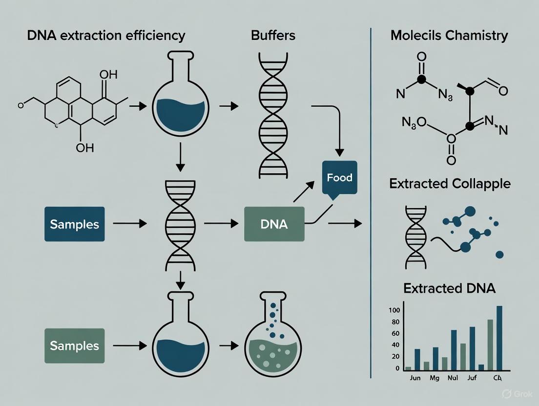
Optimizing DNA Extraction for Enhanced Allergen Detection in Processed Foods: A Guide for Researchers and Developers
This article provides a comprehensive resource for researchers and drug development professionals focused on overcoming the significant challenge of DNA degradation and the presence of PCR inhibitors during the extraction...
Optimizing DNA Extraction for Enhanced Allergen Detection in Processed Foods: A Guide for Researchers and Developers
Abstract
This article provides a comprehensive resource for researchers and drug development professionals focused on overcoming the significant challenge of DNA degradation and the presence of PCR inhibitors during the extraction of genetic material from processed foods for allergen detection. It explores the foundational impact of food processing on DNA integrity, details current and emerging methodological approaches for efficient DNA recovery, outlines targeted optimization and troubleshooting strategies for complex matrices, and discusses the critical role of validation and comparative analysis in ensuring method reliability. By synthesizing recent scientific advances, this review aims to guide the development of robust, sensitive, and accurate DNA-based assays, ultimately contributing to improved food safety and the protection of allergic consumers.
The DNA Extraction Challenge: How Food Processing Impacts Allergen Detectability
The Growing Public Health Concern of Food Allergies and Need for Accurate Detection
FAQs: Troubleshooting DNA-Based Allergen Detection
Q1: Why is my PCR detection failing for highly processed foods like baked goods? DNA degradation during high-temperature processing is a primary cause. As processing intensity increases, genomic DNA breaks down, making amplification of long DNA fragments difficult. For reliable detection in processed foods, target short, specific allergen gene fragments of approximately 200-300 base pairs [1]. Using shorter amplicons significantly improves the success rate for samples exposed to high heat.
Q2: How does food processing impact my choice between protein-based and DNA-based detection methods? DNA-based PCR methods are often more reliable for processed foods because DNA is more stable than proteins under harsh conditions like high heat. Protein structures can be damaged, altering their epitopes and making them undetectable by immunological methods like ELISA. However, the extraction of intact DNA must also be optimized for the specific food matrix [2].
Q3: What are the critical factors for optimizing DNA extraction from complex, challenging matrices? The key is the extraction buffer composition. Factors such as buffer pH, high salt concentration (e.g., 1 M NaCl), and the addition of additives like fish gelatine (to prevent non-specific binding) and Polyvinylpyrrolidone (PVP, to bind and remove polyphenols) are crucial for efficient DNA recovery from complex foods like chocolate or baked biscuits [3]. A 1:10 sample-to-buffer ratio is a common starting point [3].
Q4: My allergen recovery is low from chocolate-based matrices. How can I improve it? Chocolate is a notoriously challenging matrix due to interfering compounds like polyphenols and high fat content. To optimize recovery, use an extraction buffer containing additives such as 1% PVP to bind polyphenols and 10% fish gelatine. Even with optimized buffers, recovery from chocolate matrices may be lower than from other foods, so method validation is essential [3].
Experimental Protocol: DNA Extraction and PCR Detection for Processed Foods
This protocol is adapted from published research for detecting wheat and maize allergens in baked goods [1].
1. Sample Preparation and DNA Extraction (CTAB-based method)
- Sample Homogenization: Grind 100 mg of the processed food sample (e.g., baked crispbread) to a flour-like consistency using an electric grinder [1].
- Cell Lysis: Incubate the sample with CTAB buffer and proteinase K at 65°C [1].
- RNA Removal: Treat the lysate with RNase A [1].
- Purification: Perform chloroform extraction to separate proteins and other contaminants from the DNA [1].
- DNA Precipitation: Precipitate DNA using isopropanol, wash the pellet with 70% ethanol, air-dry, and finally re-suspend in 100 μL sterile deionized water [1].
- Quality Control: Assess DNA concentration and purity using a UV-Vis spectrophotometer (e.g., NanoDrop). Check DNA integrity via 1% agarose gel electrophoresis [1].
2. PCR Amplification of Allergen Genes
- Primer Design: Design primers to target short, specific fragments (~200-300 bp) of the allergen genes of interest. For example:
- Wheat: High-molecular-weight glutenin subunit (HMW-GS) and low-molecular-weight glutenin subunit (LMW-GS) genes.
- Maize: Zea m 14, Zea m 8, and zein genes [1].
- PCR Setup: Use standard PCR reagents and a thermal cycler. The cycling conditions will depend on the primer design and polymerase used.
- Analysis: Analyze PCR products using agarose gel electrophoresis to confirm successful amplification of the target fragments [1].
Data Presentation: Impact of Processing on DNA Detection
Table 1: Detectability of Allergen Genes in Wheat and Maize After Baking [1]
| Allergen Source | Target Gene | Baking Temperature | Maximum Detectable Baking Time |
|---|---|---|---|
| Wheat | HMW-GS / LMW-GS | 220°C | 60 minutes |
| Maize | Zea m 14, Zea m 8, Zein | 220°C | 40 minutes |
Table 2: Optimized Extraction Buffer Compositions for Challenging Food Matrices [3]
| Buffer Component | Function | Example Composition 1 | Example Composition 2 |
|---|---|---|---|
| Buffer Base | pH control, protein stability | 50 mM Carbonate Bicarbonate | PBS |
| Salt (NaCl) | Increases ionic strength, disrupts matrix interactions | - | 1 M |
| Detergent (Tween) | Aids in solubilizing fats and proteins | - | 2% |
| Fish Gelatine | Blocks non-specific binding | 10% | 10% |
| PVP | Binds and removes polyphenols (e.g., in chocolate) | - | 1% |
| Recommended For | - | General allergen recovery | Complex matrices (chocolate, baked) |
The Scientist's Toolkit: Research Reagent Solutions
Table 3: Essential Reagents for Allergen DNA Extraction and Detection
| Reagent / Material | Function in the Protocol |
|---|---|
| CTAB (Cetyltrimethyl ammonium bromide) | A detergent used in the lysis buffer to disrupt cell membranes and facilitate DNA release [1]. |
| Proteinase K | An enzyme that degrades proteins and nucleases, helping to purify DNA [1]. |
| Chloroform | Used for liquid-phase separation to remove proteins and other contaminants from the DNA solution [1]. |
| Isopropanol | A solvent used to precipitate DNA from the aqueous solution [1]. |
| RNase A | An enzyme that degrades RNA, preventing it from contaminating the final DNA extract [1]. |
| Fish Gelatine | A protein-based additive in extraction buffers that blocks non-specific binding sites, improving allergen recovery in immunoassays; likely aids in DNA extraction from complex matrices by similar mechanism [3]. |
| Polyvinylpyrrolidone (PVP) | A compound that binds to polyphenols (common in chocolate, fruits), preventing them from interfering with DNA and co-purifying [3]. |
| Allergen-Specific PCR Primers | Short, custom-designed DNA sequences that bind to and allow amplification of a specific allergen gene fragment (e.g., for HMW-GS, Zea m 14) [1]. |
Experimental Workflow and Signaling Pathways
DNA Extraction and PCR Detection Workflow
IgE-Mediated Food Allergy Pathway
FAQ: How does thermal processing damage DNA and affect its detection?
Answer: Thermal processing damages DNA through several mechanisms that directly impact its integrity and your ability to amplify it. The primary damage types are:
- Strand Breaks and Fragmentation: Heat causes the cleavage of the phosphodiester bonds in the DNA backbone. This physically breaks the long DNA strands into smaller fragments. The intensity and duration of heating correlate with the degree of fragmentation [4].
- Depurination: High temperatures can cause the loss of purine bases (adenine and guanine) from the DNA backbone. This creates apurinic sites that are unstable and can lead to subsequent strand breakage [5].
- Deamination: Heat can induce the hydrolytic deamination of cytosine to uracil. This results in a change of the base identity (from a C-G base pair to a T-A base pair), potentially introducing errors during PCR amplification [6].
The direct consequence for your experiments is that the longer the DNA target you are trying to amplify (amplicon), the more likely it is that one of these damage events has occurred within that stretch of DNA, preventing successful PCR.
FAQ: My PCR is failing for a heat-processed food sample. What is the first parameter I should check?
Answer: The first and most critical parameter to check is the amplicon size of your PCR assay. Thermal processing fragments DNA, making long target sequences unrecoverable.
- Solution: Re-design your PCR assays to target shorter DNA sequences, ideally below 200 base pairs (bp), and for highly processed foods, below 100 bp is often necessary [4] [2]. Ensure that both the transgenic/allergen-specific target and the endogenous reference gene target have similar, short amplicon lengths. This ensures they degrade in parallel, allowing for reliable relative quantification [4].
FAQ: Can I still accurately quantify DNA from a heavily processed sample?
Answer: Yes, reliable relative quantification is possible if your assay is properly designed. Research shows that although absolute DNA recovery decreases significantly with intense processing (e.g., autoclaving, UV irradiation), the measured ratio between a transgenic or allergen target and an endogenous reference gene remains accurate [4]. This is because both sequences degrade at a similar rate. The key, as noted above, is using short, similarly-sized amplicons for both targets.
FAQ: Besides amplicon length, what other PCR conditions can I optimize for damaged DNA?
Answer: Optimizing your thermal cycling protocol can improve the sensitivity of detecting degraded DNA.
- Annealing Temperature: Use stringent annealing temperatures (generally 55–72°C), as they enhance discrimination against incorrectly annealed primers, which is more common with fragmented DNA templates [7].
- Polymerase and Extension Time: For short amplicons (e.g., < 2 kb), an extension time of one minute at 72°C is typically sufficient. The rate of nucleotide incorporation can vary from 35 to 100 nucleotides per second depending on buffer conditions [7].
- Denaturation: Avoid excessively long or hot denaturation steps (e.g., >95°C for long periods), as this can unnecessarily degrade the enzyme and further damage the already-fragile DNA template [7].
Table 1: Impact of Different Processing Treatments on DNA Recovery
| Processing Treatment | Impact on DNA Recovery | Key Experimental Finding |
|---|---|---|
| Autoclaving | Severe degradation, least DNA recovery | Profound fragmentation; requires very short amplicons for detection [4]. |
| UV Irradiation | Severe degradation, least DNA recovery | Causes significant DNA damage, similar to autoclaving [4]. |
| Baking / Dry Heat | Moderate to severe degradation | DNA recovery decreases with increasing temperature and duration [4]. |
| Microwaving | Moderate degradation | Can cause significant fragmentation depending on power and time [4]. |
Table 2: Key Reagent Solutions for DNA Analysis in Processed Foods
| Research Reagent | Function / Explanation | Application Note |
|---|---|---|
| Squish Buffer (with high salt) | Lysis buffer for bulk DNA extraction; high salt concentration (e.g., 125 mM NaCl) improves DNA yield and purity from complex samples [8]. | Essential for efficient extraction from difficult matrices like insect parts or processed food. |
| RNase A | Enzyme that degrades RNA. | Used during DNA extraction to remove RNA, reducing sample viscosity and potential PCR interference, leading to cleaner DNA and lower Cq values [8]. |
| Caffeine | Additive in DNA extraction buffers. | Improves DNA yield in real-time PCR applications when added to the squish buffer [8]. |
| Paramagnetic Beads | Used for post-lysis DNA purification. | Binds DNA, allowing impurities to be washed away. Can significantly increase end Relative Fluorescent Units (RFUs) in real-time PCR but adds cost and time [8]. |
| Taq DNA Polymerase | Thermostable enzyme for PCR amplification. | Active over a broad temperature range; its concentration can become critical with rapid cycling protocols [7] [9]. |
Experimental Protocol: Assessing DNA Degradation from Thermal Processing
Title: Protocol for Quantifying DNA Degradation in a Processed Food Model Using Real-Time PCR.
Background: This protocol simulates the effect of various food processing treatments on a dual-target plasmid to systematically evaluate DNA degradation and its impact on the quantitative detection of a specific gene sequence, such as an allergen marker.
Materials:
- Model System: Dual-target plasmid (e.g., pRSETMON-02) harboring two sequences in tandem (e.g., a 359 bp allergen-specific fragment and a 200 bp endogenous reference gene) in a 1:1 ratio [4].
- Equipment: Thermal cycler for real-time PCR, nanodrop or Qubit for DNA quantification, equipment for processing treatments (autoclave, microwave, UV cross-linker, dry bath).
- Reagents: Real-time PCR master mix (e.g., iTaq Universal Probes Supermix), specific primers and probes for short (e.g., 64 bp, 84 bp) and long (e.g., 356 bp) amplicons within the plasmid insert [4].
Methodology:
- Processing Treatments: Subject aliquots of the pure plasmid DNA to various processing treatments:
- Thermal: Autoclaving (e.g., 121°C for 20 min), baking (e.g., 95°C for 30 min), microwaving.
- Radiation: UV irradiation at a defined energy level [4].
- Include an untreated plasmid control.
- DNA Analysis: Dilute all processed and control DNA samples to the same concentration.
- Real-Time PCR Quantification:
- Perform real-time PCR on all samples using primer/probe sets for the short and long amplicons for both the allergen and reference gene targets.
- Run reactions in triplicate.
- Data Analysis:
- Record the quantification cycle (Cq) for each reaction. A higher Cq indicates greater DNA degradation of that specific target.
- Calculate the mean DNA copy number ratio (allergen gene / reference gene) for each processing condition.
- Compare the ratios and absolute Cq values of processed samples to the untreated control.
Expected Outcome: The recovery of longer amplicons (356 bp) will be significantly reduced compared to shorter amplicons (64/84 bp) in processed samples. However, the calculated mean DNA copy number ratio between the allergen and reference gene should remain consistent with the expected 1:1 ratio, demonstrating that accurate relative quantification is possible despite degradation [4].
Visualizing the Concepts
Diagram 1: DNA Degradation and Solution Pathway
Diagram 2: Reliable DNA Detection Workflow
For researchers focused on allergen detection in processed foods, obtaining high-quality DNA is a foundational step. The stability of DNA makes it a superior target for detecting allergenic ingredients like peanuts, soy, or shellfish in complex food matrices. However, food processing techniques—both thermal and non-thermal—can severely degrade DNA, compromising the sensitivity and accuracy of downstream molecular assays such as PCR and DNA barcoding. This technical support center provides targeted troubleshooting guides and FAQs to help you navigate the challenges of extracting analyzable DNA from processed foods, thereby enhancing the efficiency and reliability of your research.
Troubleshooting Guides
Common Problems and Solutions in DNA Extraction from Processed Foods
Problem: Low DNA Yield
| PROBLEM | CAUSE | SOLUTION |
|---|---|---|
| Low DNA Yield | Degradation from extensive mechanical/thermal processing [10] | Optimize sample input; use larger starting material if DNA is highly fragmented [10]. |
| Polysaccharide/polyphenol co-precipitation inhibiting extraction [10] | Use extraction buffers with additives like CTAB or PVP to bind and remove contaminants [10] [11]. | |
| Silica membrane clogged by indigestible tissue fibers [12] | Centrifuge lysate at max speed for 3 min before column loading to pellet fibers [12]. | |
| DNA Degradation | Sample not stored properly post-processing [12] | Flash-freeze samples in liquid nitrogen and store at -80°C; use stabilizing reagents [12]. |
| Activity of endogenous nucleases in raw material [12] | Process samples quickly on ice; use lysis buffers with chelating agents [12]. | |
| Acid-catalyzed hydrolytic destruction during processing [10] | Neutralize acidic samples with appropriate buffers early in the extraction protocol [10]. | |
| Protein Contamination | Incomplete digestion of the sample [12] | Extend Proteinase K digestion time by 30 mins to 3 hours after tissue dissolves [12]. |
| Membrane clogged with tissue fibers [12] | Centrifuge lysate to remove fibers; reduce input material for fibrous tissues [12]. | |
| PCR Inhibition | Carry-over of PCR inhibitors (polysaccharides, polyphenols, salts) [10] | Include additional wash steps; use silica-column based purification over traditional methods [10] [13]. |
| Inadequate purification post-extraction [10] | Perform a pre-wash of the sample or use a kit designed for complex matrices [11]. |
Problem: DNA Degradation
Problem: Protein Contamination
Problem: PCR Inhibition
Frequently Asked Questions (FAQs)
1. Why is DNA quality from processed foods so variable, and what are the main processing factors that affect it? DNA quality is highly variable because it is affected by a combination of processing steps. Thermal processing (e.g., baking, retorting) causes strand breakage and depurination [10]. Chemical processing, such as exposure to acidic conditions in fruit juices, leads to hydrolytic DNA destruction [10]. Mechanical processing (e.g., blending, homogenization) shears DNA into smaller fragments. The cumulative effect of these processes determines the final fragment size and purity of the DNA, which directly impacts the success of PCR amplification [10].
2. For a highly processed product like Chestnut rose juice, which DNA extraction method is most effective? A comparative study on Chestnut rose juices and beverages found that a combination method, often involving aspects of both CTAB and silica-column purification, showed the greatest performance despite being more time-consuming and costly. This method outperformed a non-commercial modified CTAB method (which had high yield but poor purity) and other commercial kits in terms of DNA quality and amplifiability in qPCR [10].
3. My downstream PCR assay for a peanut allergen is failing. Should I switch to a protein-based method like ELISA? Not necessarily. While food processing also affects proteins, DNA-based methods retain significant advantages. DNA is more stable than proteins during food processing and extraction [14]. Furthermore, DNA-based methods like PCR can be highly sensitive and specific, and are not as strongly misled by cross-reactivity with other nuts as some immunoassays can be [14]. The solution often lies in optimizing the DNA extraction and purification to remove PCR inhibitors, rather than abandoning the DNA-based approach.
4. We work with novel feed ingredients like insect hydrolysates. Are standard DNA extraction kits sufficient? Novel ingredients often require validated protocols. A study on processed animal by-products (including hydrolysates) found that the conventional CTAB-based method and the commercial kits Invisorb Spin Tissue Mini and NucleoSpin Food demonstrated superior extraction efficiency and DNA quality ratios. Commercial kits generally enable faster processing, but the CTAB method can be optimized for specific, complex matrices [11].
5. How can I quickly assess the quality and extent of degradation of my extracted DNA? Beyond spectrophotometric measurements (A260/A280), you can use gel electrophoresis to visually check for DNA smearing (indicating degradation) versus distinct high-molecular-weight bands. A more quantitative approach is to use TaqMan real-time PCR with primers that generate amplicons of different sizes. A significant drop in amplification efficiency with longer amplicons is a clear indicator of DNA fragmentation, helping you assess the utility of the DNA for your intended assay [10].
Experimental Protocols
Protocol 1: Combined CTAB and Silica-Column Method for Processed Plant-Based Foods (e.g., Juices, Jams)
This protocol, adapted from research on Chestnut rose juices, is designed for challenging, polysaccharide-rich matrices [10].
- Sample Preparation: Homogenize 100-200 mg of the sample (or 1-2 mL for liquids after centrifugation) using a sterile pestle.
- Lysis: Add 1 mL of pre-warmed CTAB extraction buffer (2% CTAB, 100 mM Tris-HCl pH 8.0, 20 mM EDTA, 1.4 M NaCl, 0.2% β-mercaptoethanol added fresh) and 20 µL of Proteinase K (20 mg/mL). Mix by vortexing and incubate at 65°C for 60-120 minutes with occasional gentle mixing.
- Decontamination: Add an equal volume of Chloroform:Isoamyl Alcohol (24:1). Mix thoroughly by inversion for 10 minutes. Centrifuge at 12,000 x g for 15 minutes at room temperature.
- DNA Precipitation: Carefully transfer the upper aqueous phase to a new tube. Add 0.7 volumes of isopropanol, mix by inversion, and incubate at -20°C for 30 minutes. Centrifuge at 12,000 x g for 15 minutes to pellet the DNA.
- Wash and Resuspend: Wash the pellet with 1 mL of 70% ethanol. Centrifuge again, carefully discard the ethanol, and air-dry the pellet. Resuspend the DNA in 50-100 µL of TE buffer or nuclease-free water.
- Further Purification (Combination Step): Apply the resuspended DNA to a silica-column from a commercial kit (e.g., NucleoSpin Food). Follow the manufacturer's protocol for binding, washing, and elution. This step helps remove any remaining PCR inhibitors [10] [11].
Protocol 2: Commercial Kit-Based Extraction for Highly Processed Animal By-Products
This protocol is validated for novel ingredients like animal meals and hydrolysates [11].
- Sample Input: Use 60-200 mg of the processed sample.
- Recommended Kits: Invisorb Spin Tissue Mini Kit or NucleoSpin Food Kit.
- Procedure: Follow the manufacturer's instructions precisely. For difficult-to-lyse samples, the protocol can be modified by:
- Extending Lysis Time: Increase the incubation time with lysis buffer and Proteinase K.
- Initial Homogenization: For solid samples, an initial homogenization step with liquid nitrogen may improve yield [11].
- Elution: Elute the purified DNA in a small volume (e.g., 50-100 µL) to increase the final DNA concentration.
Workflow Visualization
The following diagram illustrates the critical decision-making process for selecting and optimizing a DNA extraction method based on the sample's processing history.
The Scientist's Toolkit: Research Reagent Solutions
| Reagent / Kit | Function / Application |
|---|---|
| CTAB (Cetyltrimethylammonium bromide) | A cationic detergent effective in lysing cells and precipitating polysaccharides while co-precipitating DNA. Crucial for plant-based and polysaccharide-rich processed foods [10] [11]. |
| Proteinase K | A broad-spectrum serine protease used to digest proteins and degrade nucleases that could otherwise degrade DNA during extraction. Essential for all sample types, especially tissues [12]. |
| Silica-based Spin Columns (e.g., from NucleoSpin Food kit) | The core of many commercial kits; DNA binds to the silica membrane in the presence of high salt, allowing impurities to be washed away, resulting in high-purity DNA [11] [13]. |
| Polyvinylpyrrolidone (PVP) | Used to bind and remove polyphenols during extraction, preventing them from co-purifying with DNA and inhibiting downstream PCR. Important for plant and juice samples [10]. |
| RNase A | An enzyme that degrades RNA, preventing RNA contamination from affecting DNA quantification and downstream applications [12]. |
| Chelex-100 Resin | A chelating resin that binds metal ions, inhibiting nucleases. Used in rapid, low-cost boiling methods, though with lower purity than column-based methods [13]. |
The reliable detection of food allergens in processed foods is critically dependent on the efficiency of DNA extraction and the subsequent performance of the polymerase chain reaction (PCR). Complex food matrices often contain inherent compounds that potently inhibit enzymatic reactions, leading to false-negative results and compromising food safety assessments. Among these, polyphenols and polysaccharides represent two of the most pervasive and challenging classes of PCR inhibitors. These compounds can co-purify with DNA during extraction, interfering directly with DNA polymerase activity and preventing the amplification of target allergen genes [15] [16]. This guide provides targeted troubleshooting strategies to help researchers overcome these barriers, thereby improving the accuracy and sensitivity of DNA-based allergen detection methods.
Understanding the Inhibitors
To effectively troubleshoot, it is essential to understand the nature and source of common inhibitors.
- Polyphenols: These compounds are abundant in plant tissues and are readily oxidized during cell lysis, often forming complexes with nucleic acids. This binding can render DNA insoluble or inhibit polymerase enzymes directly. Contamination with polyphenols can often be identified by a brownish discoloration in the purified DNA sample [15] [16].
- Polysaccharides: Acidic polysaccharides are particularly problematic as they are structurally similar to nucleic acids and thus co-precipitate during isolation. They are among the strongest PCR inhibitors and can impart a sticky, gelatinous texture to the DNA extract, affecting downstream applications like restriction digestion and PCR [15].
Table 1: Common PCR Inhibitors in Food Matrices
| Inhibitor Class | Common Sources | Impact on PCR | Visible Clues in DNA Extract |
|---|---|---|---|
| Polyphenols | Grape, birch (Betula), chocolate, woody plants, medicinal plants [15] [16] | Bind to nucleic acids and enzymes; inhibit polymerase activity [15] [16] | Brownish color [15] |
| Polysaccharides | Grape, birch, maize, processed cereals [15] | Co-purify with DNA; interfere with polymerases, ligases, and restriction enzymes [15] | Sticky, gelatinous consistency; brown color [15] |
| Proteins | Various food matrices (e.g., milk, eggs) | Can co-purify and inhibit enzyme active sites [15] | - |
| Chaotropic Salts | Carryover from silica-based purification kits [16] | Inhibit polymerase activity [16] | - |
Troubleshooting Guide: Common Issues and Solutions
This section addresses specific experimental problems related to inhibitor carryover.
Issue 1: No Amplification or Low Yield
Possible Causes:
- Carryover of PCR inhibitors like polyphenols or polysaccharides in the DNA template [17] [18].
- Degraded or insufficient DNA template quantity [17].
- Suboptimal PCR reaction conditions.
Recommendations:
- Re-purify DNA: Precipitate and wash the DNA with 70% ethanol to remove residual salts and inhibitors [17].
- Use Inhibitor-Robust Enzymes: Select DNA polymerases with high processivity and tolerance to inhibitors commonly found in plant tissues and processed foods [17].
- Apply PCR Additives: Incorporate additives like * Bovine Serum Albumin (BSA)* or betaine into the PCR mix. BSA can bind to inhibitors, reducing their impact on the polymerase, while betaine can help denature GC-rich templates and stabilize the reaction [18].
- Validate Template: Check DNA concentration and integrity using gel electrophoresis to rule out degradation [17].
Issue 2: Non-Specific Amplification or Primer-Dimer Formation
Possible Causes:
- PCR conditions are not stringent enough, often due to low annealing temperatures or excess magnesium [17] [18].
- High primer concentration [18].
- Use of a non-hot-start DNA polymerase, allowing activity at room temperature [17] [18].
Recommendations:
- Optimize Annealing Temperature: Increase the annealing temperature stepwise in 1–2°C increments. Use a gradient thermal cycler to determine the optimal temperature, which is typically 3–5°C below the primer's Tm [17] [18].
- Optimize Mg²⁺ Concentration: Review and lower the Mg²⁺ concentration to prevent nonspecific products [17] [18].
- Use Hot-Start Polymerases: Employ hot-start DNA polymerases that remain inactive until a high-temperature activation step, preventing nonspecific priming and primer-dimer formation at lower temperatures [17] [18].
Issue 3: Smeared Bands on Agarose Gel
Possible Causes:
- Degraded DNA template [17] [18].
- Contamination from previous PCR amplifications ("carryover contamination") [18].
- Excessively long extension times or low annealing temperatures [18].
Recommendations:
- Assess DNA Integrity: Evaluate the template DNA by gel electrophoresis before PCR. Use intact, high-quality DNA [17].
- Prevent Contamination: Implement strict laboratory practices, including physical separation of pre- and post-PCR areas and using dedicated equipment and reagents [18].
- Optimize Cycling Conditions: Shorten the extension time and/or increase the annealing temperature [18].
Table 2: Summary of Troubleshooting Solutions
| Problem | Solution Category | Specific Action |
|---|---|---|
| No/Low Yield | DNA Template | Re-purify DNA (ethanol precipitation); use inhibitor-tolerant polymerases [17] |
| PCR Additives | Add BSA or betaine to the reaction mix [18] | |
| Non-Specific Bands | Reaction Conditions | Increase annealing temperature; optimize Mg²⁺ concentration [17] [18] |
| Enzyme Choice | Switch to a hot-start DNA polymerase [17] [18] | |
| Primer-Dimer | Primer Design & Concentration | Re-design primers to avoid 3'-end complementarity; optimize primer concentration [18] |
| Smeared Bands | Laboratory Practice | Decontaminate workspace; use physical separation of pre- and post-PCR areas [18] |
| Template & Conditions | Use intact DNA template; optimize cycling conditions [17] [18] |
Optimized Experimental Protocols
CTAB-Based DNA Extraction Protocol for Recalcitrant Plant Tissues
This protocol is specifically designed to remove polyphenols and polysaccharides and has been successfully applied to difficult samples like birch and grape [15].
Buffers:
- Buffer 1: 200 mM Tris-HCl, 1.4 M NaCl, 0.5% (v/v) Triton X-100, 3% (w/v) CTAB, 0.1% (w/v) PVP (add PVP just before use).
- Buffer 2: 50 mM Tris-HCl, 2 M guanidine thiocyanate, 0.2% (v/v) mercaptoethanol (add before use), 0.2 mg/mL Proteinase K (add before use).
Procedure:
- Homogenization: Grind 50 mg of leaf or root tissue in a 2 mL tube.
- Initial Lysis: Add 400 µL of Buffer 1, vortex for 20 seconds, and incubate at 60°C for 30 minutes.
- De-proteinization: Add 400 µL chloroform-isoamyl alcohol (24:1, v/v), shake vigorously for 2 minutes, and centrifuge at 10,000 rpm for 15 minutes.
- Supernatant Transfer: Transfer 300 µL of the upper aqueous phase to a fresh 2 mL tube.
- Enzymatic Digestion: Add 150 µL (1/2 volume) of Buffer 2 and incubate at 40°C for 15 minutes.
- Polysaccharide Precipitation: Add 1/2 volume of 4 M NaCl, shake, and place the tube on ice for 5 minutes.
- DNA Precipitation: Add 2 volumes of cold isopropanol and leave at room temperature for 2 minutes. Centrifuge at 8,000 rpm for 15 minutes to pellet the DNA.
- Wash: Discard the supernatant. Gently wash the pellet with 75% (v/v) ethanol, wait 2 minutes, and centrifuge at 8,000 rpm for 2 minutes.
- Dissolution: Dry the pellet and dissolve the DNA in 100 µL TE buffer. Incubate at 70°C for 10 minutes to ensure complete dissolution [15].
Workflow for DNA Extraction and Allergen Detection in Processed Foods
The following diagram illustrates the logical workflow from sample preparation to detection, highlighting key steps for overcoming inhibition.
The Scientist's Toolkit: Essential Research Reagents
Selecting the right reagents is fundamental to successfully extracting inhibitor-free DNA and achieving robust PCR amplification.
Table 3: Key Reagent Solutions for Overcoming PCR Inhibition
| Reagent / Tool | Function / Purpose | Specific Examples / Notes |
|---|---|---|
| CTAB (Cetyltrimethylammonium bromide) | A cationic detergent that facilitates cell lysis and forms complexes with polysaccharides to prevent their solubilization [15] [1]. | Used in high-salt extraction buffers (e.g., 1.4 M NaCl) [15]. |
| PVP (Polyvinylpyrrolidone) | Binds to and neutralizes polyphenols, preventing their oxidation and complexation with DNA [15] [3]. | Often added to extraction buffers at 0.1-1% concentration [15] [3]. |
| Proteinase K | Broad-spectrum serine protease that degrades contaminating enzymes and other proteins [15]. | Used to remove nucleases and other proteins [15]. |
| High-Salt Solutions | Prevents the co-solubilization of acidic polysaccharides with DNA [15]. | 1.4 M NaCl in lysis buffer; 4 M NaCl for post-lysis precipitation [15]. |
| Inhibitor-Tolerant DNA Polymerases | Engineered enzymes with high affinity for DNA templates, resistant to common plant and food-derived inhibitors [17] [16]. | KOD One Master Mix; polymerases marketed for high processivity and tolerance [17] [16]. |
| PCR Additives | Compounds that counteract inhibitors or improve amplification efficiency of difficult targets. | BSA: Binds to inhibitors [18]. Betaine: Destabilizes secondary structures [17] [18]. |
Frequently Asked Questions (FAQs)
Q1: My DNA extract from chocolate is brown. What does this mean, and what should I do? A brown color strongly suggests contamination with polyphenols, which are abundant in cocoa [3]. You should re-extract the DNA using a protocol that includes PVP in the lysis buffer to bind these compounds. Additionally, consider using a silica-column based purification kit designed for complex matrices, as these can be more effective than CTAB alone for certain samples [16].
Q2: How does food processing affect DNA-based allergen detection? Food processing, especially thermal treatments like baking, boiling, and autoclaving, causes DNA fragmentation and degradation [19] [1]. This degradation limits the size of the DNA target that can be amplified. To ensure detection in processed foods, design your PCR assays to amplify short target sequences (100-200 bp) [1]. Chloroplast DNA targets can sometimes offer an advantage due to their multiple copies per cell [19].
Q3: I've followed an optimized protocol, but my PCR still fails. What are my next steps? First, systematically verify each component:
- Test DNA Quality: Run a fresh aliquot of your DNA on a gel to confirm it is not degraded.
- Check Reagents: Prepare fresh working stocks of PCR reagents, particularly dNTPs and primers.
- Use a Control Template: Perform a PCR with a known, control DNA template and a separate set of validated primers. If this control works, the issue lies with your sample DNA or the allergen-specific primers. If it fails, the problem is likely in your PCR master mix or cycling conditions [18].
- Consider Advanced Methods: For absolute quantification and superior tolerance to inhibitors, explore digital PCR (dPCR), which has been shown to improve sensitivity for allergenic foods like sesame by an order of magnitude compared to real-time PCR [20].
Core Concepts: Proteins, Genes, and Detection
What is the fundamental relationship between an allergenic protein and its encoding gene?
An allergenic protein is a specific molecule, typically a protein, that triggers an abnormal immune response in sensitized individuals. The genetic information for producing this protein is contained within a specific gene—a sequence of DNA. The gene is transcribed into messenger RNA (mRNA), which is then translated into the amino acid sequence that forms the allergenic protein. Therefore, detecting the gene provides an indirect, yet highly reliable, method for identifying the potential presence of the allergenic protein itself [21].
Why target DNA for allergen detection in processed foods?
Targeting DNA is particularly advantageous for detecting allergens in processed foods. While the structure and detectability of allergenic proteins can be damaged or altered by factors such as heat, pressure, or chemical treatments during food processing, DNA is often more stable and retains its molecular integrity under these conditions [2]. In such cases, DNA-based detection provides an effective and reliable alternative when protein-based immunological assays may fail [2].
What are the major allergenic foods, and what are some key allergen examples?
Certain foods are responsible for the majority of allergic reactions. A foundational group, often referred to as the 'Big 8', includes peanuts, eggs, milk, soy, wheat/cereals containing gluten, crustaceans, fish, and tree nuts [21]. Key allergen genes and their proteins have been extensively studied for many of these. For example:
- Peanuts: Ara h 2 and Ara h 6 are two of the most potent allergens, belonging to the prolamin superfamily (2S albumin family) [22]. Their genes have been explored across various Arachis species, revealing natural mutations that can reduce allergenicity [22].
- Wheat & Gluten: Celiac disease is triggered by an autoimmune response to gluten proteins, which is distinct from a wheat allergy, though both involve specific protein fractions and their encoding genes [21].
Methodologies & Data Comparison
The following table summarizes the primary methods used for allergen detection, highlighting the role of DNA-based techniques [2] [21].
Table 1: Comparison of Major Allergen Detection Methods
| Method Type | Principle | Target | Advantages | Limitations |
|---|---|---|---|---|
| Protein-Based (e.g., ELISA, Lateral Flow) | Immunological binding of antibodies to specific protein epitopes [21]. | Allergenic Protein | Directly detects the causative agent; high sensitivity and specificity; well-standardized [2]. | Protein structure can be denatured during processing, leading to false negatives [2]. |
| DNA-Based (e.g., PCR) | Amplification of specific DNA sequences unique to the allergenic source [21]. | Allergen-Encoding Gene | DNA is more stable in processed foods; highly specific and sensitive [2]. | Does not directly quantify the protein; results may not always correlate with protein amount [2]. |
| Biosensors | Biological recognition element (e.g., antibody, aptamer) coupled to a signal transducer [2]. | Protein or Gene | Potential for rapid, on-site, and high-throughput analysis [2]. | Still emerging technology; can be complex to develop and validate [2]. |
Detailed Protocol: Real-Time PCR for Allergen Detection
This protocol is adapted from methods described for detecting allergenic foods such as lobster, fish, and nuts [2].
Principle: Real-time PCR (Polymerase Chain Reaction) allows for the detection and quantification of specific DNA sequences. It monitors the amplification of a target gene in real time using a fluorescent reporter, allowing researchers to determine the presence and quantity of an allergen-encoding gene in a sample.
Workflow:
The following diagram illustrates the key steps in the DNA-based detection workflow, from sample preparation to final analysis.
Materials & Reagents:
- Sample: Processed food product (e.g., chocolate, cookie, sauce).
- Lysis Buffer: For breaking down cells and releasing DNA.
- DNA Purification Kit: For isolating high-quality DNA from the complex food matrix.
- Primers and Probes: Sequence-specific oligonucleotides designed to bind to a unique region of the target allergen gene (e.g., Ara h 2 or a fish parvalbumin gene). These are the core reagents that define the assay's specificity.
- Real-Time PCR Master Mix: Contains DNA polymerase, dNTPs, buffer, and salts necessary for the amplification reaction. Often includes a fluorescent dye (e.g., SYBR Green) or is compatible with dual-labeled probes (TaqMan).
- Real-Time PCR Instrument: The equipment used to perform thermal cycling and fluorescence detection.
Procedure:
- DNA Extraction: Homogenize the food sample. Extract genomic DNA using a validated purification method, ensuring the removal of inhibitors (e.g., polyphenols, fats) that can affect PCR efficiency. Quantify and assess the purity of the extracted DNA.
- Reaction Setup: Prepare reactions in a 96-well plate or strips. A typical reaction mix includes:
- Real-Time PCR Master Mix: 10 µL
- Forward Primer (specific to target gene): 0.5 µL
- Reverse Primer (specific to target gene): 0.5 µL
- Probe (if using a TaqMan assay): 0.5 µL
- Template DNA (extracted sample): 2-5 µL
- Nuclease-Free Water: to a final volume of 20 µL
- Include appropriate controls: no-template control (NTC), positive control (DNA from known allergenic source), and potentially an internal amplification control.
- Thermal Cycling: Place the plate in the real-time PCR instrument and run a program similar to:
- Initial Denaturation: 95°C for 2-5 minutes.
- 40-50 Cycles of:
- Denaturation: 95°C for 15-30 seconds.
- Annealing/Extension: 60°C for 30-60 seconds (acquire fluorescence at this step).
- Data Analysis: Analyze the amplification curves. The cycle threshold (Ct) value, at which the fluorescence exceeds a background threshold, is used for qualitative detection or quantitative estimation of the target DNA concentration.
Troubleshooting FAQs
Q: We are getting inconsistent results between our DNA-based and protein-based allergen tests on the same processed food sample. What could be the cause?
A: This is a common challenge. The most likely cause is the differential impact of food processing on the targets.
- Scenario 1 (Positive DNA / Negative Protein): The food has been subjected to high heat or harsh chemicals, which denatured the allergenic protein so that antibodies can no longer bind to it. However, the DNA, while potentially fragmented, remains amplifiable for a detectable target sequence. This suggests the allergenic ingredient was present, but the protein's immunoreactivity has been destroyed [2].
- Scenario 2 (Unexpected Quantification Discrepancy): The number of gene copies in the raw material may not always directly correlate with the concentration of the expressed protein. Furthermore, DNA degradation can occur with extreme processing, potentially leading to an underestimation. Always ensure your DNA extraction method is optimized for your specific food matrix to recover sufficient, amplifiable DNA.
Q: Our PCR assays are failing, showing no amplification even in positive controls. What are the first steps in troubleshooting?
A: A systematic approach is key.
- Check Reagent Integrity: Ensure primers and master mix have been stored correctly and are not expired. Prepare fresh aliquots if necessary.
- Verify DNA Quality: Re-evaluate the extracted DNA. Is the concentration sufficient? Is it degraded (check on a gel)? Are there contaminants (assess A260/A280 ratio)? Re-purify the DNA if needed.
- Inspect Thermal Cycler Protocol: Confirm the correct thermal cycling program was selected and executed. Ensure the instrument is calibrated.
- Test Components Systematically: Run a gel electrophoresis on your PCR product to confirm a lack of amplification. Set up a new reaction with a known, high-quality control DNA template to isolate the problem to either the sample DNA or the PCR reagents themselves.
Q: What is the difference between LOD and LOQ, and why are they critical for my DNA-based allergen assay?
A: Understanding these parameters is fundamental for validating your method.
- LOD (Limit of Detection): The lowest amount of the target allergen gene that can be detected by your assay, but not necessarily quantified precisely. It answers the question: "Is it there?"
- LOQ (Limit of Quantification): The lowest amount of the target allergen gene that can be measured with acceptable accuracy and precision. It answers the question: "How much is there?" For allergen management, the LOD is often the most critical parameter for ensuring that even trace amounts of an allergenic food can be detected to prevent cross-contact. However, for quantitative risk assessment, a robust LOQ is essential [21].
The Scientist's Toolkit: Key Research Reagents
Table 2: Essential Reagents for DNA-Based Allergen Detection
| Reagent / Material | Function | Key Considerations |
|---|---|---|
| Sequence-Specific Primers & Probes | Binds to and enables amplification/detection of a unique sequence within the allergen-encoding gene. | Specificity is paramount; must be designed to avoid cross-reactivity with non-target species. |
| DNA Polymerase (e.g., Taq) | Enzyme that synthesizes new DNA strands during PCR. | Should be robust and efficient for amplifying DNA from complex, potentially inhibitor-rich food matrices. |
| DNA Purification Kit | Isolates and purifies DNA from the food sample, removing proteins, fats, and PCR inhibitors. | Extraction efficiency is critical; the kit must be validated for the specific food type being tested. |
| dNTPs (Deoxynucleotide Triphosphates) | The building blocks (A, T, C, G) for synthesizing new DNA. | Quality and concentration must be consistent to ensure efficient and accurate amplification. |
| Real-Time PCR Master Mix | A pre-mixed, optimized solution containing buffer, salts, dNTPs, polymerase, and fluorescent dye. | Simplifies assay setup and improves reproducibility. Choose a mix suited to your detection chemistry (e.g., SYBR Green, TaqMan). |
| Positive Control DNA | Genomic DNA from a known source of the allergen (e.g., peanut, shrimp). | Essential for validating that the entire assay, from extraction to detection, is functioning correctly. |
Advanced DNA Extraction Protocols and Techniques for Complex Food Matrices
The selection of an appropriate DNA extraction method is a critical first step in the reliable detection of food allergens via PCR-based techniques. The efficiency of this process directly influences the sensitivity, accuracy, and reproducibility of the entire analytical workflow. This guide provides a comparative analysis of traditional CTAB, commercial kits, and emerging rapid protocols, focusing on their application within research and development for allergen detection in complex food matrices.
Table 1: Overall Comparison of DNA Extraction Method Characteristics
| Method | Typical Protocol Duration | Relative Cost per Sample | Best Suited For | Key Limitations |
|---|---|---|---|---|
| CTAB | 3-4 hours [23] | Low | High-quality DNA for demanding applications (e.g., NGS) [24]; high polysaccharide samples [23] | Time-consuming; multiple steps increase contamination risk; requires hazardous chemicals [25] [23] |
| Commercial Kits (e.g., Qiagen, Mericon, Promega) | ~1 - 1.5 hours [23] [26] | High | Routine, high-throughput analysis; consistent quality; processed foods [23] [26] | Higher cost; potential for low yield if column is overloaded [27] [23] |
| Rapid Protocols (e.g., HSD, Nucleic Acid Releasers) | 4 - 60 minutes [25] [28] | Medium | On-site screening; rapid quality control; simple allergen presence/absence tests [25] [28] | May be less effective with highly complex or inhibitory matrices; not always suitable for quantification |
Table 2: Quantitative Performance in Allergen Detection
| Method | Reported Limit of Detection (LOD) in Food | Key Allergens Successfully Detected | Impact of Food Processing |
|---|---|---|---|
| CTAB | 0.01% walnut (0.01%, LOQ 0.05%) [29] | Walnut [29], Soybean [25], Maize [23] | DNA quality and amplification reduced by autoclaving; HHP has minimal effect [29] |
| Commercial Kits | 1 ppm (mg/kg) celery protein in five product groups [26] | Celery [26], Soybean [25] | Clear matrix effect observed; quantification can be challenging [26] |
| Rapid Protocols | 10 mg/kg soybean in processed food [25]; 0.0001% shrimp in meat [28] | Soybean [25], Shrimp [28] | Designed for robustness in processed foods; performance may vary by matrix [25] |
Detailed Methodologies & Experimental Protocols
Traditional CTAB Protocol with Modifications
The CTAB (cetyltrimethylammonium bromide) method is a well-established, customisable protocol for plant-based materials. It is particularly effective for precipitating DNA while removing polysaccharides and other contaminants common in cereal grains and allergenic foods [23].
Key Reagents:
- CTAB Buffer: 20 g/L CTAB, 2.56 M NaCl, 0.1 M Tris-HCl, 20 mM EDTA, pH 8.0 [23]. Sometimes supplemented with 1-2% PVP (polyvinylpyrrolidone) to bind phenolics [23].
- Proteinase K: For enzymatic digestion of proteins.
- RNase A: For digesting RNA.
- Chloroform:Isoamyl Alcohol (24:1): For denaturing and separating proteins.
- Isopropanol and Ethanol (70%): For precipitating and washing DNA.
Validated Protocol for Cereal Grains (e.g., Maize): [23]
- Tissue Disruption: Grind 100 mg of grain to a fine powder using liquid nitrogen.
- Lysis: Incubate the powder with 500 µL CTAB buffer, 300 µL sterile water, and 20 µL Proteinase K (20 mg/mL) at 65°C for 1.5 hours.
- RNA Removal: Add 20 µL RNase A (10 mg/mL) and incubate at 65°C for 10 minutes.
- Centrifugation: Centrifuge at 16,000×g for 10 minutes. Transfer the supernatant to a new tube.
- Chloroform Extraction: Add 500 µL chloroform, mix thoroughly, centrifuge at 16,000×g for 10 minutes, and transfer the upper aqueous phase to a new tube. Repeat this step twice.
- DNA Precipitation: Mix the supernatant with 2 volumes of CTAB precipitation solution (5 g/L CTAB, 0.04 M NaCl). Incubate at room temperature for 1 hour.
- Centrifugation & Dissolution: Centrifuge at 16,000×g for 5 minutes, discard the supernatant, and dissolve the pellet in 350 µL of 1.2 M NaCl.
- Final Precipitation: Add 0.6 volumes of isopropanol to precipitate the DNA. Centrifuge at 16,000×g for 10 minutes.
- Wash & Elute: Wash the pellet with 500 µL of 70% ethanol, dry, and resuspend in 100 µL sterile water or TE buffer.
Commercial Kit Workflow (e.g., Mericon, SureFood PREP)
Commercial kits provide standardized, user-friendly protocols that minimize hands-on time and improve reproducibility.
Validated Protocol for Soybean in Processed Foods: [25]
- Homogenization: Weigh 100 ± 2 mg of homogenized food sample.
- Rapid Lysis: Add the sample to a tube containing 580 µL of proprietary lysis buffer (from SureFood PREP Advanced kit), 20 µL Proteinase K, a lysis matrix bead, and ~1.5 mg sea sand.
- Mechanical Disruption: Process using a high-speed benchtop homogenizer (e.g., FastPrep-24) at 6.5 m/s for 60 seconds at room temperature.
- Purification: Follow the manufacturer's instructions for the subsequent purification steps, which typically involve binding DNA to a silica membrane, washing with ethanol-based buffers, and eluting in a low-salt buffer [25].
Emerging Rapid Protocol (Nucleic Acid Releaser)
This protocol represents the frontier in speed, designed for near-on-site detection.
Validated Protocol for Shrimp Allergen Detection: [28]
- Rapid Nucleic Acid Release: Add the new nucleic acid releaser reagent directly to the food sample.
- Incubation: A brief incubation step of only 4 minutes is sufficient to release DNA, bypassing lengthy lysis and purification.
- Direct Amplification: The released DNA is used directly in a fast qPCR assay without further purification. The entire process from sample to result is completed in approximately 30 minutes [28].
Technical Support Center: Troubleshooting Guides & FAQs
Troubleshooting Common DNA Extraction Problems
PROBLEM: Low DNA Yield
| Cause | Solution |
|---|---|
| Incomplete tissue homogenization | Grind tissue to the smallest possible pieces with liquid nitrogen. For fibrous tissues, centrifuge the lysate to remove fibers before binding [27]. |
| Overloaded DNA binding column | Do not exceed the recommended input amount of tissue, especially for DNA-rich organs (e.g., liver, spleen) [27]. |
| DNA pellet overdried | Limit drying time after ethanol wash to less than 5 minutes. Overdried DNA is difficult to resuspend [30]. |
| Incorrect handling of cell pellets | Thaw frozen cell pellets slowly on ice and resuspend gently in cold PBS to avoid clumping [27]. |
PROBLEM: DNA Degradation
| Cause | Solution |
|---|---|
| Improper sample storage | Flash-freeze samples in liquid nitrogen and store at -80°C. Avoid long-term storage at 4°C or -20°C [27]. |
| High nuclease activity in tissues (e.g., liver, pancreas) | Process samples quickly, keep frozen, and maintain on ice during preparation. Ensure rapid contact with lysis buffer [27]. |
| Old or thawed blood samples | Use fresh whole blood (less than one week old). For frozen blood, add lysis buffer and Proteinase K directly to the frozen sample to inhibit DNases [27]. |
PROBLEM: Contaminants in DNA Eluate
| Cause | Solution |
|---|---|
| Carryover of guanidine salts from binding buffer | Avoid pipetting lysate onto the upper column wall. Ensure wash buffers contain the correct ethanol concentration and are thoroughly removed [27]. |
| Protein contamination (low A260/A280) | Extend Proteinase K digestion time. For fibrous tissues, ensure centrifugation to remove indigestible fibers [27]. |
| Polysaccharide or phenolic contamination | The CTAB method is specifically designed to remove polysaccharides. Adding PVP to the CTAB buffer can help remove phenolic compounds [23]. |
Frequently Asked Questions (FAQs)
Q1: Which DNA extraction method is most suitable for highly processed foods? DNA in highly processed foods can be fragmented and damaged. Studies show that while autoclaving (thermal treatment with pressure) reduces DNA quality and amplifiability, high hydrostatic pressure (HHP) processing has minimal effect [29]. In such cases, commercial kits optimized for processed foods (like the SureFood PREP Advanced kit) or rapid protocols that employ robust mechanical lysis (e.g., with lysing matrix beads) have demonstrated high sensitivity, detecting down to 10 mg/kg of soybean in complex matrices like sausage and chocolate [25].
Q2: Why is my DNA concentration good according to the spectrophotometer, but my PCR fails? A good concentration with PCR failure often indicates the presence of PCR inhibitors. Common inhibitors include polysaccharides, polyphenols, or carryover salts from the extraction process [31]. To overcome this:
- Check the A260/A230 and A260/A280 ratios. A low A260/A230 ratio suggests salt or organic solvent contamination.
- Dilute the DNA template to reduce the concentration of the inhibitor.
- Use a more thorough purification method, such as an additional chloroform extraction for CTAB preps or a second wash step for column-based kits [30].
- Incorporate a blocker protein like BSA into your PCR mix, which can bind to and neutralize some inhibitors.
Q3: Can I use these methods to extract DNA from refined oils for allergen detection? Yes, but it is challenging. DNA is present in crude and refined oils in very low amounts and is often co-extracted with PCR inhibitors. A study comparing CTAB, a commercial MBST kit, and a manual hexane-based method found that the manual hexane-based method provided DNA of sufficient quality and quantity for successful PCR amplification. The key is effectively separating the DNA from the lipid and inhibitory components [31].
Q4: How does the choice of extraction method impact quantitative allergen detection (qPCR)? The extraction method is critical for reliable quantification. Inconsistent DNA yield or purity between samples will lead to inaccurate results. A study on celery detection found that while commercial DNA kits could detect celery at low levels, quantification was challenging across different food matrices due to matrix effects. This highlights that for quantitative work, the extraction method must be thoroughly validated for the specific food product being tested [26].
The Scientist's Toolkit: Essential Research Reagents
Table 4: Key Reagents and Their Functions in DNA Extraction
| Reagent / Kit | Primary Function | Application Note |
|---|---|---|
| CTAB Buffer | Surfactant that dissociates and precipitates DNA from histone proteins; effective at removing polysaccharides [23]. | The classic, customizable workhorse for difficult plant tissues. |
| Proteinase K | Serine protease that digests proteins and inactivates nucleases. | Essential for efficient lysis; concentration and incubation time may need optimization for different tissues [27]. |
| Silica Membrane Columns | Bind DNA in high-salt conditions; impurities are washed away, and pure DNA is eluted in low-salt buffer. | The core of most commercial kits; provides a good balance of speed, yield, and purity [25] [23]. |
| Lysing Matrix Beads | Mechanically disrupt cell walls through high-speed shaking. | Crucial for efficient lysis of tough food matrices in rapid protocols [25]. |
| Nucleic Acid Releaser | A proprietary reagent that rapidly disrupts cells and releases DNA in a PCR-compatible form. | Enables ultra-fast (<5 min) extraction, ideal for on-site screening, but may be less pure [28]. |
| PVP (Polyvinylpyrrolidone) | Binds to and removes polyphenolic compounds that can co-precipitate with DNA and inhibit enzymes. | A valuable additive to CTAB buffer for polyphenol-rich plant species [23]. |
Workflow & Decision-Making Visualizations
Diagram 1: DNA Extraction Method Selection Workflow
Diagram 2: Method Performance Radar Chart Analogy
For researchers in food safety and allergen detection, obtaining high-quality genomic DNA from processed foods is a significant hurdle. Complex food matrices often contain potent PCR inhibitors like polysaccharides, polyphenols, and fats, which compromise detection sensitivity. The HotShot Vitis protocol, a method derived from plant pathology research for detecting grapevine pathogens, presents an innovative model for addressing these challenges. This case study explores how this rapid, cost-effective DNA extraction technique can be adapted to improve detection efficiency for allergen traces in processed foods, enabling more reliable monitoring and safeguarding public health.
The HotShot Vitis (HSV) method is a modified Hot Sodium Hydroxide and Tris (HotSHOT) protocol specifically optimized for difficult plant tissues [32]. Its core principle involves a two-step chemical process: an alkaline lysis step to release and denature DNA, followed by a neutralization step that renders the DNA suitable for PCR [33] [34].
Key Characteristics and Advantages
- Speed: The HSV method reduces DNA extraction time to approximately 30 minutes, a significant improvement over traditional CTAB methods (2 hours) and comparable commercial kits (40 minutes) [32].
- Cost-Effectiveness: It avoids expensive commercial silica columns and specialized enzymes, relying on common laboratory chemicals [32].
- Effectiveness: Despite its simplicity, the protocol efficiently extracts DNA suitable for amplifying target genes and detecting low-abundance targets, performing comparably to lengthier methods in phytoplasma detection [32].
- Scalability: The basic HotSHOT method is easily scaled to 96-well plates, facilitating high-throughput processing essential for large-scale food monitoring [35].
Experimental Protocol: Adapted HotShot Vitis for Food Matrices
The following methodology is adapted from the research conducted on grapevine tissues and can be tailored for processed food samples [32].
Reagent Preparation
| Solution | Composition | Preparation Notes |
|---|---|---|
| Alkaline Lysis Buffer (pH ~12) | 25 mM NaOH, 0.2 mM Disodium EDTA, 1% (w/v) PVP-40, 0.1% (w/v) SDS, 0.5% (w/v) Sodium Metabisulfite [32] | Add PVP-40 to combat polyphenols. SDS and Sodium Metabisulfite aid in breaking down complex food matrices. Solution is stable for 1-2 months at room temperature [33] [36]. |
| Neutralization Buffer (pH ~5) | 40 mM Tris-HCl [32] [33] | Stable at room temperature for long periods (months to years) [36]. |
Step-by-Step Workflow
- Homogenization: Place up to 500 mg of homogenized processed food sample in a tube with 3 mL of Alkaline Lysis Buffer [32]. For very dense or fatty foods, a pre-wash or defatting step may be necessary.
- Incubation: Transfer a 500 µL aliquot of the homogenate to a microcentrifuge tube. Incubate at 95°C for 10 minutes with mild shaking (e.g., 300 rpm in a thermo-mixer) [32]. This step lyses cells and denatures DNA.
- Cooling: Cool the samples on ice for 3 minutes [32].
- Neutralization: Add an equal volume (500 µL) of Neutralization Buffer. Mix gently and centrifuge at 10,000 × g for 5 minutes at 12°C [32].
- Recovery: Carefully transfer the supernatant to a new tube, avoiding disturbance of the pelleted debris [32].
- Storage: Store DNA extracts at 4°C for immediate use (within a week) or at -20°C for long-term preservation [32].
The following diagram illustrates the streamlined workflow of the adapted HotShot Vitis protocol:
The Scientist's Toolkit: Essential Research Reagent Solutions
The effectiveness of the HotShot Vitis protocol relies on a carefully formulated set of reagents, each serving a specific purpose to counteract inhibitors and ensure DNA quality.
Key Reagents and Their Functions
| Reagent | Function in Protocol | Consideration for Food Analysis |
|---|---|---|
| Sodium Hydroxide (NaOH) | Alkaline agent for cell lysis and DNA release [32] [33]. | Concentration is critical; too high can damage DNA, too low reduces yield. |
| Tris-HCl | Neutralizes the alkaline lysate, creating a pH-stable environment for PCR [32] [33]. | pH must be ~5 to effectively neutralize the lysate (pH ~12) to a PCR-compatible range [36]. |
| Polyvinylpyrrolidone (PVP-40) | Binds polyphenols, preventing them from co-purifying and inhibiting PCR [32]. | Essential for chocolate, spice, or plant-based ingredients high in polyphenols. |
| Sodium Dodecyl Sulfate (SDS) | Ionic detergent that disrupts lipid membranes and solubilizes proteins [32]. | Crucial for breaking down fat-containing and emulsified processed foods. |
| Sodium Metabisulfite (Na₂S₂O₅) | Antioxidant that helps prevent oxidation of phenolic compounds [32]. | Enhances DNA quality from samples prone to oxidative browning. |
| Disodium EDTA | Chelates divalent cations (Mg²⁺), inhibiting DNase activity [32] [33]. | Note: This may require increasing MgCl₂ concentration in the subsequent PCR master mix [33]. |
Technical Support Center
Troubleshooting Guide
This guide addresses common problems encountered when adapting the HotShot Vitis protocol for complex food samples.
| Problem | Possible Cause | Solution |
|---|---|---|
| Low DNA Yield | Sample too large for buffer volume [33] [34]. | Ensure sample mass to buffer volume ratio is optimal (e.g., 500 mg per 3 mL buffer [32]). For dense foods, reduce sample input. |
| Incomplete homogenization or lysis. | Increase homogenization time/effort. For tough matrices, extend the 95°C incubation in 5-minute increments (max 15-20 min) [36]. | |
| Excessive fat or fiber content. | Centrifuge homogenate after lysis step to pellet fats/fibers before neutralization [37]. | |
| PCR Inhibition | Polyphenols or polysaccharides carried over. | Increase PVP concentration (1-2% w/v) [32]. Dilute the DNA template 1:5 or 1:10 in the PCR reaction. |
| High fat content. | Perform a defatting step (e.g., with hexane or ether) on the food sample prior to homogenization. | |
| DNA Degradation | Sample contained active nucleases. | Ensure samples are kept on ice during preparation. Add Sodium Metabisulfite to the lysis buffer as per the HSV protocol [32]. |
| Food processing (e.g., high heat, hydrolysis) fragmented DNA. | Target shorter amplicons (<150 bp) in your PCR assays, as they are more likely to be preserved in processed foods. | |
| Inconsistent Results | Incomplete neutralization. | Verify the pH of the final extract; it should be close to neutral. Ensure neutralization buffer is at correct pH (~5) and is added in exact equal volume [36]. |
| Pipetting errors or uneven heating. | Ensure precise pipetting and that all samples are fully submerged in the thermo-mixer during incubation [32]. |
Frequently Asked Questions (FAQs)
Q1: Can the HotShot Vitis protocol be used for quantitative PCR (qPCR) applications in allergen detection? Yes. The original study validated the HSV method for FDp detection using two qPCR assays [32]. While the buffer composition may preclude accurate spectrophotometric quantification (e.g., Nanodrop), the DNA is of sufficient quality and purity for reliable qPCR amplification. For quantification, use a fluorescent dye-based method (e.g., Qubit) and standard curves from known allergen concentrations.
Q2: How does this method compare to commercial DNA extraction kits for processed foods? The HotShot Vitis protocol offers a compelling balance of speed (≈30 min), low cost, and high yield. While commercial kits provide high purity, they often give lower DNA yields from difficult samples and are more expensive, which is a constraint for large-scale screening [32]. HotShot Vitis is a robust, cost-effective alternative, though it may require more optimization for novel food matrices.
Q3: What is the most common cause of complete PCR failure with this method? Using too much starting tissue relative to the volume of alkaline lysis reagent is a frequently cited cause of failure [33] [36]. The liquid volume must be sufficient to fully submerge the tissue. If you encounter failure, first try significantly reducing the amount of food sample.
Q4: Can this protocol be automated for high-throughput labs? Absolutely. The original HotSHOT method is "easily scaled up to 96-well plates" [35]. The simple, few-step workflow involving homogenization, heating, and neutralization is ideally suited for automation using liquid handling robots, dramatically increasing throughput for food safety monitoring.
The HotShot Vitis protocol stands as a powerful model for revolutionizing DNA extraction in processed food allergen detection. Its core advantages of speed, cost-efficiency, and effectiveness with inhibitor-rich matrices directly address the critical needs of modern food safety laboratories. By leveraging this adaptable protocol and its associated troubleshooting framework, researchers can enhance the sensitivity and reliability of allergen detection, contributing to safer food supplies and improved public health outcomes.
Troubleshooting Guide: Common Issues in DNA Extraction from Complex Food Matrices
This guide addresses frequent challenges researchers face when extracting DNA for allergen detection from processed foods.
Problem: Low DNA Yield
| Potential Cause | Recommended Solution |
|---|---|
| Incomplete cell lysis due to complex, processed matrix [25]. | • Use a high-speed benchtop homogenizer (e.g., 6.5 m/s for 60 s) with lysing matrix beads and sea sand [25].• For fibrous tissues, extend lysis time or use a more aggressive lysing matrix [38] [39]. |
| Carryover of inhibitors (polysaccharides, polyphenols) from plant material [40]. | • Add Polyvinylpyrrolidone (PVP) to the lysis buffer to adsorb polyphenols [40] [3].• Use a chloroform-isoamyl alcohol purification step post-lysis [25].• Employ high-salt concentration (e.g., 1.4M NaCl) in the CTAB buffer to inhibit polysaccharide co-precipitation [40]. |
| Sample is too old or degraded [38] [39]. | • Use fresh or properly frozen samples. For blood, use unfrozen samples within a week [39].• Flash-freeze plant/animal tissues in liquid nitrogen and store at -80°C [38]. |
Problem: DNA Degradation
| Potential Cause | Recommended Solution |
|---|---|
| Endogenous nuclease activity, common in organ tissues (e.g., liver, pancreas) [38]. | • Process samples rapidly and keep them on ice during preparation [38].• Ensure samples are flash-frozen in liquid nitrogen immediately after collection [38]. |
| Tissue pieces are too large, allowing nucleases to degrade DNA before lysis [38]. | • Cut starting material into the smallest possible pieces or grind under liquid nitrogen before lysis [38]. |
Problem: Co-extraction of PCR Inhibitors
| Potential Cause | Recommended Solution |
|---|---|
| Co-purification of contaminants like salts or heme [38] [39]. | • For salt carryover, avoid touching the upper column area during pipetting and close caps gently to prevent splashing [38].• For hemoglobin precipitates in blood, reduce Proteinase K lysis time or centrifuge to pellet precipitates before purification [38] [39]. |
| Polysaccharide contamination from plant-based foods [40]. | • Use the CTAB extraction method, which is specifically designed to remove polysaccharides [40] [25].• Consider a PEG precipitation step to selectively precipitate DNA while leaving sugars in the supernatant [40]. |
Frequently Asked Questions (FAQs)
Q1: What is the most critical step in preparing plant material for efficient DNA extraction? The most critical step is the complete and rapid disruption of the rigid plant cell wall while simultaneously inactivating nucleases and sequestering secondary metabolites like polyphenols and polysaccharides. This is often achieved by grinding the material to a fine powder in liquid nitrogen and immediately using a buffer system like CTAB, which contains additives like β-mercaptoethanol to inhibit oxidation [40].
Q2: How does food processing impact my choice of pre-treatment strategy? Food processing, especially thermal treatment (e.g., baking), can cross-link proteins and DNA with other matrix components (e.g., fats, sugars), making them more difficult to extract [3]. For these challenging matrices, you may need to:
- Increase the extraction temperature (e.g., incubate at 60°C [3]).
- Use a denaturing or high-salt buffer to disrupt interactions. Buffers containing 1M NaCl or fish gelatine have been shown to improve recovery [3].
- Combine physical lysis (bead beating) with aggressive chemical lysis [25].
Q3: Are there any universal pre-treatment strategies for different food matrices? While a single universal method remains elusive, research indicates that a combination of physical homogenization and chemical treatment with a buffer containing additives offers the broadest utility. For multiplex allergen detection, two buffers have shown promising recovery for many allergens across matrices: 50 mM carbonate bicarbonate with 10% fish gelatine, and PBS with 2% Tween, 1 M NaCl, 10% fish gelatine, and 1% PVP [3].
Q4: How can I quickly assess the success of my DNA extraction? The most rapid assessment is to use spectrophotometry (e.g., Nanodrop) to check the concentration (A260) and purity via the A260/A280 and A260/A230 ratios. A low A260/A230 ratio may indicate carryover of salts or organic compounds [38]. For a visual check of DNA integrity, agarose gel electrophoresis can confirm the presence of high-molecular-weight DNA and the absence of degradation [39].
Experimental Protocol: Rapid DNA Extraction for Allergen Detection
This optimized protocol for detecting soybean allergen in processed foods significantly reduces extraction time from overnight to minutes [25].
Method: FastPrep Homogenization and Silica Column Purification
Reagents and Equipment:
- High-speed benchtop homogenizer (e.g., FastPrep-24, MP Biomedicals)
- Lysing Matrix A tubes (MP Biomedicals)
- Commercial silica-based DNA purification kit (e.g., SureFood PREP Advanced, R-Biopharm)
- Proteinase K
- Sea sand (approx. 1.5 mg per sample)
Step-by-Step Procedure:
- Homogenization: Weigh 100 ± 2 mg of homogenized sample material into a Lysing Matrix A tube.
- Lysis: Add 580 µL of the kit's lysis buffer, 20 µL of Proteinase K, one lysis matrix bead, and approximately 1.5 mg of sea sand.
- Mechanical Disruption: Process the sample in the homogenizer at a speed of 6.5 m/s for 60 seconds at ambient temperature.
- Purification: Centrifuge the lysate and transfer the supernatant to a new tube. Complete the DNA purification following the manufacturer's instructions for the silica column kit (e.g., binding, washing, and elution) [25].
Workflow: Pre-treatment for Optimal DNA Extraction
The diagram below outlines the logical decision process for selecting and applying pre-treatment methods.
Research Reagent Solutions
This table details key reagents used in the featured protocols and their specific functions in overcoming extraction challenges.
| Reagent/Chemical | Function in Pre-treatment & Extraction |
|---|---|
| CTAB (Cetyltrimethylammonium bromide) | A cationic detergent effective in lysing plant cells and precipitating polysaccharides while keeping nucleic acids in solution [40]. |
| Polyvinylpyrrolidone (PVP) | Binds to and removes polyphenolic compounds that can co-purify with DNA and inhibit downstream PCR reactions [40] [3]. |
| β-mercaptoethanol | A reducing agent added to CTAB buffer to break disulfide bonds in proteins and inhibit polyphenol oxidation by tannins [40]. |
| Proteinase K | A broad-spectrum serine protease that degrades nucleases and other cellular proteins, facilitating the release of intact DNA [38] [25]. |
| Fish Gelatine | Used as a proteinaceous additive in extraction buffers to compete for binding sites on the matrix, improving the recovery and solubility of allergens [3]. |
| Silica Columns/Magnetic Beads | Solid-phase matrices that bind DNA in high-salt conditions, allowing for efficient washing to remove salts, proteins, and other impurities [40] [25]. |
For researchers in food safety and drug development, the efficient extraction of biomolecules is a critical first step in accurately detecting allergens in processed foods. Complex food matrices, particularly those that are chocolate-based or have undergone thermal processing, present significant challenges. They can trap allergens or introduce interfering compounds that lead to false negatives in immunoassays. The strategic use of buffer additives such as Polyvinylpyrrolidone (PVP), Fish Gelatine (FG), and Sodium Dodecyl Sulfate (SDS) is paramount to overcoming these obstacles. This guide provides targeted troubleshooting and methodologies to optimize your extraction protocols, enhancing the accuracy and reliability of your results for both DNA and protein-based allergen detection.
Core Additives and Their Functions
The following table summarizes the key additives used to optimize extraction buffers for challenging food samples.
Table 1: Key Additives for Optimizing Allergen Extraction Buffers
| Additive | Primary Function | Common Use Cases | Key Mechanism |
|---|---|---|---|
| Fish Gelatine (FG) | Protein-blocking agent; reduces non-specific binding [3] [41]. | Complex, processed matrices; immunoassay quantification [3] [41]. | Saturates binding sites on surfaces and sample components, preventing analyte loss [3] [41]. |
| Polyvinylpyrrolidone (PVP) | Binds and removes polyphenols and other secondary metabolites [40] [42]. | Matrices rich in polyphenols (e.g., cocoa, tea, grapes) [40] [42]. | Prevents oxidation and co-precipitation of polyphenols with DNA/proteins, which can inhibit downstream assays [40] [42]. |
| SDS | Ionic detergent; disrupts lipid membranes and denatures proteins [42]. | General cell lysis; component of Edwards-based DNA extraction method [42]. | Solubilizes membranes and proteins by breaking hydrophobic interactions, releasing cellular contents [42]. |
| NaCl (Sodium Chloride) | Increases ionic strength [3] [42]. | CTAB-based DNA extraction; immunoassay extraction buffers [3] [42]. | Neutralizes charges on molecules like polysaccharides, preventing co-precipitation with DNA and disrupting matrix interactions [3] [42]. |
| BSA & NFDM | Protein-blocking agents [43]. | Reducing non-specific binding (NSB) in ELISA [43]. | Like FG, they saturate hydrophobic surfaces on microplates and sample components to minimize background noise [43]. |
Optimized Experimental Protocols for Allergen Extraction
Protocol 1: Optimized Buffer Formulations for Multiplex Allergen Immunoassay
This protocol is derived from a recent study that successfully recovered 14 specific allergens from challenging incurred food matrices like chocolate dessert and baked biscuits [3] [41].
1. Buffer Preparation: Prepare one of the two optimized buffers identified for broad-spectrum recovery [3] [41]:
- Buffer D (Alkaline Buffer): 50 mM sodium carbonate/sodium bicarbonate with 10% fish gelatine, pH 9.6 [41].
- Buffer J (Neutral Salt Buffer): PBS with 2% Tween-20, 1 M NaCl, 10% fish gelatine, and 1% PVP, pH 7.4 [41].
2. Extraction Procedure:
- Sample-to-Buffer Ratio: Use a 1:10 ratio (e.g., 1 g sample to 10 mL extraction buffer) [3] [41].
- Homogenization: Vortex the mixture for 30 seconds to ensure thorough mixing [3] [41].
- Incubation: Incubate for 15 minutes in an orbital shaker at 60°C and 175 rpm [3] [41].
- Clarification: Centrifuge at 1,250 rcf for 20 minutes at 4°C [3] [41].
- Collection: Carefully collect the clarified supernatant from the middle of the tube, avoiding the pellet and any separated insoluble material on the surface [3] [41].
3. Analysis: The extracted allergens are now ready for quantification using specific immunoassays, such as a multiplex bead-based array (e.g., MARIA) or ELISA [3].
Protocol 2: CTAB-Based DNA Extraction for Plant-Derived and Processed Foods
This classic method is highly effective for plant-based and processed food ingredients, which are often rich in polysaccharides and polyphenols that interfere with DNA extraction [42] [44].
1. Reagent Preparation:
- CTAB Extraction Buffer: 2% CTAB, 1.4 M NaCl, 100 mM Tris-HCl, 20 mM EDTA, and 0.2% β-mercaptoethanol (add just before use) [42]. For polyphenol-rich samples, add 1% PVP [42].
2. Extraction Procedure:
- Cell Lysis: Grind the sample to a fine powder in liquid nitrogen. Transfer the powder to a warm CTAB buffer and incubate at 65°C for 30-60 minutes with occasional mixing [42].
- Deproteinization: Add an equal volume of chloroform:isoamyl alcohol (24:1), mix thoroughly, and centrifuge to separate the phases. The DNA will be in the upper aqueous phase [42].
- DNA Precipitation: Transfer the aqueous phase to a new tube. Precipitate the DNA by adding 1 volume of isopropanol or ethanol. Recover the DNA by spooling or centrifugation [42].
- Wash and Resuspend: Wash the DNA pellet with 70% ethanol to remove salts, air-dry, and resuspend in TE buffer or nuclease-free water [42].
The workflow below summarizes the key steps for optimizing and troubleshooting the allergen and DNA extraction process.
Frequently Asked Questions (FAQs) & Troubleshooting
Table 2: Common Extraction Problems and Solutions
| Problem | Possible Cause | Recommended Solution |
|---|---|---|
| Low Allergen/DNA Yield | Inefficient lysis due to matrix complexity (e.g., chocolate, baked goods). | Increase extraction temperature (e.g., 60°C) [3] [41]. Incorporate detergents (SDS, Tween-20) and 1 M NaCl to disrupt interactions [3] [41] [42]. For DNA, ensure tissue is ground to a fine powder in liquid nitrogen [45]. |
| High Background in Immunoassays | Non-specific binding (NSB) of proteins to surfaces or sample components. | Include protein blockers like 10% Fish Gelatine or 1-5% BSA/NFDM in the extraction and/or assay buffer [3] [43]. Optimize the blocking step and select appropriate microplate binding type [43]. |
| Inhibitors in DNA Extract | Co-extraction of polysaccharides or polyphenols from plant materials. | Add 1% PVP to the CTAB extraction buffer to bind polyphenols [42]. Use high salt concentration (1.4 M NaCl) to prevent polysaccharide co-precipitation [42]. |
| Low Recovery from Processed Foods | Allergen/protein denaturation and aggregation due to heat. | Use a combination of fish gelatine and high ionic strength (e.g., Buffer J) to solubilize aggregates [3] [41]. Note that recovery from baked/chocolate matrices may be lower; use matrix-matched controls [3] [41]. |
| DNA Degradation | Action of endogenous nucleases. | Ensure samples are flash-frozen and stored at -80°C [45]. Include EDTA in the extraction buffer to chelate Mg²⁺, a necessary cofactor for DNases [42] [45]. Process samples quickly and keep them on ice [45]. |
Research Reagent Solutions: An Essential Toolkit
Table 3: Essential Reagents for Allergen and DNA Extraction
| Reagent | Function | Specific Example & Notes |
|---|---|---|
| Blocking Agents | Reduce non-specific binding in immunoassays and extractions. | Fish Gelatine (10%): Preferred for its effectiveness across matrices [3] [41]. BSA/NFDM: Common alternatives; validation is required to avoid cross-reactivity [43]. |
| Polyphenol Scavengers | Bind phenolic compounds to prevent inhibition. | PVP (1%): Critical for cocoa, tea, and many plant-based matrices [41] [42]. |
| Detergents | Disrupt membranes and solubilize proteins. | SDS (0.5-1%): Powerful ionic detergent for cell lysis [42]. Tween-20 (0.05-2%): Non-ionic detergent used in wash and extraction buffers [41] [43]. |
| Salt Solutions | Increase ionic strength to disrupt matrix interactions. | NaCl (1M - 1.4M): A key component of both immunoassay and CTAB DNA extraction buffers [3] [42]. |
| Chelating Agents | Inhibit nuclease activity to protect DNA. | EDTA (20mM): Essential for DNA integrity; chelates divalent cations [42] [45]. |
| Reducing Agents | Clean tannins and polyphenols; dissolve proteins. | β-Mercaptoethanol (0.2%): Added to CTAB buffer to improve purity [42]. |
FAQs: Addressing Common Experimental Challenges
Question: Why is my DNA yield low from heat-processed baked goods, and how can I improve it? Answer: Heat processing causes significant DNA degradation, fragmenting the molecules and reducing yield. To improve results:
- Use Smaller Amplicons: Target shorter DNA fragments (e.g., 183-bp vs. 374-bp) in your PCR, as they are more likely to survive degradation [46].
- Optimize Extraction: Certain kits, like the DNeasy mericon Food Kit, are specifically validated for heat-treated muscles and can provide more consistent results than traditional methods for these matrices [47].
- Confirm with QC: Always check DNA concentration and purity with a spectrophotometer (A260/A280 ratio of 1.7-2.0 is desirable) and confirm usability with PCR amplification [47].
Question: My PCR analysis is inhibited when testing chocolate-based products. What is the cause and solution? Answer: Chocolate is rich in polyphenols, tannins, and lipids that co-extract with DNA and inhibit DNA polymerase activity [48] [25].
- Solution 1 (DNA-based detection): Incorporate additional purification steps. A protocol combining a high-speed benchtop homogenizer (e.g., FastPrep-24) with a commercial purification kit (e.g., SureFood PREP Advanced) has been shown to effectively remove these contaminants for subsequent LAMP analysis [25].
- Solution 2 (Protein-based detection): If using mass spectrometry for allergen detection, optimize the extraction buffer composition to include denaturation agents and use purification aids like RapiGest SF to improve protein recovery from chocolate [48].
Question: For canned meat products, which DNA extraction method offers the best balance of cost, efficiency, and effectiveness? Answer: The optimal method depends on your priorities. A comparative study of eight extraction procedures for processed meat and pet food found:
- For maximum effectiveness: The DNeasy mericon Food Kit was identified as the optimal choice for heat-treated and mechanically treated meat products, providing high-quality DNA suitable for PCR [47].
- For a cost-effective alternative: The traditional Phenol-Chloroform-Isoamyl Alcohol (PCIA) method can be an excellent, low-cost option that yields high DNA concentration and purity, detecting porcine DNA in raw meat mixtures down to a 1% level [49]. However, it is more laborious and involves hazardous chemicals [47].
The table below summarizes the performance of different DNA extraction methods evaluated in a study on processed meat products [47].
Table 1: Comparison of DNA Extraction Methods for Processed Meat Products
| Extraction Method | Best Suited For | Key Performance Findings |
|---|---|---|
| DNeasy mericon Food Kit | Raw & heat-treated muscle, multi-species products | Optimal choice; reliable DNA for PCR. |
| Phenol-Chloroform Extraction | High DNA concentration yield (but may have contaminants) | Highest reported DNA concentrations; potential for chemical contaminants affecting purity. |
| Food DNA Isolation Kit (Norgen) | General Use | Lower DNA yields compared to other methods. |
| UltraPrep Genomic DNA Food Mini Prep Kit | General Use | Lower DNA yields compared to other methods. |
Troubleshooting Guides
Issue: Failing PCR Amplification After DNA Extraction from Canned Products
Problem: DNA is extracted, but no amplification occurs in subsequent PCR. Potential Causes and Solutions:
Cause: PCR Inhibitors Present. Canned products may contain fats, proteins, or salts that inhibit polymerase.
- Solution: Dilute the DNA template 1:10 or 1:100 and re-run the PCR. The dilution can reduce inhibitor concentration below a critical threshold. Alternatively, use a DNA cleanup kit or increase the amount of DNA polymerase in the reaction mix [47].
Cause: Excessive DNA Fragmentation. The harsh canning process (high heat and pressure) can shear DNA into very small fragments.
- Solution: Redesign your PCR assay to target shorter amplicons (less than 150-200 bp). Research shows that shorter targets are significantly more detectable in processed samples [46].
Cause: Low DNA Purity.
Issue: Low Allergen Protein Recovery from Chocolate for Mass Spectrometry Analysis
Problem: Low signal for marker peptides in LC-MS/MS analysis of chocolate. Potential Causes and Solutions:
Cause: Inefficient Protein Extraction from Matrix.
Cause: Incomplete Protein Digestion.
- Solution: Optimize the digestion protocol by increasing the trypsin-to-protein ratio or extending the digestion time. Ensure the digestion pH is optimal for trypsin activity (pH ~8) [48].
Experimental Protocols for Key Workflows
Protocol 1: Optimized DNA Extraction from Complex Matrices (e.g., Chocolate, Sausage)
This protocol is adapted from a method validated for soybean detection in boiled sausage and chocolate using LAMP [25].
Principle: A rapid, efficient mechanical lysis followed by silica-membrane purification to obtain PCR-quality DNA while removing common inhibitors.
Research Reagent Solutions:
- Lysis Buffer & Proteinase K: From SureFood PREP Advanced kit (R-Biopharm). Function: Disrupts cell membranes and degrades proteins.
- Lysing Matrix A: Contains ceramic and silica particles. Function: Provides mechanical shearing for efficient cell disruption.
- Sea Sand: Function: Augments the mechanical lysis process.
- Binding & Wash Buffers: From SureFood PREP Advanced kit. Function: Bind DNA to silica membrane and remove impurities.
- Elution Buffer: Low-salt Tris or TE buffer. Function: Rehydrates and elutes purified DNA from the membrane.
Procedure:
- Weigh 100 ± 2 mg of homogenized sample material.
- Add it to a tube containing 580 µL of lysis buffer, 20 µL of proteinase K, one lysing matrix bead, and approximately 1.5 mg of sea sand.
- Homogenize using a high-speed benchtop homogenizer (e.g., FastPrep-24) at 6.5 m/s for 60 seconds at room temperature.
- Centrifuge the lysate to pellet debris.
- Transfer the supernatant to a new tube and complete the DNA purification following the manufacturer's instructions for the SureFood PREP Advanced kit (Protocol 1) [25].
- Elute DNA in 50-100 µL of pre-warmed elution buffer.
The workflow for this protocol is summarized in the following diagram:
Protocol 2: Optimized Protein Extraction for Allergen Detection in Chocolate via MS
This protocol is adapted from the sample preparation workflow optimized in the ThRAll project for multi-allergen detection in chocolate using LC-MS/MS [48] [50].
Principle: Efficient extraction and denaturation of proteins bound to matrix components, followed by reduction, alkylation, and tryptic digestion to generate peptides for analysis.
Research Reagent Solutions:
- Tris HCl Buffer (200 mM, pH 8): Function: Maintains optimal pH for extraction and digestion.
- Urea / RapiGest SF: Function: Denaturants that unfold proteins and disrupt protein-polyphenol bonds.
- Dithiothreitol (DTT): Function: Reducing agent that breaks disulfide bonds.
- Iodoacetamide (IAA): Function: Alkylating agent that caps cysteine residues, preventing reformation of disulfide bonds.
- Trypsin (Mass Spectrometry Grade): Function: Protease that cleaves proteins at lysine and arginine residues to generate peptides.
- Solid-Phase Extraction (SPE) Cartridges (e.g., Strata-X): Function: Clean up and concentrate peptide samples before LC-MS/MS analysis.
Procedure:
- Grinding & Extraction:
- Carefully grind 15g of chocolate under refrigerated conditions and sieve through a 1 mm mesh.
- Weigh 2g of ground sample into a tube.
- Add 20 mL of Tris HCl extraction buffer (200 mM, pH 8). The buffer may be supplemented with a denaturant like RapiGest SF [50].
- Mix thoroughly to extract proteins.
- Protein Purification & Digestion:
- Isolate the protein fraction. This may involve precipitation or filtration.
- Reduction: Add DTT to a final concentration of 10-20 mM and incubate at 60°C for 30-60 minutes.
- Alkylation: Add IAA to a final concentration of 20-50 mM and incubate in the dark at room temperature for 30 minutes.
- Digestion: Add trypsin at an enzyme-to-protein ratio of 1:20 to 1:50 and incubate at 37°C for 4-16 hours.
- Peptide Purification:
- Stop the digestion by adding acid (e.g., formic acid).
- Purify and concentrate the resulting peptides using a reversed-phase SPE cartridge (e.g., Strata-X) [48].
- Elute peptides in a solvent compatible with LC-MS/MS (e.g., acetonitrile/water with formic acid).
- Analysis: Analyze the purified peptides by LC-MS/MS.
The workflow for this protocol is summarized in the following diagram:
Troubleshooting Low Yield and Purity: Optimization Strategies for Reliable PCR
Troubleshooting FAQs
Q1: My DNA extraction yield is consistently low. What are the most common causes?
Low DNA yield can stem from several points in the extraction process. The table below summarizes frequent issues and their solutions.
Table 1: Troubleshooting Low DNA Yield
| Problem Area | Specific Cause | Recommended Solution |
|---|---|---|
| Sample Input & Lysis | Input amount below recommended range [51] | Use recommended input amounts. Recovery efficiency drops drastically with very low inputs. |
| Lysis volume too large for the sample amount [51] | Use the appropriate lysis volume or a "low input" protocol to establish optimal DNA binding conditions. | |
| Incomplete tissue homogenization [51] [52] | Homogenize tissue into the smallest possible pieces for efficient lysis and rapid nuclease inactivation. | |
| Binding & Recovery | DNA did not attach to purification beads/matrix [51] | Ensure proper mixing during binding. For pellets, resuspend in buffer, incubate at 56°C, and use wide-bore tips to homogenize. |
| Column overload or clogged membrane [53] [52] | Reduce the amount of input material, especially for DNA-rich tissues like liver and spleen. Centrifuge lysate to remove fibers before binding. | |
| Sample Quality | Blood sample is too old [51] [53] [52] | Use fresh, unfrozen whole blood within a week. Older samples show progressive DNA degradation and yield loss. |
| Cells lost during pelleting [51] | When removing supernatant, keep the pellet side facing downward and leave a small volume of liquid behind to avoid disturbing the pellet. |
Q2: My extracted DNA has poor purity (low A260/A280 ratio). How can I improve it?
A low A260/A280 ratio typically indicates protein contamination. A low A260/A230 ratio suggests carryover of salts or other chemical contaminants [52].
Table 2: Troubleshooting Poor DNA Purity
| Contaminant | Source of Contamination | Solution |
|---|---|---|
| Proteins | Incomplete digestion of the sample [52] | Cut samples into small pieces and consider extending lysis time. Ensure Proteinase K is active and added in the correct order. |
| High hemoglobin in blood samples [53] [52] | Extend lysis incubation time by 3–5 minutes to improve purity. | |
| Membrane clogged with tissue fibers [52] | Centrifuge the lysate at maximum speed for 3 minutes to remove indigestible fibers before loading it onto the column. | |
| Salts (e.g., Guanidine) | Binding buffer contacted upper column area or cap [52] | Pipet lysate carefully onto the center of the membrane, avoid transferring foam, and close caps gently to prevent splashing. |
| Carryover Inhibitors | Incomplete washing of silica matrix [54] | Ensure wash buffers contain the correct alcohol concentration. Consider adding an extra wash or using a two-phase wash method to reduce inhibitor carryover [54]. |
Q3: My DNA extract looks fine, but it inhibits my downstream PCR. What's happening?
This is a classic sign of inhibitor carryover. Chaotropic salts from lysis buffers and alcohols from wash buffers can co-elute with your DNA if not thoroughly removed [54]. These substances are potent inhibitors of polymerase enzymes.
- Confirm the Issue: Test for inhibition by spiking a known amount of target DNA into your extract and into a clean control. A delayed or absent signal in the spiked sample indicates inhibition.
- Improve Wash Efficiency: Standard protocols may leave residual inhibitors, especially when low elution volumes are used for high-sensitivity applications [54]. To combat this, you can:
- Increase Wash Volume/Steps: Add an extra wash step with the provided wash buffer.
- Use a Two-Phase Wash (TPW): Introduce a wash with a hydrophobic liquid (like hexane) between the final ethanol wash and elution. This immiscible phase effectively removes residual inhibitors and phase-separates from the aqueous eluent, leading to purer DNA and significantly improved amplification [54] [55].
- Post-Extraction Cleanup: If the DNA is already extracted, use a standard PCR cleanup kit to re-purify the sample and dilute the inhibitors.
Experimental Protocol: Two-Phase Wash for Reducing Inhibitor Carryover
This protocol, adapted from a method validated in Scientific Reports, can be integrated into silica-column or magnetic-bead based extractions to minimize carryover of PCR inhibitors [54].
Principle: A hydrophobic liquid acts as an immiscible barrier, removing residual water-soluble inhibitors from the solid phase (column or beads) more effectively than standard washes, resulting in higher purity eluates.
Materials:
- Your standard DNA extraction kit (silica-column or magnetic beads)
- Hexane or Dichloroethane (compatible with plasticware) [54]
- Microcentrifuge tubes (if modifying a column protocol)
Workflow:
Procedure:
- Perform your DNA extraction according to the manufacturer's protocol until the final ethanol-based wash step is complete. Remove all wash buffer as usual.
- Add Hydrophobic Solvent: Add 500 µL - 1 mL of the hydrophobic wash solvent (e.g., hexane) to the column or the tube containing the magnetic beads.
- Agitate and Centrifuge:
- For columns: Cap the column, vortex or invert to mix, and centrifuge at full speed for 1 minute to pass the solvent through the membrane. Discard the flow-through.
- For magnetic beads: Agitate the tube to resuspend the beads in the solvent. Pellet the beads with a magnet and remove the solvent.
- Ensure Complete Removal: Briefly spin the column or tube again and remove any residual solvent. The solvent will not interfere with subsequent elution as it is immiscible with water and will evaporate quickly.
- Elute DNA: Proceed with the standard elution step using water or TE buffer. The resulting DNA will have significantly reduced inhibitor content [54].
Research Reagent Solutions
Table 3: Essential Reagents for Optimized DNA Extraction
| Reagent / Tool | Function in Extraction | Technical Notes |
|---|---|---|
| Chaotropic Salts (e.g., Guanidine HCl) | Denatures proteins and nucleases; enables DNA binding to silica matrix in high-salt conditions [56] [54]. | Critical for efficient lysis and binding. Carryover is a major source of PCR inhibition. |
| Paramagnetic Silica Beads | Solid phase for nucleic acid binding; enables automation and avoids centrifugation [56] [55]. | A "mobile solid phase" that can be resuspended during washes to enhance contaminant removal. |
| Proteinase K | Broad-spectrum serine protease that digests proteins and inactivates nucleases [51] [52]. | Essential for challenging samples (tissues, blood). Order of addition relative to lysis buffer is critical for efficiency. |
| Wide-Bore Pipette Tips | For handling High Molecular Weight (HMW) DNA to prevent mechanical shearing [51]. | Necessary when DNA integrity and long fragment length are priorities. |
| RNase A | Degrades contaminating RNA to increase DNA purity and accurate quantification [56]. | Can be added during lysis or directly to the elution buffer. |
| Two-Phase Wash Solvents | Hydrophobic liquids (e.g., hexane) that remove residual inhibitors more effectively than standard washes [54]. | Improves downstream assay performance, especially in sensitive applications with low eluent dilution. |
| UNG (Uracil-N-Glycosylase) | Enzyme used in PCR to prevent amplicon carryover contamination from previous reactions [57]. | A post-extraction safeguard; hydrolyzes contaminating uracil-containing PCR products. |
FAQs on Buffer Optimization for DNA Extraction
How does ionic strength affect my DNA extraction buffer? Ionic strength, often controlled with salts like NaCl, influences how molecules interact within your solution. In DNA extraction, appropriate ionic strength promotes binding to purification membranes or beads by shielding negative charges on the DNA backbone, reducing electrostatic repulsion [58]. However, excessive ionic strength can lead to salt contamination in the final eluate, which can interfere with downstream applications like PCR [59].
Why is adjusting the pH of my lysis buffer so critical? The pH of your buffer determines the charge and stability of proteins, DNA, and other cellular components. For DNA extraction, an optimal pH helps maintain DNA integrity while ensuring efficient lysis and protein denaturation. Using a buffer outside its effective range (pKa ±1) results in poor buffering capacity and rapid acidification, especially in samples with high metabolic activity [60]. This can expose DNA to nucleases, leading to degradation [59].
What is a common mistake when preparing pH-adjusted buffers? A frequent error is diluting a concentrated, pH-adjusted stock solution. The pH of a buffer is temperature-dependent and can change upon dilution. Good working practice is to prepare the buffer at its final working concentration and pH rather than diluting a pH-adjusted stock [60]. Furthermore, always measure the pH at the temperature at which it will be used.
My DNA yields are low from processed foods. What buffer component should I investigate? Processed foods are complex matrices that often contain fats, proteins, and polysaccharides. Investigate adjusting the detergent concentration in your lysis buffer. Detergents like SDS are crucial for disrupting robust structures and emulsifying lipids. Incomplete lysis due to insufficient detergent can trap DNA, reducing yield. Furthermore, ionic detergents help denature contaminating proteins and nucleases.
Troubleshooting Guide for Buffer-Related Issues
| Problem | Possible Cause | Solution |
|---|---|---|
| Low DNA Yield | Rapid acidification of the system; high nuclease activity [59]. | Optimize buffering capacity. For high solid-phase systems, use phosphate buffer at pH 7 to stabilize the system [61]. Keep samples on ice, and ensure complete and rapid lysis. |
| DNA Degradation | Lysis buffer pH is incorrect or has poor buffering capacity, failing to inactivate nucleases [59]. | Ensure buffer pH is optimal for your sample type. For DNase-rich tissues (e.g., liver, pancreas), use a robust buffer, flash-freeze samples, and process them on ice [59]. |
| Salt Contamination | Guanidine salts from binding buffer are carried over into the eluate [59]. | Avoid touching the upper column area with the pipette tip during transfers. Close caps gently to avoid splashing. Invert columns with Wash Buffer as per protocol [59]. |
| Protein Contamination | Incomplete digestion or lysis due to suboptimal detergent concentration or ionic strength [59]. | Extend Proteinase K digestion time. For fibrous tissues, centrifuge lysate to remove indigestible fibers. Ensure adequate detergent is used for complete membrane disruption [59]. |
| Inconsistent Results | Vague buffer preparation leading to irreproducible ionic strength and pH [60]. | Record and follow exact preparation procedures: specify salt forms (e.g., disodium phosphate vs. sodium borate) and the molarity of acids/bases used for pH adjustment [60]. |
Experimental Protocols for Buffer Optimization
Protocol 1: Method for Evaluating Buffer Ionic Strength on DNA Recovery
- Objective: To determine the optimal NaCl concentration for DNA binding from a processed food matrix.
- Materials: Your standard lysis buffer, Proteinase K, binding spin columns, Wash Buffer, Elution Buffer, and a 5M NaCl stock solution.
- Method:
- Prepare five identical aliquots of your sample (e.g., 20 mg of finely ground food product).
- To each aliquot, add your standard lysis buffer and Proteinase K.
- Variable: After lysis, add different volumes of 5M NaCl to the lysates to create a concentration series (e.g., 0.25 M, 0.5 M, 0.75 M, 1.0 M, 1.25 M final concentration).
- Complete the purification protocol as per your standard method (binding, washing, elution).
- Quantify the DNA yield and purity (A260/A280 and A260/A230 ratios) for each eluate.
- Expected Outcome: A bell-shaped curve where yield increases to an optimal point then potentially decreases as excessive salt interferes with binding or elution. The A260/A230 ratio will indicate if salt carryover is a problem.
Protocol 2: Systematic pH Adjustment for Inhibitor Removal
- Objective: To optimize the pH of the lysis buffer to improve DNA purity from a lipid-rich food.
- Materials: Lysis buffer components, high-purity acid (e.g., HCl) and base (e.g., NaOH) for adjustment, pH meter.
- Method:
- Prepare your standard lysis buffer but omit the detergent.
- Divide the buffer into several aliquots. Adjust each aliquot to a different target pH across a relevant range (e.g., 7.0, 7.5, 8.0, 8.5, 9.0). Precisely record the volume and molarity of acid/base used.
- Add the same amount of detergent to each pH-adjusted aliquot.
- Use these different pH buffers to process identical sample replicates.
- Proceed with the full extraction protocol and measure DNA yield, purity, and performance in a downstream assay (e.g., PCR).
- Expected Outcome: A specific pH window will provide the best combination of high yield, good purity (A260/A280 ~1.8), and minimal PCR inhibition.
Research Reagent Solutions
| Reagent | Function in DNA Extraction |
|---|---|
| Chaotropic Salts (e.g., Guanidine Thiocyanate) | Denature proteins, inactivate nucleases, and promote DNA binding to silica membranes [59]. |
| Proteinase K | A broad-spectrum serine protease that digests contaminating proteins and nucleases [59]. |
| Detergents (e.g., SDS, Triton X-100) | Disrupt lipid membranes, emulsify fats, and solubilize proteins. Critical for lysing processed food matrices. |
| Ethanol / Isopropanol | Precipitates nucleic acids from solution or facilitates binding in spin-column protocols. |
| EDTA | Chelates Mg2+ and other divalent cations, which are essential cofactors for many nucleases, thus protecting DNA from degradation [59]. |
| Tris-HCl Buffer | A common "biological buffer" used to maintain a stable pH in the slightly alkaline range (7-9), which is optimal for DNA stability [60]. |
Buffer Optimization Workflow
The diagram below outlines a logical workflow for systematically optimizing your extraction buffer.
Troubleshooting Guide: Common DNA Recovery Issues and Solutions
This guide addresses frequent challenges in DNA extraction for allergen detection in processed foods, providing targeted solutions to improve yield and quality.
| Problem | Possible Cause | Recommended Solution |
|---|---|---|
| Low DNA Yield | Incomplete tissue lysis due to large tissue pieces or inefficient homogenization. [62] | Cut tissue into the smallest possible pieces or use liquid nitrogen grinding. For fibrous tissues, ensure adequate mechanical disruption. [62] |
| Incubation time or temperature for reverse-crosslinking is suboptimal. [63] | Optimize incubation time and temperature; consider a formalin scavenger like Tris. The HiTE method uses high Tris concentration for efficient reverse-crosslinking. [63] | |
| Column membrane clogged by tissue fibers or proteins. [62] | Centrifuge the lysate at maximum speed for 3 minutes post-lysis to remove indigestible fibers before column loading. [62] | |
| DNA Degradation | High nuclease activity in tissues like liver, kidney, or pancreas. [62] | Process samples quickly, flash-freeze with liquid nitrogen, and store at -80°C. Keep samples on ice during preparation. [62] |
| Sample was stored improperly or for too long before extraction. [62] | Avoid long-term storage at 4°C or -20°C without stabilizers. Use stabilizing reagents like RNAlater or store at -80°C. [62] | |
| Protein Contamination | Incomplete digestion of the tissue sample. [62] | Extend Proteinase K digestion time by 30 minutes to 3 hours after the tissue appears dissolved. [62] |
| Membrane clogged with tissue fibers. [62] | Centrifuge lysate to remove fibers and do not overload the column with input material. [62] | |
| PCR Inhibition | Co-purification of PCR inhibitors from complex food matrices like chocolate. [64] [25] | Use extraction methods proven effective for complex foods (e.g., Nucleospin kit, CTAB-PVP). These methods remove polyphenols and polysaccharides. [64] |
Frequently Asked Questions (FAQs)
1. Why is homogenization particularly critical for DNA extraction from processed foods? Processed foods like chocolate, sausages, and soups are complex matrices that contain PCR inhibitors such as polyphenols, fats, and polysaccharides. [64] [25] Efficient mechanical homogenization is the first key step to breaking down the matrix, ensuring the lysis buffer and enzymes can access the target cells uniformly. Inadequate homogenization will trap DNA within the matrix, leading to low yield and poor-quality DNA that is unsuitable for downstream detection methods like PCR. [25]
2. How does incubation temperature impact the recovery of DNA from formalin-fixed samples? While high-temperature incubation (e.g., 90°C) is commonly used to reverse formaldehyde-induced crosslinks in FFPE samples, it is a double-edged sword. Excessive heat can cause DNA damage, including fragmentation, denaturation, and base modifications, which reduces yield and introduces sequencing artifacts. [63] Optimization is therefore essential. The HiTE extraction method demonstrates that balancing temperature with other factors, like the use of a high concentration of the formalin scavenger Tris, can yield three times more DNA with less damage compared to standard kit-based methods. [63]
3. What is the role of a "formalin scavenger" like Tris, and why is its concentration important? Tris (tris(hydroxymethyl)aminomethane) acts as a formalin scavenger by competing with DNA for formaldehyde binding, thereby promoting the reverse-crosslinking process. [63] Research on the HiTE method shows that using a highly concentrated Tris solution is a key factor in its success. This high concentration significantly improves the efficiency of reverse-crosslinking, leading to a substantial increase in both DNA yield and the quality of subsequent sequencing libraries. [63]
4. For allergen detection in chocolate, which DNA extraction method is most effective? A comparative study of seven DNA extraction protocols for detecting almond and hazelnut in chocolate found that the Nucleospin kit performed the best. [64] It achieved a low limit of detection (0.005% w/w) with high PCR efficiency, reproducibility, and linearity. It outperformed in-house methods like CTAB-PVP and Wizard variants, which struggled with sensitivity and reproducibility in this challenging matrix. [64]
Experimental Protocols for Enhanced DNA Recovery
Protocol 1: HiTE DNA Extraction from FFPE Tissues
This protocol is optimized for reversing formalin-induced crosslinks, yielding high-quality DNA suitable for sequencing. [63]
- Key Principle: Use of high-concentration Tris as a formalin scavenger during a controlled incubation.
- Materials:
- FFPE tissue sections
- Mineral oil (for deparaffinization)
- Lysis Buffer (e.g., Buffer ATL from Qiagen)
- Proteinase K
- Highly concentrated Tris buffer (concentration optimized in the HiTE method)
- Binding buffer (e.g., Buffer AL from Qiagen)
- Ethanol (96-100%)
- Silica membrane-based purification columns and wash buffers
- Procedure:
- Deparaffinization: Immerse FFPE curls in mineral oil and incubate at 56°C for 10 min. Centrifuge, discard supernatant, and repeat twice. [63]
- Lysis & Reverse-Crosslinking: Add Lysis Buffer and Proteinase K to the deparaffinized tissue. Incubate at 56°C for 1 hour.
- Tris-Mediated Incubation: Add a high concentration of Tris buffer to the lysate. Incubate at an optimized temperature (e.g., 90°C) for a defined period to reverse crosslinks. [63]
- DNA Purification: Add binding buffer and ethanol to the lysate. Load onto a silica column. Wash columns sequentially with provided wash buffers.
- Elution: Elute pure DNA in elution buffer or nuclease-free water.
Protocol 2: Rapid DNA Extraction from Complex Food Matrices for LAMP
This protocol is designed for speed and effectiveness, enabling rapid screening of allergens like soybean in processed foods. [25]
- Key Principle: High-speed mechanical homogenization combined with a rapid purification kit.
- Materials:
- Homogenized food sample (e.g., sausage, soup, chocolate)
- SureFood PREP Advanced kit (R-Biopharm) or similar
- Proteinase K
- Lysis matrix beads and sea sand
- High-speed benchtop homogenizer (e.g., FastPrep-24, MP Biomedicals)
- Microcentrifuge
- Procedure:
- Homogenization: Weigh 100 ± 2 mg of sample into a tube containing a lysis matrix bead and ~1.5 mg sea sand.
- Rapid Lysis: Add 580 µL of lysis buffer and 20 µL of Proteinase K. Homogenize at 6.5 m/s for 60 seconds at room temperature. [25]
- Centrifugation: Centrifuge the lysate to pellet debris.
- DNA Purification: Transfer the supernatant to the purification kit's column and follow the manufacturer's instructions for binding, washing, and elution. The entire process from sample to DNA can be completed in under 15 minutes. [25]
DNA Recovery Optimization Workflow
The following diagram illustrates the logical relationship and optimization pathway for the three critical parameters in DNA recovery.
Research Reagent Solutions for DNA Extraction
This table details key reagents and their critical functions in optimizing DNA recovery for allergen detection.
| Item | Function & Role in Optimization |
|---|---|
| Tris Buffer | A formalin scavenger that competes with DNA for formaldehyde binding. Using a highly concentrated Tris solution (HiTE method) can yield three times more DNA from FFPE tissues by efficiently reversing crosslinks. [63] |
| Proteinase K | A broad-spectrum serine protease essential for digesting contaminating proteins and nucleases. The amount and digestion time must be optimized based on tissue type (e.g., 3 µL for brain tissue vs. 10 µL for others) to prevent degradation or incomplete lysis. [62] |
| CTAB (Cetyltrimethylammonium bromide) | A cationic detergent used in lysis buffers, particularly effective for plant tissues and complex foods. It helps in removing polysaccharides and polyphenols (common PCR inhibitors in chocolate and herbs) by forming insoluble complexes with them. [64] [25] |
| PVP (Polyvinylpyrrolidone) | Often used in conjunction with CTAB. It binds to and helps remove polyphenols during extraction, preventing them from oxidizing and degrading DNA. This is crucial for maintaining DNA quality in polyphenol-rich matrices. [64] |
| Lysis Matrix Beads | Used in conjunction with high-speed homogenizers. The beads (e.g., silica or ceramic) provide mechanical shearing to break open tough cell walls in plant and food samples, ensuring complete homogenization and maximum DNA release in a very short time (e.g., 60 seconds). [25] |
| Silica Membrane Columns | The core of most commercial kits for DNA purification. DNA binds to the silica membrane in the presence of a high-concentration chaotropic salt (e.g., guanidine thiocyanate). Impurities are washed away, and pure DNA is eluted in a low-salt buffer. [62] |
Technical Support Center: FAQs & Troubleshooting
FAQ 1: Why is it critical to design PCR assays with short amplicons (~200-300 bp) for processed food analysis?
Processed foods undergo intensive physical and chemical treatments (e.g., high heat, pressure, enzymatic hydrolysis) that fragment and degrade DNA. Long DNA strands are sheared into small pieces. Targeting a short amplicon ensures a higher probability that an intact template strand is present for polymerase binding and amplification, drastically increasing the assay's sensitivity and reliability for detecting trace allergens.
FAQ 2: My PCR assay for a baked good allergen is inconsistent. What are the primary factors to check?
Inconsistency in complex matrices like baked goods typically points to two main issues:
- Amplicon Length: Verify your amplicon is within the 200-300 bp range. Longer targets will fail or yield weak signals.
- Inhibition: Co-extracted compounds (e.g., polysaccharides, fats, polyphenols) from the food can inhibit the PCR polymerase. Use an internal control and consider diluting the DNA template.
FAQ 3: How does DNA fragmentation in processed foods relate to my DNA extraction method?
The efficiency of your DNA extraction is paramount. Harsh processing breaks DNA into small fragments. If your extraction method is inefficient at recovering these small fragments, or if it co-purifies inhibitory substances, your PCR will fail regardless of amplicon design. The choice of extraction kit must be optimized for the specific food matrix to maximize the yield of short, amplifiable DNA.
Troubleshooting Guide: Poor PCR Sensitivity
| Symptom | Possible Cause | Solution |
|---|---|---|
| High Ct values or false negatives | Amplicon too long for degraded DNA | Redesign primers to generate a shorter product (~200 bp). |
| PCR inhibition from food matrix | Dilute DNA template 1:10 and re-run. Add BSA (0.1-0.5 µg/µL) to the reaction. | |
| Low DNA yield/quality from extraction | Use a validated extraction kit for your specific matrix (e.g., high-fat, high-sugar). Include an RNA carrier. | |
| Non-specific amplification (multiple bands) | Primer dimers or mis-priming | Increase annealing temperature in 2°C increments. Use a hot-start polymerase. Verify primer specificity in silico. |
Table 1: Impact of Amplicon Length on PCR Detection Sensitivity in Processed Food Models Data simulated from current literature on allergen detection in baked goods and infant formula.
| Food Matrix | Processing Condition | Target Amplicon Length (bp) | Resulting Ct Value (Mean) | Detection Rate (%) |
|---|---|---|---|---|
| Wheat Flour (Control) | None | 150 | 22.1 | 100 |
| 300 | 22.5 | 100 | ||
| 500 | 23.0 | 100 | ||
| Bread | Baked, 220°C | 150 | 25.3 | 100 |
| 300 | 28.7 | 100 | ||
| 500 | Undetected | 0 | ||
| Infant Formula | Spray-Dried | 150 | 26.8 | 100 |
| 300 | 32.5 | 80 | ||
| 500 | Undetected | 0 |
Table 2: Efficacy of PCR Additives in Mititating Inhibition Comparison of common additives used to overcome PCR inhibition in complex food DNA extracts.
| Additive | Typical Concentration | Function | Effect on Ct Value (Inhibited Sample) |
|---|---|---|---|
| None (Control) | - | - | Undetected |
| BSA (Bovine Serum Albumin) | 0.4 µg/µL | Binds inhibitors | 31.2 |
| T4 Gene 32 Protein | 0.2 ng/µL | Stabilizes ssDNA | 30.5 |
| Formamide | 2% (v/v) | Lowers melting temp, destabilizes secondary structure | 33.1 |
| PCR Enhancer (Commercial) | 1X | Proprietary mix of stabilizers | 29.8 |
Experimental Protocols
Protocol: Standard Workflow for Validating a Short-Amplicon PCR Assay in Processed Foods
Objective: To establish a robust DNA extraction and PCR detection method for a specific allergen (e.g., peanut) in a processed food matrix.
Materials:
- Test food matrix (e.g., chocolate bar, cookie) with and without the target allergen.
- DNA extraction kit validated for complex foods (e.g., with silica membrane and inhibitor removal steps).
- Taq DNA Polymerase (hot-start recommended), dNTPs, primer pairs for short (~200 bp) and long (~500 bp) amplicons of the target allergen, and a universal eukaryotic internal control (e.g., 18S rRNA, ~150 bp).
- Thermal cycler, real-time PCR instrument, and gel electrophoresis equipment.
Methodology:
- Sample Preparation: Homogenize 200 mg of the test food sample.
- DNA Extraction: Extract genomic DNA following the manufacturer's protocol. Include a negative control (extraction blank). Elute in 50-100 µL of elution buffer.
- DNA Quantification & Quality Assessment: Measure DNA concentration and purity (A260/A280). Note that for highly processed foods, fluorometric methods are superior to spectrophotometry.
- PCR Amplification:
- Set up two parallel reaction sets for each DNA sample.
- Set 1: Primers for short amplicon (200 bp) and internal control.
- Set 2: Primers for long amplicon (500 bp) and internal control.
- Use a standardized real-time PCR master mix. Cycle conditions: Initial denaturation 95°C for 5 min; 40 cycles of 95°C for 15 sec, 60°C for 30 sec, 72°C for 30 sec; with fluorescence acquisition at the 72°C step.
- Data Analysis:
- Compare Ct values between the short and long amplicon assays.
- The short amplicon assay should yield a significantly lower Ct value in processed samples.
- Confirm the absence of inhibition by verifying the internal control Ct is consistent across samples.
Visualizations
Title: Why Short Amplicons Work in Processed Foods
Title: PCR Sensitivity Troubleshooting Flow
The Scientist's Toolkit
Table 3: Research Reagent Solutions for Processed Food PCR
| Reagent / Material | Function in the Context of Processed Food Analysis |
|---|---|
| Silica-Membrane Spin Column Kits | Efficiently binds short-fragment DNA while washing away common PCR inhibitors (polyphenols, polysaccharides). |
| RNA Carrier (e.g., Poly-A RNA) | Added during extraction to improve the yield of very short, fragmented DNA by providing a binding partner. |
| Hot-Start Taq Polymerase | Prevents non-specific amplification and primer-dimer formation at low temperatures, crucial for complex samples. |
| PCR Enhancers (e.g., BSA, T4 GP32) | Binds to or neutralizes residual inhibitory compounds that co-purify with DNA from the food matrix. |
| Synthetic DNA Internal Control | A non-target DNA sequence added to the PCR mix to distinguish true target negativity from PCR failure due to inhibition. |
| Locked Nucleic Acid (LNA) Probes | Increases the melting temperature and specificity of TaqMan probes, allowing for shorter, more robust probe designs. |
Within the framework of research aimed at improving DNA extraction efficiency for allergen detection in processed foods, assessing DNA degradation is a critical preliminary step. Processed foods are subject to various conditions, such as high temperatures during baking, which can fragment genomic DNA. This degradation directly impacts the sensitivity and reliability of subsequent DNA-based detection methods for allergens. The use of multi-size amplicons, which involves the simultaneous amplification of DNA targets of varying lengths, provides a powerful tool to evaluate the quality of the extracted DNA and predict the success of downstream analytical processes. This technical support center provides guidelines and troubleshooting advice for implementing this essential quality control technique.
Core Concepts: DNA Degradation and Multi-Size Amplicon Analysis
What is DNA Degradation and Why Does it Matter for Allergen Detection? DNA degradation is the process whereby DNA fragments into smaller pieces due to exposure to damaging agents. In the context of processed foods, factors like high-temperature treatment (e.g., baking at 180°C or 220°C), pH, and pressure can cause this fragmentation [1]. For allergen detection, which often relies on polymerase chain reaction (PCR) methods to amplify specific allergen genes, the integrity of the DNA template is crucial. Degraded DNA may lack the full-length template required to amplify larger DNA targets, leading to false-negative results and an underestimation of the allergen presence [1] [2]. DNA-based methods are particularly valuable for detecting allergens in processed foods where proteins, the direct triggers of allergies, may have been denatured but their coding genes remain detectable [2].
How Multi-Size Amplicons Assess DNA Quality The principle behind this assay is that more degraded DNA will have a lower probability of containing intact templates for longer PCR amplicons. A qualitative multiplex PCR is performed using primer sets designed to amplify the same genetic locus but producing fragments of increasing length [65]. High-quality, high-molecular-weight DNA will yield all amplicons. As degradation increases, the longer amplicons will fail to amplify or will appear as faint bands on a gel, providing a visual profile of the DNA's integrity [65]. This method is a well-established quality control tool for predicting the success of downstream processes like array comparative genomic hybridization (aCGH) or quantitative PCR (qPCR)-based single nucleotide polymorphism (SNP) analysis [65].
Key Amplicon Sizes for Food Analysis Research on detecting wheat and maize allergens in baked goods demonstrates that the degree of DNA fragmentation correlates with increasing baking temperature and time [1]. For reliable PCR analysis of processed foods, it is generally recommended that target amplicons be limited to approximately 200–300 base pairs (bp) [1]. The table below summarizes amplicon sizes from a established multiplex PCR protocol for assessing damaged DNA.
Table 1: Example Amplicon Sizes in a Multiplex PCR DNA Quality Assay
| Amplicon Name | Length (Base Pairs) |
|---|---|
| Amplicon 1 | 132 bp |
| Amplicon 2 | 150 bp |
| Amplicon 3 | 196 bp |
| Amplicon 4 | 235 bp |
| Amplicon 5 | 295 bp |
Source: Adapted from Sigma-Aldrich technical documentation [65].
Experimental Protocols
Detailed Protocol: Gel-Based Multiplex PCR for DNA Quality
This protocol is adapted from a established method for assessing DNA quality from formalin-fixed paraffin-embedded (FFPE) tissues and other sources of damaged DNA, which is directly applicable to processed food samples [65].
Materials and Reagents
- DNA Template: 10-100 ng of genomic DNA extracted from a food sample. Alternatively, 5 µL of a tissue lysate or whole genome amplification (WGA) product can be used [65].
- PCR Ready-Mix: A pre-mixed PCR solution such as JumpStart REDTaq ReadyMix.
- Primer Mix: A multiplex primer mix containing all ten primers (forward and reverse for all targets) with each primer at a final concentration of 10 µM in the mix [65].
- Equipment: Thermal cycler and agarose gel electrophoresis system.
Procedure
- Prepare Master Mix: For a single 50 µL reaction, combine the following components in a tube. Scale up for multiple reactions.
- 25 µL of 2X JumpStart REDTaq ReadyMix
- 5 µL of the 10 µM Multiplex Primer Mix
- 15 µL of Nuclease-Free Water
- Total Master Mix Volume: 45 µL [65]
Add DNA Template: Add 5 µL of your genomic DNA sample to the master mix. Mix thoroughly until homogenous.
PCR Amplification: Place the tube in a thermal cycler and run the following program:
- Initial Denaturation: 94°C for 2 minutes
- 35 Cycles of:
- Denaturation: 94°C for 1 minute
- Annealing: 60°C for 1 minute
- Extension: 72°C for 1 minute
- Final Extension: 72°C for 7 minutes
- Hold: 4°C indefinitely [65]
Analysis: Resolve 5 µL of the PCR products on a 4% agarose gel. A DNA ladder (e.g., 100 bp ladder) should be included for size comparison.
- Interpretation: High-quality DNA will show all five bands (132 bp, 150 bp, 196 bp, 235 bp, 295 bp). Low-quality, degraded DNA will show a loss of the longer amplicons, with only the shorter bands appearing or appearing faint [65].
Workflow Diagram
The following diagram illustrates the logical workflow of the DNA degradation assessment process.
Troubleshooting Guides and FAQs
Common Experimental Issues and Solutions
Table 2: Troubleshooting Common Problems in Multiplex PCR for DNA Quality
| Problem | Possible Causes | Recommended Solutions |
|---|---|---|
| No bands or very faint bands for all amplicons | Insufficient DNA template; PCR inhibitors present; suboptimal reaction components [17]. | - Increase the amount of input DNA (e.g., up to 100 ng). - Re-purify DNA to remove inhibitors (e.g., phenol, EDTA, proteins) [17]. - Ensure fresh reagents and proper primer concentrations. |
| Long amplicons are missing, short amplicons are present | DNA is degraded [65]. | - This is the expected result for degraded samples. Confirm with a high-quality DNA control. - For downstream applications, prioritize assays that target shorter amplicons (<200 bp) [1]. |
| Non-specific amplification (smearing or extra bands) | Annealing temperature is too low; primer concentrations too high; excess Mg2+ [17]. | - Optimize annealing temperature, increasing in 1-2°C increments. - Lower primer concentrations. - Review and optimize Mg2+ concentration [17]. |
| Poor DNA yield from processed food | Extensive degradation due to processing; inefficient extraction [1]. | - Optimize extraction protocol for the specific food matrix (e.g., use CTAB-based methods for plants) [1]. - Focus on maximizing the recovery of short DNA fragments. |
Frequently Asked Questions (FAQs)
Q1: My food sample is highly processed (e.g., baked at 220°C for 60 minutes). Can I still detect allergen genes? Yes, detection is often still possible, but it requires targeting very short DNA sequences. Studies on wheat and maize show that genomic DNA degrades with increasing baking temperature and time. Success relies on using primers that generate amplicons shorter than 200-300 bp [1]. For severely processed samples, you may need to identify the shortest possible target region of the allergen gene for reliable detection.
Q2: What is the difference between this gel-based method and a qPCR-based degradation assessment? The gel-based multiplex PCR is a qualitative tool that provides a visual profile of DNA integrity across several fragment sizes [65]. In contrast, qPCR methods (e.g., using kits like PowerQuant) are quantitative. They calculate a Degradation Index (DI) by comparing the amplification efficiency of a short autosomal target to a longer one, providing a numerical value that correlates with the level of degradation [66] [67]. qPCR is more sensitive and quantitative, but the gel-based method is a cost-effective and straightforward qualitative alternative.
Q3: Why is a "degraded" DNA sample still useful for some allergen detection assays? The usefulness of DNA depends on the downstream application. While a highly fragmented sample may be unsuitable for techniques requiring long, intact DNA strands (e.g., some types of aCGH), it can still be perfectly adequate for qPCR-based SNP analysis or allergen detection assays that are designed to amplify very short targets [65]. The multiplex assay helps you match the DNA sample quality to the appropriate downstream application.
Q4: How can I improve the quality of DNA I extract from processed foods?
- Minimize shearing: Use gentle isolation methods to avoid mechanical breakage during extraction [17].
- Use appropriate buffers: Storage in TE buffer (pH 8.0) or molecular-grade water can prevent nuclease degradation [17].
- Optimize extraction protocol: For plant-based foods like wheat and maize, CTAB-based extraction methods are often effective [1].
The Scientist's Toolkit: Key Research Reagent Solutions
Table 3: Essential Materials for DNA Degradation and Allergen Detection Research
| Item | Function / Application | Examples / Notes |
|---|---|---|
| CTAB-Based DNA Extraction Kit | Efficiently isolates genomic DNA from complex plant-based matrices (wheat, maize) and processed foods [1]. | Often used manually; effective for removing polysaccharides and polyphenols. |
| Multiplex PCR ReadyMix | A pre-mixed solution containing DNA polymerase, dNTPs, and buffer optimized for simultaneous amplification of multiple targets. | Kits like JumpStart REDTaq ReadyMix include a gel-loading dye for direct analysis [65]. |
| qPCR Quantification Kits with Degradation Index | Pre-optimized assays for quantifying human and male DNA, while also assessing inhibition and degradation. | PowerQuant, Quantifiler Trio. These provide a quantitative Degradation Index (DI) [66] [67]. |
| DNA Polymerases with High Processivity | Enzymes with high affinity for DNA templates, useful for amplifying difficult targets (e.g., GC-rich sequences) or in the presence of PCR inhibitors [17]. | Ideal for robust amplification from challenging food samples. |
| PCR Additives / Co-solvents | Assist in denaturing GC-rich DNA and resolving secondary structures that can hinder amplification. | Additives like DMSO or GC Enhancer can improve assay performance for complex targets [17]. |
Ensuring Accuracy: Method Validation, Comparative Analysis, and Emerging Technologies
Technical Support Center: FAQs and Troubleshooting Guides
This technical support center provides targeted guidance for researchers establishing robust DNA extraction methods for allergen detection in complex, processed food matrices. The following FAQs and troubleshooting guides address critical parameters for ensuring method repeatability and reproducibility within a research context.
Frequently Asked Questions (FAQs)
Q1: What are the key parameters to define when establishing robustness for a DNA extraction method? A robust DNA extraction method for allergen detection should have clearly defined and controlled parameters to ensure consistent performance. Key parameters include [26] [68]:
- Input Sample Amount: Precise and consistent sample mass is critical. Overloading can lead to column clogging and incomplete purification, while underloading may yield insufficient DNA [68].
- Lysis Conditions: This includes the type of lysis buffer, incubation time, and temperature. Incomplete lysis reduces yield, while overly harsh conditions can shear DNA [25].
- Sample Homogeneity: The degree to which the sample is ground or homogenized significantly impacts the consistency of DNA recovery, especially for fibrous tissues or heterogeneous food products [68].
- Inhibition Testing: The presence of co-extracted compounds (e.g., polyphenols, fats, salts) from the food matrix can inhibit downstream PCR analysis. Methods should include controls to detect inhibition [26] [3].
- Operator Variability: The protocol should be detailed enough to minimize differences in technique between different personnel.
Q2: Why does my DNA yield vary significantly between different processed food matrices? Variation in yield is often due to the matrix effect. Different food matrices present unique challenges for DNA extraction [26] [3]:
- High-Fat/High-Sugar Matrices (e.g., chocolate): Can co-purify with DNA, leading to inhibition in downstream applications and reducing effective yield [3].
- Processed Foods: Thermal processing can fragment DNA, making it more difficult to purify in high molecular weight form and reducing amplifiable targets [25].
- Complex Spices and Herbs: Often contain high levels of polysaccharides and secondary metabolites that can co-precipitate with DNA or inhibit enzymes [26].
- Acidic Foods: Can lead to DNA degradation during processing or storage if not neutralized by the extraction buffer.
Q3: How can I troubleshoot low DNA yield from a food sample? Low yield is a common issue. The table below outlines potential causes and solutions [68].
| Problem | Possible Cause | Solution |
|---|---|---|
| Incomplete Lysis | Tissue pieces too large; insufficient lysis time. | Homogenize sample thoroughly (e.g., with liquid nitrogen or bead beating). Increase lysis incubation time [68] [25]. |
| Overloaded Column | Too much starting material. | Reduce the amount of input sample, especially for DNA-rich tissues [68]. |
| Enzyme Inefficiency | Incorrect amount or inactive Proteinase K. | Ensure enzymes are stored properly and added in the recommended volume. For tough tissues, increase enzyme concentration [68]. |
| Incomplete Elution | DNA not fully released from the silica membrane. | Ensure elution buffer is pre-warmed and applied directly to the membrane. A second elution step can increase yield [68]. |
Q4: What steps can I take to minimize variability and improve reproducibility between experiments? To maximize reproducibility [26] [68] [25]:
- Standardize Protocols: Use a single, detailed, step-by-step protocol across all experiments.
- Control Sample Preparation: Use the same grinding technique, sample size, and storage conditions (-80°C or with stabilizers like RNAlater) for all samples [68].
- Use Reference Materials: Include a control sample with a known DNA concentration in every extraction batch to monitor kit performance and technician technique.
- Calibrate Equipment: Regularly calibrate pipettes, centrifuges, and spectrophotometers.
- Replicate Extractions: Always perform extractions in triplicate to assess variability inherent to the method and matrix.
Troubleshooting Guide for Common Experimental Issues
This guide addresses specific problems encountered during genomic DNA extraction and purification.
| Problem & Symptom | Cause | Solution [68] |
|---|---|---|
| DNA DEGRADATIONDNA appears smeared on gel; low A260/A280 ratio. | - Tissues high in nucleases (e.g., liver, pancreas) not processed quickly enough.- Sample stored improperly or for too long. | - Flash-freeze tissue in liquid nitrogen immediately after collection. Store at -80°C.- Keep samples on ice during preparation.- Reduce tissue piece size for rapid lysis. |
| PROTEIN CONTAMINATIONLow A260/A280 ratio (~1.6-1.7). | - Incomplete digestion by Proteinase K.- Fibrous tissues (muscle, skin) releasing indigestible fibers that clog the column. | - Extend lysis incubation time by 30 min - 3 hours after tissue dissolves.- Centrifuge lysate at high speed to pellet fibers before loading the column. |
| SALT CONTAMINATIONLow A260/A230 ratio. | - Carry-over of guanidine salts from the lysis/binding buffer.- Pipetting lysate onto the column's upper wall or cap. | - Pipette carefully directly onto the center of the silica membrane.- Avoid transferring foam from the lysate.- Ensure wash buffers contain ethanol as specified. |
| RNA CONTAMINATIONRNA bands visible on gel. | - Insufficient digestion by RNase A, often due to high sample viscosity. | - Do not exceed recommended input material.- Extend lysis time to improve RNase A efficiency.- Ensure RNase A is added to the protocol. |
Experimental Protocols for Robust DNA Extraction
Protocol 1: Standardized CTAB-Phenol-Chloroform Extraction
This elaborate in-house protocol is useful for achieving high-purity DNA from challenging, processed matrices [25].
- Lysis: Add 100 mg of homogenized sample to 1.4 mL of CTAB buffer (55 mM CTAB, 1.4 M NaCl, 20 mM EDTA, 100 mM Tris, pH 8.0) and 20 µL of Proteinase K. Incubate at 65°C for 1 hour with agitation.
- Purification: Add an equal volume of chloroform, mix thoroughly, and centrifuge to separate phases. Transfer the upper aqueous phase to a new tube.
- Precipitation: Add 0.6-0.7 volumes of isopropanol and mussel glycogen as a carrier. Mix gently to precipitate DNA. Pellet DNA by centrifugation.
- Wash: Wash the pellet with 70% ethanol, then air-dry.
- Resuspension and Clean-up: Re-suspend the DNA pellet in TE buffer overnight. Further purify using a commercial PCR purification kit (e.g., QIAquick from Qiagen) [25].
Protocol 2: Optimized Rapid DNA Extraction for LAMP-based Detection
This protocol significantly reduces extraction time for rapid screening, ideal for assessing method robustness with speed [25].
- Rapid Lysis: Weigh 100 ± 2 mg of homogenized sample. Add it to a tube containing 580 µL of commercial lysis buffer (e.g., from SureFood PREP Advanced kit), 20 µL Proteinase K, one lysis matrix bead, and ~1.5 mg of sea sand.
- Homogenization: Process the sample using a high-speed benchtop homogenizer (e.g., FastPrep-24) at 6.5 m/s for 60 seconds at room temperature.
- Purification: Transfer the lysate to a microcentrifuge tube and centrifuge. Purify the supernatant using a commercial silica-membrane kit (e.g., SureFood PREP Advanced) according to the manufacturer's instructions [25].
The workflow below illustrates the key decision points in these protocols.
DNA Extraction Protocol Workflow
The Scientist's Toolkit: Key Research Reagent Solutions
The selection of appropriate reagents is fundamental to the robustness of any DNA extraction method. The following table details key materials and their functions in the context of allergen detection research [69] [70] [68].
| Item | Function in Research | Example Use-Case |
|---|---|---|
| CTAB Buffer | A cationic detergent effective in lysing plant cells and denaturing proteins; crucial for stabilizing DNA in polysaccharide-rich matrices. | Used in the standard CTAB protocol for extracting DNA from high-fiber foods, spices, and herbs [25]. |
| Silica-Membrane Columns | Selective binding of DNA in the presence of high-salt buffers, enabling efficient purification from contaminants like proteins and salts. | Core component of most commercial kits (e.g., Promega, Qiagen) for rapid, reproducible clean-up [26] [70]. |
| Proteinase K | A broad-spectrum serine protease that digests contaminating proteins and nucleases, protecting DNA and facilitating lysis. | Essential for digesting tough animal tissues (e.g., meat in sausages) and inactivating DNases in organ tissues [68] [25]. |
| Lysis Matrix Beads | Microbeads used in homogenizers to provide mechanical disruption of tough sample materials, enhancing lysis efficiency and reproducibility. | Used in rapid protocols to break down fibrous foods (e.g., boiled sausage) and seeds within minutes [25]. |
| RNase A | An enzyme that degrades RNA, preventing RNA contamination from affecting DNA quantification and downstream PCR analysis. | Added during lysis to ensure only genomic DNA is purified and quantified [68]. |
| Guanidine Salts | Chaotropic agents that disrupt cell membranes, denature proteins, and promote binding of DNA to silica surfaces. | Key component of the binding buffer in many silica-based kits [68]. |
Quantitative Parameters for Method Validation
When reporting method robustness, include quantitative data from validation studies. The table below summarizes key performance metrics, drawing from studies on celery allergen detection [26].
| Parameter | Target Performance Metric | Experimental Approach for Assessment |
|---|---|---|
| Limit of Detection (LOD) | DNA from 1 ppm spiked allergenic protein in a food matrix [26]. | Analyze samples spiked with decreasing concentrations of the target allergen. The LOD is the lowest concentration consistently detected. |
| Matrix Effects | Minimal variation in LOD across different food product groups [26]. | Test the method on a panel of matrices representing different segments of the AOAC food-matrix triangle (e.g., meats, sauces, snacks, spices) [26]. |
| Repeatability (Intra-assay Precision) | Low coefficient of variation (e.g., < 10-15%) for DNA yield/concentration. | Perform multiple extractions (n≥5) from the same homogenized sample within the same assay run. |
| Reproducibility (Inter-assay Precision) | Consistent LOD and DNA yield across different days, operators, and instrument lots. | Perform extractions and analyses on different days, with different analysts, and using different reagent lots. |
| Extraction Efficiency | High and consistent recovery of DNA. | Spike the matrix with a known amount of the target species' DNA pre-extraction and measure the percentage recovered post-extraction. |
Frequently Asked Questions (FAQs)
Q1: Why is the choice of DNA extraction method so critical for detecting allergens in processed foods? The effectiveness of DNA extraction is foundational to accurate allergen detection. Processed foods present unique challenges as manufacturing steps—such as high-temperature baking, high-pressure treatment, or the use of acidic ingredients—can fragment and degrade DNA, while food components like polyphenols, polysaccharides, and fats can co-purify with DNA and inhibit downstream PCR reactions [26] [10] [71]. An inefficient extraction will yield low quantities of poor-quality DNA, leading to false-negative results in PCR analysis, which can have serious implications for consumer safety [72].
Q2: What are the main types of DNA extraction methods compared in recent studies? Comparative studies typically evaluate several core methodologies:
- CTAB-Based Methods: Traditional protocols using Cetyltrimethylammonium bromide (CTAB) to separate DNA from polysaccharides and proteins. These often involve organic extraction and ethanol precipitation [71] [72].
- Silica-Membrane Column Kits: Commercial kits where DNA binds to a silica membrane in the presence of a chaotropic salt, followed by washing and elution (e.g., QIAGEN DNeasy series) [73] [72].
- Magnetic Bead-Based Kits: Commercial kits using magnetic beads coated with a DNA-binding surface (e.g., Promega Wizard Magnetic systems) [73] [72].
- Rapid/Combined Protocols: Optimized methods that may combine physical lysis (e.g., bead beating) with commercial kit purification to save time and improve yield from difficult matrices [25].
Q3: How does food processing impact DNA extraction and subsequent analysis? Food processing significantly damages DNA. Thermal processing causes strand breakage, and the extent of degradation correlates with increasing temperature and duration [71]. For instance, one study noted that genomic DNA from wheat and maize was severely degraded after baking at 220°C for 60 minutes [71]. Furthermore, ingredients like cocoa or acidic fruit juices can introduce PCR inhibitors. To ensure reliable detection in processed foods, target amplicon sizes in PCR should ideally be limited to short fragments, typically between 200-300 base pairs [71].
Q4: My DNA yields are good according to the spectrophotometer, but my PCR fails. What could be the cause? This is a classic sign of PCR inhibition. Spectrophotometers like the NanoDrop measure nucleic acid concentration but cannot distinguish between amplifiable DNA and co-extracted contaminants. Substances such as polyphenols, humic acids, salts, or detergents from the extraction process can inhibit DNA polymerase activity [10] [72]. Assessing DNA quality by gel electrophoresis to check for high molecular weight smears and performing a dilution series PCR can help overcome mild inhibition. Using a qPCR assay that includes an internal control is the best practice to detect the presence of inhibitors [26].
Troubleshooting Guides
Common Problems and Solutions in DNA Extraction
| Problem | Possible Cause | Recommended Solution |
|---|---|---|
| Low DNA Yield | Incomplete tissue lysis due to large particle size [74]. | Grind samples to a fine powder using liquid nitrogen or a high-speed homogenizer [25] [71]. |
| Column overloadation or clogging from too much starting material or tissue fibers [74]. | Reduce the amount of input sample material. For fibrous tissues, centrifuge the lysate to remove particulates before loading it onto the column [74]. | |
| DNA Degradation | Activity of endogenous nucleases, especially in certain tissues [74]. | Process samples quickly, flash-freeze in liquid nitrogen, and store at -80°C. Keep samples on ice during preparation [74]. |
| Sample aging or improper storage [74]. | Use fresh samples where possible. For blood, do not use fresh whole blood older than one week [74]. | |
| PCR Inhibition | Co-purification of PCR inhibitors (polyphenols, polysaccharides, lipids) [10] [72]. | Use a commercial kit designed for your specific food matrix. Incorporate additional purification steps, such as the CTAB method or kit-based clean-up protocols [10] [25] [72]. |
| Protein Contamination | Incomplete digestion of the sample [74]. | Ensure sufficient Proteinase K is used and extend lysis incubation time, particularly for tough or fibrous tissues [74] [71]. |
| High Salt Contamination | Carry-over of binding buffer containing guanidine salts [74]. | Be careful during pipetting to avoid touching the side of the column. Ensure wash buffers are thoroughly removed before elution [74]. |
Decision Diagram for Method Selection
When selecting a DNA extraction method, follow this workflow to guide your decision based on sample matrix and processing levels.
Comparative Performance Data
The following table consolidates key findings from published comparative studies on DNA extraction methods, highlighting their performance across different food matrices.
Table 1: Comparative Performance of DNA Extraction Methods in Food Analysis
| Extraction Method | Food Matrix | Key Performance Findings | Study Reference |
|---|---|---|---|
| DNeasy mericon Food Kit (Silica Column) | Various processed foods (Sausages, Snacks, Grains) | Achieved the highest proportion of successful PCR amplifications for endogenous genes compared to CTAB and magnetic methods. DNA purity (A260/A280) was close to the optimal 1.8 [72]. | [72] |
| Wizard Magnetic DNA Purification (Magnetic Beads) | Various food and feed samples | Demonstrated high efficiency for vegetable matrices. However, for complex and processed matrices, the silica column method (DNeasy) was more effective [73]. | [73] |
| CTAB-Based Protocol | Wheat & Maize dough, baked goods | Effective for raw materials, yielding high DNA concentration. However, DNA from highly processed (baked) samples showed significant degradation, complicating PCR [71] [72]. | [71] [72] |
| Combined Method (Bead Beating + SureFood PREP Kit) | Processed foods: boiled sausage, soup, chocolate | Enabled detection of soybean at or below 10 mg/kg. The high-speed homogenization step was critical for efficient lysis in challenging matrices, providing a rapid and robust protocol [25]. | [25] |
| NucleoSpin Tissue Kit (Silica Column) | Bovine Milk | Identified as the most suitable method for milk somatic cells in terms of DNA quality and amplificability for downstream qPCR applications, despite milk being a challenging matrix with inhibitors [75]. | [75] |
Detailed Experimental Protocols
Protocol 1: CTAB-Based DNA Extraction from Plant-Based Foods
This is a widely used traditional method, particularly for removing polysaccharides, and is effective for raw materials and some processed foods [71] [72].
Key Reagents:
- CTAB Extraction Buffer: 20 g/L CTAB, 1.4 M NaCl, 0.1 M Tris-HCl, 20 mM Na₂-EDTA, pH 8.0.
- Proteinase K (20 mg/mL)
- RNase A
- Chloroform:Isoamyl Alcohol (24:1)
- Isopropanol
- 70% Ethanol
Step-by-Step Methodology:
- Sample Preparation: Grind 100 mg of sample to a fine powder using liquid nitrogen or a high-speed benchtop homogenizer.
- Lysis: Incubate the powder with CTAB buffer and Proteinase K (e.g., 20 µL) at 65°C for 30-60 minutes with occasional mixing.
- RNase Treatment: Add RNase A and incubate at room temperature for 10 minutes.
- Purification: Add an equal volume of Chloroform:Isoamyl Alcohol, mix thoroughly, and centrifuge to separate phases. Transfer the upper aqueous phase to a new tube.
- Precipitation: Add 0.6 - 1.0 volume of isopropanol to the aqueous phase to precipitate the DNA. Centrifuge to pellet the DNA.
- Wash: Wash the DNA pellet with 70% ethanol, then air-dry.
- Resuspension: Re-dissolve the purified DNA in 100 µL of sterile deionized water or TE buffer [71] [72].
Protocol 2: Optimized Rapid Extraction Using Bead Beating and Silica Columns
This combined method is recommended for tough, processed matrices where standard lysis is insufficient [25].
Key Reagents:
- SureFood PREP Advanced Kit (R-Biopharm) or equivalent.
- Proteinase K
- Lysing Matrix A beads (MP Biomedicals)
- Sea sand (optional, for added abrasion)
Step-by-Step Methodology:
- Homogenization: Weigh 100 ± 2 mg of homogenized sample into a tube containing a lysis matrix bead and ~1.5 mg of sea sand.
- Rapid Lysis: Add 580 µL of the kit's lysis buffer and 20 µL of Proteinase K. Securely cap the tube and lyse using a high-speed benchtop homogenizer (e.g., FastPrep-24) at 6.5 m/s for 60 seconds at room temperature.
- Centrifugation: Centrifuge the lysate to pellet debris.
- DNA Binding and Washing: Transfer the supernatant to the kit's silica spin column and follow the manufacturer's instructions for binding, washing, and elution. This typically involves a series of centrifugation steps with wash buffers.
- Elution: Elute the purified DNA in 50-100 µL of the provided elution buffer or nuclease-free water [25].
The Scientist's Toolkit: Research Reagent Solutions
Table 2: Essential Reagents and Kits for DNA Extraction from Allergenic Foods
| Reagent / Kit | Function / Principle | Application Notes |
|---|---|---|
| CTAB (Cetyltrimethylammonium bromide) | A cationic detergent that effectively precipitates DNA while removing polysaccharides and other contaminants. | Core component of classical plant DNA extraction protocols. Highly effective for raw and high-polysaccharide matrices but can be time-consuming [71] [72]. |
| Proteinase K | A broad-spectrum serine protease that degrades nucleases and other proteins, facilitating the release of intact DNA. | Critical for efficient lysis, especially for animal tissues and processed foods. Incubation temperature is typically 55-65°C [74] [71]. |
| Silica-Membrane Spin Columns | DNA binds to the silica membrane in the presence of high concentrations of chaotropic salts (e.g., guanidine HCl) and is eluted in low-salt buffer. | Basis for many commercial kits (e.g., QIAGEN DNeasy). Offer a good balance of speed, purity, and ease of use for many matrices [73] [75] [72]. |
| Magnetic Bead Systems | DNA binds to paramagnetic beads coated with silica in a similar binding chemistry to columns. Separation is achieved using a magnet. | Amenable to automation and high-throughput workflows. Can perform well with liquid and semi-solid samples [73] [72]. |
| Lysing Matrix Tubes | Tubes containing a mixture of ceramic and silica beads. Mechanical disruption via bead beating enhances cell wall breakdown. | Essential for breaking down tough plant and animal tissues, as well as processed food matrices, ensuring high DNA yield [25]. |
| Polyvinylpyrrolidone (PVP) / PVPP | Binds to and removes polyphenolic compounds that can co-purify with DNA and inhibit PCR. | A crucial additive in extraction buffers for matrices rich in polyphenols, such as chocolate, berries, and some spices [41]. |
FAQs on DNA and Protein-Based Allergen Detection
Q1: Why use DNA-based detection (like PCR) for allergens when the hazard is a protein? DNA-based methods, such as PCR, detect specific DNA sequences unique to an allergenic source (e.g., a fish species) [76]. They are highly specific and can be more robust for detecting allergens in processed foods, where proteins may become denatured or aggregated, making them difficult to extract and detect with immunoassays [2] [77]. However, since they do not detect the allergenic protein itself, a positive DNA signal indicates the potential presence of the allergen but does not directly quantify the hazard or its ability to trigger an allergic reaction [78]. Therefore, PCR is best used as a complementary tool for identifying the allergenic source, especially when protein-based methods are ineffective [77].
Q2: In what scenarios might DNA and protein-based detection methods yield conflicting results, and how should this be interpreted? Conflicting results are common and can be interpreted as follows [76] [77]:
- Positive DNA / Negative Protein: This suggests that the allergenic material is present, but the proteins have been degraded or altered by processing (e.g., high heat, fermentation) such that they are no longer detectable by ELISA or may have reduced allergenicity. Alternatively, the protein may be present below the ELISA's limit of detection.
- Negative DNA / Positive Protein: This could occur if the food product contains a highly refined allergen protein (e.g., oil, isolate) where the DNA has been extensively degraded or removed during processing, but the protein persists.
Q3: What are the key challenges in correlating DNA copy number to allergenic protein content? Directly correlating DNA copy number to protein content is complex due to several factors [77]:
- Variable Gene Copy Number: The number of DNA copies (genes) coding for an allergen can vary between species, cultivars, and even tissues.
- Differential Protein Expression: The amount of protein expressed from a gene can vary based on the biological source, growth conditions, and season.
- Impact of Food Processing: Processing methods affect DNA and proteins differently. DNA may fragment, making amplification less efficient, while proteins can denature or aggregate, affecting their detectability in immunoassays.
Q4: How can I improve DNA extraction efficiency from heavily processed foods? Heavily processed foods can fragment and degrade DNA. To improve extraction [76]:
- Use Robust Kits: Employ DNA extraction kits validated for complex and processed food matrices.
- Optimize Sample Homogenization: Ensure the sample is finely and uniformly ground to maximize cell lysis.
- Include Purification Steps: Methods involving chloroform-isoamyl alcohol or commercial purification columns can help remove PCR inhibitors like polysaccharides, fats, and polyphenols that are common in processed foods [76].
- Validate Recovery: Use spike-and-recovery experiments with a known quantity of target DNA to validate the efficiency of your extraction protocol for each matrix type.
Experimental Protocols for Integrated Allergen Detection
The following protocol provides a methodology for the parallel detection of fish allergens using both DNA and protein-based methods, as adapted from a study on eight commonly consumed fish species [76].
1. Sample Preparation and Protein Extraction
- Material: Fresh or processed fish tissue, neutral food matrix (e.g., vegetarian soup for spiked samples).
- Protocol:
- Homogenize the food sample.
- Prepare protein extracts by suspending the homogenate in a suitable extraction buffer (e.g., phosphate-buffered saline with 0.5 mM CaCl₂, pH 7.2).
- Perform a buffer exchange to ensure compatibility with downstream ELISA.
- For spiked samples, mix fish tissue into the neutral matrix at defined concentrations (e.g., 1–200 ppm).
2. DNA Extraction and Purification
- Materials: Lysis buffer (e.g., 0.8% sarkosyl, 823 mM NaCl, 23 mM EDTA, 125 mM Tris-HCl pH 7.5), Proteinase K, chloroform-isoamyl alcohol (24:1), isopropanol, ethanol.
- Protocol [76]:
- Incubate 700 mg of sample with 750 µL lysis buffer and 40 µL Proteinase K at 56°C for 3 hours.
- Add one volume of chloroform-isoamyl alcohol, mix thoroughly, and centrifuge to separate phases.
- Precipitate DNA from the aqueous phase using isopropanol at –20°C for 6 hours.
- Wash the DNA pellet with ethanol, air-dry, and resuspend in sterile water.
- Assess DNA purity by measuring the A260/A280 ratio.
3. Protein-Based Detection: Sandwich ELISA for Parvalbumin
- Key Reagents:
- Capture Antibody: Polyclonal anti-parvalbumin rabbit antibodies (e.g., 300 ng/well).
- Detection Antibody: Polyclonal anti-parvalbumin mouse antibodies.
- Enzyme Conjugate: Horseradish peroxidase (HRP)-labeled anti-mouse IgG antibody.
- Protocol [76]:
- Coat a 96-well plate with the capture antibody.
- Block remaining binding sites with 3% BSA.
- Incubate overnight at 4°C with purified parvalbumin standards (2–600 ng/mL) or sample protein extracts.
- After washing, add the detection antibody (e.g., 1:10,000 dilution), followed by the enzyme conjugate.
- Develop the color reaction using a substrate like ABTS and measure the optical density at 405 nm.
- Quantify parvalbumin content by interpolating from the standard curve.
4. DNA-Based Detection: Endpoint PCR
- Key Reagents: Parvalbumin gene-specific primers, dNTPs, MgCl₂, Taq polymerase, reaction buffer.
- Protocol [76]:
- Prepare a 25 µL PCR reaction mix containing 400 nM of each primer, 250 µM dNTPs, 3 mM MgCl₂, reaction buffer, 0.5 U Taq polymerase, and 0.1–300 ng of sample DNA.
- Perform amplification in a thermal cycler with the following conditions: 1 cycle of 2 min at 94°C; 30–40 cycles of 30 sec at 94°C, 30 sec at 61°C, 1 min at 72°C; and a final extension.
- Analyze PCR products by electrophoresis on a 4% agarose gel.
Quantitative Data Comparison of Detection Methods
The sensitivity of both ELISA and PCR can vary significantly depending on the fish species and whether the sample is fresh or processed. The table below summarizes exemplary detection limits from a model study [76].
Table 1: Detection Limits for Fish Allergens in Spiked Food Samples
| Fish Species | Parvalbumin Content in Fresh Muscle (µg/g) | ELISA LOD (ppm Fresh Fish in Matrix) | PCR LOD (ppm Fresh Fish in Matrix) | ELISA LOD (ppm Processed Fish in Matrix) | PCR LOD (ppm Processed Fish in Matrix) |
|---|---|---|---|---|---|
| Tuna | Lowest | 1 | 3 | 30 | 30 |
| Mackerel | Low | 5 | 3 | 50 | 50 |
| Cod | Moderate | 5 | 3 | 50 | 70 |
| Salmon/Trout | Moderate | 10 | 3 | 100 | 100 |
| Carp | High | 15 | 3 | 170 | 150 |
| Herring | Highest | 15 | 3 | 170 | 150 |
Table 2: Key Reagent Solutions for Allergen Detection Experiments
| Reagent / Solution | Function | Example / Specification |
|---|---|---|
| Polyclonal Anti-Parvalbumin Antibodies | Capture and detect the target allergenic protein in ELISA. | Raised in rabbits (capture) and mice (detection) against a mix of fish parvalbumins [76]. |
| Protein Extraction Buffer | Extract soluble proteins from the food matrix while maintaining immunoreactivity. | Phosphate-buffered saline (PBS) with 0.5 mM CaCl₂, pH 7.2 [76]. |
| DNA Lysis Buffer | Lyse cells and release DNA, inactivating nucleases. | Contains sarkosyl, NaCl, EDTA, and Tris-HCl [76]. |
| Proteinase K | Digest proteins and nucleases that may degrade DNA or inhibit downstream PCR. | Incubated with sample at 56°C for 3 hours [76]. |
| Parvalbumin Gene-Specific Primers | Amplify a unique DNA sequence for the target allergenic source via PCR. | Designed from cloned and sequenced parvalbumin genes; used at 400 nM in PCR [76]. |
| HRP-Labeled Conjugate & Chromogenic Substrate | Generate a measurable signal in ELISA proportional to the amount of allergen. | Anti-mouse IgG-HRP with ABTS substrate [76]. |
Integrated Workflow for Allergen Detection
This workflow illustrates the complementary use of DNA and protein-based methods for comprehensive allergen detection, aligning with the thesis context of improving DNA extraction for processed foods.
Relationship Between DNA, Protein, and Allergenicity
This diagram conceptualizes the core thesis challenge: the indirect and complex relationship between detected DNA and the actual allergenic risk, which is influenced by multiple biological and technological factors.
The Role of Public Allergen Databases (e.g., COMPARE) in Sequence Verification and Assay Design
The COMprehensive Protein Allergen REsource (COMPARE) is a publicly accessible, high-quality allergen database essential for evaluating the potential allergenicity of proteins, particularly in the safety assessment of genetically modified (GM) foods and feeds [79]. It is collaboratively developed and updated annually by academic experts, regulatory agencies (including the U.S. FDA and EPA), and industry partners under the coordination of the Health and Environmental Sciences Institute (HESI) [80] [79].
For researchers focusing on DNA extraction and allergen detection in processed foods, COMPARE provides a curated list of clinically relevant allergens, complete with source organism identification, amino acid sequences, and peer-reviewed citation support [80]. Its integration with the bioinformatics tool COMPASS (COMPare Analysis of Sequences with Software) enables real-time, website-based bioinformatics comparative sequence analysis, which is critical for sequence verification during assay development [80].
FAQs: Leveraging COMPARE for Experimental Design
1. How can COMPARE assist in verifying the sequence of a target allergen gene before designing a PCR assay? COMPARE provides peer-reviewed, clinically relevant allergen sequences. Before designing primers, you can use the database to:
- Confirm the exact amino acid sequence of your target protein (e.g., wheat glutenins or maize Zea m 14) [79] [71].
- Identify all known isoform sequences within a species to ensure your DNA-based assay (e.g., PCR) targets a conserved and relevant genomic region [79].
- Use the companion tool COMPASS to run a bioinformatics comparative sequence analysis, ensuring your chosen DNA sequence is unique to the target allergen and does not unintentionally cross-react with non-target species [80].
2. What is the advantage of using a DNA-based method like PCR for detecting allergens in processed foods? While allergenicity resides with proteins, DNA is often more stable during high-temperature food processing. Proteins can denature, losing their immunological properties and making antibody-based detection (like ELISA) less effective. DNA, however, often retains its integrity, making PCR a highly sensitive and reliable method for detecting allergenic ingredients in baked or processed foods [2] [71]. For instance, PCR can detect wheat and maize allergen genes even after baking at 220°C for 40-60 minutes [71].
3. My PCR assay for a baked food product is failing. What steps should I take to troubleshoot? PCR failure in processed foods is often linked to DNA degradation. Follow this systematic approach:
- Verify DNA Quality: Check your extracted DNA on an agarose gel. High degradation (a smeared appearance) indicates poor template quality [71].
- Redesign Amplicon Size: A key factor for success is using a short amplicon. Due to DNA fragmentation during processing, target a PCR product between 200-300 base pairs [71].
- Check Primer Specificity: Re-run your primer sequences against the COMPARE database to ensure they are specific to the target and have not bound to a non-target region due to a sequence similarity you initially missed [80] [79].
- Use a Positive Control: Always include a positive control (e.g., DNA from raw flour) to confirm your PCR reagents and conditions are working properly [71].
Troubleshooting Guide: DNA Extraction from Processed Foods
Problem: Low Yield or Degraded DNA from Heat-Treated Samples
This is a common challenge when analyzing baked goods or other processed foods, leading to failed downstream PCR assays.
Investigation and Resolution:
| Step | Action | Rationale & Technical Protocol |
|---|---|---|
| 1. Assess Input Material | Grind the sample to a fine, homogeneous powder using an electric grinder (e.g., at 5000 rpm for 2 minutes). | This critical first step disrupts the compact food matrix, maximizing the surface area for lysis and ensuring representative sampling [71]. |
| 2. Optimize Extraction | Use a CTAB-based DNA extraction method. | Cetyltrimethyl ammonium bromide is particularly effective for plant-based materials, as it helps remove polysaccharides and polyphenols that can inhibit PCR [71]. The protocol involves incubation with CTAB buffer and proteinase K at 65°C, followed by RNase A treatment, chloroform extraction, and isopropanol precipitation [71]. |
| 3. Validate DNA Quality | Evaluate DNA concentration and purity using a spectrophotometer (e.g., NanoDrop). Assess integrity via agarose gel electrophoresis. | A 260/280 ratio of ~1.8 indicates pure DNA. A clear band on the gel, even if smeared, confirms the presence of DNA; a heavy smear indicates significant fragmentation [71]. |
| 4. Adapt the Assay | If DNA is degraded, redesign your PCR assay to amplify a shorter target (<300 bp). | Shorter DNA fragments are more likely to survive harsh processing. Research shows that primer sets producing shorter amplicons can successfully detect allergens in foods baked at 220°C, while those for longer targets fail [71]. |
The following workflow outlines the complete experimental process from database query to final result interpretation:
Comparison of Allergen Detection Methods
When planning your experiments, selecting the appropriate detection method is crucial. The table below summarizes the primary approaches, their principles, and applications relevant to allergen detection in processed foods.
| Method | Principle | Key Application in Allergen Detection |
|---|---|---|
| Protein-based (ELISA) | Uses antibodies to detect and bind to specific allergen proteins [2]. | Official method for gluten detection (CAC). Best for unprocessed or mildly processed foods where protein structure is intact [2]. |
| DNA-based (PCR) | Amplifies species-specific DNA sequences unique to the allergenic food [2] [71]. | Highly effective for processed foods (e.g., baked goods) where DNA stability is higher than that of proteins. Ideal for verifying the presence of allergenic ingredients [71]. |
| Mass Spectrometry | Identifies and quantifies proteins based on their mass-to-charge ratio [2]. | Used for multi-allergen detection and confirmation. Can detect specific allergen markers but requires specialized equipment and expertise [2]. |
The Scientist's Toolkit: Research Reagent Solutions
The following reagents and materials are essential for developing and executing reliable DNA-based allergen detection protocols.
| Reagent / Material | Function in Experimental Protocol |
|---|---|
| CTAB Buffer | A key component in the DNA extraction protocol for plant-based materials. It helps in lysing cells and separating DNA from polysaccharides and proteins, which are common PCR inhibitors [71]. |
| Proteinase K | An enzyme used during DNA extraction to digest and remove contaminating proteins, leading to a purer DNA sample [71]. |
| PCR Primers | Short, single-stranded DNA sequences designed to be complementary to the boundaries of the target DNA region. Their specificity is critical and should be verified against allergen databases like COMPARE [71]. |
| COMPARE Database | A fundamental in-silico tool for verifying allergen sequences and ensuring the specificity of PCR primers before assay development and experimental work begins [80] [79]. |
This technical support resource is designed for researchers working to enhance DNA extraction efficiency for allergen detection in processed foods. The integration of artificial intelligence (AI), advanced biosensors, and high-throughput sequencing (HTS) is revolutionizing this field, yet it introduces new experimental challenges. The following guides and FAQs address specific technical issues you might encounter, providing targeted solutions to ensure the success of your experiments.
Frequently Asked Questions (FAQs) & Troubleshooting
1. FAQ: My DNA yields from processed food samples are consistently low. What are the primary factors affecting extraction efficiency?
- Answer: DNA degradation and fragmentation during food processing are major challenges. Processing techniques involving heat, high pressure, or enzymatic treatment can severely damage DNA, making it difficult to extract high-quality, amplifiable material [81] [2].
- Troubleshooting Guide:
- Problem: Low DNA yield and quality.
- Potential Cause 1: Severe DNA fragmentation from thermal processing (e.g., baking, canning).
- Solution: Optimize your protocol by incorporating a pre-digestion step to remove proteins and starches. Consider using silica-membrane or magnetic bead-based kits specifically validated for fragmented DNA.
- Potential Cause 2: Co-purification of PCR inhibitors such as polyphenols, polysaccharides, or fats.
- Solution: Include additional purification washes in your protocol. For difficult matrices, a diluted DNA template or the use of inhibitor-resistant polymerase enzymes in downstream PCR can improve results [2].
- Troubleshooting Guide:
2. FAQ: My biosensor signals are inconsistent when analyzing complex food matrices. How can I improve accuracy?
- Answer: Complex food matrices can cause significant interference, leading to false positives or negatives. This is often due to non-specific binding or fouling of the sensor surface [82] [83].
- Troubleshooting Guide:
- Problem: Inconsistent or noisy biosensor signals.
- Potential Cause 1: Non-specific binding of non-target food components to the biosensor's biorecognition element.
- Solution: Optimize the blocking step during biosensor fabrication. Use a high-quality blocking agent (e.g., BSA, casein) tailored to your specific food matrix. Increase the stringency of the washing steps post-sample application.
- Potential Cause 2: The bioreceptor (antibody, aptamer) is denatured or unstable.
- Solution: Ensure proper storage conditions for the biosensor. For lab-built sensors, test the stability of the immobilized bioreceptor over time. Consider using more robust aptamers or peptide receptors as alternatives to antibodies [83].
- Troubleshooting Guide:
3. FAQ: How can I leverage AI to improve my allergen detection workflow?
- Answer: AI, particularly machine learning (ML) and deep learning, can enhance your workflow in two key areas: data analysis and predictive modeling. AI models can interpret complex signals from biosensors and spectroscopic tools, improving sensitivity and reducing false results. For instance, AI can analyze hyperspectral imaging data to non-destructively detect allergen contamination [84]. Furthermore, AI can predict the potential allergenicity of novel protein sequences or how processing might alter protein immunoreactivity, helping to prioritize lab experiments [85] [83].
4. FAQ: What are the key considerations when choosing between HTS and PCR for allergen detection?
- Answer: The choice depends on the goal of your experiment. PCR is ideal for targeted, sensitive detection of one or a few known allergens. HTS is a powerful, untargeted approach that can identify unknown or unexpected allergens and provide comprehensive data on allergen sequences and abundance [86].
- Troubleshooting Guide:
- Problem: Deciding between PCR and HTS.
- Application Need: Routine monitoring for a defined set of allergens (e.g., peanut, egg).
- Recommended Method: Use real-time PCR. It is cost-effective, rapid, and highly sensitive for detecting specific allergen DNA sequences [2].
- Application Need: Discovery of unknown allergens, de novo characterization, or detailed analysis of complex samples.
- Recommended Method: Use HTS (e.g., Illumina, PacBio). It provides a holistic view of the entire allergen profile without prior target knowledge, though it requires significant bioinformatics expertise [86].
- Troubleshooting Guide:
Experimental Protocols & Data Presentation
Protocol: AI-Enhanced Analysis of Hyperspectral Imaging for Non-Destructive Allergen Detection
This protocol outlines a method for using AI to detect allergen traces on food production equipment surfaces, complementing DNA-based methods.
- Sample Preparation: Wipe designated critical control points (e.g., cutting blades, conveyor belts) using standardized swabs.
- Image Acquisition: Capture hyperspectral images of the swab or surface using a hyperspectral camera across the visible and near-infrared (VNIR) range (400-1000 nm).
- Data Preprocessing: Extract spectral signatures from the images. Perform noise reduction and normalization to standardize the data.
- AI Model Training: Train a convolutional neural network (CNN) using a labeled dataset of spectral images known to be positive or negative for the target allergen (e.g., peanut powder).
- Validation: Test the trained model on a separate set of validation images to determine its accuracy, sensitivity, and specificity. The model can then be deployed for real-time, non-destructive monitoring [84].
Quantitative Data on Sequencing Platforms for Allergen Research
The table below summarizes the key specifications of modern HTS platforms, which are crucial for designing experiments to detect and characterize allergen genes in complex food matrices.
Table 1: Comparison of High-Throughput Sequencing Platforms
| Platform | Technology | Maximum Output (per run) | Key Strengths | Typical Read Length |
|---|---|---|---|---|
| Illumina NovaSeq 6000 [86] | Sequencing by Synthesis (SBS) | 3 Terabases (TB) | Very high throughput, low cost per base, high accuracy (Q30) | Short (150-300 bp) |
| PacBio Sequel IIe [86] | Single Molecule, Real-Time (SMRT) | Not specified in source | Long reads, high consensus accuracy (>99.999%), useful for sequencing through repetitive regions | Long (median ~15 kb) |
| Oxford Nanopore PromethION [86] | Nanopore Sequencing | 14 Terabases (TB) | Very long reads, real-time analysis, direct RNA and DNA sequencing | Very long (median ~30 kb) |
Workflow Diagram: Integrating AI, Biosensors, and HTS for Allergen Detection
The following diagram illustrates a consolidated experimental workflow for advanced allergen detection, showing how these technologies can be combined.
Workflow for Advanced Allergen Detection
The Scientist's Toolkit: Essential Research Reagents & Materials
The table below lists key reagents and materials used in the development and application of biosensors and HTS for allergen detection.
Table 2: Key Research Reagent Solutions for Advanced Allergen Detection
| Item Name | Function/Application | Technical Notes |
|---|---|---|
| Biorecognition Elements (Antibodies, Aptamers) [83] [2] | Key component of biosensors; binds specifically to target allergen proteins or DNA sequences. | Aptamers can offer superior stability over antibodies in some conditions. Select based on affinity for the specific allergenic epitope. |
| ELISA Kits [87] [2] | Gold-standard immunological method for quantifying specific allergen proteins. | Used for validating biosensor performance. Ensure the kit is validated for your specific food matrix. |
| PCR & qPCR Reagents [87] [2] | Amplifies and detects specific DNA sequences of allergenic foods. | More reliable than protein-based methods for highly processed foods where protein structure is denatured but DNA is still detectable. |
| HTS Library Prep Kits (e.g., Nextera) [86] | Prepares DNA fragments for sequencing on platforms like Illumina by adding adapters and barcodes. | Critical for successful sequencing. Choose kits designed for fragmented DNA from processed samples. |
| Blocking Agents (BSA, Casein) [83] | Reduces non-specific binding on biosensor surfaces, minimizing background noise. | Optimization of blocking conditions is essential for achieving high signal-to-noise ratios in complex food matrices. |
Conclusion
Efficient DNA extraction is the cornerstone of reliable allergen detection in processed foods, a necessity in the face of rising global food allergy prevalence. This synthesis of current knowledge confirms that a one-size-fits-all approach is inadequate; success hinges on selecting and optimizing extraction protocols tailored to specific food matrices and processing conditions. Key takeaways include the superiority of combined or modified CTAB methods for high inhibitor matrices, the non-negotiable need for short PCR amplicons, and the critical importance of rigorous validation. For biomedical and clinical research, these advancements pave the way for more sensitive diagnostic tools, improved safety assessments for novel foods, and ultimately, the development of more effective strategies for allergy prevention and management. Future efforts should focus on standardizing methods, developing universal extraction buffers, and further integrating DNA-based detection with complementary protein analysis for a holistic safety approach.
References
- 1. Development of PCR Methods for Detecting Wheat and Maize ... [https://pmc.ncbi.nlm.nih.gov/articles/PMC12551070/]
- 2. Research progress on detection methods for food allergens [https://www.sciencedirect.com/science/article/abs/pii/S0889157524009402]
- 3. Optimising Extraction of Specific Food Allergens from ... [https://www.mdpi.com/2304-8158/14/20/3501]
- 4. Assessment of DNA degradation induced by thermal and ... [https://www.sciencedirect.com/science/article/abs/pii/S0308814613006158]
- 5. Thermal stability of DNA - PMC [https://pmc.ncbi.nlm.nih.gov/articles/PMC147704/]
- 6. DNA stability: a central design consideration for DNA data ... [https://www.nature.com/articles/s41467-021-21587-5]
- 7. DNA Amplification | Thermal Cycling Parameters & Optimization [https://nij.ojp.gov/nij-hosted-online-training-courses/dna-amplification/overview/thermal-cycling-parameters-optimization]
- 8. An Improved Bulk DNA Extraction Method for Detection of ... [https://www.mdpi.com/2075-4450/15/8/585]
- 9. Minimizing the time required for DNA amplification by ... [https://pubmed.ncbi.nlm.nih.gov/2363506/]
- 10. Comparative evaluation of various DNA extraction methods ... [https://pmc.ncbi.nlm.nih.gov/articles/PMC11748555/]
- 11. Enhancing the authenticity of animal by-products [https://www.frontiersin.org/journals/chemistry/articles/10.3389/fchem.2024.1350433/full]
- 12. Troubleshooting Guide for Genomic DNA Extraction & ... [https://www.neb.com/en-us/tools-and-resources/troubleshooting-guides/troubleshooting-guide-for-t3010?srsltid=AfmBOoooqeNtOZ0O12qAE9EdTGMRgCrBfFbkqHMg0s3Ff8Hb69O9sjvE]
- 13. Comparison and Optimization of DNA Extraction Methods ... [https://www.mdpi.com/2036-7503/17/2/30]
- 14. Advanced DNA-based methods for the detection of peanut ... [https://www.sciencedirect.com/science/article/abs/pii/S0165993618305648]
- 15. An efficient protocol for isolation of inhibitor-free nucleic ... [https://pmc.ncbi.nlm.nih.gov/articles/PMC4752943/]
- 16. Plant-Based PCR Inhibitors & Master Mix Performance [https://www.sigmaaldrich.com/US/en/technical-documents/protocol/genomics/dna-and-rna-purification/plant-based-pcr-inhibitors-dna-extraction-pcr-master-mix-performance?srsltid=AfmBOooA4K_C5sc22KyO_zGFElS1NIhSkQb2QNWaliz3Ck70wnF6cO5-]
- 17. PCR Troubleshooting Guide | Thermo Fisher Scientific - US [https://www.thermofisher.com/us/en/home/life-science/cloning/cloning-learning-center/invitrogen-school-of-molecular-biology/pcr-education/pcr-reagents-enzymes/pcr-troubleshooting.html]
- 18. PCR Troubleshooting: Common Problems and Solutions [https://www.mybiosource.com/learn/pcr-troubleshooting-common-problems-and-solutions/]
- 19. Detection of Peanut Allergen by Real-Time PCR [https://pmc.ncbi.nlm.nih.gov/articles/PMC8234062/]
- 20. First nanoplate digital PCR method to trace allergenic foods [https://www.sciencedirect.com/science/article/pii/S030881462400298X]
- 21. Have Questions About Food Allergen Testing? [https://www.romerlabs.com/en/food-allergens-frequently-asked-questions]
- 22. Molecular evolution of genes encoding allergen proteins in ... [https://pmc.ncbi.nlm.nih.gov/articles/PMC6824556/]
- 23. Comparison of three genomic DNA extraction methods to ... [https://pmc.ncbi.nlm.nih.gov/articles/PMC5209869/]
- 24. Comparison and optimization of DNA Isolation protocols for ... [https://www.sciencedirect.com/science/article/pii/S2215016122001790]
- 25. Rapid DNA extraction and colorimetric amplicon ... [https://link.springer.com/article/10.1007/s00217-023-04334-6]
- 26. Comparison of commercial DNA kits for allergen detection ... [https://pmc.ncbi.nlm.nih.gov/articles/PMC11419853/]
- 27. Troubleshooting Guide for Genomic DNA Extraction & ... [https://www.neb.com/en-us/tools-and-resources/troubleshooting-guides/troubleshooting-guide-for-t3010?srsltid=AfmBOoqSSWGCx--sQHxtc46sq08XK_BID2ZuGSdKFHckVMNTTzsUIJCA]
- 28. Rapid and sensitive detection of shrimp allergens within 30 ... [https://www.sciencedirect.com/science/article/pii/S0023643824006728]
- 29. Detection by real time PCR of walnut allergen coding ... [https://pubmed.ncbi.nlm.nih.gov/26920302/]
- 30. Genomic DNA Purification Support—Troubleshooting [https://www.thermofisher.com/us/en/home/technical-resources/technical-reference-library/nucleic-acid-purification-analysis-support-center/genomic-dna-purification-support/genomic-dna-purification-support-troubleshooting.html]
- 31. Quantitative and qualitative analysis of three DNA ... [https://www.sciencedirect.com/science/article/pii/S266656622400008X]
- 32. An optimized DNA extraction protocol for reliable PCR-based ... [https://pmc.ncbi.nlm.nih.gov/articles/PMC12532460/]
- 33. HOTSHOT Method of DNA Preparation - UConn Health [https://health.uconn.edu/mouse-genome-modification/protocols/hotshot-method-of-dna-preparation/]
- 34. HotSHOT genomic DNA prep [https://wahoo.cns.umass.edu/book/export/html/1052]
- 35. HotSHOT DNA extraction [https://www.protocols.io/view/hotshot-dna-extraction-ewov1dbovr24/v1]
- 36. HotSHOT DNA Extraction [https://hackmd.io/@nanoporenetwork/B1AEhUkw2]
- 37. Troubleshooting Guide for Genomic DNA Extraction & ... [https://www.neb.com/en-us/tools-and-resources/troubleshooting-guides/troubleshooting-guide-for-t3010?srsltid=AfmBOorDW6uyCtHtGe8fVOcoRI-9qrRCHSaCTtIE_JpzP3an5LAspofQ]
- 38. Troubleshooting Guide for Genomic DNA Extraction & ... [https://www.neb.com/en-us/tools-and-resources/troubleshooting-guides/troubleshooting-guide-for-t3010?srsltid=AfmBOorvZbz6P5puP4tepUJfJW_bIwND1YqXDvHPckQg65jaalidJ16G]
- 39. Troubleshooting DNA Extraction from Blood [https://www.mpbio.com/troubleshooting-dna-extraction-from-blood?srsltid=AfmBOoqGKRbZpg1tl_1T0zfD-wcaodiO9N-EDsZieLBCZMv38cjRpH-k]
- 40. Advances in DNA Extraction: Methods, Improvement and ... [https://www.cd-genomics.com/blog/dna-extraction-methods-optimization-troubleshooting/]
- 41. Optimising Extraction of Specific Food Allergens from ... [https://pmc.ncbi.nlm.nih.gov/articles/PMC12563046/]
- 42. Guide to Plant DNA Extraction [https://www.goldbio.com/blogs/articles/guide-to-plant-dna-extraction?srsltid=AfmBOoqwaDbYHaHJnkL3HQgEo9sTB4oK0CovcPN8u9ynX6Feafq9mDH4]
- 43. Non-Specific Binding and Cross-Reaction of ELISA [https://www.mdpi.com/2304-8158/10/8/1708]
- 44. harmonization of DNA extraction methods from novel ... [https://pmc.ncbi.nlm.nih.gov/articles/PMC10912508/]
- 45. Troubleshooting Guide for Genomic DNA Extraction & ... [https://www.neb.com/en-us/tools-and-resources/troubleshooting-guides/troubleshooting-guide-for-t3010?srsltid=AfmBOopUpEgbkDm0iNmAWGMc5rTFpxO5qBpeZch-kbp2uDQm9Yxk_3vJ]
- 46. Effect of heat processing on DNA quantification of meat species - PubMed [https://pubmed.ncbi.nlm.nih.gov/22900921/]
- 47. The Quality of DNA Isolated from Processed Food and Feed via Different Extraction Procedures - PMC [https://pmc.ncbi.nlm.nih.gov/articles/PMC6471455/]
- 48. Optimization of a sample preparation workflow based on ... [https://www.sciencedirect.com/science/article/abs/pii/S0956713522004492]
- 49. of traditional Efficiency method in... dna extraction [https://journals.utm.my/index.php/jurnalteknologi/article/view/14636]
- 50. In-house validation of an LC–MS method ... [https://pmc.ncbi.nlm.nih.gov/articles/PMC10766722/]
- 51. Troubleshooting Guide for Monarch HMW DNA Extraction ... [https://www.neb.com/en-us/tools-and-resources/troubleshooting-guides/troubleshooting-guide-for-monarch-hmw-dna-extraction-kits-neb-t3050-and-t3060?srsltid=AfmBOorxDQWD4yAkSNSIHE6X99sNNdz6ORCNBGqml4AK2BpPlG0NRYT0]
- 52. Troubleshooting Guide for Genomic DNA Extraction & ... [https://www.neb.com/en-us/tools-and-resources/troubleshooting-guides/troubleshooting-guide-for-t3010?srsltid=AfmBOorwUtRNe0o1i8mx9L5_CaQr9PMKZ8Prgzyv9uovU7jBCUdYwzZm]
- 53. Troubleshooting DNA Extraction from Blood [https://www.mpbio.com/troubleshooting-dna-extraction-from-blood?srsltid=AfmBOor-7EHxLfY4vjkYCoen_tvcRbs_COPJFlwbtFrK3EhdmYqKnxHP]
- 54. Two-phase wash to solve the ubiquitous contaminant- ... [https://www.nature.com/articles/s41598-020-58586-3]
- 55. Immiscible Phase Nucleic Acid Purification Eliminates PCR ... [https://pmc.ncbi.nlm.nih.gov/articles/PMC2928426/]
- 56. DNA Purification | DNA Extraction Methods [https://www.promega.com/resources/guides/nucleic-acid-analysis/dna-purification/]
- 57. Preventing PCR Amplification Carryover Contamination in ... [https://www.annclinlabsci.org/content/34/4/389.long]
- 58. The Effect of Ionic Strength on the Formation and Stability ... [https://www.mdpi.com/2304-8158/13/2/218/review_report]
- 59. Troubleshooting Guide for Genomic DNA Extraction & ... [https://www.neb.com/en-us/tools-and-resources/troubleshooting-guides/troubleshooting-guide-for-t3010?srsltid=AfmBOooQzaPeU3pV9-A5YKlrlDQ5P9vdqVL6uT-7uGwnMI6rukusUQVz]
- 60. Buffer Preparation — Hints, Tips and Common Errors [https://www.chromatographyonline.com/view/buffer-preparation-hints-tips-and-common-errors-0]
- 61. Optimization of buffer adjustment strategies for enhancing ... [https://www.sciencedirect.com/science/article/abs/pii/S221334372504000X]
- 62. Troubleshooting Guide for Genomic DNA Extraction & ... [https://www.neb.com/en-us/tools-and-resources/troubleshooting-guides/troubleshooting-guide-for-t3010?srsltid=AfmBOoq04bF5RdHwJtX2lI6U4u9dbCDqrYcU1H-1dUR5WtsxQ2eJ3eF_]
- 63. An efficient procedure for the recovery of DNA ... [https://pmc.ncbi.nlm.nih.gov/articles/PMC9351614/]
- 64. Tracing tree nut allergens in chocolate: A comparison of ... [https://www.sciencedirect.com/science/article/abs/pii/S0308814615006196]
- 65. Multiplex PCR Assay for Damaged DNA [https://www.sigmaaldrich.com/US/en/technical-documents/technical-article/genomics/pcr/qualitative-multiplex?srsltid=AfmBOoq_wuEdOnTfdk1kP7yfQEGJtPJEnr3oU4pJu_WAGsOUunJdSVvw]
- 66. Assessing DNA Degradation through Differential ... [https://pmc.ncbi.nlm.nih.gov/articles/PMC11120943/]
- 67. Effective use of the degradation index from human DNA ... [https://www.sciencedirect.com/science/article/pii/S187249732500122X]
- 68. Troubleshooting Guide for Genomic DNA Extraction & ... [https://www.neb.com/en-us/tools-and-resources/troubleshooting-guides/troubleshooting-guide-for-t3010?srsltid=AfmBOorjjH1oN0cSEyIqANqBQVY-pmXjYOeBdWksTV97whzCqIDfMPr3]
- 69. Kit in the Real World: 5 Uses You'll Actually See... Food DNA Extraction [https://www.linkedin.com/pulse/food-dna-extraction-kit-real-world-5-uses-youll-actually-bphbe]
- 70. DNA Extraction Protocols | Thermo Fisher Scientific - US [https://www.thermofisher.com/us/en/home/references/protocols/nucleic-acid-purification-and-analysis/dna-extraction-protocols.html]
- 71. Development of PCR Methods for Detecting Wheat and ... [https://www.mdpi.com/2673-6284/14/4/78]
- 72. Comparison of three DNA extraction methods for the detection ... [https://bmcresnotes.biomedcentral.com/articles/10.1186/s13104-017-3083-x]
- 73. A comparison of DNA extraction methods for food analysis [https://www.sciencedirect.com/science/article/abs/pii/S0956713505001878]
- 74. Troubleshooting Guide for Genomic DNA Extraction & ... [https://www.neb.com/en-us/tools-and-resources/troubleshooting-guides/troubleshooting-guide-for-t3010?srsltid=AfmBOop5qHumYp-LWHk7Fv3IZL9LLtn_ChqM1oHwSMKwDhvY4slPdVNW]
- 75. DNA extraction procedures and validation parameters of a ... [https://www.sciencedirect.com/science/article/pii/S0956713522004522]
- 76. Protein and DNA-based assays as complementary tools for ... [https://pmc.ncbi.nlm.nih.gov/articles/PMC6040006/]
- 77. Detection of Allergenic Proteins in Foodstuffs: Advantages ... [https://pmc.ncbi.nlm.nih.gov/articles/PMC8949930/]
- 78. Allergen Analysis – key considerations (including gluten & ... [https://www.ifst.org/resources/information-statements/allergen-analysis-%E2%80%93-key-considerations-including-gluten-food]
- 79. The COMPARE Database: A Public Resource for Allergen ... [https://pmc.ncbi.nlm.nih.gov/articles/PMC8974746/]
- 80. Compare Database – Allergen Database [https://comparedatabase.org/]
- 81. Advances in Food Processing Techniques for Allergenicity ... [https://pmc.ncbi.nlm.nih.gov/articles/PMC12651889/]
- 82. Electrochemical biosensors: The beacon for food safety ... [https://www.sciencedirect.com/science/article/pii/S0308814625005357]
- 83. Integration of Artificial Intelligence in Biosensors for ... [https://www.mdpi.com/2079-6374/15/10/690]
- 84. Innovations shaping the future of food allergen testing [https://allergenbureau.net/innovations-shaping-the-future-of-food-allergen-testing/]
- 85. AI Revolutionizes Food Safety and Quality Control Market [https://www.bccresearch.com/pressroom/ift/ai-revolutionizes-food-safety-and-quality-control-market?srsltid=AfmBOoq_zdWSC1jDQj_zxhAZ3C7uTOtilQdBTVaMl2CdklzdZrMBEUxD]
- 86. The power, potential, benefits, and challenges of ... [https://www.nature.com/articles/s41538-022-00150-6]
- 87. How US Food Allergen Testing Works — In One Simple ... [https://www.linkedin.com/pulse/how-us-food-allergen-testing-works-one-simple-flow-r8dme/]