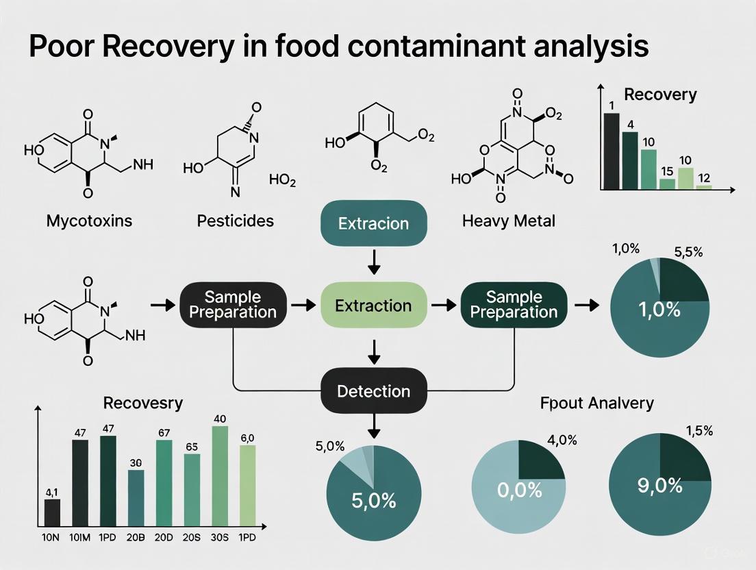
Optimizing Analyte Recovery in Food Contaminant Analysis: Strategies for Researchers and Scientists
This article provides a comprehensive guide for researchers and drug development professionals addressing the critical challenge of poor recovery in food contaminant analysis.
Optimizing Analyte Recovery in Food Contaminant Analysis: Strategies for Researchers and Scientists
Abstract
This article provides a comprehensive guide for researchers and drug development professionals addressing the critical challenge of poor recovery in food contaminant analysis. It explores the foundational causes of low recovery rates, details advanced methodological approaches for improvement, offers systematic troubleshooting and optimization protocols, and establishes robust validation and comparative frameworks. By integrating the latest 2025 research on techniques like LC-MS/MS, ICP-MS, and rapid molecular detection, this resource aims to enhance analytical accuracy, ensure regulatory compliance, and strengthen the reliability of data used in safety assessments and biomedical research.
Understanding the Root Causes of Poor Recovery in Food Contaminant Testing
Defining Analyte Recovery and Its Impact on Data Integrity and Regulatory Compliance
Fundamental Concepts: Understanding Analyte Recovery
What is analyte recovery and why is it a critical measurement in analytical chemistry?
Analyte recovery is a measure of the efficiency of an analytical process, representing the percentage of a known amount of an analyte that is successfully carried through the sample extraction and processing steps of the method and ultimately measured [1]. In practical terms, it compares the signal from an extracted sample to a standard solution of the same concentration, indicating what proportion of the target substance was successfully extracted and detected from the original sample [2].
High recovery rates (typically >70%) are essential for method validation as they ensure quantification accuracy, improve reproducibility, and prevent data misinterpretation. Low recovery indicates significant analyte loss, which compromises the reliability of analytical results and their suitability for regulatory decision-making [2].
How does poor analyte recovery directly impact data integrity and regulatory compliance?
Poor analyte recovery directly undermines multiple principles of the ALCOA+ framework, which is the standard for data integrity in regulated industries:
- Accuracy: Low recovery means reported concentrations do not reflect true values, violating the "Accurate" principle [3].
- Complete: When analytes are lost during processing, the dataset is incomplete as it doesn't represent the full sample composition [3].
- Consistent: Variable recovery between samples and batches creates inconsistencies that make results unreliable for trend analysis [2] [3].
Regulatory agencies like the FDA cite data integrity issues in 61% of warning letters, with inaccurate data due to poor recovery being a significant concern. Maintaining acceptable recovery rates is therefore essential for demonstrating compliance with Good Laboratory Practices (GLP) and Good Manufacturing Practices (GMP) [3].
Troubleshooting Low Analyte Recovery
Comprehensive Troubleshooting Guide
Table 1: Common Causes and Solutions for Low Analyte Recovery
| Problem Category | Specific Issue | Recommended Solution | Expected Outcome |
|---|---|---|---|
| Sample Preparation | Non-specific adsorption to labware surfaces [2] [1] | Use low-binding plasticware, silanized glassware, or add anti-adsorptive agents (e.g., BSA, 0.01% Tween 20) [2] [1] | Reduced analyte loss to container walls |
| Chemical or biological degradation [1] | Optimize storage conditions, use stabilizers, reduce processing time | Improved analyte stability | |
| SPE Sorbent Selection | Mismatched sorbent chemistry [2] | Hydrophobic compounds → Reversed-phase (C18, C8); Polar compounds → HILIC; Ionizable compounds → Ion exchange [2] | Improved retention and elution |
| Overloaded SPE column [2] | Reduce sample volume or concentration; Use cartridge with higher binding capacity | Prevents breakthrough | |
| Solvent & pH Conditions | pH mismatch with analyte ionization state [2] | Adjust sample pH to ensure analytes are in optimal state for retention/elution (e.g., pH > pKa for basic drugs) [2] | Enhanced binding efficiency |
| Over-aggressive washing [2] | Reduce wash solvent strength or change composition | Prevents premature elution | |
| Incomplete elution [2] | Use stronger elution solvents, increase volume, adjust elution pH | More complete analyte recovery | |
| Method Execution | Inconsistent flow rates or drying steps [2] | Standardize procedures with defined parameters; Use positive pressure or vacuum manifolds [2] | Improved reproducibility |
| Inefficient liberation of analyte bound to matrix [1] | Optimize extraction conditions (solvent, time, temperature); Use protein precipitation | Better extraction efficiency |
Systematic Protocol for Identifying Recovery Loss Points
How can I systematically identify at which step analyte loss is occurring?
For a structured investigation of recovery issues, follow this diagnostic protocol adapted from current bioanalytical research [1]:
Objective: To identify and quantify sources of analyte loss during sample preparation and analysis.
Experimental Setup: Prepare four sets of samples in replicates of six:
- Set A (Control): Pure analyte in solution (no matrix, no extraction)
- Set B (Pre-extraction Spike): Analyte spiked into biological matrix, then immediately processed with extraction
- Set C (Post-extraction Spike): Blank matrix extracted, then analyte spiked into the extracted solution
- Set D (Standard Curve): Analyte in pure solution for comparison
Measurement and Calculation:
- Analyze all samples and compare peak responses.
- Overall Recovery = (Set B / Set A) × 100
- Extraction Efficiency = (Set B / Set C) × 100
- Matrix Effect = (Set C / Set D) × 100
- Pre-extraction Loss = 100% - (Set B response relative to Set A)
Table 2: Interpretation of Recovery Investigation Results
| Scenario | Overall Recovery | Extraction Efficiency | Matrix Effect | Primary Issue Identified |
|---|---|---|---|---|
| 1 | Low | High | ~100% | Pre-extraction losses (degradation, NSB) |
| 2 | Low | Low | ~100% | Inefficient extraction process |
| 3 | Low | High | Low | Significant matrix suppression |
| 4 | Low | Low | Low | Combined extraction and matrix issues |
This systematic approach allows researchers to pinpoint exactly where analyte losses occur and apply targeted solutions rather than generalized troubleshooting [1].
Advanced Techniques for Complex Matrices
What specialized techniques improve recovery in complex food matrices like acrylamide analysis?
Analysis of food contaminants like acrylamide presents particular challenges due to complex matrices. Advanced techniques include:
- Selective SPE Sorbents: Use optimized sorbent materials for specific contaminant classes. For acrylamide, methods utilize solid-phase extraction for purification and concentration from food samples [4].
- Matrix-Specific Extraction: For fatty foods, initial defatting with non-polar solvents improves recovery. Protein precipitation with Carrez solutions or acetonitrile removes interfering compounds [4].
- Solvent Optimization: Acidified acetonitrile has demonstrated superior efficacy in extracting acrylamide across diverse food samples compared to other solvents [4].
What role does LC-MS/MS play in achieving reliable recovery for trace-level contaminants?
Liquid chromatography-tandem mass spectrometry (LC-MS/MS) provides the sensitivity and selectivity needed for accurate recovery determination at trace levels:
- Trace-Level Detection: LC-MS/MS enables accurate trace acrylamide detection across complex food matrices, essential for validating mitigation strategies [4].
- Matrix Effect Assessment: LC-MS/MS with electrospray ionization allows identification of ion suppression/enhancement caused by co-eluting matrix components [1].
- Specificity in Complex Mixtures: Advanced chromatographic techniques separate target analytes from matrix interferences, providing more accurate recovery measurements [4].
Data Integrity and Regulatory Framework
Essential Documentation for Recovery Validation
What documentation is essential to demonstrate recovery validation for regulatory purposes?
- Standard Operating Procedures (SOPs): Detailed protocols for sample preparation, extraction, and analysis [2] [5].
- Recovery Validation Data: Results from systematic recovery experiments demonstrating consistency and accuracy [1].
- Audit Trails: Secure, time-stamped electronic records of all sample processing steps, including any reprocessing [3] [5].
- Quality Control Records: Documentation of QC samples analyzed alongside test samples to demonstrate ongoing recovery performance [5].
How does the ALCOA+ framework apply specifically to recovery data?
The ALCOA+ framework provides specific guidance for maintaining data integrity in recovery studies [3]:
- Attributable: Record who performed each extraction and analysis, with timestamps for critical steps like pH adjustment or solvent addition [3].
- Legible: All recovery data, including chromatograms and calculations, must be readable throughout the data lifecycle [3].
- Contemporaneous: Document extraction conditions, observations, and results at the time of the activity [3].
- Original: Preserve raw data (chromatograms, peak areas) in their original format [3].
- Accurate: Ensure recovery calculations are error-free and reflect what was actually observed [3].
- Complete: Include all recovery data, including failed experiments or outliers, with appropriate justification for exclusion [3].
Frequently Asked Questions (FAQs)
What are the most common misconceptions about analyte recovery?
Myth: "High variability in recovery is acceptable for difficult analytes."
Myth: "Recoveries above 100% are better than 100% recovery."
- Fact: Recoveries significantly >100% typically indicate matrix enhancement, interference, or calibration problems that compromise data accuracy [1].
How often should recovery experiments be performed in an ongoing analysis?
- Initial Validation: Complete recovery assessment during method development and validation.
- Routine Monitoring: Include quality control samples at low, medium, and high concentrations with each batch (typically 5-10% of total samples).
- Change Events: Re-assess recovery whenever critical parameters change (new reagent lot, different instrument, matrix change) [5].
Can automated systems improve recovery reproducibility?
Yes, automated SPE and liquid handling systems significantly improve recovery reproducibility by:
- Minimizing human error and variability in timing and solvent volumes [2]
- Providing consistent flow rates and pressure application [2]
- Reducing analyst-to-analyst variability, especially in high-throughput environments [2] [5]
The Scientist's Toolkit: Research Reagent Solutions
Table 3: Essential Materials for Optimizing Analyte Recovery
| Reagent/Material | Function | Application Examples |
|---|---|---|
| Mixed-mode SPE sorbents (e.g., HLB, MCX, MAX) | Combined reversed-phase and ion-exchange mechanisms for complex analyte profiles [2] | Pharmaceutical compounds, multi-class contaminant analysis |
| Low-binding plasticware (silanized glass, polypropylene) | Reduce non-specific binding of hydrophobic analytes [2] [1] | Biological samples, hydrophobic compounds |
| Anti-adsorptive agents (BSA, CHAPS, Tween 20) | Block analyte absorption to labware surfaces [2] [1] | Urine, CSF, protein-free matrices |
| Buffering systems (ammonium formate, phosphate buffers) | Control pH to optimize analyte ionization state [2] | Ionizable compounds, pH-dependent extraction |
| Protein precipitation solvents (acetonitrile, methanol) | Remove protein-bound analytes and precipitate interfering compounds [1] [4] | Plasma, tissue homogenates, food matrices |
Workflow Visualization
Diagram 1: This troubleshooting workflow provides a structured approach to diagnosing and resolving analyte recovery issues, beginning with systematic problem identification and proceeding through targeted solutions based on the specific type of loss identified.
Diagram 2: This diagram illustrates how the ALCOA+ framework principles ensure data integrity throughout recovery studies, ultimately leading to reliable data and regulatory compliance.
Troubleshooting Guides
Matrix Effects in Food Contaminant Analysis
Matrix effects occur when co-extracted compounds from the sample interfere with the ionization of the target analyte during analysis, leading to signal suppression or enhancement and inaccurate quantitation [6] [7] [8]. This is a predominant challenge in liquid chromatography–tandem mass spectrometry (LC–MS/MS) analysis of complex food matrices [6] [8].
Diagnosis and Identification:
- Observed Symptoms: Inconsistent calibration curves, poor recovery of internal standards, inaccurate quantification despite proper sample preparation, high variability in replicate analyses.
- Diagnostic Test: Perform a post-column infusion test. Infuse a standard solution of the analyte into the mobile post-column while injecting a blank sample extract. A dip or rise in the baseline at the analyte's retention time confirms matrix suppression or enhancement [6].
- Quantitative Assessment: Compare the analyte response in a pure standard solution to the response of the same amount of analyte spiked into a blank matrix extract. A significant difference (typically > 25%) indicates substantial matrix effects [7].
Resolution Strategies: The following table summarizes the primary strategies to overcome matrix effects:
| Strategy | Core Principle | Application Example | Key Advantage | Key Limitation |
|---|---|---|---|---|
| Stable Isotope Dilution (SIDA) [6] | Uses a stable isotopically labeled analog of the analyte as an internal standard. | Analysis of mycotoxins in corn and peanut butter using 13C-labeled homologs [6]. | Provides the most effective compensation, as the labeled standard co-elutes and experiences identical matrix effects. | Isotopically labeled standards can be expensive and are not available for all analytes. |
| Matrix-Matched Calibration [6] [7] | Calibration standards are prepared in a blank matrix extract that is free of the analyte. | Pesticide residue analysis in various fruits and vegetables [6]. | Practically accounts for the net matrix effect. | Requires a reliable source of blank matrix; can be difficult for some food types. |
| Improved Sample Cleanup [6] [7] [9] | Removes interfering matrix components before instrumental analysis. | Using graphitized carbon SPE to clean up perchlorate analysis in food samples [6]. | Reduces the source of the interference, addressing the root cause. | May add steps to the workflow; potential for analyte loss. |
| Alternative Ionization [6] | Switching from electrospray ionization (ESI), which is highly susceptible, to atmospheric pressure chemical ionization (APCI). | Can be applied to less polar compounds that are amenable to APCI. | Can significantly reduce ionization suppression for certain classes of compounds. | Not suitable for all analytes, particularly ionic and thermally labile ones. |
Analyte Degradation During Analysis
Analyte degradation leads to a progressive loss of the target compound from the time of sample collection until analysis, resulting in underestimation of its true concentration.
Diagnosis and Identification:
- Observed Symptoms: Poor recovery, appearance of new chromatographic peaks (degradants), inconsistent results over time, failure in stability-indicating methods.
- Diagnostic Test: Re-inject a standard or sample extract after 24-48 hours of storage in the autosampler and compare the peak area/height to the initial injection. A significant decrease suggests degradation in the solution.
Resolution Strategies:
| Strategy | Core Principle | Application Example | Key Advantage | Key Limitation |
|---|---|---|---|---|
| Stabilization during Storage | Optimizes storage conditions to minimize degradation. | Keeping light-sensitive samples in amber vials; storing thermolabile extracts at -20°C. | Preserves sample integrity from collection to analysis. | Requires foresight into analyte stability. |
| Optimized Extraction Solvents | Uses solvents and pH conditions that enhance analyte stability. | Extracting glyphosate with a buffer containing EDTA to prevent metal-complexation [6]. | Addresses chemical instability during the sample preparation process. | May require extensive method development. |
| Use of Protectants (GC-MS) [6] | Adds chemicals to the sample that mask active sites in the GC system. | Using analyte protectants to shield pesticides from degradation in the GC inlet. | Improves peak shape and quantitative response. | Adds another component to the method; may contaminate the system. |
Inefficient Extraction
Inefficient extraction fails to quantitatively release the analyte from the complex food matrix into the solution, leading to low recovery.
Diagnosis and Identification:
- Observed Symptoms: Consistently low recovery in spiked samples, poor method precision, inability to detect incurred residues known to be present.
- Diagnostic Test: Perform a recovery study by spiking the analyte into the sample matrix prior to extraction. Low recovery (<70% or outside acceptable method validation criteria) indicates inefficient extraction.
Resolution Strategies:
| Strategy | Core Principle | Application Example | Key Advantage | Key Limitation |
|---|---|---|---|---|
| Pressurized Liquid Extraction (PLE) [10] [11] | Uses high temperature and pressure to achieve efficient and rapid extraction with less solvent. | Extraction of organic contaminants from solid food matrices like grains and soil. | Higher efficiency, automation, and reduced solvent consumption compared to traditional techniques. | Initial equipment cost can be high. |
| Supercritical Fluid Extraction (SFE) [10] [11] | Uses supercritical fluids (e.g., CO2) as the extraction solvent. | Extraction of lipids and non-polar contaminants; decaffeination of coffee. | Highly tunable selectivity, no organic solvent waste, fast diffusion. | Best for non-polar to moderately polar analytes. |
| Ultrasound-Assisted Extraction (UAE) [12] [11] | Uses ultrasonic energy to disrupt cell walls and enhance mass transfer. | Pretreatment (30 min) for drying apples to improve efficiency and preserve antioxidants [12]. | Simple equipment, effective for disrupting tough plant and animal tissues. | Potential for heat generation; may require optimization of ultrasonic parameters. |
| Novel Green Solvents [10] | Employs solvents like Deep Eutectic Solvents (DES) with low toxicity and high biodegradability. | Extraction of bioactive compounds from food. | Safer for analysts and the environment; can be tailored for specific analytes. | Still an emerging technology; may require custom synthesis. |
Frequently Asked Questions (FAQs)
Q1: What are the most common sources of matrix effects in LC-MS/MS for food analysis? Matrix effects primarily arise from co-extracted compounds that alter ionization efficiency in the ESI source. Common culprits in food include lipids, proteins, salts, carbohydrates, and organic acids [6] [8]. The complexity of the matrix directly influences the severity, with fatty foods, spices, and dark-colored products often posing the greatest challenges.
Q2: How can I quickly check if my method is suffering from significant matrix effects?
The most straightforward test is the post-extraction spike experiment [6]. Prepare three solutions: A) a neat standard in solvent, B) the blank matrix extract spiked with the same amount of analyte after extraction, and C) the blank matrix extract. Inject all three. Calculate the matrix effect (ME) as: ME% = (Peak Area B / Peak Area A) × 100. A value of 100% indicates no effect; significant suppression is indicated by values <75-80%, and enhancement by values >115-120% [7].
Q3: My recovery is low. How do I determine if the problem is inefficient extraction or analyte degradation? Design a recovery and stability test [6]:
- Spike before extraction: Measure recovery to assess extraction efficiency + any degradation during the entire process.
- Spike after extraction: Spike the analyte into the final extract just before injection. This measures losses due primarily to degradation during analysis or matrix effects. If recovery is low in (1) but good in (2), the issue is inefficient extraction. If recovery is low in both (1) and (2), the issue is likely analyte degradation in the final solution or during analysis.
Q4: Are there "greener" alternatives to traditional extraction methods like QuEChERS that still perform well? Yes, the field is moving towards miniaturized and greener techniques. Miniaturized Solid-Phase Extraction (SPE) techniques, such as Solid-Phase Microextraction (SPME) and Stir-Bar Sorptive Extraction (SBSE), significantly reduce or eliminate organic solvent use [9]. Additionally, techniques like Pressurized Liquid Extraction (PLE) and Supercritical Fluid Extraction (SFE) are recognized as green alternatives as they reduce solvent consumption and energy usage while maintaining high performance [10] [11].
Q5: What is the single most effective way to compensate for matrix effects in quantitative LC-MS/MS? The most effective single approach is the use of Stable Isotope Dilution Assay (SIDA) [6]. By using a stable isotopically labeled internal standard (e.g., 13C-, 15N-labeled) that is identical in chemical behavior to the analyte but distinguishable by mass, it perfectly compensates for both matrix effects and losses during sample preparation, as it experiences the same suppression/enhancement and extraction efficiency as the native compound.
Experimental Protocols
Protocol: Stable Isotope Dilution Method for Mycotoxins in Corn
This protocol is adapted from a validated procedure used by the FDA for the simultaneous determination of multiple mycotoxins (e.g., aflatoxins, ochratoxin A) [6].
1. Scope and Application: For the quantification of 12 mycotoxins in corn, peanut butter, and wheat flour using LC-MS/MS.
2. Experimental Procedure:
- Weighing: Accurately weigh 5 g of homogenized sample into a 50 mL centrifuge tube.
- Isotope Addition: Add a known concentration of a mixed 13C-labeled internal standard solution for each target mycotoxin.
- Extraction: Add 20 mL of acetonitrile/water (50:50, v/v). Vortex mix vigorously for 1 minute, then shake or sonicate for 20 minutes.
- Centrifugation: Centrifuge at ≥4000 rpm for 10 minutes.
- Filtration: Transfer an aliquot of the supernatant to an autosampler vial through a 0.2 µm syringe filter.
- LC-MS/MS Analysis: Inject the filtered extract directly for analysis.
3. Key Parameters and Calculations:
- Quantitation: Use the internal standard method for calibration. The calibration curve is constructed by plotting the peak area ratio (analyte / internal standard) against the concentration ratio.
- Compensation: The 13C-labeled internal standard compensates for both matrix effects during ionization and for minor losses during the sample preparation steps, ensuring high accuracy.
Protocol: Pulsed Electric Field (PEF) and Extraction for Plant Proteins
This protocol outlines an innovative approach to improve extraction efficiency from plant tissues, as demonstrated for stinging nettle [12].
1. Scope and Application: For the efficient extraction of proteins from stinging nettle (Urtica dioica L.) leaves, with simultaneous reduction of chlorophyll.
2. Experimental Procedure:
- Sample Preparation: Wash and comminute fresh stinging nettle leaves.
- Cell Disruption (PEF): Subject the plant slurry to Pulsed Electric Field treatment (e.g., field strength: 1-3 kV/cm; specific energy: 10-100 kJ/kg). This electroporates the cell membranes without significant heating.
- Extraction: Mix the PEF-treated slurry with a suitable aqueous buffer.
- Separation: Apply a separation technique such as Ultrafiltration to separate proteins from smaller molecules like chlorophyll.
- Analysis: Determine protein yield (e.g., via Kjeldahl or Bradford assay) and chlorophyll content (via spectrophotometry).
3. Key Parameters and Findings:
- Optimal Combination: The study found that coupling PEF with Ultrafiltration effectively reduced chlorophyll content from 4781.41 µg/g in raw leaves to 15.07 µg/g in the final extract [12].
- Advantage: This combination enhances the sustainability and efficiency of protein recovery, yielding a cleaner product suitable for food fortification.
Signaling Pathways and Workflows
Troubleshooting Poor Recovery Workflow
The following diagram outlines a systematic decision-making process for diagnosing the root cause of poor recovery in food contaminant analysis.
Matrix Effect Mechanism in LC-ESI-MS
This diagram visualizes the mechanism of ion suppression in electrospray ionization (ESI), a common type of matrix effect.
Research Reagent Solutions
The following table lists key reagents and materials essential for implementing the troubleshooting strategies discussed in this guide.
| Reagent/Material | Function/Application | Key Consideration |
|---|---|---|
| Stable Isotopically Labeled Internal Standards (e.g., 13C-, 15N-, 2H-labeled) [6] | Gold standard for compensating for matrix effects and analyte losses during sample preparation and analysis. | Select an isotope that has sufficient mass difference from the native analyte to avoid cross-talk. Ensure the labeled standard is added at the very beginning of the extraction. |
| Graphitized Carbon SPE Cartridges [6] | Cleanup of sample extracts to remove colored pigments, organic acids, and other polar interferences that cause matrix effects. | Useful for planar molecules but can strongly retain target analytes if not carefully conditioned; requires method optimization to prevent analyte loss. |
| QuEChERS Extraction Kits [7] | A standardized, quick, and efficient sample preparation method for pesticide residues and other contaminants in food. | Available in different buffered versions (e.g., citrate, acetate) to suit specific analyte and matrix pH stability requirements. |
| Deep Eutectic Solvents (DES) [10] | Novel, green extraction solvents with low toxicity and high biodegradability for replacing conventional organic solvents. | Can be tailored by combining hydrogen bond donors and acceptors to selectively extract target compounds, but may require custom synthesis. |
| Analyte Protectants (e.g., Gulonolactone, Sorbitol) [6] | Used in GC-MS to mask active sites in the inlet and column, reducing adsorption and degradation of target analytes. | Improves peak shape and quantification for sensitive compounds but can contaminate the GC system over time. |
| Molecularly Imprinted Polymers (MIPs) [9] | Synthetic sorbents with high selectivity for a specific analyte or class of analytes, used in SPE for efficient cleanup. | Provide high selectivity, reducing matrix effects, but require development and synthesis for each specific target. |
The analysis of food contaminants is fundamentally complicated by the food matrix—the intricate physical and chemical structure where components like fats, proteins, and carbohydrates interact. This complex architecture can bind, trap, or chemically modify contaminants and analytical targets, leading to the critical problem of poor recovery during analysis. Recovery rates are compromised when target compounds are not fully released or are degraded during extraction from the matrix, yielding inaccurate results that underestimate true contaminant levels. Understanding these matrix-contaminant interactions is essential for developing accurate, reliable methods to ensure food safety.
Frequently Asked Questions (FAQs)
Q1: Why do I get low recovery rates when analyzing contaminants in high-fat foods? High-fat matrices pose significant challenges. Lipids can co-extract with target analytes, causing interference in chromatographic systems (e.g., column fouling, signal suppression) and necessitating extensive clean-up that can lead to the loss of the target contaminant. Furthermore, many lipophilic contaminants (e.g., persistent organic pollutants, some pesticides) partition into the fat phase, making them difficult to isolate completely during solvent extraction. The amphiphilic nature of lipids also complicates standardized extraction protocols [13] [14].
Q2: How do protein-rich matrices interfere with contaminant quantification? Proteins can bind strongly to contaminants, including heavy metals and certain mycotoxins, through various interactions (e.g., covalent, ionic, hydrophobic). This binding can make it difficult to release the contaminant during standard extraction procedures. Additionally, proteins may precipitate during sample preparation, co-precipitating the bound analytes and removing them from the solution meant for analysis. The intricate folding and conformational variability of proteins, sensitive to denaturation, further complicate achieving consistent recovery [13] [14].
Q3: What specific issues do complex carbohydrates present? Complex carbohydrates, such as starch, cellulose, and hemicellulose, create a physical entrapment network. Their diverse branching patterns, varying degrees of polymerization, and water-binding capacity can hinder solvent penetration and analyte diffusion. Starch can form gels upon heating, further trapping contaminants. Dietary fibers can also bind specifically to certain analytes. The structural heterogeneity of polysaccharides means a single extraction method is often insufficient for different food types [14].
Q4: Which advanced detection techniques help overcome matrix effects? Advanced detection technologies are crucial for mitigating matrix effects and improving recovery accuracy.
- Liquid Chromatography-Mass Spectrometry (LC-MS): Provides high selectivity and sensitivity for a wide range of chemical contaminants, allowing for the identification and quantification of co-eluting compounds that would interfere with less specific detectors [13] [15].
- Inductively Coupled Plasma Mass Spectrometry (ICP-MS): Offers exceptional detection limits for elemental contaminants like heavy metals (lead, mercury, cadmium, arsenic), even in complex matrices, by effectively ionizing the sample [13].
- Multidimensional Nuclear Magnetic Resonance (NMR) and Advanced Mass Spectrometry (MS): These tools are increasingly used to probe the molecular architecture, interactions, and dynamics of food biomacromolecules and their association with contaminants at high resolutions [14].
Q5: How does the food matrix itself influence contaminant bioavailability and toxicity? The matrix does not just interfere with analysis; it directly influences a contaminant's biological impact. For example, contaminants bound tightly to dietary fiber or protein may have reduced bioavailability during human digestion compared to the same contaminants in a simple solution. Conversely, the presence of fat can increase the absorption of lipophilic toxins. This means that analytical methods which accurately determine the "bioaccessible" fraction of a contaminant, not just its total concentration, are critical for a realistic toxicological risk assessment [16] [17].
Troubleshooting Guide: Poor Recovery in Contaminant Analysis
Table 1: Common Recovery Problems and Solutions in Food Matrix Analysis
| Observed Problem | Potential Root Cause | Recommended Corrective Action |
|---|---|---|
| Consistently low recovery across all analytes in a complex matrix. | Inefficient extraction due to strong analyte-matrix binding or physical entrapment. | - Optimize extraction solvent (e.g., use of surfactants or more polar solvents).- Incorporate enzymatic hydrolysis (e.g., proteases for proteins, amylases for starch) to break down the matrix.- Increase extraction temperature or use pressurized liquid extraction (PLE). |
| High variability in recovery between sample replicates. | Inhomogeneous sample or inconsistent sample clean-up. | - Improve sample homogenization (e.g., cryogenic milling).- Standardize and automate sample clean-up steps (e.g., Solid-Phase Extraction - SPE).- Use internal standards to correct for losses during preparation. |
| High background noise and interference during chromatographic analysis. | Incomplete removal of co-extracted matrix components (e.g., lipids, pigments). | - Implement more rigorous clean-up steps (e.g., gel permeation chromatography - GPC for lipid removal, dispersive SPE).- Optimize chromatographic separation parameters to resolve analytes from interferences. |
| Low recovery of specific analyte classes (e.g., heavy metals). | Strong chelation with matrix components or loss during digestion. | - Use a more robust acid digestion mixture (e.g., HNO₃ + H₂O₂) for complete mineralization.- Employ chelating agents during extraction to compete for metal binding. |
| Recovery satisfactory in simple solutions but poor in real food samples. | Pronounced matrix effect (suppression or enhancement) in the detection system. | - Use matrix-matched calibration standards.- Dilute the sample extract to reduce the matrix concentration.- Employ standard addition method for quantification. |
Detailed Experimental Protocols for Mitigating Matrix Effects
Protocol 1: Enzymatic Hydrolysis for Protein-Rich Matrices (e.g., Meat, Dairy)
Objective: To break down protein structures and release protein-bound contaminants for improved recovery.
- Homogenization: Precisely weigh 2.0 g of homogenized sample into a 50 mL centrifuge tube.
- Protein Denaturation: Add 10 mL of a 0.1 M Tris-HCl buffer (pH 7.5) and vortex. Heat the mixture in a water bath at 80°C for 10 minutes to denature proteins.
- Enzymatic Digestion: Cool the sample to 37°C. Add 50 mg of protease (e.g., pronase) and incubate in a shaking water bath at 37°C for 4-16 hours.
- Extraction: After incubation, add 10 mL of acetonitrile or a suitable solvent, vortex vigorously for 1 minute, and then centrifuge at 5000 x g for 10 minutes.
- Clean-up: Transfer the supernatant to a clean tube for subsequent clean-up (e.g., SPE, QuEChERS) before analysis [14].
Protocol 2: Integrated HACCP/TACCP/FMEA for Risk Assessment in Complex Supply Chains
Objective: To systematically identify, assess, and prioritize vulnerabilities in the food supply chain that could lead to contamination and poor analytical recovery.
- Hazard Identification (HACCP): Map the entire process flow. Identify potential points of biological, chemical, and physical contamination.
- Threat Assessment (TACCP): Evaluate the identified points for vulnerability to intentional adulteration or fraud.
- Risk Quantification (FMEA): For each identified vulnerability, score three parameters on a scale of 1-5:
- Vulnerability (V): How susceptible is the point to contamination?
- Impact (W): What would be the consequence of contamination?
- Probability (PR): How likely is contamination to occur?
- Risk Calculation: Calculate the risk priority number (RPN):
RPN = V x W x PR. - Prioritization & Control: Prioritize control measures for points with the highest RPNs. Implement targeted countermeasures like enhanced surveillance, strict access control, and staff training [18].
Essential Research Reagent Solutions
Table 2: Key Reagents for Analyzing Contaminants in Complex Food Matrices
| Reagent / Material | Function in Analysis | Application Example |
|---|---|---|
| Protease Enzymes | Hydrolyzes peptide bonds in proteins, releasing bound contaminants and reducing matrix viscosity. | Recovery of mycotoxins or heavy metals from protein-rich matrices like meat and dairy products [14]. |
| Amylase Enzymes | Breaks down starch molecules (amylose and amylopectin) into simpler sugars, disrupting gel structures that trap analytes. | Analysis of pesticides in starchy foods like potatoes or cereals to prevent physical entrapment [14]. |
| Solid-Phase Extraction (SPE) Cartridges | Selective clean-up of sample extracts to remove interfering lipids, pigments, and other co-extracted matrix components. | Purification of extract prior to LC-MS analysis to reduce ion suppression and column fouling. Various phases (C18, Florisil, NH₂) are used based on the analyte [13]. |
| De-fatting Agents (e.g., n-hexane, GPC) | Removal of lipid content from samples to prevent interference in chromatographic analysis and concentrate non-lipid analytes. | Essential pre-treatment for accurate analysis of contaminants in high-fat matrices like oils, nuts, and fatty fish [13]. |
| Isotopically Labeled Internal Standards | Added to the sample at the beginning of preparation to correct for analyte loss during extraction, clean-up, and for matrix effects during detection. | Crucial for achieving accurate quantification in LC-MS and ICP-MS, as the standard experiences the same matrix effects as the analyte [13] [19]. |
| Matrix-Matched Calibration Standards | Calibration standards prepared in a blank matrix extract that is free of the target analyte. This compensates for signal suppression/enhancement during analysis. | Used when complete elimination of the matrix effect is impossible, ensuring the calibration curve mirrors the sample's behavior [18]. |
Analytical Workflow and Pathway Visualization
Workflow for contaminant analysis in complex food matrices
Relationship between matrix challenges and mitigation strategies
For researchers in food safety and drug development, addressing poor recovery during the extraction and analysis of contaminants is a fundamental challenge. Poor recovery directly compromises data accuracy, leading to an underestimation of contaminant levels and a misrepresentation of the true risks in food products. At the heart of this problem lie the complex molecular interactions between contaminants and food components. These interactions, whether covalent, ionic, or hydrophobic, can strongly bind contaminants to proteins, starches, or lipids, effectively sequestering them and reducing the yield available for detection. This technical support guide is designed to help you troubleshoot these issues by providing a deeper understanding of the binding mechanisms and offering practical, actionable protocols to overcome them.
Frequently Asked Questions (FAQs)
1. What are the primary molecular mechanisms by which contaminants bind to food matrices, leading to low analytical recovery?
Contaminants bind to food components through several specific mechanisms, which can occur simultaneously:
- Covalent Bonding: Certain processing contaminants, such as acrolein, can form stable covalent adducts with proteins and DNA. For instance, acrolein reacts with lysine and cysteine residues in proteins, creating strong cross-links that are difficult to break during standard extraction [20].
- Complexation with Proteins: Heavy metals like lead (Pb) and cadmium (Cd) often chelate with functional groups in proteins, such as sulfhydryl (-SH) and carboxyl (-COOH) groups. This binding can alter the protein's structure and mask the metal, making it unavailable for extraction [21] [13].
- Integration within Starch Helices: Lipophilic contaminants, including certain pesticides and persistent organic pollutants (POPs), can become entrapped within the helical structure of amylose in starch. This physical encapsulation requires methods that disrupt the crystalline starch matrix to release the contaminant [22].
- Partitioning into Lipids: Non-polar contaminants, such as dioxins and many mycotoxins, readily dissolve and partition into the fat droplets of lipid-rich food matrices. Recovery depends on the complete dissolution of the fat and the use of solvents with appropriate polarity [13].
- Ionic and Hydrogen Bonding: Polar contaminants, including various herbicides and arsenic species, can form strong ionic bonds with charged sites on food components or engage in hydrogen bonding with polysaccharides like cellulose and pectin [21].
2. Beyond the binding mechanisms, what are common methodological pitfalls that cause poor recovery?
Even with an understanding of the binding, experimental error is common. Key pitfalls include:
- Inconsistent Sample Preparation: Variability in sample particle size, shape, and homogeneity is a leading cause of non-reproducible recovery. Using non-standardized preparation methods introduces significant random error [23].
- Improper Solvent Selection: Using a solvent with insufficient strength to disrupt the specific contaminant-matrix bond will result in low recovery. The solvent must be matched to the chemical nature of both the contaminant and the food matrix [24].
- Inadequate Calibration: An improperly calibrated instrument, such as a texture analyser or chromatograph, will produce inaccurate measurements, making it impossible to distinguish between true recovery issues and instrumental drift [23].
- Overlooking Matrix Effects: Failing to use matrix-matched calibration standards can lead to severe quantification errors. The food matrix can suppress or enhance the analytical signal, giving a false recovery value [24].
3. My recovery is low for a known contaminant-matrix pair. What is the first parameter I should optimize?
The first and most critical parameter to optimize is the extraction solvent system. The solvent must be powerful enough to compete with and break the dominant molecular interaction. A systematic approach is recommended:
- For protein-bound metals, consider adding a chelating agent (e.g., EDTA) or using a mild denaturant to unfold the protein and expose binding sites.
- For starch-encapsulated pesticides, employing a solvent like DMSO that can disrupt hydrogen bonding and swell the starch granules is often effective.
- For mycotoxins in lipid matrices, a mixture of non-polar (hexane) and polar (acetonitrile) solvents is frequently needed to address both the lipid and the toxin.
Troubleshooting Guides
Guide 1: Diagnosing and Solving Poor Recovery of Heavy Metals from Protein-Rich Foods
Problem: Consistently low recovery of heavy metals (e.g., Pb, Cd, As) from animal- and plant-based protein powders.
Root Cause: The metals are strongly chelated by functional groups (thiols, amines, carboxylates) within the protein's three-dimensional structure. Standard acidic extraction may not be sufficient to denature the protein and release the metal.
Solution: A Sequential Extraction and Digestion Protocol
- Initial Solubilization: Homogenize 1 g of sample in 10 mL of a neutral pH buffer (e.g., 50 mM Tris-HCl).
- Mild Denaturation: Add a denaturing agent (e.g., 1 mL of 1% SDS) and incubate at 60°C for 30 minutes. This begins to unfold the protein.
- Chelation Competition: Add a chelating agent (e.g., 2 mL of 0.1 M EDTA) and vortex vigorously. EDTA competes with protein binding sites for the metal ions.
- Enzymatic Digestion (if needed): For stubborn matrices, add a protease (e.g., proteinase K) and incubate at 37°C for 2-4 hours to fully digest the protein.
- Final Acid Extraction: Acidify the mixture with concentrated nitric acid to a final concentration of 2% and incubate for a further 15 minutes at 80°C.
- Analysis: Centrifuge, filter (0.45 µm), and analyze the supernatant via ICP-MS [21] [13].
Table 1: Expected Recovery Improvements for Heavy Metals Using the Sequential Protocol
| Metal | Matrix | Standard Acid Digestion Recovery (%) | Sequential Protocol Recovery (%) |
|---|---|---|---|
| Cadmium (Cd) | Soy Protein | 65-75 | 90-102 |
| Lead (Pb) | Whey Protein | 60-70 | 88-95 |
| Arsenic (As) | Rice Protein | 70-80 | 92-98 |
Guide 2: Troubleshooting Low Recovery of Hydrophobic Contaminants from Starchy Matrices
Problem: Low and variable recovery of lipophilic pesticides (e.g., organochlorines) from powdered starchy foods like flours and infant cereals.
Root Cause: The contaminants are physically trapped within the helical structure of amylose, preventing their contact with the extraction solvent.
Solution: Using a Starch-Disrupting Solvent with Thermal Assistance
- Sample Weighing: Precisely weigh 2 g of homogenized sample into a centrifuge tube.
- Solvent Addition: Add 10 mL of a DMSO-Water mixture (90:10 v/v). DMSO is highly effective at breaking hydrogen bonds and swelling starch granules.
- Thermal Treatment: Heat the mixture in a water bath at 80°C for 20 minutes, vortexing every 5 minutes. This thermal energy helps to dissolve the starch and release the entrapped contaminants.
- Liquid-Liquid Extraction: After cooling, add 10 mL of hexane, cap the tube, and shake vigorously for 2 minutes. The target contaminants will partition into the hexane layer.
- Separation and Concentration: Centrifuge to separate layers. Carefully collect the hexane (top) layer. Evaporate it to dryness under a gentle nitrogen stream and reconstitute the residue in 1 mL of an appropriate solvent for GC-MS analysis [22].
- Validation: Validate the method by comparing against a reference method, such as LC-MS, and determine recovery using spiked samples [24].
Guide 3: A General Workflow for Systematic Method Development
The following diagram outlines a logical, step-by-step workflow for diagnosing and resolving poor recovery, based on the principles outlined in the FAQs and guides.
Diagram 1: A logical workflow for troubleshooting poor recovery in contaminant analysis.
Research Reagent Solutions
The following table details key reagents and their specific functions in overcoming molecular binding interactions to improve recovery.
Table 2: Essential Reagents for Mitigating Contaminant Binding in Food Analysis
| Reagent / Material | Function in Troubleshooting | Example Application |
|---|---|---|
| Ethylenediaminetetraacetic Acid (EDTA) | A chelating agent that competes with protein binding sites for metal ions, forming stable, soluble complexes. | Improving recovery of lead and cadmium from protein-rich powders [21]. |
| Dimethyl Sulfoxide (DMSO) | A powerful, aprotic solvent that disrupts hydrogen bonding networks, effectively swelling and dissolving starch helices. | Releasing encapsulated lipophilic pesticides from starchy food matrices [22]. |
| Solid-Phase Extraction (SPE) Cartridges | Used to separate and concentrate target contaminants from complex extracts, minimizing matrix effects and improving detection limits. | Clean-up and concentration of phthalates or mycotoxins prior to LC-MS analysis [24]. |
| Proteinase K | A broad-spectrum protease that enzymatically digests proteins, breaking them down and releasing covalently or ionically bound contaminants. | Recovering acrolein adducts or protein-bound metals in a quantitative manner [20] [13]. |
| Sodium Dodecyl Sulfate (SDS) | An ionic detergent that denatures proteins by breaking hydrophobic interactions and disrupting tertiary structure. | Unfolding globular proteins to expose internal binding sites for heavy metals [13]. |
| Immunoaffinity Columns | Contain immobilized antibodies that provide highly specific binding for target contaminants, offering superior clean-up from complex matrices. | Selective extraction of aflatoxins or ochratoxin A for accurate quantification [22]. |
Regulatory Implications of Poor Recovery in FSMA and EU Maximum Residue Levels (MRLs)
Troubleshooting Guides
Guide 1: Addressing Poor Recovery in Pesticide Residue Analysis for EU MRL Compliance
Problem: Inconsistent or low recovery rates for pesticides like difenoconazole and flonicamid during analysis, jeopardizing compliance with new EU MRLs [25].
Explanation: Poor recovery indicates inefficiency in your sample preparation method, leading to inaccurate quantification. Under-reporting due to low recovery poses a significant compliance risk, as your product may appear to meet MRLs when it does not. The recent EU Regulation (EU) 2024/2612 sets specific MRLs that must be reliably verified [25].
Solution: A step-by-step method optimization and troubleshooting protocol.
Step 1: Quick Assessment Checklist
- Internal Standard: Verify the internal standard is added at the correct stage and shows no signs of degradation.
- Solvent Compatibility: Ensure extraction solvents are matched to the chemical properties of the analyte (e.g., polarity, pH stability).
- Cartridge Conditioning: Confirm that Solid-Phase Extraction (SPE) cartridges are properly conditioned and not overloaded.
- Evaporation Temperature: Check that solvent evaporation steps use minimal heat to prevent loss of volatile analytes.
Step 2: Systematic Investigation & Protocol Adjustment Follow the workflow below to diagnose and correct the root cause of poor recovery.
Step 3: Post-Correction Validation
- After implementing changes, re-analyze certified reference materials (CRMs) or spiked samples to validate recovery improvements.
- Re-calibrate your instrument using matrix-matched standards to account for any remaining matrix effects and ensure accurate quantification against the EU MRLs [25].
Guide 2: Managing Traceability Data Gaps Under FSMA Rule 204
Problem: Inability to provide complete traceability records, including Key Data Elements (KDEs) for Critical Tracking Events (CTEs), as required by the FSMA Food Traceability Rule [26].
Explanation: The FSMA Rule 204 mandates specific recordkeeping for foods on the Food Traceability List (FTL) to enable rapid tracing during a food safety incident. Incomplete data for a "transformation" CTE (e.g., repacking peanuts) can result in non-compliance and regulatory action [26]. Poor data "recovery" from your supply chain is a critical system failure.
Solution: A procedure to identify and remediate gaps in your traceability data system.
Step 1: Data Gap Self-Assessment Checklist Answer these questions for your FTL products:
- Can you identify all immediate suppliers and recipients (one step back, one step forward)?
- For each Transformation event, are you recording all required KDEs, including:
- Traceability Lot Code (TLC) you assigned?
- Product description and quantity?
- Location of transformation?
- Date of activity?
- Reference to the source TLC(s) from received ingredients? [26]
- For each Shipping event, are you recording:
- TLC(s) shipped?
- Shipment date?
- Recipient name and address?
- Recipient phone number and email? [26]
Step 2: Implementation of a Traceability Plan The FDA requires a documented Traceability Plan. Follow this logical sequence to build a compliant system [26].
Step 3: Proactive Gap Remediation
- Conduct a Mock Trace: Test your system by performing an internal trace forward and trace backward for a specific TLC. Time yourself; the FDA can request information within 24 hours [26].
- Audit Partner Compliance: Periodically request traceability information from your suppliers and customers to ensure their systems are compatible and data exchange is seamless.
Frequently Asked Questions (FAQs)
Q1: What is the compliance deadline for the FSMA Food Traceability Rule, and what happens if I can't provide the data? The final rule has a proposed extended compliance date of July 20, 2028 [26]. If you cannot provide the required records to the FDA within 24 hours of a request, your firm would be in violation of the rule, which could lead to regulatory actions such as product detention, suspension of facility registration, or a mandated recall [26] [27].
Q2: For which foods do I need to maintain these enhanced traceability records? The requirements apply to foods on the Food Traceability List (FTL). This includes specific items like fresh herbs, certain vegetables, fruits, and peanuts. It also covers foods that contain listed ingredients if the ingredient remains in the same form (e.g., fresh) as it appears on the FTL [26].
Q3: The EU has set a new MRL for difenoconazole in wheat at 0.1 mg/kg. How can I ensure my analytical method is accurate at this level? To ensure accuracy at the 0.1 mg/kg level, you must validate your method's Limit of Quantification (LOQ) to be sufficiently below the MRL, typically by a factor of 3-5. This involves confirming that your method can achieve satisfactory precision and accuracy (recovery between 70-120%) at this low concentration. Using matrix-matched calibration standards (in wheat) is crucial to correct for matrix effects that can cause inaccurate results [25].
Q4: What are the key differences in how FSMA and EU MRL regulations approach food safety?
- FSMA (U.S. Focus): Emphasizes prevention and traceability. The FSMA Rule 204 focuses on recordkeeping to enable rapid tracking and removal of contaminated food from the supply chain [26] [28].
- EU MRL Regulations: Focus on product-specific safety thresholds. Regulations set maximum legal limits for specific chemical contaminants (like pesticides) in food, based on rigorous risk assessments by EFSA to ensure consumer safety over a lifetime of exposure [25].
The Scientist's Toolkit: Research Reagent Solutions
Table: Essential materials for food contaminant analysis and traceability compliance.
| Item | Function & Application |
|---|---|
| Certified Reference Materials (CRMs) | Calibrate instruments and validate method accuracy for precise MRL verification [25]. |
| QuEChERS Extraction Kits | Streamline sample preparation for pesticide analysis; improve recovery and reproducibility. |
| Stable Isotope-Labeled Internal Standards | Correct for analyte loss during sample preparation, improving data accuracy for low-level residues. |
| Matrix-Matched Standard Solutions | Compensate for matrix suppression/enhancement effects in LC-MS/MS, ensuring accurate quantification. |
| Electronic Recordkeeping System | Maintain FSMA-mandated KDEs and CTEs; enable rapid data retrieval for FDA requests (<24 hours) [26]. |
| Traceability Lot Code (TLC) Labels | Uniquely identify traceability lots as required by FSMA, linking all KDEs throughout the supply chain [26]. |
Advanced Analytical Techniques and Protocols to Maximize Recovery Rates
Ensuring food safety hinges on the accurate detection and quantification of harmful substances, from chemical residues to toxic elements. A central challenge in this analytical process is poor recovery—a phenomenon where the measured concentration of an analyte significantly deviates from its true value in the original sample. Poor recovery can stem from various factors, including inefficient extraction, matrix interference, analyte degradation, or instrument instability, ultimately jeopardizing data integrity and regulatory compliance [29] [13]. Modern mass spectrometry (MS) techniques form the backbone of food safety monitoring. This guide focuses on three pivotal technologies—LC-MS/MS, GC-MS/MS, and ICP-MS—providing a comparative framework for their selection and offering targeted troubleshooting advice to overcome recovery challenges in food contaminant research.
Technique Comparison and Applications
The following table summarizes the core characteristics, primary applications, and primary contaminants analyzed by each technique to guide initial method selection.
Table 1: Comparison of LC-MS/MS, GC-MS/MS, and ICP-MS in Food Contaminant Analysis
| Feature | LC-MS/MS (Liquid Chromatography-Tandem Mass Spectrometry) | GC-MS/MS (Gas Chromatography-Tandem Mass Spectrometry) | ICP-MS (Inductively Coupled Plasma Mass Spectrometry) |
|---|---|---|---|
| Best For | Non-volatile, thermally labile, and polar compounds [29] | Volatile and semi-volatile, thermally stable compounds [30] | Elemental analysis; toxic metals and essential nutrients [13] [31] |
| Ionization Source | Electrospray Ionization (ESI), Atmospheric Pressure Chemical Ionization (APCI) [29] | Electron Ionization (EI), Chemical Ionization (CI) [30] | Inductively Coupled Plasma (ICP) [31] |
| Mass Analyzer | Typically triple quadrupole [29] | Typically triple quadrupole [30] | Single quadrupole, triple quadrupole, magnetic sector [32] |
| Common Food Contaminants | Mycotoxins, marine biotoxins, pesticide residues, veterinary drugs, plant toxins [29] [33] | Pesticides, environmental pollutants, processing contaminants, certain mycotoxins [30] | Heavy metals (Pb, Cd, As, Hg), essential minerals, metalloids [13] [31] |
| Key Strengths | High selectivity/sensitivity; no derivatization needed for many compounds; ideal for complex liquid matrices [29] [33] | High chromatographic resolution; powerful library searchable spectra (EI); excellent sensitivity [30] | Ultra-trace detection (ppt-ppq); high throughput; wide linear dynamic range; isotopic analysis capability [31] [32] |
| Limitations / Challenges | Matrix effects (ion suppression/enhancement); requires skilled operators [29] | Requires analyte volatility; may need derivatization; thermal degradation risk [30] | Spectral interferences; high instrument cost; requires ultra-clean labs for trace analysis [31] [32] |
Table 2: Quantitative Performance and Regulatory Context
| Aspect | LC-MS/MS | GC-MS/MS | ICP-MS |
|---|---|---|---|
| Detection Limits | Ultralow levels (e.g., ppt for some toxins) [29] | Ultralow levels (e.g., ppt for some pesticides) | Sub-ppt to ppt levels for most elements [31] [32] |
| Example Contaminant & Regulatory Limit | Aflatoxin B1 (MRLs in low ppb range) [13] | – | Lead in candy (0.1 ppm), Arsenic in apple juice (10 ppb) [13] |
| Typical Sample Prep | QuEChERS, Solid-Phase Extraction (SPE), dilution [33] | QuEChERS, Liquid-Liquid Extraction, derivatization [30] | Microwave-assisted acid digestion [31] [32] |
| Tolerance to Complex Matrices | Moderate (requires careful cleanup to mitigate matrix effects) [33] | Moderate (requires cleanup) | Low (high total dissolved solids require dilution or specialized introduction systems) [32] |
Essential Research Reagents and Materials
Successful analysis and optimal recovery depend on using the correct reagents and materials throughout the workflow.
Table 3: Key Research Reagent Solutions for Food Contaminant Analysis
| Reagent / Material | Function | Application Notes |
|---|---|---|
| QuEChERS Kits | Quick, Easy, Cheap, Effective, Rugged, Safe; a standardized sample preparation method for extracting pesticides and other contaminants from food matrices [33]. | Critical for cleaning up complex, high-chlorophyll leafy plants; helps prevent matrix effects in LC-MS/MS and GC-MS/MS [33]. |
| Derivatization Reagents | Chemicals that react with functional groups to increase analyte volatility and thermal stability. | Used in GC-MS/MS for compounds that are not naturally volatile, improving recovery and sensitivity [30]. |
| Certified Reference Materials | Materials with certified concentrations of specific analytes, used for method validation and quality control. | Essential for verifying accuracy and identifying poor recovery in all MS techniques [31]. |
| ICP-MS Tuning Solution | A solution containing elements covering a wide mass range for instrument performance optimization. | Ensures optimal sensitivity, resolution, and stability, which are foundational for accurate recovery [34]. |
| High-Purity Acids & Gases | Nitric acid for digestions; argon for plasma; reaction/collision gases for MS/MS. | Purity is paramount to avoid contamination and high backgrounds, especially in ultra-trace ICP-MS analysis [31] [32]. |
Troubleshooting Guides and FAQs
This section addresses common experimental issues that lead to poor recovery, organized in a question-and-answer format.
LC-MS/MS Troubleshooting
Q: My analysis shows significant ion suppression and poor recovery. What steps should I take? A: Ion suppression is a major cause of poor recovery in LC-MS/MS, often due to co-eluting matrix components.
- Check Sample Preparation: Improve sample clean-up. For pesticide analysis in chlorophyll-rich plants, ensure the QuEChERS protocol is optimized for this difficult matrix [33].
- Optimize Chromatography: Alter the LC gradient to separate the analyte from matrix interferences. A longer run time or a different stationary phase can improve separation.
- Use Internal Standards: Isotopically labeled internal standards (IS) are the most effective way to correct for ion suppression. They co-elute with the analyte and compensate for signal loss [29].
Q: I am observing a loss of sensitivity and precision in my LC-MS/MS data. What is the likely cause? A: This is a common performance issue. A structured approach is needed.
- Isolate the Problem: First, determine if the issue lies with the LC system or the MS detector. Check system pressure and inject a standard directly into the MS (if possible) to check baseline MS response [35].
- Check the Ionization Source: Contamination of the ESI source is a frequent culprit. Inspect and clean the source components, including the capillary, according to the manufacturer's guidelines. Properly controlling ionization is key to stable signal [35].
GC-MS/MS Troubleshooting
Q: My target analytes are showing poor peak shape (tailing) and low recovery. What should I investigate? A: Poor peak shape often points to activity or degradation in the inlet or column.
- Inspect the Inlet Liner: An old, dirty, or deactivated liner can cause adsorption and degradation of analytes. Replace it with a new, properly deactivated liner. "Does the deactivation of a column or liner REALLY matter? The short answer is Yes!" [30].
- Check the GC Column: Cut a small section (e.g., 0.5 meters) from the inlet side of the column to remove contamination. If the problem persists, the column may need to be replaced. "When we observe peak tailing, do we immediately change a column?" - It is better to check the inlet and syringe first [30].
Q: I am getting unexpected peaks or a high background in my chromatogram. A: This indicates system contamination.
- Check the Syringe and Sample Vials: Ensure the syringe is clean and the sample vials/septa are not contaminated.
- Avoid Jumping to Conclusions: "If we observe peaks that are ¼ expected size, do we immediately vent the GC/MS to clean the source and change the column? Or, is it better to check the samples, syringe, and inlet before jumping into those time-intensive actions?" A systematic check of the samples and introduction system can save time [30].
ICP-MS Troubleshooting
Q: My calibration standards are unstable, and analyte signals are drifting upwards or downwards over time. How can I fix this? A: Signal drift is a common issue that severely impacts recovery accuracy.
- "Drift up" is most often a sign of poor cone conditioning. New or cleaned sampler and skimmer cones should be conditioned by aspirating a conditioning solution before running samples to stabilize their surface [34].
- "Drift down" is often associated with a build-up of matrix on the sample introduction components (nebulizer, torch injector, cones). This is typical when running samples with high total dissolved solids [34].
- General Steps: Check the sample introduction system for wear or damage. Inspect and clean the nebulizer, spray chamber, and pump tubing. Ensure all gas connections are tight. Verify proper grounding of the connector block to minimize static charge buildup [34].
Q: The internal standard (ISTD) recovery is poor and unstable in my analysis. What does this indicate? A: Poor ISTD mixing is a critical problem that invalidates data.
- Symptom: High relative standard deviation (RSD) for the ISTD signal. You may observe large dips or rises in the ISTD signal even minutes into stabilization [34].
- Solutions: Check that the ISTD is being added consistently and is mixing completely with the sample. Inspect the peristaltic pump tubing for wear and ensure there are no kinks or leaks in the line delivering the ISTD. Purge the ISTD line to remove any bubbles [34].
Experimental Workflows for Optimal Recovery
The following diagrams outline generalized experimental workflows for each technique, highlighting critical steps that influence recovery.
LC-MS/MS Workflow for Pesticides in Leafy Greens
LC-MS/MS Analysis Workflow
This workflow for analyzing pesticide residues in high-chlorophyll leafy plants emphasizes two critical steps for mitigating matrix effects and ensuring good recovery: effective QuEChERS extraction and SPE clean-up [33]. The final data analysis should always use a matrix-matched calibration to correct for residual ionization effects.
ICP-MS Workflow for Metals in Food
ICP-MS Analysis Workflow
This ICP-MS workflow highlights microwave-assisted digestion as a best practice for complete and consistent recovery of elements from the solid food matrix [31] [32]. The precise addition of an internal standard (ISTD) after digestion is crucial for correcting for signal drift and physical interferences during analysis [34].
Method Selection and Troubleshooting Pathway
Method Selection and Troubleshooting Pathway
This decision pathway provides a logical starting point for selecting the appropriate mass spectrometry technique based on the nature of the target analyte. It then directs the user to the specific troubleshooting FAQs relevant to the recovery issues encountered with that technique.
Frequently Asked Questions (FAQs)
Q1: What is the primary advantage of using the QuEChERS method over traditional techniques like Soxhlet extraction? QuEChERS offers a streamlined, greener alternative to traditional sample preparation. It significantly reduces solvent consumption (e.g., ~95% less than some traditional methods), shortens processing time, lowers consumable costs, and requires less laboratory space while maintaining high efficiency for multiresidue analysis [36] [37]. It is a high-throughput method that minimizes errors through its simple two-step process [37].
Q2: My analyte recovery is low with QuEChERS. What are the first parameters I should investigate? Begin by checking the sample hydration level and the order of reagent addition. Samples must be at least 80% hydrated for effective extraction. Always mix the sample with the solvent before adding extraction salts; adding salts directly to the sample can significantly reduce recovery [38]. Also, consider whether your target analytes are pH-sensitive and if the protocol needs buffering [38] [36].
Q3: How can I reduce matrix effects in complex samples during QuEChERS and SPE? Using matrix-matched calibration standards is the most effective way to ensure accuracy and compensate for matrix effects [38] [39]. For QuEChERS, optimizing the d-SPE clean-up step with sorbents like PSA, C18, or GCB can remove specific interferents like fatty acids, pigments, and sugars [40] [38] [36]. The use of internal standards is also highly recommended [38] [39].
Q4: Can these extraction methods be applied to new types of analytes or matrices? Yes, these methods are highly adaptable. QuEChERS, for instance, was originally developed for pesticides in produce but has been successfully modified and validated for a wide range of contaminants (e.g., pharmaceuticals, PAHs, antibiotics) in diverse matrices such as soils, blood, meat, and infant food [40] [41] [42]. The key is method optimization for the specific analyte-matrix combination [36].
Troubleshooting Guides
QuEChERS Method Troubleshooting
Table 1: Common QuEChERS Issues and Solutions
| Problem | Potential Causes | Recommended Solutions |
|---|---|---|
| Low Analyte Recovery | • Insufficient sample hydration• Incorrect order of reagent addition• Degradation of base-sensitive compounds• Use of Graphitized Carbon Black (GCB) for planar analytes | • Ensure sample is >80% hydrated [38]• Mix sample with solvent before adding salts [38]• Add buffer for pH-sensitive analytes; add dilute formic acid to final extract for LC analysis [38] [36]• Minimize GCB amount; use a two-phase column eluted with acetone/toluene (3:1) [38] |
| Poor Clean-up/Matrix Effects | • Co-extraction of interferents (fatty acids, pigments)• Inappropriate d-SPE sorbents | • Use matrix-matched calibration and internal standards [38] [39]• Optimize d-SPE: Use C18 for lipids, PSA for fatty acids/sugars, and GCB for chlorophyll (with caution) [36] [42] |
| Chromatography Issues (GC) | • Use of acetic acid causing fronting/tailing• Loss of thermally labile pesticides | • Choose a QuEChERS method without acetic acid [38]• Perform solvent exchange of final extract into toluene [38] |
Solid-Phase Extraction (SPE) Troubleshooting
Table 2: Common SPE Issues and Solutions
| Problem | Potential Causes | Recommended Solutions |
|---|---|---|
| Poor & Irreproducible Recovery | • Incomplete sample loading• Sorbent channeling• Improper sorbent conditioning• Analyte not fully eluted | • Ensure sample is properly dissolved and loaded slowly• Check for dry columns; keep sorbent bed wet during loading [43]• Follow manufacturer's conditioning instructions precisely• Use a stronger elution solvent; perform multiple elutions [43] |
| Excessive Background/Contamination | • Inadequate washing steps• Carryover from previous samples• Impure solvents or sorbents | • Optimize wash solvent strength to remove impurities without eluting analyte [43]• Use clean apparatus and sufficient washing between samples• Use high-purity (HPLC/MS-grade) solvents and reputable SPE cartridges |
Soxhlet Extraction Troubleshooting
Table 3: Common Soxhlet Extraction Issues and Solutions
| Problem | Potential Causes | Recommended Solutions |
|---|---|---|
| Low Extraction Efficiency | • Exhausted solvent in the thimble• Incorrect solvent selection• Extraction time too short | • Ensure proper siphon cycle and solvent replenishment• Choose a solvent with high affinity for the target analyte• Increase the number of extraction cycles or total time |
| Long Extraction Time | • Inherent method limitation | • This is a known drawback of Soxhlet. Consider modern alternatives like QuEChERS or PLE for faster results [44] [37] |
| Co-extraction of Interferents | • Low solvent selectivity | • Incorporate a clean-up step post-extraction (e.g., SPE, GPC) [37] |
Detailed Experimental Protocols
Optimized QuEChERS for PAHs in Powder Aerosol Particles
This protocol, optimized using a response surface method, is effective for polycyclic aromatic hydrocarbons (PAHs) in powder-form samples and can be adapted for other challenging matrices [44].
Workflow Diagram:
Materials and Reagents:
- Extraction Solvent: Acetonitrile/Dichloromethane (7:1, v/v) [44]
- Extraction Salts: Anhydrous Na₂SO₄ and NaCl (1:1, w/w) [44]
- Clean-up Sorbent: Primary Secondary Amine (PSA) [44]
- Centrifuge Tubes
Step-by-Step Procedure:
- Weighing: Accurately weigh a representative sample (e.g., 1-2 g) into a 50-mL centrifuge tube.
- Solvent Addition: Add the extraction solvent (ACN/DCM, 7:1, v/v).
- Salting-out: Add the mixture of Na₂SO₄ and NaCl (1:1, w/w). Immediately shake the tube manually for 1 minute to prevent salt clumping [44] [38].
- Centrifugation: Centrifuge the tubes for approximately 4-5 minutes at >3000 rpm to achieve clear phase separation.
- Clean-up: Transfer an aliquot of the upper organic layer to a d-SPE tube containing PSA sorbent. Shake to disperse.
- Final Centrifugation: Centrifuge again to separate the sorbent. The supernatant is ready for analysis by GC-MS or LC-MS.
Key Data: This optimized method demonstrated good PAH recoveries of 76–118%, reduced solvent usage by 97%, and significantly shortened sample pretreatment time compared to the Soxhlet method [44].
Modified QuEChERS for Pharmaceuticals in Soils
This protocol has been validated for extracting multiple classes of pharmaceuticals (e.g., carbamazepine, diclofenac) from soils, a complex and challenging matrix [41].
Workflow Diagram:
Materials and Reagents:
- Soil Sample: Air-dried and sieved (<2 mm) [41].
- Water: Milli-Q water for hydration.
- Extraction Solvents: LC-MS grade Acetonitrile (ACN) and Methanol (MeOH) [41].
- Buffering Salts: Acetate-buffered QuEChERS salts (6 g MgSO₄ + 1.5 g NaAc) to control pH and improve recovery of pH-dependent analytes [41].
- d-SPE Tubes: Containing 150 mg PSA and 900 mg MgSO₄ for clean-up [41].
Step-by-Step Procedure:
- Hydration: Add 10 mL of Milli-Q water to 5 g of soil in a 50-mL centrifuge tube. Shake vigorously for 1 minute using a vortex mixer [41].
- Solvent Addition: Add 15 mL of ACN and 2 mL of MeOH.
- Buffered Extraction: Add the acetate buffering salts (MgSO₄ and NaAc). Hand-shake the tube for 1 minute and then centrifuge for 4 minutes at 3700 rpm [41].
- Clean-up: Transfer a 6 mL aliquot of the extract to a d-SPE tube containing PSA and MgSO₄. Shake and centrifuge.
- Analysis: The final extract can be analyzed by LC-MS/MS.
Key Data: The method showed satisfactory recoveries (80–99%) for most pharmaceuticals with RSDs ≤11.8% and LOQs in the low μg kg⁻¹ range, making it suitable for trace analysis [41].
The Scientist's Toolkit: Key Research Reagent Solutions
Table 4: Essential Materials for QuEChERS and Related Extraction Methods
| Reagent/Sorbent | Primary Function | Key Considerations |
|---|---|---|
| Acetonitrile (ACN) | Primary extraction solvent for QuEChERS. | Versatile; can be injected into both GC and LC systems after appropriate preparation [41]. |
| Primary Secondary Amine (PSA) | d-SPE sorbent for removing fatty acids, organic acids, sugars, and pigments. | Can reduce recovery of certain polar pesticides if used in large amounts [37] [36]. |
| C18 (Octadecyl silica) | d-SPE sorbent for removing non-polar interferents like lipids and sterols. | Essential for cleaning up fatty matrices (e.g., avocado, meat) [36]. |
| Graphitized Carbon Black (GCB) | d-SPE sorbent for removing pigments (chlorophyll, carotenoids). | Can strongly adsorb planar analytes (e.g., some PAHs, certain pesticides), reducing their recovery [38] [36]. Use with caution. |
| Anhydrous MgSO₄ | Salting-out agent and desiccant. Removes residual water from the organic extract. | Adding it directly to the sample (before solvent) can reduce recovery. Always add after solvent [38]. |
| Buffering Salts (Acetate/Citrate) | Control pH during extraction to stabilize pH-sensitive compounds. | AOAC 2007.01 uses acetate; CEN 15662 uses citrate. Both are crucial for recovering base-sensitive pesticides [36] [41]. |
Frequently Asked Questions (FAQs)
FAQ 1: What are the most effective strategies to minimize matrix effects in complex samples like dried herbs or biosolids? Matrix effects, where co-extracted compounds interfere with analyte detection, are a major challenge in complex matrices. Three proven approaches include:
- Analyte Protectants (APs): These are compounds added to samples or injected into the GC system to block active sites in the instrument, preventing analyte adsorption and degradation. Injecting a mixture of APs at the start of a sequence has been shown to minimize matrix effects to acceptable levels for over 80% of 236 pesticides tested in dried herbs and fruits [45].
- Standard Addition Method: This method involves adding known amounts of the analyte to the sample itself to create a calibration curve. It is particularly effective for compensating for matrix effects, especially for endogenous compounds or in complex matrices like biosolids where internal standards alone may be insufficient [46].
- Enhanced Cleanup Sorbents: Using selective solid-phase extraction (SPE) cartridges designed for specific interferences is highly effective. For instance, Enhanced Matrix Removal (EMR) cartridges can selectively remove lipids and other matrix components from fatty food samples, while dual-bed cartridges with sorbents like graphitized carbon black (GCB) and weak anion exchange (WAX) are optimized for removing organic interferences in PFAS analysis from water and solid samples [47].
FAQ 2: My analyte recovery is poor in my multi-residue method. What should I check first in my sample preparation protocol? Poor recovery often stems from inefficient extraction or analyte loss during clean-up. Focus on these areas:
- Evaluate Extraction Efficiency: Ensure your extraction technique is appropriate for your analyte and matrix. Pressurized Liquid Extraction (PLE) and other compressed fluid techniques use high temperature and pressure to achieve faster and more efficient extraction from solid samples compared to traditional methods [10].
- Review Clean-up Selectivity: Overly aggressive clean-up can remove your analytes along with the matrix. Consider using pass-through cleanup cartridges (e.g., Captiva EMR) which are designed to remove specific classes of matrix interferences without retaining the analytes, thus improving recovery and simplifying the workflow [47].
- Verify the Use of Internal Standards: For quantitative LC-MS analysis, a stable isotope-labeled internal standard (SIL-IS) is the gold standard for correcting losses during preparation and matrix effects. If these are unavailable or costly, a well-chosen structural analog that co-elutes with the analyte can be a viable alternative [48].
FAQ 3: Are there modern, sustainable alternatives to traditional solvent-based extraction? Yes, the field is moving towards greener sample preparation techniques aligned with Green Analytical Chemistry (GAC) principles. Key innovations include:
- Compressed Fluids: Techniques like Supercritical Fluid Extraction (SFE), often using CO₂, and Pressurized Liquid Extraction (PLE) significantly reduce or eliminate the need for large volumes of toxic organic solvents. They offer high selectivity, shorter extraction times, and lower environmental impact [10].
- Novel Green Solvents: Deep Eutectic Solvents (DES) are emerging as a sustainable, biodegradable, and safe alternative to conventional solvents. They can be tailored for specific applications and improve the overall safety and environmental footprint of the analytical workflow [10].
FAQ 4: How can I handle the high-throughput analysis of a large number of samples from a contaminated site? High-throughput screening requires a combination of rapid sample preparation, fast analysis, and streamlined data management.
- Simplified, Rapid Extraction: Develop or adopt a simple, robust extraction protocol, such as a modified QuEChERS (Quick, Easy, Cheap, Effective, Rugged, and Safe) method, which is widely used for multi-residue analysis in food and environmental samples [49] [45].
- Fast Analytical Techniques: Consider techniques with high analytical speed. MALDI-TOF MS has been successfully used for the high-throughput quantification of explosives in soil samples, providing results comparable to LC-MS/MS but with much faster analysis times, enabling the characterization of large areas [50].
- Automation: Utilize automated sampling systems and liquid handlers to perform unattended, reproducible sample preparation and injection, minimizing manual labor and variability [47].
Troubleshooting Guides
Problem: Low Analytical Recovery in Complex Food Matrices
Background: Low recovery indicates a failure to fully extract the analyte or a loss during clean-up, leading to inaccurate quantification. This is common in complex, dry, or fatty matrices.
Step-by-Step Investigation:
- Assess the Extraction Step:
- Action: Confirm that the extraction solvent and conditions (time, temperature) are optimal for your analyte-matrix combination. For dry matrices, a pre-wetting step might be necessary.
- Protocol: Weigh 2 g of sample into a centrifuge tube. Add 4 mL of an appropriate solvent (e.g., acetonitrile). Shake vigorously for 1 hour. Centrifuge at 10,000×g for 5 minutes and collect the supernatant [50].
- Evaluate the Clean-up Step:
- Action: If using a clean-up sorbent (e.g., PSA, C18, EMR), test the recovery with and without the clean-up step. A significant increase without clean-up points to analyte adsorption by the sorbent.
- Protocol: For a d-SPE clean-up, add the sorbent mixture (e.g., 150 mg MgSO₄, 25 mg PSA) to 1 mL of extract. Vortex for 30 seconds, then centrifuge. Analyze the supernatant [45].
- Verify Calibration and Standardization:
- Action: Use matrix-matched calibration or the standard addition method to account for matrix effects that can suppress or enhance the signal, making recovery appear low.
- Protocol for Standard Addition: Split the sample extract into four aliquots. Spike three of them with increasing known concentrations of the analyte. Analyze all four and plot the signal against the added concentration. The absolute value of the x-intercept gives the original sample concentration [48] [46].
Problem: Severe Matrix Effects in LC-MS/MS Causing Ion Suppression
Background: Matrix effects occur when co-eluting compounds alter the ionization efficiency of the analyte in the mass spectrometer, leading to inaccurate results.
Step-by-Step Resolution:
- Detect and Quantity the Matrix Effect:
- Action: Use the post-extraction addition method to quantify the matrix effect (ME).
- Protocol: Prepare a neat standard in mobile phase and a post-extracted blank matrix sample spiked at the same concentration. Calculate ME% = (Peak area of post-spiked sample / Peak area of neat standard - 1) × 100. An ME% beyond ±20% is typically significant [48].
- Improve Sample Clean-up:
- Action: Implement a more selective clean-up procedure. For fatty samples, use Captiva EMR-Lipid HF cartridges. For complex organic mixtures, consider dual-layer SPE cartridges (e.g., WAX/GCB) [47].
- Optimize Chromatography:
- Action: Improve the separation to prevent co-elution of interferences. Adjust the gradient, use a different column chemistry, or increase the run time to shift the analyte's retention time away from the suppression zone identified via post-column infusion [48].
- Apply Mathematical Correction:
- Action: If the above steps are insufficient, use a stable isotope-labeled internal standard (SIL-IS). It undergoes the same sample preparation and matrix effects as the analyte, providing a reliable correction factor [48].
Experimental Protocols & Data
Detailed Protocol: Minimizing Matrix Effects in GC-MS/MS Using Analyte Protectants
This protocol is adapted from a study on pesticides in dried herbs and fruits [45].
1. Reagents and Materials:
- Sample: Dried complex matrices (e.g., thyme, chamomile, currants).
- Extraction Solvent: Acetonitrile.
- QuEChERS Salts: MgSO₄, NaCl, Na₃Citrate, Na₂Citrate.
- Clean-up Sorbents: PSA, C18, EMR sorbent as needed.
- Analyte Protectants (APs) Mixture: Prepare a solution of 20 mg/mL of D-sorbitol and 20 mg/mL of L-gulonic acid γ-lactone in water. A third AP, e.g., 3-(ethoxycarbonyl)-2,6-pyridinecarboxylic acid, can be added [45].
2. Sample Preparation Workflow:
- Extraction: Weigh 5 g of homogenized sample into a 50 mL tube. Add 10 mL of acetonitrile and shake vigorously. Add the QuEChERS salt packet and shake immediately. Centrifuge.
- Clean-up: Transfer an aliquot of the extract (e.g., 1 mL) to a d-SPE tube containing the chosen sorbents. Vortex and centrifuge.
- AP Addition: Either add the AP mixture directly to the final extract, or inject the AP mixture at the beginning of the GC sequence.
3. Instrumental Analysis:
- GC-MS/MS Conditions: Use a triple quadrupole mass spectrometer with a programmed temperature vaporization (PTV) injector. A common column is a 30 m × 0.25 mm i.d., 0.25 μm film BPX-35 or equivalent.
- Injection: Inject 1-5 μL in splitless mode.
- Key Step: Prior to running the sample sequence, perform an injection of the AP mixture to "prime" the system and coat the active sites.
Quantitative Data: Comparison of Matrix Effect Minimization Approaches
The table below summarizes data from a study comparing three methods for minimizing matrix effects for 236 pesticides in dried herbs and fruits using GC-MS/MS [45].
Table 1: Efficacy of Different Strategies for Minimizing Matrix Effects
| Minimization Strategy | Description | Key Findings | % of Pesticides with Acceptable ME* |
|---|---|---|---|
| Matrix-Matched Calibration | Calibration standards prepared in a blank sample extract. | Common but requires blank matrix. Effective but not for all pesticides. | 60-70% |
| Analyte Protectants (APs) Added to Extract | AP mixture is added to every sample extract before injection. | Improves peak shape and response. More effective than matrix matching for some pesticides. | 70-75% |
| APs Injected Prior to Sequence | A single injection of AP mixture coats the system before the analytical run. | A simpler, more effective "priming" method. Significantly reduces ME. | >80% |
*Acceptable Matrix Effect (ME) is defined as -20% to +20%.
Research Reagent Solutions
Table 2: Key Reagents and Materials for Advanced Sample Preparation
| Reagent / Material | Function | Example Applications |
|---|---|---|
| Enhanced Matrix Removal (EMR) Cartridges | Pass-through cleanup sorbents that selectively retain specific matrix interferences (lipids, pigments) while allowing analytes to pass through. | PFAS in food [47]; Mycotoxins in feed [47]; Multiclass pesticides in fatty matrices [47]. |
| Dual-layer SPE Cartridges (e.g., WAX/GCB) | Combines multiple sorbent chemistries (e.g., Weak Anion Exchange + Graphitized Carbon Black) for comprehensive removal of various interferents. | PFAS in water, soil, and tissue via EPA Method 1633 [47]. |
| QuEChERS Kits | A standardized, streamlined method for dispersive Solid-Phase Extraction, offering rapid sample preparation for multi-residue analysis. | Pesticide residues in fruits and vegetables [45]; Veterinary drugs in meat [49]. |
| Analyte Protectants (APs) | Compounds like D-sorbitol and gulonolactone that mask active sites in the GC system, reducing analyte adsorption and improving signal. | GC analysis of pesticides in complex, dry matrices like herbs and spices [45]. |
| Deep Eutectic Solvents (DES) | Green, biodegradable solvents used as eco-friendly alternatives to traditional organic solvents in extraction processes. | Sustainable extraction of bioactive compounds from food [10]. |
| Stable Isotope-Labeled Internal Standards (SIL-IS) | Isotopically labeled versions of the analyte that behave identically during sample prep and analysis, used for precise quantification and correcting matrix effects. | Quantitative LC-MS/MS analysis of contaminants in any complex matrix [48] [46]. |
Workflow and Strategy Visualization
Salmonella detection in food products is critical for public health, yet traditional culture-based methods, as outlined in ISO 6579-1, can take up to 5 days to confirm the presence of the pathogen. This timeline is incompatible with the need to prevent the distribution of contaminated foods and to respond rapidly to epidemic outbreaks [51]. This case study, framed within a broader thesis on overcoming poor recovery in food contaminant analysis, details a rapid, cost-effective method for achieving same-day detection of Salmonella in various food matrices using an optimized DNA recovery strategy and Real-Time PCR [51].
Technical Support Center: FAQs & Troubleshooting
This section addresses specific, high-priority issues researchers might encounter when implementing rapid Salmonella detection protocols.
Frequently Asked Questions (FAQs)
FAQ 1: What is the most critical step for achieving same-day Salmonella detection from food samples?
The most critical step is the combination of a selective pre-enrichment and an efficient DNA extraction method. Using a preheated Buffered Peptone Water (BPW) enrichment broth and extracting DNA via the Chelex 100 method enables detection of Salmonella within 4 hours of enrichment, making same-day results feasible [51].
FAQ 2: My PCR results are inconsistent when testing complex food matrices. What could be the cause?
Inconsistent results often stem from PCR inhibitors present in the food sample or inefficient DNA extraction. The Chelex 100 resin method is particularly effective as it chelates metal ions that are co-factors for nucleases, thereby protecting the DNA and reducing inhibitors [51] [52]. For tough matrices, combining chemical lysis with optimized mechanical homogenization can ensure complete cell disruption and consistent DNA recovery [52].
FAQ 3: Can I use this rapid method for all food types?
The method has been validated in multiple food matrices, including leafy greens, minced meat, mozzarella cheese, and mussels [51]. However, the protocol can be adapted. For samples with high background microflora, such as poultry environmental samples, incorporating selective agents like malachite green and bile salts directly into the pre-enrichment broth (selective preenrichment) can improve Salmonella recovery without extending the process [53].
Troubleshooting Guide
| Problem | Possible Cause | Solution |
|---|---|---|
| Low DNA yield | Incomplete bacterial cell lysis. | - Optimize homogenization parameters (speed, time, bead type) [52].- For fibrous samples, combine chemical (EDTA) and mechanical homogenization [52]. |
| PCR Inhibition | Co-purification of contaminants from food (e.g., fats, polyphenols) or reagents (e.g., EDTA). | - Use Chelex 100 extraction, which is less prone to carrying over inhibitors [51].- Dilute the DNA template or use a DNA clean-up kit. Ensure EDTA is properly balanced in lysis buffers [52]. |
| High background noise in detection | Overgrowth of competing microbiota during enrichment, outcompeting Salmonella. | - Use selective preenrichment broth to suppress background flora [53].- Optimize the enrichment incubation temperature (e.g., 42°C) to favor Salmonella growth [51]. |
| Failure to detect low-level contamination | Salmonella quantity below the detection limit of the method. | - Ensure a sufficient pre-enrichment period (at least 4-6 hours) to allow bacteria to multiply to detectable levels [51].- Use a high-sensitivity detection method like qPCR [54]. |
Experimental Protocols for Key Workflows
Protocol 1: Rapid Salmonella Detection with Chelex 100 DNA Extraction
This protocol enables detection within approximately 7 hours, including enrichment time [51].
Sample Preparation and Enrichment:
- Homogenize 25 g of food sample with 225 mL of pre-warmed (41.5 °C) Buffered Peptone Water (BPW) [51].
- Incubate at 37 °C. For same-day detection, DNA can be extracted after just 4 hours of enrichment.
DNA Extraction (Chelex 100 Method):
- Transfer 1-10 mL of enrichment culture to a tube and centrifuge at 10,000 × g for 10 minutes at 4 °C [51].
- Discard the supernatant and resuspend the pellet in 300 µL of a 6% Chelex 100 solution.
- Incubate the suspension at 56 °C for 20 minutes, followed by 8 minutes at 100 °C.
- Immediately chill on ice for 1 minute, then centrifuge at 10,000 × g for 5 minutes at 4 °C.
- The supernatant containing the purified DNA is now ready for PCR analysis. Use 5 µL as a template in a 25 µL qPCR reaction [51].
Protocol 2: Alternative Boiling Method for DNA Extraction
A simpler, low-cost alternative, though potentially less robust than the Chelex method [51].
- Sample Preparation:
- Centrifuge 1 mL of enrichment culture for 10 minutes at 14,000 × g.
- Discard the supernatant and resuspend the pellet in 100 µL of DNase/RNase-free water.
- Boiling Lysis:
- Incubate the resuspended pellet for 10 minutes at 100 °C.
- Centrifuge for 5 minutes at 14,000 × g at 4 °C.
- Use 5 µL of the supernatant as the template for qPCR [51].
Workflow Visualization: Traditional vs. Rapid Method
The following diagram illustrates the significant time savings achieved by the optimized rapid detection method.
Research Reagent Solutions
The table below details key reagents and materials essential for implementing the described rapid Salmonella detection protocol.
| Item | Function/Application in the Protocol |
|---|---|
| Buffered Peptone Water (BPW) | A non-selective pre-enrichment broth that allows recovery of stressed Salmonella cells and promotes growth [51]. |
| Chelex 100 Resin | A chelating resin used in DNA extraction to bind metal ions, protecting DNA from nucleases and removing PCR inhibitors [51] [52]. |
| RAPID'Salmonella Capsules | Selective supplements that can be added to BPW to suppress the growth of competing background flora, enhancing Salmonella recovery [51]. |
| Real-Time PCR Master Mix | A optimized ready-to-use mix containing DNA polymerase, dNTPs, salts, and buffer necessary for the qPCR detection of Salmonella DNA [51]. |
| Salmonella-specific Primers/Probes | Short, specific DNA sequences designed to target a unique region of the Salmonella genome, enabling specific amplification and detection during qPCR [51] [54]. |
| Mechanical Homogenizer (e.g., Bead Ruptor) | Instrument used to efficiently lyse bacterial cells in complex food samples, ensuring high DNA yield and minimizing shearing through controlled parameters [52]. |
The following table consolidates key performance metrics from the featured study, providing a clear comparison of the methodological parameters.
Table 1: Performance Metrics of the Rapid Salmonella Detection Method [51]
| Parameter | Result / Description |
|---|---|
| Total Analysis Time | Approximately 7 hours (from sample to result). |
| Optimal Enrichment Time | 4 hours in preheated BPW at 41.5°C. |
| DNA Extraction Method | Chelex 100 (superior to the boiling method for this application). |
| Detection Technology | Real-Time PCR (qPCR). |
| Food Matrices Tested | Leafy greens, minced meat, mozzarella cheese, mussels. |
| Contamination Levels Detected | Low (1–10 CFU/25 g) and High (10–100 CFU/25 g). |
This case study demonstrates that achieving same-day detection of Salmonella is feasible through the strategic optimization of pre-enrichment conditions and DNA recovery methods. The integration of selective pre-enrichment with the Chelex 100 DNA extraction method and Real-Time PCR provides a robust, rapid, and cost-effective analytical strategy. This approach directly addresses the challenge of poor recovery in food contaminant analysis, offering food business operators and regulatory authorities a powerful tool to enhance product control, ensure food safety, and enable rapid diagnosis during public health emergencies [51].
This technical support center provides troubleshooting guidance for researchers facing the challenge of poor recovery in food contaminant analysis. Recovery issues, which indicate the inefficiency of extracting and detecting target analytes, can compromise data accuracy and risk assessment. This guide explores how integrating New Approach Methodologies (NAMs) and AI-driven workflows can identify contamination sources, optimize protocols, and significantly improve analytical precision and reliability in food safety research.
FAQs & Troubleshooting Guides
Q1: What are the most common causes of poor recovery in food contaminant analysis, and how can NAMs help? Poor recovery often stems from inefficient extraction, matrix interference, analyte degradation, or methodological incompatibility. NAMs offer targeted solutions:
- Inefficient Extraction: Utilize in chemico or in vitro assays (e.g., biomimetic extraction systems) to model and optimize contaminant release from complex food matrices more accurately than traditional methods [55].
- Matrix Interference: Employ omics technologies (metabolomics, proteomics) to comprehensively characterize the food matrix. This data can identify specific interfering compounds, allowing for the development of tailored sample clean-up protocols [56] [55].
- Analyte Degradation: Apply in silico computational models to predict the degradation pathways and stability of contaminants under various storage and processing conditions, informing better sample handling procedures [56].
Q2: How can AI and machine learning improve the accuracy of my recovery rates? AI enhances recovery by moving from reactive to predictive science.
- Predictive Modeling: Machine learning algorithms can analyze historical experimental data (e.g., solvent types, pH, temperature) to predict the optimal extraction parameters for maximizing recovery of specific contaminants in specific foods [57].
- Real-time Analysis: AI-driven sensor technologies and spectroscopic methods can monitor the extraction process in real-time, providing immediate feedback and allowing for on-the-fly adjustments to improve recovery yields [57].
- Pattern Recognition: AI excels at identifying subtle, non-linear patterns in large datasets that humans might miss. It can correlate low recovery rates with specific instrument states or subtle matrix components, diagnosing previously elusive problems [57].
Q3: My lab is new to NAMs. What are the initial steps for integrating them into existing workflows? Start with a phased, targeted approach to build confidence and expertise.
- Begin with Defined Approaches (DAs): Implement OECD-approved Defined Approaches, which are validated combinations of NAMs data and fixed interpretation procedures. For example, use DAs for specific endpoints to supplement or replace parts of your hazard identification process [55].
- Pilot a Hypothesis-Driven Project: Apply NAMs to address a specific, mechanistic question in your recovery investigation. For instance, use a microphysiological gut-on-a-chip model (in vitro) to study the bioavailability and metabolic transformation of a contaminant [55] [58].
- Build Internal Confidence: Run traditional methods and NAMs in parallel on well-characterized samples to benchmark performance and demonstrate equivalence, which is crucial for regulatory acceptance [58].
Q4: What are the key regulatory considerations when submitting data generated using NAMs and AI? Regulatory acceptance is evolving, and proactive engagement is key.
- Focus on Fit-for-Purpose: Clearly demonstrate that the NAMs data is relevant and reliable for the specific safety decision you are supporting (e.g., demonstrating equivalence to a known substance) [58].
- Embrace Transparency: Provide comprehensive documentation of your AI models, including training data, algorithms, and validation procedures. For NAMs, detail the experimental protocols and data interpretation procedures [55] [59].
- Engage Early: Regulatory agencies like the FDA are actively developing frameworks for these new methodologies [59]. Seek early dialogue and refer to emerging guidelines from bodies like WHO and OECD to align your submissions with current regulatory thinking [56] [55].
Key Experimental Protocols for Enhancing Recovery
Protocol 1: Using an AI-Powered Spectroscopic Method for Mycotoxin Detection
This protocol uses machine learning to enhance the accuracy of spectroscopic data, mitigating matrix effects that cause poor recovery.
- 1. Sample Preparation & Spectral Acquisition:
- Prepare a calibration set of samples with known mycotoxin concentrations across a range of relevant food matrices (e.g., corn, wheat).
- Collect spectral data (e.g., using Near-Infrared or Raman spectroscopy) from all samples.
- 2. Model Training & Validation:
- Input the spectral data and reference concentration values into a machine learning algorithm (e.g., a convolutional neural network).
- The algorithm will learn the unique spectral fingerprints of the mycotoxin within different food matrices.
- Validate the trained model using a separate set of samples not used in training.
- 3. Prediction & Quantification:
- Introduce unknown samples into the system.
- The AI model analyzes the new spectral data, identifies the mycotoxin fingerprint, and predicts its concentration, effectively accounting for matrix interference and improving recovery accuracy [57].
Protocol 2: Applying a Defined Approach (DA) for Skin Sensitization Assessment
This OECD-approved protocol replaces an animal test and provides a standardized, reliable method for hazard identification, relevant for assessing contaminants with dermal exposure risk.
- 1. Conduct Individual In Vitro Assays:
- Perform the following three OECD-approved tests:
- Direct Peptide Reactivity Assay (DPRA): Measures the chemical's reactivity to skin proteins.
- KeratinoSens: Assesses the activation of specific antioxidant response pathways in human keratinocytes.
- h-CLAT: Measures changes in surface markers on human dendritic-like cells.
- Perform the following three OECD-approved tests:
- 2. Apply the Fixed Data Interpretation Procedure:
- Input the results from the three assays into the OECD-defined Bayesian network.
- The network uses a pre-established statistical model to integrate the data and weight the results.
- 3. Generate Prediction:
- The Defined Approach outputs a final prediction of the substance's skin sensitization potential (e.g., sensitizer or non-sensitizer) and may include a potency classification [55].
Data Presentation
Table 1: Impact of AI on Drug Development Efficiency Metrics [60]
| Metric | Traditional Workflow | AI-Optimized Workflow | Improvement |
|---|---|---|---|
| Time to Preclinical Candidate | ~5 years | 12-18 months | Up to 40% reduction |
| Cost to Preclinical Candidate | High | ~$2.6 billion total drug cost | Up to 30% reduction |
| Probability of Clinical Success | ~10% | Increasing | Significant potential increase |
Table 2: Research Reagent Solutions for NAMs-based Food Safety Research
| Research Reagent / Tool | Function in Experiment |
|---|---|
| Microphysiological Systems (Organ-on-a-Chip) | Provides a human-relevant, dynamic model to study systemic toxicity and contaminant absorption in organs like the liver or gut [55]. |
| Omics Assays (Transcriptomics, Metabolomics) | Enables unbiased profiling of molecular changes in response to contaminant exposure, identifying novel biomarkers of effect and mechanisms of toxicity [56] [55]. |
| High-Content Screening (HCS) Assays | Uses automated microscopy and multi-parameter analysis to detect complex phenotypic changes in cell cultures exposed to contaminants, providing rich data for hazard assessment [55]. |
| Biomimetic Extraction Phases | Synthetic phases that mimic biological membranes or tissues, used to predict the bioavailability and partitioning of contaminants from food [55]. |
| AI-Driven Spectral Libraries | Curated databases of spectral fingerprints for contaminants, used to train machine learning models for rapid identification and quantification in complex food matrices [57]. |
Workflow & Pathway Visualizations
NAM-based recovery troubleshooting workflow
NAM-AI data integration for safety
Systematic Troubleshooting and Optimization of Recovery Workflows
Developing a Systematic Diagnostic Framework for Recovery Failure Analysis
Frequently Asked Questions
What is the primary purpose of a Failure Mode Analysis (FMA) in analytical research? The primary purpose of a Failure Mode Analysis (FMA) is to build reliability into a system by proactively identifying potential failure points. This process, integral to the architecture and design phases, ensures that both resiliency (withstanding failures) and recoverability (restoring functionality after a failure) are incorporated from the beginning [61].
How is a Root Cause Analysis (RCA) different from a general problem-solving investigation? Root Cause Analysis (RCA) is a systematic method to determine the fundamental underlying reasons for how and why an event occurred. Specifically, had the root causes not occurred, the event would have been prevented or had a lesser impact. It focuses on clarifying the steps needed to correct the core problem to prevent recurrence, moving beyond merely addressing superficial or contributing factors [62].
A bad update was deployed to our automated analysis system. How can we recover? For software or system-related failures due to a bad update, a standard recovery method is to use multiple deployment slots and roll back to the last-known-good deployment. Monitoring application health through dashboards or implementing a health endpoint monitoring pattern is crucial for early detection of such issues [61].
Our data analysis pipeline cannot process a particular data file. What should we do? This is analogous to a message processing failure in a queue. The recommended approach is to move the problematic "message" or data file to a separate, isolated queue or directory. A separate process can then examine these files later to determine the cause of the failure, preventing the entire pipeline from halting [61].
What is a proven methodology for structuring a diagnostic review of a failure? A robust methodology involves employing a structured, dual-reviewer process. This can be guided by established tools like the Quality Assessment of Diagnostic Accuracy Studies-2 (QUADAS-2) for evaluation and the Standards for Reporting of Diagnostic Accuracy Studies (STARD) checklist to ensure comprehensive and transparent reporting [63] [64]. Pre-registering the review protocol in a database like PROSPERO and adhering to PRISMA guidelines further enhances rigor [64].
Troubleshooting Guides
Guide 1: Conducting a Failure Mode Analysis (FMA) for Your Experimental Setup
This guide helps you proactively identify and mitigate potential failures in your laboratory workflows.
- Step 1: Component Identification. List all components in your experimental system. Include external dependencies such as instrumentation software, third-party reagents, data storage systems, and power sources [61].
- Step 2: Failure Mode Identification. For each component, brainstorm all potential ways it could fail. Consider different types of failures separately (e.g., a spectrometer could fail to calibrate, fail to read a sample, or produce corrupted data) as their impacts and mitigations will differ [61].
- Step 3: Risk Assessment. Rate each failure mode based on its overall risk. Consider [61]:
- Likelihood: How common is this failure?
- Impact: What is the effect on availability, data integrity, and project timeline?
- Step 4: Mitigation and Recovery Planning. For each high-risk failure mode, define how the system or operator will respond and recover. Document specific corrective actions and consider trade-offs in cost and complexity [61].
Guide 2: Performing a Root Cause Analysis (RCA) for a Contamination Incident
This protocol is adapted from food safety best practices for investigating experimental failures, such as inconsistent recovery rates or sample contamination [62].
- Step 1: Assemble the Investigation Team. Form a multidisciplinary team with knowledge of the entire experimental process, from sample preparation to data analysis [62].
- Step 2: Define the Problem. Clearly state what happened, when and where it occurred, and the magnitude of the problem. Gather all relevant data from lab notebooks, instrument logs, and reagent batch records.
- Step 3: Apply the "Five Whys" Technique. For the defined problem, ask "why" it happened. Repeat this process for each answer, typically around five times, to drill down from superficial symptoms to the fundamental root cause [62].
- Step 4: Identify Root Causes and Contributing Factors. Distinguish the root causes (the most fundamental reasons that, if removed, would have prevented the failure) from contributing factors. A root cause is rarely the fault of a single individual but often a systemic or procedural issue [62].
- Step 5: Develop and Implement Corrective Actions. Design actionable steps to eliminate the root causes. Communicate findings and revised protocols to all relevant personnel to prevent recurrence [62].
Experimental Protocols for Failure Analysis
Protocol 1: Systematic Review for Diagnostic Method Accuracy
This protocol provides a framework for rigorously assessing the accuracy of new diagnostic or analytical tools before implementation, which is critical for validating methods in recovery failure analysis [63] [64].
- 1. Protocol Registration: Register the review protocol with a prospective register of systematic reviews, such as PROSPERO [63] [64].
- 2. Search Strategy: Develop a comprehensive search strategy using precise keyword combinations across multiple relevant databases (e.g., PubMed, Scopus, Web of Science) [63] [64].
- 3. Study Selection: Define distinctive inclusion and exclusion criteria. Employ a dual-reviewer methodology to screen titles, abstracts, and full texts to minimize bias and ensure the selection of high-quality studies [63] [64].
- 4. Quality Assessment: Use the Quality Assessment of Diagnostic Accuracy Studies-2 (QUADAS-2) tool to evaluate the risk of bias and applicability of the included studies [63] [64].
- 5. Data Extraction and Synthesis: Extract relevant data on sensitivity, specificity, and other accuracy metrics. Synthesize the evidence qualitatively and, if appropriate, quantitatively via meta-analysis [64].
- 6. Reporting: Adhere to the Preferred Reporting Items for Systematic Reviews and Meta-Analyses (PRISMA) guidelines and the Standards for Reporting of Diagnostic Accuracy Studies (STARD) checklist to ensure transparent and complete reporting [63] [64].
Protocol 2: Data Augmentation for Failure Recovery Modeling
Inspired by advanced robotic failure recovery research, this protocol outlines a method for generating data to train systems to recognize and correct errors, which can be analogously applied to automated analytical systems [65].
- 1. Generate Erroneous Trajectories: Systematically record or simulate data from experimental runs that result in failure. This includes both obvious failures and sub-optimal outcomes [65].
- 2. Annotate with Rich Language: Augment the failure data with detailed, fine-grained language annotations. These should describe the failure mode, analyze the cause, and specify the corrective action required, providing a rich dataset for model training [65].
- 3. Develop a Supervisor-Actor Framework: Implement a two-part system. A supervisor model (e.g., a model trained on the annotated data) analyzes the ongoing experiment and provides detailed correction instructions. An actor model (the primary analytical system) then executes these corrections [65].
The table below summarizes key quantitative findings from failure recovery research.
Table 1: Performance Metrics of Failure Recovery Frameworks
| Framework / Model | Domain | Performance Metric | Result | Context |
|---|---|---|---|---|
| RoboFAC Model [65] | Robotic Manipulation | Evaluation Benchmark Score | Outperformed GPT-4o by 34.1% | Benchmark included standard, dynamic, and unseen tasks. |
| RACER-rich [65] | Robotic Manipulation | Relative Performance Improvement | 29.1% improvement on average | Integrated into a control pipeline for four real-world tasks. |
| NTSB [62] | Civil Transportation | Safety Recommendation Implementation Rate | >80% implemented | Contributed to a decade without fatal U.S. airline crashes (as of 2019). |
The Scientist's Toolkit: Research Reagent Solutions
Table 2: Essential Materials for Diagnostic and Failure Analysis Research
| Item | Function in Research |
|---|---|
| QUADAS-2 Tool | A critical "reagent" for ensuring quality. It is used to assess the risk of bias and applicability in diagnostic accuracy studies, ensuring that evidence used for validation is reliable [63] [64]. |
| STARD Checklist | Ensures complete and transparent reporting of diagnostic studies, which is vital for replicability and peer evaluation in method development [63] [64]. |
| PRISMA Guidelines | Provides a structured framework for conducting and reporting systematic reviews, essential for synthesizing existing evidence on a diagnostic method's performance [63] [64]. |
| RCA Framework | A methodological tool for investigating failures. It enables researchers to move beyond symptoms and identify the fundamental, systemic reasons for an experimental or process failure [62]. |
Workflow Visualization
Systematic Root Cause Analysis Workflow
Systematic Review for Diagnostic Accuracy
Troubleshooting Guides
Troubleshooting Guide 1: Poor Analytic Recovery in Solid-Phase Extraction (SPE)
| Problem & Cause | Diagnostic Signs | Solution | Preventive Measures |
|---|---|---|---|
| Sorbent/analyte polarity mismatch [66] | Analyte detected in load-through fraction; low signal across all fractions. | Select sorbent with matching mechanism: Reversed-phase for non-polar, ion-exchange for charged analytes [66]. | Pre-screen sorbent chemistry against analyte properties (log P, pKa). |
| Insufficient elution strength [66] | Analyte not in load-through, but absent or low in elution fraction. | Increase organic solvent percentage; adjust pH to neutralize analyte charge for effective elution [66]. | Perform elution profile test with sequential, stronger solvents. |
| Sorbent overloading [66] | Recovery decreases with higher sample loading; poor reproducibility. | Reduce sample load or use a cartridge with higher capacity (e.g., polymeric sorbents) [66]. | Estimate capacity (e.g., ~5% of sorbent mass for silica) before experiment [66]. |
| Incomplete sample cleanup | High background noise in chromatography; co-elution of matrix interferents. | Optimize wash solvent composition (organic %, pH) for selective impurity removal; use more selective sorbents [66]. | Employ a selective wash protocol; use a pre-filter for particulate-heavy samples [66]. |
Troubleshooting Guide 2: Emulsion Formation in Liquid-Liquid Extraction (LLE)
| Problem & Cause | Diagnostic Signs | Solution | Preventive Measures |
|---|---|---|---|
| High surfactant compounds (proteins, phospholipids) [67] | Stable emulsion layer between organic and aqueous phases; phase separation failure. | Gently swirl (don't shake) separatory funnel; add brine (salting out); use centrifugation [67]. | For high-fat samples, switch to Supported Liquid Extraction (SLE) [67]. |
| Mutual solubility of solvents [67] | Emulsion forms immediately upon mixing; phases fail to separate clearly. | Add small amount of different organic solvent to adjust properties and break emulsion [67]. | Select solvent pairs with greater immiscibility for the target matrix. |
Troubleshooting Guide 3: Inefficient Recovery from Complex or Low-Water-Activity Food Matrices
| Problem & Cause | Diagnostic Signs | Solution | Preventive Measures |
|---|---|---|---|
| Matrix effects in low-water-activity foods [68] | Low pathogen recovery; presence of antimicrobials, PCR inhibitors, or fatty matrices. | Use tailored enrichment media; incorporate neutralizers; apply mechanical lysis for cell disruption [68]. | Pre-determine matrix-specific inhibitors and apply counteracting agents. |
| Inefficient extraction from solid food samples | Low yield of target analyte; inconsistent results. | Adopt advanced techniques: Pressurized Liquid Extraction (PLE) or Ultrasound-Assisted Extraction (UAE) [10] [69]. | Optimize solvent polarity (e.g., hydro-ethanol for phenolics) [69] and temperature. |
Frequently Asked Questions (FAQs)
What are the most critical parameters to optimize for improving recovery in food contaminant analysis?
The four most critical parameters are solvent selection, pH, temperature, and extraction time. Their optimization is interdependent. For instance, selecting a solvent like hydro-ethanol (70%) is highly effective for recovering phenolic compounds, but its efficiency is maximized only when paired with the correct pH (to ensure the analyte is in its neutral form for retention/elution) and an appropriate extraction time and temperature (e.g., Soxhlet vs. cold maceration) [69] [66]. The optimal combination is highly specific to the analyte's chemical nature and the food matrix.
How can I prevent the formation of emulsions during Liquid-Liquid Extraction?
The best strategy is prevention. Instead of vigorous shaking, gently swirl the separatory funnel to reduce agitation while maintaining contact between phases [67]. If an emulsion forms, you can break it by:
- Salting Out: Adding brine to increase the ionic strength of the aqueous layer [67].
- Centrifugation: To isolate the emulsion material [67].
- Filtration: Using phase separation filter paper or a glass wool plug [67].
- Switching Techniques: For samples consistently prone to emulsions, Supported Liquid Extraction (SLE) is a robust alternative that avoids emulsion formation entirely [67].
Why is my method suffering from poor reproducibility, and how can I improve it?
Poor reproducibility in extraction often stems from inconsistent procedures or uncontrolled variables.
- In Solid-Phase Extraction: Ensure the sorbent bed does not dry out before sample loading, control the flow rate during all steps (too fast a flow reduces interaction time), and avoid overloading the cartridge capacity [66].
- In General Extractions: The age and physical state (e.g., grind size) of the sample can introduce variability. Always use fresh, uniformly prepared material [70]. For techniques like SLE, ensure the aqueous sample is evenly absorbed by the solid support before solvent application [67].
What are some green and sustainable alternatives to traditional solvent extraction?
The field is moving towards Green Analytical Chemistry (GAC) principles. Promising alternatives include:
- Pressurized Liquid Extraction (PLE): Uses high temperature and pressure for fast, efficient extraction [10].
- Supercritical Fluid Extraction (SFE): Often using CO₂, it provides high selectivity and leaves no toxic solvent residue [10].
- Gas-Expanded Liquid Extraction (GXL) [10].
- Novel Green Solvents: Such as Deep Eutectic Solvents (DES) and bio-based solvents, which offer improved biodegradability and safety profiles [10].
Experimental Protocols
Protocol 1: Systematic Optimization of SPE for Maximum Recovery
This protocol outlines a step-by-step approach to optimize Solid-Phase Extraction parameters.
1. Goal: To determine the optimal sorbent, conditioning, wash, and elution conditions for high recovery of a target analyte from a food matrix.
2. Materials:
- SPE manifolds (e.g., Welch SPE Manifold) [66].
- Various SPE cartridges (e.g., C18 for reversed-phase, HLB for mixed-mode, Ion-exchange for charged analytes) [66].
- Solvents (methanol, acetonitrile, water, buffers, ethyl acetate).
- Standard solutions of the target analyte.
- LC-MS or GC-MS system for analysis.
3. Workflow:
4. Key Steps:
- Sorbent Selection: Test different sorbents (e.g., C18, HLB, ion-exchange) with your spiked sample. The one with the highest analyte recovery in the elution fraction and the least in the load-through is optimal [66].
- Conditioning and Equilibration: Pass 5-10 mL of conditioning solvent (e.g., methanol) followed by 5-10 mL of equilibration solvent (e.g., water or buffer). Do not let the sorbent bed dry out [66].
- Sample Loading: Load the sample at a controlled flow rate of 1-5 mL/min to ensure sufficient interaction time [66].
- Wash Optimization: Test wash solvents with increasing strength (e.g., 5%, 10%, 20% methanol in water). The ideal wash removes impurities without causing significant analyte loss (<5%) [66].
- Elution Optimization: Elute with 2 x 1-2 mL of strong solvent (e.g., methanol, acetonitrile, often with modifiers). Test compositions from 50% to 100% organic. Collect multiple fractions to profile the elution [66].
5. Data Analysis: Calculate recovery for each tested condition: (Amount in Eluate / Total Amount Loaded) * 100. The condition with the highest recovery and minimal interference is selected.
Protocol 2: Evaluating Extraction Techniques and Solvents for Plant Matrices
This protocol compares different extraction methods for recovering bioactive compounds from a plant matrix (e.g., Mentha longifolia), evaluating yield, phenolic content, and antioxidant activity [69].
1. Goal: To compare Soxhlet, Ultrasound-Assisted Extraction (UAE), and Cold Maceration using solvents of varying polarity.
2. Materials:
- Dried, ground plant material.
- Solvents: Water, Ethyl Acetate, 70% Ethanol (v/v) [69].
- Equipment: Soxhlet apparatus, Ultrasonic bath (40 kHz), Rotating shaker (for maceration), Rotary evaporator [69].
- Analytical: HPLC-DAD, Spectrophotometer for Total Phenolic Content (TPC) and DPPH antioxidant assay [69].
3. Workflow:
4. Key Steps:
- Sample Preparation: Mechanically grind the aerial parts of the plant to a fine powder for consistency [69].
- Soxhlet Extraction: Place 10 g of powder in a cartouche. Extract with 150-250 mL of each solvent for 1-4 hours under reflux. Concentrate the filtrate [69].
- Ultrasound-Assisted Extraction (UAE): Mix 1 g of powder with 20 mL of solvent (1:20 ratio). Sonicate in a 40 kHz bath for 20 minutes at 25°C. Filter, centrifuge, and concentrate [69].
- Cold Maceration: Soak 1 g of powder in 20 mL of solvent for 24 hours with occasional shaking. Filter and concentrate [69].
- Analysis:
- Calculate extraction yield:
(Weight of dry extract / Weight of dry plant material) * 100. - Measure Total Phenolic Content (TPC) using the Folin-Ciocalteu method (mg Gallic Acid Equivalents/g extract).
- Assess Antioxidant Activity with the DPPH radical scavenging assay (% Inhibition or IC50).
- Identify and quantify specific phenolic compounds (e.g., rosmarinic acid, caffeic acid) via HPLC-DAD [69].
- Calculate extraction yield:
The Scientist's Toolkit: Research Reagent Solutions
| Reagent / Material | Function & Application | Key Considerations |
|---|---|---|
| Hydrophilic-Lipophilic Balanced (HLB) Sorbent [66] | A polymeric reversed-phase sorbent for retaining a wide range of acidic, basic, and neutral compounds. Ideal for multi-residue analysis of contaminants. | High capacity (~15% of sorbent mass). Retains analytes from water, can be eluted with organic solvents like methanol or acetonitrile [66]. |
| Deep Eutectic Solvents (DES) [10] | Novel, green solvents formed from eutectic mixtures of hydrogen bond donors and acceptors. Used as a sustainable alternative for extracting polar compounds. | Biodegradable, low toxicity, and tunable. Their properties can be designed for specific extraction tasks, aligning with Green Chemistry principles [10]. |
| C18 Sorbent [71] [66] | The most common reversed-phase sorbent (silica-based, bonded with C18 chains). Used for extracting non-polar to moderately polar analytes. | Lower capacity than HLB (~5% of sorbent mass). Can be unstable at extreme pH. Suitable for targeted analysis of stable, non-polar molecules [66]. |
| Pressurized Liquid Extraction (PLE) Equipment [10] | Automated system using high pressure and temperature to rapidly extract analytes from solid samples with less solvent. | Excellent for difficult matrices. High temperature improves solubility and kinetics, but must be below the degradation temperature of target analytes [10]. |
| Ion-Exchange Sorbents [66] | Sorbents functionalized with charged groups (e.g., SCX, SAX) to selectively retain ionizable analytes based on their charge. | Crucial for selective cleanup. Requires careful pH control to manipulate analyte charge. Capacity is measured by exchange capacity (mmol/g) [66]. |
| QuECHERS Kits | (Quick, Easy, Cheap, Effective, Rugged, Safe) A standardized method for multi-pesticide residue analysis in food. Involves extraction and a dispersive-SPE cleanup step. | Provides high throughput. Kits are available with different sorbent mixtures (e.g., PSA, C18, GCB) tailored to specific matrix types (e.g., high water, high fat). |
Leveraging Internal Standards and Isotope Dilution for Accurate Recovery Quantification
Fundamental Concepts: FAQs
What are the primary causes of low analyte recovery in sample preparation?
Low analyte recovery can stem from multiple sources throughout the sample preparation and analysis workflow. The most common causes include:
- Non-specific adsorption (NSB): Analytes adhere to labware surfaces (vials, tubes, pipette tips) due to hydrophobic or electrostatic interactions. This can cause losses exceeding 90% for some hydrophobic compounds [2] [72].
- Inappropriate sorbent selection: Using a sorbent chemistry mismatched to analyte properties (e.g., reversed-phase for highly polar compounds) leads to poor retention and breakthrough [2].
- pH mismatch: Failure to adjust sample pH to ensure analytes are in the optimal ionization state for retention or elution [2].
- Over-aggressive washing: Wash solvents that are too strong may prematurely elute weakly retained analytes [2].
- Incomplete elution: Elution solvents may lack sufficient strength or be used in inadequate volumes [2].
- Matrix effects: Co-eluting matrix components can suppress or enhance analyte ionization in mass spectrometry [72].
How do internal standards correct for recovery losses?
Stable isotope-labeled internal standards (SIL-IS) are added in equal amounts to all samples, calibrators, and controls. They normalize variations by tracking the same losses as the native analyte throughout the entire analytical process [73]. The ratio of analyte signal to internal standard signal is used for quantification, compensating for:
- Inefficient extraction recovery
- Variations in sample preparation
- Chromatographic injection volume differences
- Ionization efficiency changes in the mass spectrometer source [73] [74]
When is isotope dilution mass spectrometry (IDMS) preferred over other quantification methods?
IDMS is considered a primary analytical method offering superior accuracy because:
- It provides results directly traceable to the International System of Units (SI) [75]
- It is virtually free from matrix effects when the isotopically labeled standard behaves identically to the analyte [76]
- It is particularly valuable for certifying reference materials and validating methods [76] [75]
- It is especially suited for complex, variable sample matrices where traditional calibration curves may fail [76]
What criteria define an ideal internal standard?
An effective internal standard should meet these key criteria [73]:
- Structurally identical to the target analyte except for isotopic composition
- Chemically stable throughout sample preparation and analysis
- Easily distinguishable mass-to-charge ratio by mass spectrometry
- Absent from the sample matrix naturally
- Co-elute chromatographically with the target analyte to experience identical matrix effects [74]
Troubleshooting Guides
Problem: Consistently Low Recovery Across Multiple Samples
Potential Causes and Solutions
| Problem Area | Specific Issue | Diagnostic Steps | Solution Strategies |
|---|---|---|---|
| Non-Specific Binding | Hydrophobic analytes adsorbing to labware [72] | Compare recovery in low-binding vs. regular plasticware | • Use low-binding plasticware or silanized glassware [2]• Add anti-adsorptive agents (BSA, CHAPS, Tween 20) [2] [72]• Increase organic solvent content in samples [72] |
| SPE Sorbent Selection | Mismatched sorbent chemistry [2] | Test different sorbent types with standard solutions | • Hydrophobic compounds → Reversed-phase (C18, C8)• Polar compounds → Normal-phase or HILIC• Ionizable compounds → Ion-exchange sorbents• Complex profiles → Mixed-mode sorbents (HLB, MCX, MAX) [2] |
| SPE Conditioning | Incomplete sorbent activation [2] | Add dye to conditioning solvent to visualize flow | • Ensure sorbent is fully solvated before sample loading• Do not let sorbent run dry before sample application [2] |
| Elution Conditions | Incomplete analyte desorption [2] | Perform multiple elutions and analyze separately | • Increase elution solvent strength (higher % organic)• Use larger elution volume• Adjust elution pH to promote desorption [2] |
Experimental Protocol: Diagnosing NSB Losses
- Prepare analyte solutions in expected matrix and concentration
- Aliquot into different labware materials (polypropylene, glass, low-bind)
- Store at analysis temperature for various time periods (immediate, 1h, 4h, 24h)
- Analyze without any sample preparation to isolate NSB from other losses
- Compare peak areas to identify optimal labware and maximum storage time [72]
Problem: Variable Recovery Between Samples and Batches
Potential Causes and Solutions
| Problem Area | Specific Issue | Diagnostic Steps | Solution Strategies |
|---|---|---|---|
| Internal Standard Selection | Non-coeluting IS or different chemical structure [74] | Check chromatography and IS behavior | • Switch to stable isotope-labeled version of analyte [73]• Ensure IS co-elutes with analyte [74]• Verify IS doesn't naturally occur in matrix [73] |
| Sample Loading Flow Rate | Inconsistent flow causing variable retention [2] | Measure and control flow rates | • Use positive pressure or vacuum manifolds• Develop SOPs with defined flow rates• Consider automated SPE systems [2] |
| pH Control | Variable analyte ionization affecting retention [2] | Measure pH of sample before loading | • Use adequate buffering capacity• Adjust sample to optimal pH (2 units from pKa for ionizable compounds) [2] |
| Matrix Effects | Ion suppression/enhancement in MS detection [72] | Perform post-column infusion experiment | • Improve sample cleanup• Enhance chromatographic separation• Use alternative ionization polarity (negative mode often less affected) [72] |
Case Example: Improving Recovery of Basic Drugs from Plasma
A bioanalytical lab observed poor recovery (~40%) of a basic pharmaceutical compound using reversed-phase SPE [2]:
- Problem identified: Sample pH not adjusted (drug largely ionized), and wash solvent (20% methanol) was eluting a portion of the analyte
- Solutions implemented:
- Adjusted sample to pH 9 (non-ionized form of basic drug)
- Replaced methanol wash with aqueous buffer
- Used 5% NH₄OH in methanol for elution
- Result: Recovery improved to >85% with good reproducibility [2]
Problem: Poor Linearity in Calibration Curves Despite Using Internal Standards
Potential Causes and Solutions
| Problem Area | Specific Issue | Diagnostic Steps | Solution Strategies |
|---|---|---|---|
| IS Concentration | IS concentration too high or low relative to analyte [74] | Evaluate response factor across concentration range | • Adjust IS concentration to be near mid-range of calibration curve• Ensure IS concentration is constant across all standards [74] |
| Detector Saturation | Non-linear detector response at high concentrations [74] | Analyze calibration standards without IS | • Dilute samples to remain in linear range• Use quadratic fitting for calibration curve• Choose less abundant transition for quantification [74] |
| Chromatographic Separation | Partial separation of IS and analyte [74] | Closely examine chromatograms for peak shape | • Optimize chromatography to achieve co-elution• Adjust mobile phase composition• Consider column with different selectivity [74] |
Experimental Protocols
Protocol 1: Isotope Dilution Mass Spectrometry for Protein Quantification (Bottom-Up Approach)
This protocol describes the accurate quantification of proteins (e.g., Interleukin-6) using signature peptides and IDMS [77].
Workflow Diagram
Step-by-Step Methodology
Internal Standard Addition
Protein Denaturation
- Use heat (95°C for 10 min) or chaotropic agents (urea, guanidine HCl) to unfold protein structure
- This exposes cleavage sites for enzymatic digestion
Reduction and Alkylation
- Add dithiothreitol (DTT) to final concentration of 5mM, incubate at 37°C for 30min to reduce disulfide bonds
- Add iodoacetamide (IAA) to final concentration of 15mM, incubate in dark at 25°C for 30min to alkylate cysteine residues
Enzymatic Digestion
- Add trypsin at 1:20-1:50 enzyme-to-protein ratio
- Digest at 37°C for 48 hours to ensure complete digestion [77]
- Stop reaction by acidification with formic acid
LC-MS/MS Analysis
- Analyze using reverse-phase chromatography with C18 column
- Use multiple reaction monitoring (MRM) to detect specific transitions for native and isotope-labeled signature peptides
- The signature peptides should be unique to the target protein and avoid missed cleavage sites or post-translational modifications [77]
Quantification
- Plot peak area ratio (native:labeled) against concentration to generate calibration curve
- Calculate protein concentration in unknown samples from calibration curve
Critical Parameters for Success
- Signature peptide selection: Choose peptides that are unique to the target protein, without missed cleavage sites, glycosylation sites, or disulfide bonds [77]
- Complete digestion: Verify digestion completeness by testing different time points (2, 8, 16, 24, 48, 72, 168h); 48h is typically sufficient for complete digestion [77]
- Matrix effects: Validate method in appropriate biological matrix (serum, plasma) as digestion efficiency may differ from buffer alone [77]
Protocol 2: Standard Addition Method for Complex Matrices
This protocol is particularly useful when analyzing samples with variable or unknown matrices where traditional calibration curves may fail [76].
Workflow Diagram
Step-by-Step Methodology
Sample Splitting
- Accurately split the final sample solution into separate containers (e.g., take exactly 50.00g of a 100.00g sample solution) [76]
Spike Addition
- Perform a quick semi-quantitative analysis to estimate analyte levels
- Spike one aliquot with a concentrate of the analyte to achieve 2x to 3x the estimated unknown concentration [76]
- Keep spiking volumes low (<0.2% of total volume) to minimize dilution errors
- If larger spiking volumes are required, add equal volume of water to the unspiked portion [76]
Analysis Sequence with Drift Monitoring
- Analyze samples in sequence to account for instrumental drift: blank → sample → blank → spiked sample → blank → sample → blank → spiked sample → blank → sample → blank [76]
- Average all measurements for final calculation
Calculation
- Subtract the intensity of the spiked from the unspiked sample: Yₖ - Yᵢ = mxₛ
- Calculate slope: m = (Yₖ - Yᵢ) / xₛ
- Determine unknown concentration: xᵤ = Yᵢ / m [76]
- Where Yᵢ = intensity of sample, Yₖ = intensity of spiked sample, xₛ = concentration contribution from spike, xᵤ = unknown concentration
Critical Parameters for Success
- Linear range: Ensure analyte response is linear across the concentration range of interest [76]
- Background correction: Accurately correct for background signals as the method assumes zero intensity at zero concentration [76]
- Multiple spike levels: While a single spike can be used, multiple spike levels (2x, 3x, 4x, 5x estimated concentration) provide more reliable results [76]
The Scientist's Toolkit: Essential Research Reagents and Materials
| Category | Item | Function & Application Notes |
|---|---|---|
| Internal Standards | Stable Isotope-Labeled Analogs (²H, ¹³C, ¹⁵N) | Normalize for losses throughout analysis; should be structurally identical to analyte except for isotopes [73] [74] |
| SPE Sorbents | Reversed-Phase (C18, C8, HLB) | Retain hydrophobic compounds; HLB (hydrophilic-lipophilic balanced) for broad spectrum [2] |
| Ion-Exchange (MCX, MAX, WCX, WAX) | MCX (mixed-mode cation exchange) for bases; MAX (mixed-mode anion exchange) for acids [2] | |
| Anti-Adsorption Agents | Bovine Serum Albumin (BSA) | Blocks adsorption sites in biological samples [2] [72] |
| CHAPS, Tween 20/80 | Surfactants that reduce hydrophobic adsorption; may interfere with MS ionization [72] | |
| Cyclodextrins | Form inclusion complexes with hydrophobic compounds [72] | |
| Digestion Reagents | Trypsin | Proteolytic enzyme for protein digestion in bottom-up proteomics; cleaves C-terminal to Lys and Arg [77] |
| Acidic Reagents (HCl, TFA) | Peptide hydrolysis for amino acid analysis; microwave-assisted reduces time to 150min [75] | |
| Mobile Phase Additives | Formic Acid, Ammonium Formate | Volatile salts and acids for LC-MS compatibility; improve ionization efficiency |
| Labware Materials | Low-Binding Polypropylene | Minimizes non-specific binding of hydrophobic analytes [72] |
| Silanized Glassware | Masked silanol groups reduce adsorption [2] |
FAQs: Troubleshooting Poor Recovery in Contaminant Analysis
1. Why am I getting low recoveries for multiple mycotoxins in my cereal samples, and how can I improve it?
Low recoveries in multi-mycotoxin analysis often stem from inefficient extraction or cleanup, especially when analyzing a wide range of compounds with diverse chemical properties. To ensure high recovery across all analytes, you must optimize your cleanup sorbents.
- Recommended Protocol: Implement a dispersive Solid-Phase Extraction (d-SPE) cleanup step. Research shows that a C18 end-capped (C18 EC) sorbent provides highly effective cleanup for a broad spectrum of mycotoxins in cereal matrices, including ergot alkaloids, aflatoxins, and ochratoxins. This sorbent effectively removes co-extracted interferents, leading to high recoveries and repeatability for up to 35 different mycotoxins and their derivatives [78].
- Validation Data: A validated method using this approach demonstrated excellent performance, with recovery rates and precision meeting the quality criteria set by European Commission Decision 2002/657/EC [78].
2. How do I mitigate strong matrix effects that cause inaccurate quantification of pesticide residues in complex, dry food matrices?
Matrix effects (ME), which cause signal suppression or enhancement, are a major source of poor accuracy in pesticide analysis, particularly in complex, low-moisture matrices like herbs and spices. The effect depends heavily on the matrix type and analyte properties.
- Solution: Use matrix-matched calibration for quantification. This involves preparing your calibration standards in a solution of a blank, contaminant-free sample extract that matches your test samples. This compensates for the matrix-induced signal changes [79].
- Key Evidence: Studies evaluating over 200 pesticide residues found that matrix effects vary significantly:
- High-water content matrices (e.g., apples, grapes): Tend to cause strong signal enhancement for most analytes [79].
- High-starch/protein, low-water content matrices (e.g., herbs, spelt kernels): Tend to cause strong signal suppression for a majority of analytes [80] [79].
- Using matrix-matched calibration for each specific sample type successfully compensated for these effects, yielding satisfactory recoveries for up to 90% of analytes [79].
3. What is the most effective strategy for analyzing pesticides in challenging, pigment-rich matrices like chili powder?
Pigment-rich matrices require a tailored cleanup approach to remove interferents without adsorbing the target pesticides.
- Optimized d-SPE Protocol: For chili powder, a combination of d-SPE sorbents is most effective.
- Primary Secondary Amine (PSA): Removes organic acids and sugars.
- C18: Targets non-polar compounds like lipids.
- Graphitized Carbon Black (GCB): Effectively removes pigments and colored compounds.
- Critical Note: GCB is essential for removing pigments but can also adsorb planar pesticide molecules, leading to low recoveries for those specific analytes. The sorbent combination and amounts must be carefully optimized to balance effective cleanup with maximal analyte recovery [81]. A validated method using this approach successfully quantified 135 pesticides in chili powder with a limit of quantification (LOQ) of 0.005 mg/kg for all compounds [81].
4. Are there any natural agents that can simultaneously suppress mycotoxin production and chelate heavy metals for bioremediation?
Emerging research indicates that microbial melanin shows promise for this dual application.
- Experimental Findings: Melanin synthesized from Aspergillus flavus and Aspergillus carbonarius demonstrated two key functions:
- Mycotoxin Suppression: A melanin-enriched culture medium (0.3-0.4%) completely inhibited the production of aflatoxin B1 (AF-B1) and ochratoxin A (OTA) [82].
- Heavy Metal Chelation: Melanin acted as a potent ion-exchange molecule, effectively chelating heavy metals like Cd²⁺ and Cr⁶⁺. The removal efficiency was concentration-dependent, with 15 mg/mL melanin removing 60% of Cd²⁺ and 77% of Cr⁶⁺ from solution [82].
- Mechanism: The study suggested that melanin's capacity to chelate compounds and control their cellular uptake allows it to function as a trap for metal ions, providing a pipeline for bioremediation [82].
Troubleshooting Guides for Common Recovery Issues
Table 1: Troubleshooting Matrix Effects in Pesticide Analysis
| Symptom | Possible Root Cause | Corrective Action |
|---|---|---|
| Inaccurate quantification, inconsistent results between sample types | Co-extracted matrix components causing ion suppression/enhancement in LC-MS/MS or blocking active sites in GC-MS/MS [80] [79] [81] | Use matrix-matched calibration for each specific sample type [79] [81]. |
| Overestimation of analyte concentration (common in GC-MS/MS) | Matrix-induced signal enhancement; matrix components protecting analytes from adsorption in the GC system [79] | Implement a matrix-matched calibration standard to compensate for the enhancement [79]. |
| Strong signal suppression for early or late-eluting compounds | Higher competition for ionization or increased interaction with active sites at the beginning/end of chromatographic runs [80] | Evaluate and optimize chromatographic separation. Use internal standards with similar retention times for quantification [80]. |
Table 2: Optimizing d-SPE Cleanup for Complex Matrices
| Matrix Type | Major Interferents | Recommended d-SPE Sorbents | Considerations |
|---|---|---|---|
| Herbs (Roots, Leaves, Flowers) [80] | Sugars, phenolics, flavonoids, pigments, essential oils | PSA, C18, Silica Gel | MEs are strongly dependent on the medicinal part of the plant (e.g., stronger suppression in leaves and flowers) [80]. |
| Spices (e.g., Chili Powder) [81] | Pigments (carotenoids), capsinoids, oils, lipids | Optimized combination of PSA, C18, and GCB | Avoid over-use of GCB, as it can adsorb planar pesticides and reduce their recovery [81]. |
| Cereals [78] | Starches, proteins, fats | C18 end-capped (C18 EC) | Proven highly effective for the simultaneous analysis of 35 mycotoxins, including ergot alkaloids [78]. |
Detailed Experimental Protocols
Protocol 1: Simultaneous Analysis of 35 Mycotoxins in Cereals
This method is optimized for robust quantitation and high recovery of multiple mycotoxin classes in wheat and other cereals [78].
- Extraction: Weigh 2 g of homogenized sample into a 50 mL centrifuge tube. Add 10 mL of water/acetonitrile (20:80, v/v) and shake vigorously for 1 hour.
- Cleanup: Use a d-SPE cleanup with C18 end-capped (C18 EC) sorbent. Add the sorbent to the extract, vortex, and centrifuge.
- Analysis: Inject the supernatant into the UPLC-MS/MS system.
- Chromatography: Use a C18 column with a gradient elution of mobile phase A (0.1% formic acid in water) and B (0.1% formic acid in acetonitrile).
- Mass Spectrometry: Operate in positive/negative-switching electrospray ionization (ESI) mode with Multiple Reaction Monitoring (MRM).
- Quantification: Use matrix-matched calibration curves prepared in blank wheat extract to compensate for matrix effects.
Protocol 2: Evaluation and Compensation of Matrix Effects
A systematic approach to diagnose and correct for matrix effects in pesticide residue analysis [80] [79].
- Sample Preparation: Perform extraction using the standard QuEChERS (Quick, Easy, Cheap, Effective, Rugged, and Safe) method.
- ME Assessment: Prepare two sets of calibration standards: one in pure solvent and another in a matrix extract from a blank sample (matrix-matched). Post-extraction, spike both sets at known concentrations.
- Calculation: Inject both sets into the LC-MS/MS or GC-MS/MS. Calculate the Matrix Effect (ME%) for each analyte as:
ME% = [(Peak area of matrix-matched standard - Peak area of solvent standard) / Peak area of solvent standard] * 100- ME% > 0 indicates signal enhancement.
- ME% < 0 indicates signal suppression.
- Compensation: If significant MEs are observed (typically |ME%| > 10-20%), use matrix-matched calibration for all quantitative analyses of that specific sample matrix.
Research Reagent Solutions
Table 3: Essential Materials for Multi-Contaminant Analysis
| Reagent / Material | Function in Analysis | Example Application |
|---|---|---|
| C18 end-capped (C18 EC) sorbent | d-SPE cleanup; removes non-polar interferents like lipids and pigments [78] [81]. | Effective cleanup for 35 mycotoxins in cereals [78]. |
| Primary Secondary Amine (PSA) | d-SPE cleanup; removes polar organic acids, sugars, and fatty acids [80] [81]. | Reducing matrix effects in pesticide analysis of herbs and spices [80] [81]. |
| Graphitized Carbon Black (GCB) | d-SPE cleanup; effectively removes pigments (chlorophyll, carotenoids) [81]. | Decolorizing chili powder extracts for pesticide analysis [81]. |
| QuEChERS Extraction Kits | Standardized sample preparation for pesticide and contaminant analysis; involves acetonitrile extraction and partitioning salts [80] [79]. | Multi-residue analysis of >200 pesticides in various food matrices [79]. |
| Melanin (Microbial) | Natural agent for mycotoxin suppression and heavy metal chelation in bioremediation studies [82]. | Inhibition of aflatoxin B1 and ochratoxin A; removal of Cd²⁺ and Cr⁶⁺ [82]. |
Analytical Workflow and Contaminant Pathways
Diagram: Multi-Contaminant Analysis Workflow
Diagram: Heavy Metal Toxic Mechanisms
Troubleshooting Guide: Addressing Poor Analytical Recovery
Poor recovery rates during food contaminant analysis can compromise data reliability and lead to inaccurate safety assessments. The table below outlines common issues, their potential causes, and recommended solutions.
| Problem Area | Specific Issue | Possible Cause | Recommended Solution | Reference |
|---|---|---|---|---|
| Sample Preparation | Low pathogen recovery from low-water-activity foods (e.g., spices, dried foods). | Presence of antimicrobial compounds, PCR inhibitors, and fatty matrices that impede detection [68]. | Employ Pressurized Liquid Extraction (PLE) or Supercritical Fluid Extraction (SFE). These methods use high temperature and pressure for efficient, automated extraction with less solvent [10]. | |
| Low efficiency in contaminant extraction. | Reliance on traditional, inefficient solvent-based methods [10]. | Use QuEChERS method for sample preparation. It is a quick, easy, cheap, effective, rugged, and safe approach for determining pollutants in complex matrices like fish [83]. | ||
| Detection & Analysis | Inability to obtain isolate for further characterization after a positive screening test. | Use of Culture-Independent Diagnostic Tests (CIDTs) without reflex culture. CIDTs detect antigens/nucleic acids but do not provide live isolates [84]. | Implement a reflex culture practice. Following a positive CIDT result, perform a traditional culture to obtain an isolate for subtyping, antimicrobial resistance testing, and outbreak detection [84]. | |
| False negatives/positives in contaminant detection. | Limitations of traditional methods with complex food matrices and demand for speed [57]. | Integrate AI and machine learning with spectroscopic data. AI can enhance detection accuracy and enable real-time monitoring of contaminants like mycotoxins and heavy metals [57]. | ||
| Methodology | Inconsistent results between laboratories. | Use of methods that lack proper validation [85]. | Use only methods validated by official guidelines, such as those in the FDA's Bacteriological Analytical Manual (BAM) or those validated under the FDA Foods Program’s Guidelines [85]. | |
| High variability in recovery during method development. | Uncontrolled or unoptimized sample preparation parameters. | For difficult matrices like low-water-activity foods, seek new, validated approaches from sources like the FDA, which are actively developing methods to increase testing sensitivity [68]. |
Frequently Asked Questions (FAQs)
Q1: Our lab has switched to rapid CIDTs for pathogen detection. Why is public health authorities still requesting culture isolates? A1: Culture isolates are vital for public health practice. While CIDTs provide rapid results, reflex culture is necessary to obtain the live pathogen. This isolate is used for whole-genome sequencing to link cases to outbreaks, conduct antimicrobial susceptibility testing, and identify specific virulent strains, information that is not available from CIDTs alone [84].
Q2: What are the emerging, greener alternatives to traditional solvent-based extraction? A2: The field is moving towards Green Analytical Chemistry (GAC). Key sustainable techniques include:
- Pressurized Liquid Extraction (PLE): Uses high pressure and temperature for fast, efficient extraction with reduced solvent volume [10].
- Supercritical Fluid Extraction (SFE): Primarily uses supercritical CO₂, a non-toxic and recyclable solvent, offering high selectivity [10].
- Deep Eutectic Solvents (DES): These novel solvents are biodegradable, have low toxicity, and can be tailored for specific applications, improving safety and environmental impact [10].
Q3: How can we quickly screen for a specific pathogen during a suspected outbreak? A3: Nucleic acid-based techniques offer a rapid solution. For example, during a Listeria monocytogenes outbreak, a specific real-time PCR screening assay was used to quickly identify outbreak-linked strains from thousands of samples in 2-4 hours. This rapid identification allows contaminated food to be removed from the supply chain faster, minimizing the number of illnesses [83].
Q4: Are there rapid, non-destructive methods for assessing food quality and authenticity? A4: Yes, spectroscopic techniques are increasingly used. For instance, Laser-Induced Breakdown Spectroscopy (LIBS) and Fluorescence Spectroscopy, when combined with machine learning algorithms, have proven highly effective for the rapid authentication of extra-virgin olive oil and detecting adulteration with other edible oils [12].
Experimental Protocols for Key Cited Methodologies
Protocol: Reflex Culture for Pathogen Confirmation
Purpose: To obtain a viable bacterial isolate from a food sample that tested positive using a Culture-Independent Diagnostic Test (CIDT) for confirmatory analysis and subtyping.
Materials:
- Food sample homogenate that was positive by CIDT.
- Appropriate non-selective and selective enrichment broths (e.g., Buffered Peptone Water, Rappaport-Vassiliadis broth for Salmonella).
- Selective agar plates (e.g., XLD, Hektoen Enteric agar for Salmonella; Chromogenic agar for Listeria or E. coli).
- Biochemical confirmation media or kits.
- Incubator set to appropriate temperature.
Procedure:
- Enrichment: Aseptically transfer a portion of the food homogenate into the recommended non-selective enrichment broth. Incubate for a specified time and temperature (e.g., 35°C for 16-20 hours) to revive potentially stressed cells [84].
- Selective Enrichment: Transfer a loopful of the pre-enriched culture into a selective enrichment broth. Incubate again to inhibit competing flora and promote the growth of the target pathogen.
- Plating: Streak a loopful from the selectively enriched culture onto the surface of a selective and differential agar plate. Incubate for 18-24 hours.
- Colony Picking: Examine plates for colonies with typical morphology for the target pathogen. Pick suspect colonies and streak for isolation on a non-selective or secondary selective agar plate to obtain a pure culture.
- Confirmation: Perform biochemical and/or serological tests on the pure culture to confirm its identity. The confirmed isolate can then be sent for further characterization like pulsed-field gel electrophoresis (PFGE) or whole-genome sequencing.
Protocol: Loop-Mediated Isothermal Amplification (LAMP) forAnisakisDetection
Purpose: To provide a rapid, sensitive, and specific method for detecting Anisakis parasite DNA in processed fish samples, as an alternative to more time-consuming real-time PCR.
Materials:
- DNA extracted from fish muscle tissue.
- LAMP reaction kit (includes DNA polymerase with high strand displacement activity, dNTPs, and reaction buffer).
- Specifically designed primer sets (F3, B3, FIP, BIP) targeting the Anisakis ITS-1/2 rDNA region [83].
- Water bath or heat block capable of maintaining a constant temperature of 60-65°C.
- Method for visual detection (e.g., fluorescent intercalating dye and UV light, or colorimetric dye).
Procedure:
- Reaction Setup: Prepare the LAMP reaction mix on ice, containing reaction buffer, dNTPs, primers, polymerase, and the DNA template.
- Amplification: Incubate the reaction tubes at a constant temperature of 60-65°C for 45-60 minutes. Unlike PCR, no thermal cycling is required.
- Detection:
- Fluorescent: Add a fluorescent dye like SYBR Green to the reaction post-amplification. A positive reaction will fluoresce under UV light.
- Colorimetric: A colorimetric dye can be included in the master mix prior to amplification; a color change indicates a positive result.
- Validation: Include appropriate controls: a known Anisakis DNA template as a positive control, a non-target parasite DNA (e.g., Contracaecum) for specificity, and a no-template control. This LAMP assay has demonstrated 100% sensitivity and specificity for Anisakis in validation studies [83].
Workflow and Relationship Diagrams
Diagnostic Pathway for Pathogen Detection
Systematic Troubleshooting for Poor Recovery
The Scientist's Toolkit: Research Reagent Solutions
The following table details key reagents and materials essential for implementing the advanced methods discussed in this guide.
| Item | Function/Application | Key Consideration |
|---|---|---|
| Deep Eutectic Solvents (DES) | Novel, green solvents for extraction of contaminants and bioactive compounds. Replace traditional toxic organic solvents [10]. | Biodegradable, low toxicity, and tailorable physicochemical properties. Improve safety and environmental footprint of sample prep. |
| Supercritical CO₂ | Extraction fluid used in Supercritical Fluid Extraction (SFE) for contaminants and lipids. | Non-toxic, non-flammable, and easily removed from the extract. Requires specialized high-pressure equipment. |
| LAMP Primer Sets | Specifically designed primers for the loop-mediated isothermal amplification of target pathogen DNA (e.g., Anisakis, Listeria). | Enable rapid, specific detection without complex thermal cyclers. Must be rigorously validated for specificity against non-target organisms [83]. |
| Culture-Independent Diagnostic Tests (CIDTs) | Immunoassays or PCR-based tests for rapid screening of pathogens from food or clinical specimens. | Provide speed but lack isolate. Must be part of a workflow that includes reflex culture for public health purposes [84]. |
| AI/Machine Learning Algorithms | Software and models for analyzing complex data from spectroscopy or sensors to identify and predict contamination. | Enhance detection accuracy and enable real-time monitoring. Requires large, high-quality datasets for training [57]. |
| Selective & Differential Agar Media | Used in reflex culture to isolate and presumptively identify pathogens from enriched samples based on colony appearance. | Critical for obtaining a pure culture for confirmation and subtyping. Choice of media depends on the target organism [85] [84]. |
Validation Frameworks and Comparative Analysis of Method Performance
Troubleshooting Guides
Poor Analytical Recovery
Problem: Inconsistent or low recovery rates during the quantification of analytes, compromising accuracy.
Potential Cause: Matrix Effects
- Solution: Implement a matrix-matched calibration strategy. Prepare your calibration standards in a sample matrix that is free of the analyte. This technique compensates for the matrix's influence on the instrument response, leading to more accurate quantification [86]. For highly complex matrices, use isotopically labelled internal standards. These standards correct for losses during sample preparation and mitigate ionization suppression/enhancement in mass spectrometry [87].
Potential Cause: Inefficient Extraction
- Solution: Optimize sample preparation. For the analysis of pesticide residues in diverse foodstuffs, an adapted QuEChERS (Quick, Easy, Cheap, Effective, Rugged, and Safe) method has been demonstrated to provide consistent and high recovery rates (between 70 and 120%) for multiple fungicides and their metabolites [87]. Ensure the extraction protocol is tailored to your specific analyte and food matrix.
Potential Cause: Suboptimal Instrument Parameters
- Solution: Re-optimize the analytical method. For Liquid Chromatography-Tandem Mass Spectrometry (LC-MS/MS), this includes checking the chromatography (separation, peak shape) and mass spectrometer settings (source temperature, gas flows, collision energies). A well-developed and validated method is a prerequisite for achieving acceptable accuracy and precision [86].
Inconsistent Precision
Problem: High variability in results under presumed identical conditions, affecting method reliability.
Potential Cause: Lack of Protocol Standardization
- Solution: Adopt a rigorously validated and standardized operating procedure. An interlaboratory study on measuring α-amylase activity showed that switching from a basic single-point assay to an optimized, multi-time-point protocol dramatically improved reproducibility, reducing the interlaboratory coefficient of variation (CV) by up to four times [88]. Detailed, step-by-step protocols are essential.
Potential Cause: Uncontrolled Method Variables
- Solution: Conduct a robustness test during method validation. Deliberately introduce small, intentional variations in critical method parameters (e.g., pH, mobile phase composition, temperature) to understand their impact on the results. Establishing a control strategy for these parameters ensures consistent method performance [89].
Challenges in Determining LOD and LOQ
Problem: Difficulty in establishing reliable Limits of Detection (LOD) and Quantification (LOQ) for trace-level analysis.
Potential Cause: High Background Noise
- Solution: Calculate LOD and LOQ based on the signal-to-noise ratio (S/N). The LOD is typically the concentration that yields a S/N of 3:1, while the LOQ corresponds to a S/N of 10:1 [90]. This approach is practical for chromatographic methods like HPLC. Use the following formulas for calculation based on standard deviation of the blank (σ) and the slope of the calibration curve (S): LOD = 3.3σ/S and LOQ = 10σ/S [91].
- Solution: To reduce noise and improve sensitivity, use a more sensitive analytical technique. For instance, switch from UV-Vis spectroscopy to LC-MS/MS or from Atomic Absorption Spectroscopy (AAS) to Inductively Coupled Plasma Mass Spectrometry (ICP-MS) for heavy metal analysis [13] [91].
Potential Cause: Complex Sample Matrix
- Solution: Employ sample pre-concentration techniques such as solid-phase extraction (SPE) or liquid-liquid extraction to increase the analyte concentration relative to the background matrix, thereby improving the LOD and LOQ [91]. Using matrix-matched standards for calibration also helps achieve more realistic and reliable detection limits [86].
The following workflow integrates these key concepts into a structured approach for method development and validation.
Diagram: Analytical Method Lifecycle Workflow
This diagram outlines the modern, lifecycle-based approach to analytical method development and validation, as emphasized by ICH Q2(R2) and Q14 guidelines, moving from defining requirements to continuous management [89].
Frequently Asked Questions (FAQs)
Q1: What is the fundamental difference between LOD and LOQ?
- A: The Limit of Detection (LOD) is the lowest concentration of an analyte that can be reliably distinguished from background noise. It confirms the analyte's presence. The Limit of Quantification (LOQ) is the lowest concentration that can be measured with acceptable accuracy and precision (typically with ≤20% RSD). It ensures the analyte can be quantified reliably [90]. For example, if lead is detected at a level between the LOD and LOQ, it confirms its presence but the exact concentration cannot be reported with confidence [91].
Q2: According to ICH/WHO guidelines, what are the target performance criteria for accuracy and precision?
- A: For accuracy, recovery rates should generally be between 80% and 120% for the analyte at the LOQ and other concentration levels [87]. For precision, the relative standard deviation (RSD) should typically be below 20% at the LOQ level, and even lower (e.g., 3.5%-14.2% for intra-day precision) at higher concentrations [86] [87]. These criteria must be predefined in the validation protocol.
Q3: My analyte concentration falls between the LOD and LOQ. What steps can I take to improve quantification?
- A: Several strategies can be employed:
- Sample Pre-concentration: Use techniques like solid-phase extraction or evaporation to increase the analyte concentration above the LOQ [91].
- Instrument Optimization: Switch to a more sensitive technique (e.g., ICP-MS instead of AAS for metals, or LC-MS/MS instead of UV-Vis) or adjust detector settings to enhance the signal-to-noise ratio [13] [91].
- Replicate Analysis: Perform multiple analyses to average results and reduce variability [91].
- Matrix Matching: Use calibration standards prepared in the same sample matrix to correct for interference effects [86].
Q4: How do international guidelines like ICH Q2(R2) impact my validation protocol?
- A: ICH Q2(R2) provides a harmonized global framework for validating analytical procedures. Adhering to it ensures your method is recognized by regulatory bodies like the FDA. The guideline modernizes validation by promoting a science- and risk-based approach, covering parameters like accuracy, precision, specificity, LOD, LOQ, and robustness. It emphasizes that validation is a lifecycle process, integrated with method development (ICH Q14) and post-approval changes (ICH Q12) [89].
Research Reagent Solutions for Food Contaminant Analysis
The table below lists key reagents and materials used in advanced food contaminant analysis, as cited in recent research.
| Item | Function/Application | Example from Literature |
|---|---|---|
| Isotopically Labelled Internal Standards | Corrects for analyte loss during preparation and matrix effects in mass spectrometry, significantly improving accuracy and precision. | Used in the quantification of SDHI fungicides and egg allergens for highly accurate and reproducible results [86] [87]. |
| Tryptin (Sequencing Grade) | Enzymatically digests allergenic proteins into specific signature peptides for detection and quantification by LC-MS/MS. | Essential for sample preparation in the analysis of egg allergens (Gal d 1-6) [86]. |
| QuEChERS Kits | Provides a standardized, efficient methodology for sample extraction and cleanup of pesticides and other contaminants from complex food matrices. | Employed for the multi-residue analysis of SDHI fungicides in fruits, vegetables, and beverages [87]. |
| Matrix-Matched Reference Materials | Serves as calibrants to account for the influence of the sample matrix on the analytical signal, ensuring accurate quantification. | Critical for the precise quantification of egg allergens in various food products using LC-MS/MS [86]. |
The following table summarizes typical performance targets and examples from recent studies for key validation parameters.
| Parameter | Typical Target | Practical Example (from cited literature) |
|---|---|---|
| Accuracy (Recovery) | 70-120% | Recovery of 70-120% for SDHI fungicides in food [87]; 62.4-88.5% for egg allergens in food matrices [86]. |
| Precision (Repeatability, RSD) | < 20% | Intra-day precision of 3.5-14.2% for egg allergen analysis [86]; overall repeatability <15% for an optimized α-amylase activity protocol [88]. |
| Limit of Quantification (LOQ) | Fit-for-purpose, as low as possible | LOQ of 1-5 mg/kg for egg allergens [86]; LOQ of 0.003-0.3 ng/g for SDHI fungicides in various matrices [87]. |
| Linearity | R² > 0.99 | Demonstrated over more than three orders of magnitude for an SDHI fungicide method [87]. |
In food contaminant analysis, the recovery rate—the efficiency with which an analytical method can extract and detect a target analyte from a food matrix—is a fundamental performance metric. Poor recovery directly compromises food safety by leading to false negatives, inaccurate risk assessment, and potential public health threats. For decades, traditional culture-based methods for biological contaminants and laborious chemical techniques for chemical hazards have been the benchmarks. However, these methods are often time-consuming, requiring several days for results, and can be prone to low recovery due to multiple processing steps. The food industry's need for timely decisions has driven the development of rapid analytical methods that promise faster results while maintaining or even improving recovery efficiency and accuracy. This technical support document benchmarks the performance of traditional versus rapid methods, providing troubleshooting guidance for researchers addressing the critical challenge of poor recovery in food contaminant analysis.
Performance Benchmarking: Traditional vs. Rapid Methods
The following tables summarize quantitative performance data for methods used to detect biological and chemical contaminants, highlighting key differences in recovery rates, time, and sensitivity.
Table 1: Comparison of Methods for Detecting Biological Contaminants
| Method Category | Specific Method | Reported Recovery Efficiency & Sensitivity | Time to Result | Key Advantages & Limitations |
|---|---|---|---|---|
| Traditional Culture | Standard plating and enrichment [83] [92] | High sensitivity but requires high microbial loads; used as a reference standard. | 2–3 days [92] | Advantage: Standardized, inexpensive.Limitation: Time-consuming, labor-intensive. |
| Rapid Molecular | Loop-Mediated Isothermal Amplification (LAMP) [83] | Sensitivity: 100%; Specificity: No cross-reaction with similar genera [83]. | A few hours [83] | Advantage: High speed, specificity, ease of use.Limitation: Requires optimization for different matrices. |
| Rapid Molecular | Real-time PCR [83] | Lower limit of detection than culture methods [83]. | 2–4 hours (for screening) [83] | Advantage: Rapid screening of large sample sets.Limitation: Requires specialized equipment and expertise. |
| Rapid Sample Prep | Alkaline Elution + PCR [92] | Effectively elutes and enables detection of bacteria from lettuce surfaces without enrichment. | A few hours [92] | Advantage: Eliminates time-consuming culturing steps.Limitation: Performance depends on elution buffer and pH optimization. |
Table 2: Comparison of Methods for Detecting Chemical Contaminants and Sample Preparation
| Method Category | Specific Method | Reported Recovery Efficiency & Key Metrics | Time to Result | Key Advantages & Limitations |
|---|---|---|---|---|
| Traditional Chemical | Classical solvent extraction [10] | Good recovery but can be variable; uses large volumes of solvents. | Several hours [10] | Advantage: Well-established.Limitation: Laborious, uses toxic solvents, environmentally concerning. |
| Rapid Sample Prep | QuEChERS (Quick, Easy, Cheap, Effective, Rugged, Safe) [83] | Good sensitivity and recovery; reported as an "acceptable green analysis method" [83]. | Faster than traditional methods [83] | Advantage: Simplicity, high extract clean-up efficiency, low solvent use. |
| Rapid Sample Prep | Pressurized Liquid Extraction (PLE) [10] | High selectivity and efficiency. | Shorter extraction times [10] | Advantage: Automated, uses less solvent, higher throughput.Limitation: Higher initial equipment cost. |
| Rapid Detection | Gold Nanoparticle (Au-NP) Sensors [83] | Recovery: 96–103% for histamine in spiked salmon samples [83]. | Rapid (specific time not given) | Advantage: High sensitivity and selectivity, simple pre-treatment, low cost. |
Experimental Protocols for Key Rapid Methods
Protocol: Rapid Separation and Detection of Food-borne Pathogens from Lettuce Surfaces
This protocol details a method that eliminates culturing stages, reducing detection time to a few hours [92].
Workflow Diagram: Alkaline Elution and Detection
The following diagram illustrates the key steps of the protocol:
Step-by-Step Procedure
Step 1: Sample Inoculation and Preparation
- Cut lettuce into pieces of 5 ± 0.2 grams.
- Sterilize pieces by immersing in 3% sodium hypochlorite for 15 minutes.
- Rinse with sterile distilled water containing a few drops of 1% sodium thiosulfate to remove residual chlorine, then wash three times with sterile distilled water.
- Inoculate 100 µL of the bacterial suspension (e.g., Salmonella Typhimurium, E. coli O157:H7, Listeria monocytogenes) onto the lettuce surface.
- Dry inoculated samples under a laminar flow hood for 60 minutes and store at 4°C overnight [92].
Step 2: Alkaline Elution
- Transfer the inoculated sample into a stomacher bag containing 225 mL of pre-optimized elution buffer (e.g., Buffer Peptone Water, pH 9.5).
- Shake the bag for 45 minutes at 150 rpm at room temperature [92].
Step 3: Neutralization and Concentration
- Adjust the pH of the elution buffer to 7.0 ± 0.2 using 1N HCl.
- Centrifuge the sample at 10,000 rpm for 5 minutes.
- Discard the supernatant and resuspend the pellet in 100 µL of sterile normal saline [92].
Step 4: DNA Extraction
- Perform DNA extraction on the resuspended pellet using a commercial genomic DNA isolation kit, following the manufacturer's instructions [92].
Step 5: Multiplex PCR
- Prepare a PCR master mix with primers specific for the target pathogens (e.g., rfb for E. coli O157:H7, invA for Salmonella Typhimurium, prfA for L. monocytogenes).
- Run the PCR using the optimized thermal cycling conditions for the primer set [92].
Step 6: Pathogen Detection
- Analyze the PCR products by gel electrophoresis.
- Identify the presence of target pathogens based on the expected amplicon sizes (420 bp for E. coli O157:H7, etc.) [92].
Protocol: QuEChERS for Chemical Contaminant Analysis
This method is widely used for the determination of pesticides and other chemical pollutants in food [83].
Workflow Diagram: QuEChERS Method
The following diagram outlines the standard QuEChERS procedure:
Troubleshooting Guide: Addressing Poor Recovery Rates
Frequently Asked Questions (FAQs)
Q1: My rapid molecular method (e.g., LAMP or PCR) is showing false negatives, even though I know the pathogen is present. What could be causing this? A: This is a common symptom of poor recovery. The issue often lies in the sample preparation stage prior to amplification.
- Inhibitors in the Food Matrix: Complex food components (e.g., fats, proteins, polyphenols) can co-extract with DNA and inhibit the polymerase enzyme in the amplification reaction. Solution: Improve the DNA clean-up step by using specialized commercial kits designed for complex matrices or incorporate dilution of the extracted DNA.
- Inefficient Bacterial Elution/Cell Lysis: If the target microorganism is not effectively removed from the food surface or its cells are not properly lysed to release DNA, the template will be unavailable for amplification. Solution: Optimize the elution step (e.g., ensure correct buffer pH, agitation time, and use of surfactants) and validate your lysis protocol for the specific food type and target pathogen [92].
Q2: When validating a new rapid method against a traditional culture-based one, the recovery rates do not correlate well. How should I proceed? A: Discrepancies are expected as these methods measure different things (e.g., viable cells vs. DNA sequences).
- Understand the Metrics: Culture methods detect only viable, cultivable cells, while PCR detects DNA from both live and dead cells. This can lead to higher counts with molecular methods.
- Use a Reference Biomarker: For a more objective comparison, use a recovery biomarker if available. For example, in dietary assessment studies, biomarkers like doubly labeled water for energy intake and urinary nitrogen for protein intake provide objective measures to evaluate the performance of self-reported dietary assessment tools [93] [94]. While not directly applicable to pathogen detection, this principle underscores the importance of an unbiased reference.
- Focus on Validation Parameters: Systematically evaluate the rapid method's sensitivity (ability to find the target at low levels), specificity (selective detection without cross-reaction), and robustness across different conditions. Follow established validation guidelines to ensure the method is fit for its intended purpose [95].
Q3: The recovery of chemical contaminants using a new green extraction technique (e.g., PLE, QuEChERS) is low and inconsistent. What factors should I investigate? A: Low recovery in chemical analysis is often tied to the extraction efficiency and matrix effects.
- Parameter Optimization: Techniques like Pressurized Liquid Extraction (PLE) are highly dependent on temperature, pressure, and solvent selection. Use experimental design (e.g., Central Composite Design) to systematically optimize these parameters for your specific analyte-matrix combination [96] [10].
- Matrix Effects: The food matrix can bind analytes or interfere with their detection. Solution: Employ effective clean-up steps like dispersive Solid-Phase Extraction (dSPE) in the QuEChERS method to remove interferents. Using isotope-labeled internal standards for quantification can effectively correct for matrix-induced losses and signal suppression/enhancement in mass spectrometry [96].
- Chemical Stability: Verify that the target analyte is stable under the extraction conditions (e.g., high temperature in PLE). Degradation during extraction will manifest as low recovery.
The Scientist's Toolkit: Key Research Reagent Solutions
Table 3: Essential Reagents and Materials for Food Contaminant Analysis
| Item Name | Function/Application | Key Considerations |
|---|---|---|
| Buffer Peptone Water (Alkaline, pH 9.5) | Elution buffer for separating bacteria from solid food surfaces like lettuce [92]. | Optimal pH and incubation time (45 min) are critical for maximum recovery without harming bacterial cells. |
| Deep Eutectic Solvents (DES) | Green, biodegradable solvents for extracting chemical contaminants [10]. | Offer high selectivity for target analytes and are safer and more environmentally friendly than traditional organic solvents. |
| Gold Nanoparticles (Au-NPs) | Used in colorimetric and fluorescent nanosensors for detecting small molecules like histamine [83]. | Provide high sensitivity and selectivity; enable simple pre-treatment and low-cost detection platforms. |
| dSPE Sorbents (e.g., PSA, C18) | Used in the clean-up step of QuEChERS to remove interfering matrix components (e.g., fatty acids, sugars) [83]. | Choice of sorbent is crucial and depends on the specific food matrix and the analytes of interest. |
| Doubly Labeled Water (²H₂¹⁸O) | Recovery biomarker for objectively measuring total energy expenditure in validation studies [93] [94]. | Serves as a gold-standard reference to validate the accuracy of other dietary assessment methods. |
| Primers for LAMP (e.g., for Anisakis spp.) | Set of primers for isothermal amplification of specific pathogen DNA [83]. | LAMP primers are designed to recognize multiple regions of the target gene, providing high specificity and a lower detection limit compared to PCR. |
For researchers in food contaminant analysis, achieving regulatory acceptance of analytical data hinges on strict adherence to established quality guidelines. The SANTE/11312/2021 document, issued by the European Commission, outlines the analytical quality control and method validation procedures for pesticide residue analysis [97]. Concurrently, the ISO/IEC 17025 standard specifies the general requirements for the competence of testing and calibration laboratories. A laboratory's ISO 17025-certified quality system ensures samples are analyzed with the highest scientific integrity, often employing approved methods from bodies like the FDA and AOAC [98]. This technical support center provides targeted guidance to troubleshoot the common issue of poor recovery within this regulatory context.
FAQs on Regulatory Compliance and Method Performance
Q1: What are the specific method performance criteria for recovery and precision as per SANTE/11312/2021?
The SANTE/11312/2021 guide sets clear validation criteria to ensure method robustness and data reliability for pesticide residue analysis. The following table summarizes the key performance parameters [97]:
| Parameter | Acceptance Criteria | Context from Validation Studies |
|---|---|---|
| Recovery | 70 - 120% | Demonstrates that the method accurately extracts and measures the analyte from the sample matrix [97]. |
| Precision | ≤ 20% | Reflects the repeatability of the method; a relative standard deviation (RSD) of less than 20% is required [97]. |
| LOQ (Limit of Quantification) | 0.01 mg/kg | The lowest concentration that can be quantitatively determined with acceptable accuracy and precision [97]. |
Q2: Why might our method, validated according to SANTE, still show low recovery for certain food matrices?
SANTE methods are validated using specific matrices, but real-world samples can vary significantly. Low recovery often stems from matrix-specific interferences not fully accounted for during initial validation [99]. Key reasons include:
- Insufficient Extraction: The extraction solvent's polarity may not be optimal for the specific analyte-matrix combination. For instance, high-fat matrices may require lower-polarity solvents like acetonitrile over methanol [100].
- Analyte Degradation: Target compounds like vitamins or certain pesticides may be unstable and degrade during sample preparation due to light, heat, or oxygen [100].
- Losses During Cleanup: Using too much dispersive Solid-Phase Extraction (dSPE) sorbent or insufficient elution volume in SPE cleanup can cause analytes to be adsorbed and not fully recovered [100].
- Matrix Effects: In LC-MS/MS, co-extracted compounds can suppress or enhance the analyte's signal, leading to an inaccurate calculation of the recovery rate [100].
Q3: How does ISO 17025 accreditation support the generation of regulatory-accepted data?
ISO 17025 accreditation provides a framework for technical competence and quality management. It ensures that a laboratory operates under a validated quality system, uses appropriate and validated methods, and maintains traceability of measurements [98]. This independent verification gives regulators and clients confidence that the reported data, including recovery rates, are scientifically sound and reliable.
Troubleshooting Guide: Poor Recovery in Food Contaminant Analysis
Low recovery rates are a frequent challenge that can jeopardize regulatory acceptance. The following workflow provides a systematic approach to diagnose and resolve this issue.
Step 1: Investigate Extraction Efficiency If the extraction is incomplete, the recovery will be low. Adjust the polarity of the extraction solvent based on both the target analyte and the sample matrix. For high-fat samples, solvents like acetonitrile or acetone may be more effective than methanol. Techniques like water bath heating, sonication, or multiple extractions can also significantly improve yield [100].
Step 2: Verify Analyte Stability Some compounds are inherently unstable. If degradation is suspected, work under nitrogen gas, add antioxidants like EDTA to chelate catalytic metal ions, and perform procedures under light protection to preserve the target analyte [100].
Step 3: Optimize Cleanup Steps Losses can occur during the cleanup process. For dSPE, reduce the amount of sorbent and the shaking time. For SPE columns, ensure proper activation and use a larger volume of elution solvent to ensure complete elution of the analyte [100].
Step 4: Address Calculation Errors from Matrix Effects In mass spectrometry, matrix effects can cause inaccurate recovery calculations. To correct for this, use matrix-matched calibration curves or, for the highest accuracy, employ isotope-labeled internal standards for quantification [100].
The Scientist's Toolkit: Essential Reagents and Materials
The following table details key reagents and materials crucial for developing and validating robust analytical methods for food contaminants.
| Reagent/Material | Function in Analysis | Application Example |
|---|---|---|
| ISO 17025 Certified Reference Materials | Used for instrument calibration, method validation, and quality control to ensure accuracy and traceability [98]. | Verifying the accuracy of a method for pesticide quantification. |
| QuEChERS Extraction Kits | Provides a quick, easy, cheap, effective, rugged, and safe method for extracting pesticides and other contaminants from food matrices [97]. | Multi-residue extraction of 349 pesticides from tomato samples [97]. |
| LC-MS/MS Acetonitrile & Methanol | High-purity solvents are used in the mobile phase and for sample extraction. Their polarity is matched to the target analytes [100]. | Extracting analytes from low-water-activity foods and fatty matrices [68] [100]. |
| dSPE Cleanup Sorbents (e.g., PSA, C18) | Used in the cleanup step to remove interfering matrix components like fatty acids, sugars, and pigments from the sample extract [100]. | Purifying sample extracts prior to LC-MS/MS analysis to reduce matrix effects. |
| Isotope-Labeled Internal Standards | Added to the sample at the start of preparation; they correct for analyte loss during cleanup and matrix effects during MS analysis [100]. | Quantifying pesticides in complex matrices to achieve accurate results despite variable recovery. |
| Antioxidants (e.g., EDTA) | Added to sample extracts to chelate metal ions and prevent the oxidative degradation of unstable target analytes [100]. | Preserving penicillin or vitamin content during analysis. |
Validated Experimental Protocol: LC-MS/MS for Pesticides
This detailed protocol for the determination of 349 pesticides in tomatoes exemplifies adherence to SANTE/11312/2021 guidelines [97].
1. Sample Preparation
- Homogenize a representative tomato sample.
- Weigh 10.0 ± 0.1 g of the homogenized sample into a 50 mL centrifuge tube.
2. Extraction (QuEChERS)
- Add 10 mL of LC-MS grade acetonitrile to the tube and shake vigorously.
- Add a pre-packaged salt mixture (typically containing MgSO₄ and NaCl) for partitioning.
- Shake immediately and vigorously for 1 minute.
- Centrifuge the tube at >3000 RCF for 5 minutes.
3. Cleanup (dSPE)
- Transfer an aliquot (e.g., 1 mL) of the upper acetonitrile layer to a dSPE tube containing sorbents (e.g., MgSO₄, PSA, C18).
- Shake for 30 seconds and centrifuge.
- The supernatant is filtered through a 0.22 µm syringe filter prior to LC-MS/MS analysis. The method eliminates additional cleanup steps [97].
4. LC-MS/MS Analysis
- Instrument: LC-MS/MS System.
- Chromatography: Utilize a suitable C18 column with a gradient elution program. The mobile phase typically consists of water and methanol, both with additives like 5mM ammonium formate.
- Run Time: 15 minutes per sample [97].
- Mass Spectrometry: Operate in multiple reaction monitoring (MRM) mode for high selectivity and sensitivity.
5. Quantification & Validation
- Use a matrix-matched calibration curve, prepared in blank tomato extract, to correct for matrix effects.
- Validate the method by ensuring all 349 analytes meet the SANTE recovery (70-120%), precision (<20% RSD), and LOQ (0.01 mg/kg) criteria [97].
Comparative Analysis of Recovery Efficiencies Across Different Food Matrices
Welcome to the Technical Support Center for food contaminant analysis. This resource is designed to assist researchers, scientists, and drug development professionals in diagnosing and resolving the prevalent challenge of poor recovery efficiencies when analyzing contaminants across diverse food matrices. The complexity of food samples—ranging from high-fat and high-protein to acidic and fibrous compositions—can significantly interfere with analytical methods, leading to inaccurate quantification, compromised data quality, and ultimately, risks to public health. The following guides and protocols, framed within the broader thesis of addressing poor recovery in food contaminant research, provide a systematic approach to identifying, troubleshooting, and mitigating these issues to ensure reliable and reproducible results.
Fundamental Concepts: Understanding Recovery and Matrix Effects
What are recovery efficiency and matrix effects, and why are they critical in food contaminant analysis?
Recovery Efficiency refers to the percentage of an analyte that is successfully extracted and measured from a sample matrix when compared to a known reference standard. It is a direct indicator of the effectiveness of your sample preparation and extraction protocol [101].
Matrix Effects describe the phenomenon where co-extracted components from the sample matrix alter the analytical signal of the target analyte, leading to either ion suppression or ion enhancement, particularly in techniques like LC-MS/MS [102] [101]. These effects are a major source of inaccuracy in quantitative analysis.
High recovery with minimal matrix interference is essential for achieving accurate, precise, and legally defensible results in food safety testing.
What are the common causes of poor recovery in complex food matrices?
Poor recovery can stem from various factors related to the sample's inherent properties [103] [104]:
- Physical Properties: High viscosity or particulate matter can hinder analyte extraction.
- Chemical Composition: High fat, protein, carbohydrate, or fiber content can bind analytes or interfere with detection.
- pH and Ionic Strength: Can affect analyte stability and extraction efficiency.
- Naturally Occurring or Added Inhibitors: Substances like essential oils, salts, or preservatives can inhibit chemical reactions or microbial growth in certain assays [103].
Troubleshooting Guide: Diagnosing Poor Recovery
Use this step-by-step guide to diagnose the root cause of suboptimal recovery in your experiments.
Systematic diagnostic workflow for poor recovery. ME: Matrix Effect.
Step 1: Systematically Evaluate Recovery and Matrix Effects
The following experimental protocol, adapted from established bioanalytical guidelines, allows for the simultaneous determination of recovery, matrix effect, and overall process efficiency in a single experiment [102].
Experimental Protocol: Integrated Assessment of Recovery and Matrix Effects
Objective: To determine the extraction efficiency (recovery), the ion suppression/enhancement (matrix effect), and the overall process efficiency for your analyte in a specific food matrix.
Principle: This method involves comparing analyte responses in three different sample sets: a pure solvent standard, a sample spiked with the analyte after extraction, and a sample spiked with the analyte before extraction [102] [101].
Chemicals and Reagents:
- Analytical standard of the target contaminant (e.g., pesticide, mycotoxin).
- Appropriate internal standard (IS), if used.
- LC-MS grade solvents (e.g., methanol, acetonitrile, water).
- Blank samples of the target food matrix.
Sample Preparation:
- Set 1 (Solvent Standard): Prepare the analyte at low, medium, and high concentrations in a neat solvent (e.g., mobile phase). This set represents the ideal response with no matrix.
- Set 2 (Post-extraction Spiked Matrix): Take aliquots of the extracted blank matrix and spike them with the analyte at the same concentrations as Set 1 after the extraction process is complete.
- Set 3 (Pre-extraction Spiked Matrix): Spike the analyte into the blank food matrix at the same concentrations before carrying out the entire extraction and sample preparation workflow.
Instrumental Analysis:
- Analyze all three sets using your validated LC-MS/MS method within a single analytical run to maintain consistent conditions.
- Record the peak areas (or peak area ratios if using an IS) for the analyte in all samples.
Calculations and Interpretation: Calculate the following key parameters using the peak areas (A = Set 1, B = Set 2, C = Set 3) [102] [101]:
- Matrix Effect (ME):
ME (%) = (B / A) × 100- >120%: Significant ion enhancement.
- 80-120%: Acceptable range, minimal matrix effect.
- <80%: Significant ion suppression.
- Extraction Recovery (RE):
RE (%) = (C / B) × 100- >120%: Potential error, check for contamination or carryover.
- 80-120%: Typically acceptable recovery [101].
- <80%: Poor extraction efficiency.
- Process Efficiency (PE):
PE (%) = (C / A) × 100- This represents the overall efficiency, combining the losses from both extraction and matrix effects.
- Matrix Effect (ME):
Step 2: Interpret Results and Identify the Primary Issue
The calculated values from the protocol above will guide you toward the root cause of the problem. The following table outlines common scenarios and their interpretations.
Table: Diagnostic Scenarios for Poor Recovery and Matrix Effects
| Scenario | Recovery (RE) | Matrix Effect (ME) | Process Efficiency (PE) | Primary Issue & Focus for Optimization |
|---|---|---|---|---|
| 1. Inefficient Extraction | Low (<80%) | ~100% (Acceptable) | Low | The extraction method fails to release the analyte from the matrix. Focus on: harsher extraction solvents, longer extraction time, or different extraction techniques (e.g., PLE [10]). |
| 2. Strong Matrix Interference | Acceptable (80-120%) | Low (<80%) or High (>120%) | Low | Co-extracted matrix components suppress or enhance ionization. Focus on: better sample clean-up (e.g., SPE, QuEChERS), improved chromatographic separation, or using a stable isotope-labeled internal standard [102]. |
| 3. Combined Problem | Low (<80%) | Low (<80%) or High (>120%) | Very Low | Both inefficient extraction and severe matrix effects are present. Focus on: a comprehensive re-optimization of the sample preparation workflow. |
| 4. Optimal Performance | Acceptable (80-120%) | Acceptable (80-120%) | Acceptable | The method is performing well for this matrix. No action required. |
FAQs and Advanced Optimization Strategies
What strategies can I use to compensate for or reduce matrix effects?
- Improve Sample Clean-up: Utilize solid-phase extraction (SPE) or dSPE (as in QuEChERS) with selective sorbents to remove more co-extracted matrix components [101].
- Enhance Chromatographic Separation: Optimize the LC method to increase the retention time difference between the analyte and the early-eluting matrix components that typically cause ionization suppression [102].
- Use a Stable Isotope-Labeled Internal Standard (SIL-IS): This is considered the gold standard. The SIL-IS co-elutes with the analyte and experiences nearly identical matrix effects, effectively compensating for them in the quantification [102].
- Standard Dilution: Diluting the final sample extract can reduce the concentration of matrix components below the threshold where they cause significant effects [101].
- Modify the Ionization Source: Switching from electrospray ionization (ESI) to atmospheric pressure chemical ionization (APCI) can sometimes reduce susceptibility to matrix effects, as APCI is generally less prone to ion suppression [101].
How can I improve the extraction recovery for a stubborn analyte in a high-fat or high-protein matrix?
- Switch Extraction Techniques: Consider modern techniques like Pressurized Liquid Extraction (PLE) or Supercritical Fluid Extraction (SFE), which use high pressure and temperature to achieve faster and more efficient extraction from complex matrices [10].
- Employ Green Solvents: Novel solvents like Deep Eutectic Solvents (DES) have shown potential for improving the recovery of specific compounds due to their unique properties and high selectivity [10].
- Optimize Experimentally: Use multivariate statistical methods like Response Surface Methodology (RSM) to scientifically determine the optimal combination of factors affecting recovery (e.g., solvent volume, extraction time, temperature) [105]. Artificial Neural Networks (ANNs) can further model complex, non-linear relationships for even more precise optimization [105].
My method works for one food type but fails for another. How should I handle this variability?
This is a common challenge due to the vast differences in food matrix composition [104]. The solution is to perform a matrix-specific validation.
- Identify Representative Matrices: Group similar commodities (e.g., high-water content, high-acid, high-fat) and validate your method for at least one representative from each group [101].
- Determine ME and RE: Conduct the integrated assessment protocol described above for each representative matrix to establish matrix-specific acceptance criteria.
- Use Matrix-Matched Calibration: Prepare your calibration standards in blank extracts of the same matrix as your samples. This is one of the most effective ways to correct for both matrix effects and recovery losses during quantification [101].
The Scientist's Toolkit: Essential Research Reagent Solutions
Table: Key Reagents and Materials for Recovery and Matrix Effect Studies
| Reagent/Material | Function & Importance |
|---|---|
| Stable Isotope-Labeled Internal Standard (SIL-IS) | The most effective tool for compensating for matrix effects in mass spectrometry; its nearly identical chemical behavior to the native analyte allows for accurate correction of ion suppression/enhancement [102]. |
| Matrix Sorbents (e.g., PSA, C18, GCB) | Used in clean-up steps (e.g., QuEChERS) to remove specific matrix interferences like fatty acids, pigments, and sugars, thereby reducing matrix effects and improving recovery precision [101]. |
| Green Solvents (e.g., Deep Eutectic Solvents - DES) | Novel solvents that can offer superior extraction selectivity and efficiency for certain analytes while aligning with Green Chemistry principles [10]. |
| Appropriate Blank Matrix | Crucial for preparing matrix-matched calibration standards and quality control samples. Must be verified to be free of the target analytes and potential interferences [101]. |
| Reference Materials (CRMs) | Certified Reference Materials with known analyte concentrations are essential for validating the accuracy and recovery of your final method [104]. |
Workflow Visualization: From Problem to Solution
Pathway from problem identification to solution implementation.
The Role of Proficiency Testing and Inter-laboratory Comparisons in Method Validation
FAQs: Proficiency Testing and Method Validation
1. What is the purpose of participating in a Proficiency Testing (PT) scheme? Proficiency Testing (PT), a type of interlaboratory comparison (ILC), is a vital tool for laboratories to demonstrate the reliability and accuracy of their analytical results. Participation provides an external quality control measure, allowing labs to compare their test results on identical, blind samples with those of other laboratories. It is a key requirement for accreditation under standards like ISO 17025, helping to identify areas for improvement in technical procedures and internal quality assurance programs [106] [107] [108].
2. Our lab results for a PT showed a high z-score. What does this mean and how should we troubleshoot? A high z-score (typically outside the range of ±2) indicates a significant deviation from the assigned value or consensus median.
- Potential Causes and Corrective Actions:
- Calibration Issues: Review the calibration curves and standards used. Ensure standards are traceable and prepared correctly.
- Method Performance: Re-evaluate the method's precision and trueness. Check for insufficient selectivity or interferences, which can be more pronounced at low analyte levels [108].
- Sample Processing: Investigate the sample preparation procedure, including extraction efficiency and potential contamination during sample handling [108].
- Matrix Effects: For complex matrices like feed, the sample heterogeneity or matrix-induced suppression/enhancement in mass spectrometry can affect results. Review the use of internal standards and sample cleanup steps [109].
3. We consistently get satisfactory PT results for high concentrations but struggle with results near our Limit of Quantification (LOQ). Why? Analysis near the LOQ presents unique challenges. At these low levels, issues like insufficient method selectivity, unrecognized interferences, and background contamination have a more significant impact on the results [108]. Conventional z-score evaluation may not be feasible if most participants report values below their LOQ. Some PT providers use alternative evaluation methods for such cases, such as numerically sorting all quantitative and "< LOQ" results and using the median to identify false positives from contamination or interference [108].
4. How does sample homogeneity affect PT results and our method validation? Sample homogeneity is critical for a meaningful PT. PT items are typically finely ground and blended to minimize variability between units. If a sample is not homogeneous, the between-unit variation can contribute significantly to the observed variance in results, making it difficult to assess true laboratory performance. A homogeneity test is performed by PT providers to ensure the standard deviation between units is less than 30% of the target standard deviation for the study [109]. In your own laboratory, failure to properly homogenize a laboratory sample before sub-sampling is a major source of error in obtaining a representative test portion [110].
5. Are PT programs available for emerging contaminants beyond regulated mycotoxins? Yes. The scope of PT programs is expanding. While many programs focus on regulated analytes, there are studies and programs that include "emerging" or non-regulated contaminants. For example, interlaboratory comparisons have successfully included emerging mycotoxins alongside regulated ones, demonstrating that laboratories can expand their analytical portfolios effectively [109]. It is important to seek out PT providers that offer these broader scopes.
Troubleshooting Guide: Addressing Poor Recovery in Food Contaminant Analysis
Poor recovery is a common issue that can lead to unsatisfactory PT performance and inaccurate method validation data. The following guide outlines a systematic approach to diagnose and correct this problem.
Step 1: Verify the Sample Preparation Process
Action: Scrutinize each step of your extraction and clean-up protocol.
- Checklist:
- Weighing: Confirm the accuracy of sample weight.
- Solvent Volumes: Verify that extraction solvent volumes are measured and dispensed accurately.
- Internal Standards: If used, ensure they were added at the correct stage and concentration. The use of stable isotope-labeled internal standards can correct for losses during sample preparation [109].
- Extraction Time & Vigor: Ensure the extraction is performed for the specified duration and with consistent vigor (shaking, sonication, etc.).
- Clean-up: Check that solid-phase extraction (SPE) cartridges are properly conditioned and that elution solvents are fresh and of the correct volume.
Step 2: Investigate Analytical Instrumentation and Calibration
Action: Rule out issues with the analytical instrument and calibration curve.
- Checklist:
- Calibration Linearity: Re-run the calibration curve. Check for non-linearity, especially at the concentration level of the sample.
- Standard Quality: Confirm that stock solutions of standards are not degraded and have been stored correctly.
- Instrument Performance: Review system suitability data. Look for loss of sensitivity, peak shape deterioration, or retention time shifts that could indicate a problem with the LC-MS/MS system (e.g., contaminated ion source or chromatographic column) [109].
Step 3: Assess Method Selectivity and Matrix Effects
Action: Evaluate whether the sample matrix is interfering with the analysis.
- Checklist:
- Selectivity: Inject a blank sample extract to confirm the absence of interfering peaks at the analyte's retention time.
- Matrix Effects: Perform a post-column infusion experiment to detect signal suppression or enhancement. Quantify matrix effects by comparing the response of an analyte in neat solvent to the response in a blank sample extract [109].
- Correct for Matrix Effects: If matrix effects are significant, improve sample clean-up, use a more specific sample preparation technique, or employ a stable isotope-labeled internal standard which will experience the same matrix effects as the native analyte [109].
Step 4: Review Data and Calculations
Action: Perform a final check on the data handling process.
- Checklist:
- Peak Integration: Manually review the integration of all analyte and internal standard peaks for accuracy.
- Calculation Formula: Double-check the spreadsheet or software calculation used to determine the final concentration, ensuring the correct dilution factors and response factors are applied.
Experimental Protocols for Key Scenarios
Protocol 1: Conducting a Homogeneity Test for In-House Reference Material
Purpose: To ensure a test material is sufficiently homogeneous for use in internal method validation or quality control, mirroring the principles used by PT providers [109].
Materials:
- Test material (e.g., powdered food or feed)
- Analytical balance
- Appropriate extraction solvents and standards
- LC-MS/MS or other validated analytical instrument
Methodology:
- Preparation: Homogenize the bulk test material according to your standard procedure.
- Sampling: Randomly select at least 10 individual units (e.g., bottles of powder) from the entire batch.
- Analysis: From each of the 10 units, take one sub-sample and analyze it for the target analyte(s). Perform a single analysis per unit.
- Statistical Evaluation:
Protocol 2: Evaluating Method Performance via an Interlaboratory Comparison
Purpose: To validate an in-house method by comparing its performance against other laboratories, as done in formal PT schemes [109] [106].
Materials:
- Homogeneous test samples distributed to all participating laboratories.
- Validated in-house method for the target analyte (e.g., multi-mycotoxin LC-MS/MS).
- Data reporting and statistical analysis software.
Methodology:
- Sample Analysis: Each laboratory receives and analyzes the same homogeneous test samples using their own in-house methods.
- Data Collection: The organizing body collects all quantitative results.
- Statistical Analysis - Z-scoring:
- An assigned value (
X) is established, often as a robust consensus mean from all participants. - A standard deviation for proficiency assessment (
σ_p) is set, based on fitness-for-purpose criteria or a model like the Horwitz equation. - A z-score is calculated for each laboratory's result:
z = (Laboratory Result - X) / σ_p
- An assigned value (
- Performance Interpretation:
Performance Data from Research Studies
This table summarizes data from a study involving 9 laboratories analyzing 10 lots of different feed matrices for 24 mycotoxins.
| Matrix | Analyte | Average Concentration (μg/kg) | Target Standard Deviation (σ_p, μg/kg) | Z-score Performance (Success Rate) |
|---|---|---|---|---|
| Chicken Feed | Deoxynivalenol | 413 | 75.5 | Overall success rate of 70% for all tested compounds across all matrices. |
| Chicken Feed | Fumonisin B1 | 182 | 37.6 | |
| Chicken Feed | T-2 Toxin | 39.8 | 8.76 | |
| Swine Feed | Fumonisin B1 | 163 | 34.2 | |
| Swine Feed | Ochratoxin A | 16.6 | 3.65 |
This study highlights the error introduced by laboratory sampling processes (selecting test portions), which directly impacts recovery and method validation.
| Analyte | Average RSD for Item A (%) | Average RSD for Item B (%) |
|---|---|---|
| Crude Protein | 5.08 | 5.23 |
| Crude Fat | 3.45 | 5.67 |
| Non-Protein Nitrogen | 8.90 | 16.6 |
| Vitamin A | 33.9 | 26.9 |
| Calcium | 21.9 | 23.6 |
| Zinc | 17.9 | 27.9 |
| Copper | 17.4 | 27.9 |
Workflow Visualization
The Scientist's Toolkit: Key Reagents and Materials
Table 3: Essential Research Reagent Solutions for Mycotoxin and Contaminant Analysis
| Item | Function in Analysis |
|---|---|
| Stable Isotope-Labeled Internal Standards (e.g., ¹³C-labeled mycotoxins) | Corrects for analyte loss during sample preparation and matrix effects during LC-MS/MS analysis, significantly improving accuracy and precision [109]. |
| Certified Reference Materials (CRMs) | Provides a material with a certified analyte concentration for method validation, calibration verification, and ensuring trueness [109]. |
| Multi-component Mycotoxin Standard Solutions | Used for calibration and quality control in multi-analyte LC-MS/MS methods, allowing for the simultaneous quantification of regulated and emerging contaminants [109]. |
| Ready-to-Use Culture Media | Provides consistent, quality-assured media for microbiological proficiency testing and method validation in food microbiology, reducing preparation errors [106]. |
| Solid-Phase Extraction (SPE) Cartridges | Used for sample clean-up to remove interfering matrix components and pre-concentrate analytes, reducing signal suppression/enhancement in mass spectrometry [109]. |
Conclusion
Addressing poor recovery is fundamental to generating reliable, actionable data in food contaminant analysis, with direct implications for public health and biomedical research. A successful strategy requires an integrated approach, combining a deep understanding of analyte-matrix interactions, the application of advanced instrumentation like LC-MS/MS and ICP-MS, rigorous troubleshooting protocols, and comprehensive validation against regulatory standards. Future progress hinges on adopting New Approach Methods (NAMs), leveraging AI for predictive modeling and real-time monitoring, and developing standardized, matrix-specific protocols. For researchers, mastering these elements is crucial not only for ensuring food safety but also for producing the high-quality data needed to understand contaminant exposure and its impact on human health in clinical and pharmacological studies.
References
- 1. Analyte Recovery in LC-MS/MS Bioanalysis: An Old Issue ... [https://pmc.ncbi.nlm.nih.gov/articles/PMC8864627/]
- 2. Why Is Your SPE Recovery So Low? - News [https://www.alwsci.com/news/why-is-your-spe-recovery-so-low-85174255.html]
- 3. Data Integrity Takes Center Stage in Lab Quality Control ... [https://www.sartorius.com/en/knowledge/science-snippets/data-integrity-quality-control-1617952]
- 4. Advancements and obstacles in acrylamide detection and ... [https://pmc.ncbi.nlm.nih.gov/articles/PMC12173749/]
- 5. Assessing Data Integrity Risks in an R&D Environment [https://www.pharmtech.com/view/assessing-data-integrity-risks-in-an-r-and-d-environment]
- 6. Important Considerations Regarding Matrix When Developing... Effects [https://www.chromatographyonline.com/view/important-considerations-regarding-matrix-effects-when-developing-reliable-analytical-residue-method]
- 7. The Ultimate Guide to Matrix Effects [https://www.numberanalytics.com/blog/ultimate-guide-matrix-effects]
- 8. [Problems in LC-MS/MS analysis to ensure food safety] [https://pubmed.ncbi.nlm.nih.gov/21720131/]
- 9. Miniaturized solid-phase extraction techniques in sample ... [https://www.sciencedirect.com/science/article/pii/S0026265X25011488]
- 10. Innovative sample preparation techniques in food analysis [https://www.sciencedirect.com/science/article/pii/S2772582025000440]
- 11. Green chemistry methods for food analysis: Overview of ... [https://www.sciencedirect.com/science/article/pii/S2772582023000037]
- 12. New Methods in Food Processing and Analysis - PMC [https://pmc.ncbi.nlm.nih.gov/articles/PMC12608938/]
- 13. Toxicological impacts and mitigation strategies of food ... [https://www.sciencedirect.com/science/article/pii/S2666027X25000544]
- 14. Recent Advances in the Characterization of Food ... [https://pmc.ncbi.nlm.nih.gov/articles/PMC12206662/]
- 15. Current research development on food contaminants ... [https://www.sciencedirect.com/science/article/pii/S0301479725012228]
- 16. Exploring the Power of the Food Matrix in Health Outcomes [https://dairycouncilofca.org/news-media/exploring-the-power-of-the-food-matrix-in-health-outcomes]
- 17. Editorial: From nutrients, food matrices, bioactive ... [https://www.frontiersin.org/journals/nutrition/articles/10.3389/fnut.2025.1738451/full]
- 18. Evaluation of a Method for Assessing Food Contamination ... [https://www.mdpi.com/2304-8158/14/17/3110]
- 19. Food contact articles as source of micro- and nanoplastics [https://www.nature.com/articles/s41538-025-00470-3]
- 20. Chemical Contaminants and Food Quality - PMC [https://pmc.ncbi.nlm.nih.gov/articles/PMC12248491/]
- 21. Mitigating toxic metals contamination in foods [https://pmc.ncbi.nlm.nih.gov/articles/PMC11634057/]
- 22. Emerging technologies and strategies for food contaminant ... [https://www.sciencedirect.com/science/article/abs/pii/S0889157525011196]
- 23. A troubleshooting guide for texture measurement [https://www.stablemicrosystems.com/blog/2025/common-pitfalls-in-texture-measurement-and-how-to-avoid-them-troubleshooting-guide/]
- 24. Rapid Detection of Chemical Contaminants in Food Matrices ... [https://www.nal.usda.gov/research-tools/food-safety-research-projects/rapid-detection-chemical-contaminants-food-matrices]
- 25. EU Introduces New Maximum Residue Levels for Pesticides ... [https://www.useforesight.io/news/eu-introduces-new-maximum-residue-levels-for-pesticides-and-low-risk-substances]
- 26. FSMA Final Rule on Requirements for Additional ... [https://www.fda.gov/food/food-safety-modernization-act-fsma/fsma-final-rule-requirements-additional-traceability-records-certain-foods]
- 27. Food Safety Modernization Act (FSMA) [https://rfxcel.com/food-safety-modernization-act-fsma/]
- 28. FDA Food Safety Modernization Act - an overview [https://www.sciencedirect.com/topics/agricultural-and-biological-sciences/fda-food-safety-modernization-act]
- 29. Recent Advances in the Detection of Food Toxins Using Mass ... [https://pmc.ncbi.nlm.nih.gov/articles/PMC10731662/]
- 30. How to Troubleshoot and Improve your GC/MS [https://www.sepscience.com/how-to-troubleshoot-and-improve-your-gc-ms-6845]
- 31. A Comparison of ICP-OES and ICP-MS for the ... [https://www.chromatographyonline.com/view/comparison-icp-oes-and-icp-ms-determination-metals-food-0]
- 32. Best Practices for Optimizing ICP-MS Analytical Methodology [https://www.spectroscopyonline.com/view/best-practices-for-optimizing-icp-ms-analytical-methodology-impact-of-the-evolving-application-landscape]
- 33. Developments toward analysis of pesticides at residue ... [https://www.sciencedirect.com/science/article/pii/S2949771X25000131]
- 34. Troubleshooting ICP-MS Instrument Drift - Articles [https://community.agilent.com/knowledge/icp-ms-portal/kmp/icp-ms-articles/kp1144.troubleshooting-icp-ms-instrument-drift]
- 35. LC-MS Troubleshooting: From Frustration to Fix [https://www.chromatographyonline.com/view/lc-ms-troubleshooting-from-frustration-to-fix]
- 36. QuEChERS, a Sample Preparation Technique that Is " ... [https://www.chromatographyonline.com/view/quechers-sample-preparation-technique-catching-date-interview-inventors]
- 37. Pesticide-Residue Analysis in Soils by the QuEChERS Method [https://pmc.ncbi.nlm.nih.gov/articles/PMC9268078/]
- 38. The Highlights of QuEChERS and Troubleshooting Tips [https://www.hawach.com/news/the-highlights-of-hawach-quechers-products-and-troubleshooting-tips.html]
- 39. Influence of QuEChERS modifications on recovery and ... [https://pmc.ncbi.nlm.nih.gov/articles/PMC5383684/]
- 40. Updated overview of QuEChERS applications in food ... [https://www.sciencedirect.com/science/article/pii/S0165993623004624]
- 41. Validation of a modified QuEChERS method for the extraction ... [https://chembioagro.springeropen.com/articles/10.1186/s40538-022-00305-3]
- 42. Development of a highly sensitive method based on ... [https://www.frontiersin.org/journals/nutrition/articles/10.3389/fnut.2024.1403541/full]
- 43. Troubleshooting [https://www.chromacademy.com/sample-preparation/troubleshooting/]
- 44. Optimization of the QuEChERS extraction method to ... [https://www.sciencedirect.com/science/article/pii/S2352186423001372]
- 45. Three approaches to minimize matrix effects in residue ... [https://www.sciencedirect.com/science/article/abs/pii/S0308814618320776]
- 46. A high-throughput analytical method for complex ... [https://www.sciencedirect.com/science/article/pii/S0269749124002318]
- 47. New Sample Preparation Products and Accessories for ... [https://www.chromatographyonline.com/view/new-sample-preparation-products-and-accessories-for-2024-2025]
- 48. Strategies for the Detection and Elimination of Matrix ... [https://www.chromatographyonline.com/view/strategies-detection-and-elimination-matrix-effects-quantitative-lc-ms-analysis]
- 49. Advanced Sample Preparation Techniques for Multi ... [https://www.sciltp.com/journals/fm/articles/2510001759]
- 50. A novel strategy for high-throughput sample collection ... [https://link.springer.com/article/10.1007/s13762-022-04088-w]
- 51. Toward Same-Day Detection of Salmonella: A Rapid and ... [https://www.frontiersin.org/journals/microbiology/articles/10.3389/fmicb.2025.1710697/full]
- 52. How to Work with Challenging or Limited Genomic ... [https://blog.omni-inc.com/blog/how-to-work-with-challenging-or-limited-genomic-samples-updated-for-2025]
- 53. Optimizing Salmonella Recovery From Commercial Poultry ... [https://www.sciencedirect.com/science/article/pii/S0362028X25001796]
- 54. Current Progress and Future Trends of Genomics ... - PMC [https://pmc.ncbi.nlm.nih.gov/articles/PMC11989147/]
- 55. New approach methodologies (NAMs): identifying and ... [https://pmc.ncbi.nlm.nih.gov/articles/PMC10964841/]
- 56. New Approach Methodologies (NAMs) in future food safety ... [https://www.who.int/news-room/events/detail/2025/06/18/default-calendar/new-approach-methodologies-(nams)-in-future-food-safety-risk-assessment]
- 57. Integrating AI in food contaminant analysis [https://www.sciencedirect.com/science/article/pii/S2772416624001104]
- 58. Application of new approach methods (NAMs) in food ... [https://pubmed.ncbi.nlm.nih.gov/41270929/]
- 59. New Method for Ranking Chemicals in Food for Post ... [https://www.fda.gov/food/hfp-constituent-updates/fda-seeks-input-new-method-ranking-chemicals-food-post-market-assessments]
- 60. AI in Pharma and Biotech: Market Trends 2025 and Beyond [https://www.coherentsolutions.com/insights/artificial-intelligence-in-pharmaceuticals-and-biotechnology-current-trends-and-innovations]
- 61. Failure mode analysis - Azure Architecture Center [https://learn.microsoft.com/en-us/azure/architecture/resiliency/failure-mode-analysis]
- 62. A Guide for Conducting a Food Safety Root Cause Analysis [https://www.pew.org/en/research-and-analysis/reports/2020/03/a-guide-for-conducting-a-food-safety-root-cause-analysis]
- 63. Protocol for conducting a systematic review on diagnostic ... [https://www.sciencedirect.com/science/article/pii/S2215016124000232]
- 64. Protocol for conducting a systematic review on diagnostic ... [https://pubmed.ncbi.nlm.nih.gov/38304392/]
- 65. RACER: Rich Language-Guided Failure Recovery Policies for ... [https://rich-language-failure-recovery.github.io/]
- 66. Common Problems in Solid-Phase Extraction: A Practical ... [https://www.welch-us.com/blogs/knowleage-base/common-problems-in-solid-phase-extraction-a-practical-troubleshooting-guide]
- 67. Tips for Troubleshooting Liquid–Liquid Extractions [https://www.chromatographyonline.com/view/tips-troubleshooting-liquid-liquid-extractions]
- 68. Challenges in Recovering Foodborne Pathogens from Low ... [https://www.sciencedirect.com/science/article/pii/S0362028X22102449]
- 69. Optimization of extraction process and solvent polarities to ... [https://pmc.ncbi.nlm.nih.gov/articles/PMC11933538/]
- 70. Troubleshooting OOS & CAPA Investigations [https://www.eurofinsus.com/food-testing/resources/troubleshooting-oos-capa-investigations-where-to-begin-and-what-to-watch-for/]
- 71. Liquid Chromatography Analysis of Common Nutritional ... [https://pmc.ncbi.nlm.nih.gov/articles/PMC6352167/]
- 72. Analyte recovery in LC-MS/MS bioanalysis: An old issue ... [https://www.sciencedirect.com/science/article/abs/pii/S0003267022000836]
- 73. Optimizing Laboratory-Developed LC-MS/MS Tests [https://www.labroots.com/webinar/optimizing-laboratory-developed-lc-ms-ms-tests-proper-internal-standards-insights-troubleshooting-st?srsltid=AfmBOorzRvp89ko3E2rRyXlyCFRcLk90XSYBPO-BQBsOmNu-tSHEDknJ]
- 74. why internal standards? [http://www.chromforum.org/viewtopic.php?t=110114]
- 75. Evaluation of different isotope dilution mass spectrometry ... [https://link.springer.com/article/10.1007/s00216-024-05176-1]
- 76. Standard Addition, Internal Standardization and Isotope ... [https://www.inorganicventures.com/icp-guide/standard-addition-internal-standardization-and-isotope-dilution]
- 77. The Development of an Isotope Dilution Mass ... [https://www.mdpi.com/1422-0067/25/12/6777]
- 78. Optimization, Validation, and Application of Cleanup ... [https://www.mdpi.com/2304-8158/13/22/3617]
- 79. Matrix Effect Evaluation in GC/MS-MS Analysis of Multiple ... [https://pmc.ncbi.nlm.nih.gov/articles/PMC10650748/]
- 80. Evaluation of matrix effects for pesticide residue analysis ... [https://www.sciencedirect.com/science/article/abs/pii/S0308814622027170]
- 81. Overcoming Matrix Challenges in Pesticide Residue ... [https://www.chromatographyonline.com/view/tackling-the-heat-overcoming-matrix-challenges-in-pesticide-residue-analysis-of-chili-powder-using-lc-ms-ms]
- 82. Suppression of mycotoxins production and efficient chelation ... [https://pmc.ncbi.nlm.nih.gov/articles/PMC11724575/]
- 83. Rapid Methods for Assessing Food Safety and Quality - PMC [https://pmc.ncbi.nlm.nih.gov/articles/PMC7230918/]
- 84. Diagnostic Laboratory Practices Tool Guide | FoodNet [https://www.cdc.gov/foodnet/data/laboratory-practices.html]
- 85. Laboratory Methods (Food) [https://www.fda.gov/food/science-research-food/laboratory-methods-food]
- 86. An integrated calibration strategy for the development and ... [https://www.sciencedirect.com/science/article/abs/pii/S0308814623025402]
- 87. Development and validation of a highly sensitive method ... [https://www.sciencedirect.com/science/article/pii/S0308814625017832]
- 88. Interlaboratory validation of an optimized protocol for ... [https://pubmed.ncbi.nlm.nih.gov/40846714/]
- 89. ICH and FDA Guidelines for Analytical Method Validation [https://www.labmanager.com/ich-and-fda-guidelines-for-analytical-method-validation-34252]
- 90. What Are LOD and LOQ in HPLC Methods? [https://altabrisagroup.com/lodloq-in-hplc-methods/]
- 91. Practical Analysis of LOD and LOQ with Examples [https://www.chemistryjobinsight.com/2025/03/practical-analysis-lod-loq-method.html]
- 92. A rapid method for separating and concentration of food ... [https://pmc.ncbi.nlm.nih.gov/articles/PMC6414744/]
- 93. Evaluation and Comparison of Food Records, Recalls ... [https://pmc.ncbi.nlm.nih.gov/articles/PMC3202154/]
- 94. Comparison of self-reported dietary intakes from the ... [https://www.sciencedirect.com/science/article/pii/S0002916522027459]
- 95. Review Articles Validation of Rapid Microbiological Methods [https://www.sciencedirect.com/science/article/pii/S2472630322014698]
- 96. Analysis of Chemical Contaminants in Food - PMC [https://pmc.ncbi.nlm.nih.gov/articles/PMC7356903/]
- 97. Multi-residue method validation of a LC-MS ... [https://www.sciencedirect.com/science/article/abs/pii/S0889157524000838]
- 98. Frequently Asked Questions [https://foodtestinglab.com/faq/]
- 99. Analysis of Things the Way They Are Not [https://www.americanlaboratory.com/Blog/176841-Analysis-of-Things-the-Way-They-Are-Not/]
- 100. Troubleshooting Low Recovery Rates in Chromatographic ... [https://welchlab.com/blogs/news/troubleshooting-low-recovery-rates-in-chromatographic-analysis?srsltid=AfmBOoph6SVxFKmYFiSlstKrfyQoitgTYniOrjsk7SCehl1NJYuJ9Mfm]
- 101. Determining Matrix Effects in Complex Food Samples [https://www.waters.com/blog/understanding-sample-complexity-determining-matrix-effects-in-complex-food-samples/?srsltid=AfmBOopArqrMOdIoV1Lpz6ABIVJODD2WRfRzfymtYDPjL4Jp4yqpdj4N]
- 102. Systematic Assessment of Matrix Effect, Recovery, and ... [https://pmc.ncbi.nlm.nih.gov/articles/PMC12613129/]
- 103. Common microbial food challenges and why matrix ... [https://www.newfoodmagazine.com/article/166140/common-microbial-inhibitors-challenging-food-matrices-and-the-importance-of-matrix-verification/]
- 104. Tips for Handling Food Matrix Variability in Analytical ... [https://www.linkedin.com/advice/1/how-do-you-handle-variability-complexity]
- 105. Optimizing Recovery of High-Added-Value Compounds ... [https://www.mdpi.com/2076-3921/13/12/1510]
- 106. Lessons from the organization of a proficiency testing ... [https://www.sciencedirect.com/science/article/abs/pii/S0740002005000201]
- 107. Interlaboratory comparisons - The Joint Research Centre [https://joint-research-centre.ec.europa.eu/projects-and-activities/reference-and-measurement/interlaboratory-comparisons_en]
- 108. Concept for evaluating results close to the participants' ... [https://link.springer.com/article/10.1007/s00769-024-01594-x]
- 109. An Interlaboratory Comparison Study of Regulated and ... [https://pmc.ncbi.nlm.nih.gov/articles/PMC9229327/]
- 110. Piloting a Proficiency Testing Program for Laboratory ... [https://pmc.ncbi.nlm.nih.gov/articles/PMC9978575/]