Microstructure to Mouthfeel: Decoding Food Texture for Scientific Innovation
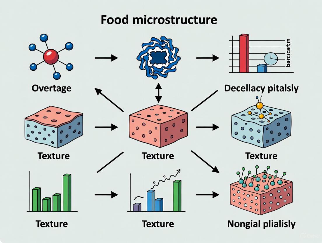
This article provides a comprehensive analysis of the fundamental relationship between food microstructure and its resulting textural properties, a critical consideration for product development and sensory science.
Microstructure to Mouthfeel: Decoding Food Texture for Scientific Innovation
Abstract
This article provides a comprehensive analysis of the fundamental relationship between food microstructure and its resulting textural properties, a critical consideration for product development and sensory science. Tailored for researchers and scientists, we explore the structural foundations of texture, detail advanced methodologies for its characterization, address common optimization challenges, and present rigorous validation frameworks. By integrating insights from cutting-edge studies on cellular foods, gels, and meat products, this resource bridges material science with practical application, offering a foundational reference for fields ranging from food technology to biomedical formulations.
The Structural Blueprint of Texture: From Molecules to Sensory Perception
In the realm of food science, the microstructure of a material—its inner organization and composition at the microscopic level—is a fundamental determinant of its macroscopic properties, particularly texture [1]. Microstructure refers to the arrangement of constituents such as proteins, carbohydrates, and fats, separated by grain and phase boundaries, which collectively influence the mechanical, thermal, and sensory characteristics of food [1]. Understanding and controlling this microstructure is therefore critical for product development and quality control.
The relationship between food microstructure and texture is profound. Food texture, defined as "all the mechanical, geometrical and surface attributes of a product perceptible utilizing mechanical, tactile and, where appropriate, visual and auditory receptors," is directly governed by the underlying physical structure [2]. This encompasses mechanical properties (e.g., hardness, cohesion), geometrical properties (e.g., particle size and shape), and surface properties (e.g., moistness, creaminess) [2]. Consequently, manipulating the microstructure allows scientists to engineer specific textural experiences, a principle that is central to advancing sustainability in food systems by creating appealing alternative protein products [3].
This whitepaper provides an in-depth technical exploration of microstructure within the context of food and biomaterial science. It is designed to equip researchers, scientists, and drug development professionals with a clear understanding of core concepts, modern evaluation techniques, and practical experimental methodologies for analyzing and designing microstructures to achieve desired functional outcomes.
Core Concepts and Definitions
Microstructure analysis involves the quantitative and qualitative examination of a material's characteristics at the microscopic level [1]. The primary components and features of a microstructure include:
- Grains: Individual crystalline areas within a material. Their size, shape, and orientation are critical; for instance, smaller grain sizes generally result in higher strength, while larger grains enhance ductility [1].
- Phase Boundaries: Surfaces that separate different microstructure constituents from one another [1].
- Porosity: The presence of pores or voids within the structure, which can affect texture and density [1].
- Inclusions: Foreign constituents, such as non-metallic particles, which can alter the material's characteristics [1].
These components are not static; they are influenced by processing conditions such as heat treatment, forging, and casting, which can permanently alter the microstructure and, consequently, the material's properties [1].
In food systems, microstructures can be broadly categorized into:
- Cellular Networks: Organized assemblies of cells, such as those found in plant tissues or engineered cell cultures in biomaterials [4] [5].
- Gel Matrices: Three-dimensional networks formed by polymers like proteins or polysaccharides that trap water, creating a semi-solid structure. These are ubiquitous in products like yogurt, jellies, and plant-based meat analogs [3].
- Protein Assemblies: Structures formed by the aggregation and organization of protein molecules, which can form gels, fibrous networks, or particulate structures [3].
The principles of microstructure analysis are universally applicable across materials science, whether the subject is a metal, a ceramic, a polymer, or a food product [1]. In the following sections, we will explore the techniques used to evaluate these structures and their direct link to sensory perception.
Microstructure-Texture Relationship in Foods
The connection between microstructure and texture is mechanistic and forms the basis for rational food design. A prime example is the development of plant-based protein foods, where mimicking the heterogeneous texture of animal tissue is a key challenge.
Recent research has demonstrated that composite gels of pea protein and curdlan gum can be tuned to achieve a range of textures by controlling thermal history [3]. The microstructure, and thus the texture, is directly modulated by processing temperature:
- At 60°C, large pea protein particles remain visible and are only partially embedded into the curdlan network.
- At 80°C, smaller protein particles are fully embedded into a unified gel network [3].
This microstructural difference translates to macroscopic mechanical properties. Isothermal incubation at 80°C for 1 hour can increase the gel's storage modulus (G') from 1.3 kPa to 288 kPa, indicating a dramatic increase in rigidity [3]. Furthermore, by creating gels with varying thermal histories in different layers, researchers can produce heterogenous structures that mimic the varying stiffness found in animal tissues [3]. This illustrates how deliberate microstructural engineering can create complex, desirable textures without the need for extrusion.
Table 1: Quantitative Microstructure-Texture Relationship in a Pea Protein-Curdlan Gum Composite Gel
| Processing Condition | Resulting Microstructure | Storage Modulus (G') | Resulting Textural Property |
|---|---|---|---|
| 55-60°C, 1h incubation | Large protein particles partially embedded in network | Similar to 80°C after incubation (e.g., ~288 kPa) | Heterogenous, tunable stiffness |
| 80°C, no incubation | Protein particles more fully integrated | 1.3 kPa | Soft gel |
| 80°C, 1h incubation | Protein particles fully embedded, developed network | 288 kPa | Rigid gel |
| Increased protein conc. (4% to 8%) | Less organized network, more space occupied by particles | Modulated | Altered firmness and organization |
Beyond mechanical properties, geometrical properties are equally important. Attributes like graininess are related to the size, shape, and orientation of particles within the food [2]. Evaluating these complex attributes requires sophisticated instrumental methods that move beyond traditional rheology [2].
Methodologies for Microstructure Analysis
A robust analysis of microstructure requires careful sample preparation, advanced imaging techniques, and quantitative image analysis.
Sample Preparation Protocols
Proper preparation is the most critical step for accurate microstructure analysis [1]. The standard protocol for solid samples in metallography, which is directly applicable to many food and biomaterials, is as follows:
- Sampling and Sectioning: Obtain a representative sample and cut it to a manageable size using a cutting system that avoids generating a heat-affected zone that could alter the native structure [1].
- Grinding and Polishing: Grind the surface flat and then polish it using progressively finer abrasives (e.g., diamond, aluminium oxide) on polishing cloths or discs. The goal is to produce a smooth, reflective surface that reveals the microscopic microstructure without distortion [1].
- Etching (if applicable): For many materials, the polished surface is treated with a chemical etchant. The etching agent reacts differently with various phases or grains, creating contrasts that make the microstructure constituents visible under a microscope [1].
For soft biological or food materials like hydrogels and cell-embedded matrices, preparation involves stabilization, often through fixation, followed by dehydration and critical point drying before imaging under scanning electron microscopy (SEM) to preserve delicate structures [4] [5].
Imaging and Visualization Techniques
- Light Microscopy: The foundational tool for qualitative analysis. It allows for the initial assessment of grain structures, phase boundaries, and overall architecture [1]. Advanced forms like Confocal Laser Scanning Microscopy (CLSM) are invaluable for visualizing specific components in food, such as proteins or fats, by using fluorescent tagging [3].
- Scanning Electron Microscopy (SEM): Provides high-resolution, topographical images of surfaces at the micro- and nanoscale. SEM is crucial for studying the surface morphology of foods like grains, crispy products, and powders, revealing details about texture, moisture retention, and overall quality [6].
- Image Processing Software: Tools like ImageJ (with plugins such as OrientationJ) and custom scripts in MATLAB or Python are used to quantitatively analyze microscopic images. These tools can calculate local collagen orientation and coherence in biomaterials [4], or perform cell segmentation and morphological analysis [5].
Quantitative Evaluation Methods
After imaging, quantitative data is extracted using standardized methods:
- Linear Intercept Method: Lines are drawn on microscopic images, and the number of intersections between these lines and microstructure constituents (e.g., grain boundaries) is counted to quantify features like grain size [1].
- Planimetry (Surface Area Analysis): The microscopic image is analyzed to determine the surface areas of various microstructure constituents, such as different phases within the material [1].
- Point Count Method: A grid of points is overlaid on the microscopic image. The number of contact points between the grid and specific microstructure constituents is counted to determine their volume fraction [1].
- Fractal Analysis: A quantitative approach used to describe the complexity and heterogeneity of a surface's structure. When applied to SEM images, fractal and multifractal analysis yields numerical descriptors of surface roughness and complexity, which can be correlated with food attributes and processing conditions [6].
Experimental Protocols for Key Analyses
Protocol: Engineering a Heterogenous Protein Gel
This protocol, adapted from a study on pea protein, details the creation of a composite gel with tunable microstructure to mimic animal tissue texture [3].
Objective: To form a composite pea protein and curdlan gum gel and modulate its gel network and texture through thermal history.
Materials:
- Pea protein isolate
- Curdlan gum
- Deionized water
- Heating bath with precise temperature control
Methodology:
- Solution Preparation: Prepare a neutralized solution of pea protein and curdlan gum in deionized water. The study used protein concentrations of 4% to 8% (w/w).
- Composite Gel Formation:
- Option A (Soft Gel): Heat the solution to 55-60°C. At this temperature, a soft gel forms.
- Option B (Rigid Gel): Heat the solution to 80°C. This temperature induces the formation of a rigid gel.
- Network Development (Isothermal Incubation): Hold the gel at the target temperature (e.g., 80°C) for a defined period (e.g., up to 1 hour). This step significantly develops the gel network, as evidenced by a massive increase in storage modulus.
- Creating Heterogeneity: To mimic a multi-layered animal tissue structure, create separate gel batches with different thermal histories (e.g., one incubated at 60°C, another at 80°C) and layer them.
Key Analysis:
- Rheology: Measure the storage modulus (G') using a rheometer to quantify gel strength.
- Microstructure Imaging: Use Confocal Laser Scanning Microscopy (CLSM) and Scanning Electron Microscopy (SEM) to visualize the integration of protein particles into the curdlan network and observe differences between gels formed at 60°C and 80°C [3].
Protocol: Patterning ECM Microstructure for 3D Cell Culture
This protocol summarizes the use of the MC3A platform to pattern 3D extracellular matrix (ECM) with controlled microstructure for cell migration studies [4].
Objective: To engineer a 3D collagen ECM with aligned microstructure to study the effects of contact guidance and chemotaxis on cell migration.
Materials:
- FITC-labeled Type I Collagen (e.g., from rat tail)
- Cell line of interest (e.g., MDA-MB-231 breast cancer cells)
- MC3A platform (or custom rotary stage and culture inserts)
- Sulfo-SANPAH
- Corona treater
- Serum-free and serum-rich media
Methodology:
- Surface Functionalization: Corona treat the culture inserts for 10 minutes. Then, immerse with Sulfo-SANPAH and expose to UV light (320-350 nm) for 2 hours to functionalize binding surfaces. Wash with PBS and deionized water.
- Cell Suspension in Collagen: Suspend cells at low density in a neutralized collagen solution (e.g., 1.5 mg/mL, pH 7.4).
- Matrix Patterning: Transfer the cell-collagen suspension to the functionalized assay. Place a treated blade at the center of the assay using the rotary stage and rotate at a defined speed (e.g., 128 RPM for 4 minutes). This rotation creates a flow that templates the alignment of the collagen fibers as they polymerize. For radial alignment, use zero RPM.
- Polymerization: Polymerize the solution for 21 minutes at room temperature, then for 25 minutes in a 5% CO₂ incubator at 37°C.
- Establishing Chemotactic Gradient: After serum starvation, replace the medium in the center reservoir with a serum-rich medium and the outer reservoir with a serum-free medium. This establishes a stable serum gradient across the channel, driving chemotaxis.
Key Analysis:
- Live-Cell Imaging: Capture time-lapse images over 18 hours using a confocal microscope to track cell migration and morphology.
- ECM Geometry Analysis: Use the ImageJ plugin OrientationJ to compute the local principal direction and coherence of the collagen fibers from the acquired images [4].
The Scientist's Toolkit: Research Reagent Solutions
Table 2: Essential Research Reagents and Materials for Microstructure Studies
| Item | Function/Application | Technical Notes |
|---|---|---|
| Curdlan Gum | A bacterial polysaccharide that forms thermal-irreversible gels; used to create composite plant protein gels with tunable texture. | Gel strength and microstructure are modulated by thermal history (55-80°C) [3]. |
| Pea Protein Isolate | A plant-derived protein used as a model alternative protein for structuring meat analogs. | Forms composite gels with curdlan; particle integration into the network is temperature-dependent [3]. |
| FITC-labeled Collagen Type I | A fluorescently conjugated extracellular matrix protein for 3D cell culture and migration assays. | Allows visualization of ECM fiber alignment and organization using fluorescence microscopy [4]. |
| Sulfo-SANPAH | A photoactivatable, water-soluble crosslinker for surface functionalization. | Used to covalently attach biomolecules (e.g., collagen) to culture surfaces under UV light [4]. |
| ImageJ / FIJI with OrientationJ | Open-source image processing software with a plugin for directional and coherence analysis. | Quantifies local fiber orientation and level of alignment (coherence) in fibrous materials like ECM or biopolymer networks [4]. |
| Scanning Electron Microscope (SEM) | High-resolution imaging instrument for topographical surface characterization at micro- and nanoscales. | Essential for visualizing surface morphology; combined with fractal analysis to quantify roughness and complexity [6]. |
| Rheometer | Fundamental instrument for measuring mechanical properties, specifically viscoelasticity. | Quantifies storage (G') and loss (G") moduli, providing direct correlation between microstructure and mechanical texture [3] [2]. |
The precise definition and control of microstructure in cellular networks, gel matrices, and protein assemblies are paramount for understanding and engineering the texture of foods and biomaterials. As demonstrated, techniques like controlled gelation and ECM patterning allow researchers to deliberately architect microstructures, which in turn dictate macroscopic sensory and functional properties like rigidity, chewiness, and creaminess.
The future of this field lies in the continued development of advanced analytical methods, including the integration of fractal analysis with SEM for quantitative surface characterization [6], and multi-modal instrumental approaches that combine rheology, tribology, and tactile sensing to better predict complex sensory texture attributes [2]. By mastering the relationship between microstructure and texture, scientists can more effectively develop sustainable food products and advanced biomaterials that meet specific quality and sensory demands.
Food texture is a critical quality attribute, defined as "all the mechanical, geometrical and surface attributes of a product perceptible utilizing mechanical, tactile and, where appropriate, visual and auditory receptors" [2]. In both food and pharmaceutical industries, texture profoundly influences product acceptability, stability, and performance. This technical guide examines four fundamental textural properties—hardness, crispness, chewiness, and spreadability—within the context of food microstructure research. Understanding the relationship between a product's microstructural organization and its macroscopic textural properties enables researchers to design foods with targeted functional and sensory characteristics. Advances in instrumental texture analysis now allow precise quantification of these properties, bridging the gap between subjective sensory perception and objective physical measurements [2] [7].
Defining Key Textural Properties
Mechanical Properties and Their Definitions
Textural properties can be categorized into primary mechanical characteristics perceived during oral processing. The table below defines the four key properties and their relationship to food microstructure.
Table 1: Fundamental textural properties and their characteristics
| Textural Property | Definition | Microstructural Basis | Common Food Examples |
|---|---|---|---|
| Hardness | The force required to achieve a given deformation or penetration; often measured as peak force during compression [8]. | Density, strength of internal bonds, and structural integrity of the food matrix. | Hard candy, firm fruits, nuts [8] [7] |
| Crispness | A combination of hardness and brittleness, characterized by a series of fractures under compressive force with audible acoustic emission. | Stiff, porous cellular structure that fractures catastrophically when stressed. | Potato chips, breakfast cereals, crackers [9] |
| Chewiness | The energy required to masticate a solid food into a state ready for swallowing; calculated from TPA parameters [2]. | Degree of cross-linking in protein or polymer networks, elasticity, and resistance to breakdown. | Chewy candy, steak, bagels [2] |
| Spreadability | The ease with which a material can be spread over a surface; related to shear resistance and adhesiveness. | Soft, plastic structure with low yield stress and appropriate adhesive properties. | Butter, margarine, cream cheese [8] |
The Texture Perception Pathway
The perception of these properties is not merely mechanical but arises from a dynamic process known as oral processing, where food is physically broken down and lubricated with saliva to form a bolus safe for swallowing [2] [10]. This process entails a complex interplay of physical processing and sensory perception, where mechanical, geometrical, and surface properties are detected by tactile, auditory, and even visual receptors [2]. The following diagram illustrates the pathway from food structure to sensory perception.
Quantitative Measurement and Instrumental Analysis
Principles of Instrumental Texture Analysis
Texture Analyzers provide objective quantification of textural properties by compressing, stretching, bending, or shearing a sample while precisely measuring the force response [8]. The key data channels collected are force, distance, and time, from which parameters like stress and strain are derived [11]. The resulting force-distance or force-time graphs provide a visual interpretation of how materials respond to applied forces, revealing characteristics like hardness (peak force), cohesiveness (the degree to which a sample deforms before rupturing), and elasticity (how well a sample springs back after deformation) [8].
Proper load cell selection is critical for measurement accuracy. Load cells should be used to measure forces from approximately 10% to 100% of their capacity. Using a high-capacity load cell to measure very small forces can result in significant electronic noise on the test graph, while a low-capacity cell may overload when testing hard samples [11].
Standardized Measurement Methodologies
Table 2: Standard experimental protocols for measuring key textural properties
| Property | Test Type | Probe/Jig Type | Typical Protocol Parameters | Key Output Metric |
|---|---|---|---|---|
| Hardness | Compression or Puncture | Cylindrical Probe or Puncture Probe | Pre-test speed: 1-2 mm/sTest speed: 1-5 mm/sPost-test speed: 10 mm/sStrain: 50-75% | Peak Force (N or g) [7] |
| Crispness | Three-Point Bending or Shear | 3-Point Bending Rig or Kramer Shear Cell | Pre-test speed: 1-2 mm/sTest speed: 1-2 mm/sDistance: until fracture | Number of force peaks,Break Force (N),Slope of curve (N/mm) [7] [9] |
| Chewiness | Texture Profile Analysis (TPA) | Cylindrical Compression Platen | Two consecutive compression cyclesStrain: 25-75% (sample dependent) | Calculated parameter:Hardness × Cohesiveness × Springiness [2] |
| Spreadability | Backwards Extrusion or Shear | Cone Probe or Spreader Fixture | Test speed: 1-2 mm/sTarget distance or force | Peak Force (N) orWork of Shear (N×mm) [8] |
Experimental Workflow for Texture Analysis
A standardized workflow is essential for generating reproducible and meaningful texture analysis data. The following diagram outlines a generalized protocol applicable to various food and pharmaceutical products.
Research-Grade Experimental Protocols
Protocol 1: Measuring Hardness and Crispness in Baked Goods
This protocol is adapted from recent studies on biscuit texture, utilizing a three-point bending test to evaluate hardness and crispness simultaneously [12] [7].
4.1.1 Research Reagent Solutions and Materials
Table 3: Essential materials and reagents for baked goods texture analysis
| Item | Specification/Function |
|---|---|
| Texture Analyzer | Stable Micro Systems or Shimadzu EZ Test equipped with a 50 kg load cell for sufficient capacity [11] [7]. |
| Three-Point Bending Rig | Jig with maximum 100 mm between fulcrums; tip of punch and fulcrum typically R1 or R2.5 [7]. |
| Flat-Base Support Rig | Used as an alternative for compression tests on thicker biscuits or crackers [12]. |
| Standardized Samples | Biscuits/cookies of uniform dimensions (e.g., 80 mm length, 10 mm thickness); conditioned to controlled humidity (e.g., 50% ± 5%) [12]. |
4.1.2 Detailed Procedure
- Sample Preparation: Condition biscuit samples at 25°C ± 2°C and 50% ± 5% relative humidity for at least 24 hours before testing to standardize moisture content [12].
- Instrument Setup: Install the three-point bending rig. Set the distance between the two support fulcrums (typically 50-80% of the sample length). Calibrate the instrument for force and distance.
- Test Parameters:
- Pre-test speed: 1.0 mm/s
- Test speed: 2.0 mm/s
- Post-test speed: 10.0 mm/s
- Target mode: Distance (set to break the sample)
- Trigger force: 5 g
- Testing: Place the sample across the two supports. Start the test. The probe will descend and fracture the sample.
- Data Analysis:
- Hardness/Firmness: Record the peak force (N) required to break the sample.
- Crispness/Brittleness: Calculate the slope (N/mm) of the initial linear region of the force-distance curve. A steeper slope indicates a stiffer, crisper product [7] [9]. The fracture pattern (a clean, sharp break) is also indicative.
Protocol 2: Texture Profile Analysis (TPA) for Chewiness
TPA is a two-bite compression test that mimics the action of the jaw, providing multiple textural parameters, including chewiness, from a single test [2].
4.2.1 Detailed Procedure
- Sample Preparation: Prepare samples with a consistent height-to-diameter ratio (often 1:1 or 1:2). Cylindrical cores are ideal.
- Instrument Setup: Use a large diameter cylindrical compression platen (e.g., 75 mm). The platen should be significantly larger than the sample to prevent edge effects.
- Test Parameters:
- Pre-test speed: 2.0 mm/s
- Test speed: 1.0 mm/s (or slower for soft samples)
- Post-test speed: 2.0 mm/s
- Strain: 50-75% (This is critical and must be optimized for the product type).
- Time between cycles: 3-5 seconds (to allow for partial recovery).
- Testing: The analyzer performs two consecutive compression cycles on the same sample.
- Data Analysis: From the TPA curve, calculate the following [2]:
- Hardness: Peak force of the first compression cycle (N).
- Cohesiveness: Ratio of the area under the second compression curve to the area under the first compression curve (Adhesiveness/Area 1).
- Springiness: The distance the sample recovers in height between the end of the first bite and the start of the second bite (mm).
- Chewiness (for solid foods): = Hardness × Cohesiveness × Springiness.
Case Study: Linking Microstructure to Texture in Food Research
Impact of Pumpkin Seed Flour on Biscuit Hardness
A 2025 study investigated the effect of supplementing wheat flour (WF) with pumpkin seed flour (PSF) on biscuit texture. The research demonstrated a direct link between ingredient composition (microstructure), dough rheology, and final product texture [12].
Table 4: Textural and sensory properties of biscuits with pumpkin seed flour (PSF) during storage
| Formulation (WF:PSF) | Hardness (N) at Day 0 | Hardness (N) at Day 60 | Overall Acceptability (9-pt scale) |
|---|---|---|---|
| 100:0 (Control) | Data from source | Data from source | Baseline |
| 95:5 | Data from source | Data from source | Score |
| 90:10 | Data from source | Data from source | Score |
| 85:15 (D3) | 59.62 | 59.69 | 8.33 |
| 80:20 | Data from source | Data from source | Score |
Key Findings: The incorporation of 15% PSF (D3 formulation) resulted in a biscuit that maintained its textural properties (hardness) over a 60-day storage period, with minimal change from 59.62 N to 59.69 N. This formulation also achieved the highest overall acceptability score of 8.33, indicating that optimizing the composite flour matrix can enhance both nutritional profile and textural stability without compromising sensory appeal [12]. The study attributed these changes to the interaction of PSF proteins and fibers with the gluten network, altering the dough's water absorption and development time, which ultimately influenced the microstructure and mechanical strength of the final baked product [12].
Molecular Structure and Extrudate Crispness
Research on extruded wheat flour establishes a direct correlation between the molecular architecture of starch and the macroscopic texture of the final puffed product. SEC analysis revealed that crispness correlates negatively with long amylopectin branches (degree of polymerization 36 < X ≤ 100). Waxy wheat (WW), with its specific amylopectin structure, displayed superior crispness, quantified as 12.22 N/mm [9]. This finding provides a powerful example of how the manipulation of a single ingredient at the molecular level, through breeding or processing, can be used to design a specific textural experience.
The Scientist's Toolkit
Table 5: Essential research reagents and instruments for textural property analysis
| Item/Category | Function in Texture Analysis | Example Applications |
|---|---|---|
| Texture Analyzer | Core instrument that applies controlled force/displacement to a sample and measures the response. | Universal testing for hardness, fracture, tension, etc. [8] [7] |
| Cylindrical Probe | Used for compression, puncture, and TPA tests. | Measuring hardness of gels, fruits, and baked goods [7]. |
| Three-Point Bending Rig | Bends a sample supported at two ends until fracture. | Evaluating crispness and breaking strength of biscuits and snack bars [7]. |
| Kramer Shear Cell | Simultaneously compresses and shears a sample with multiple blades. | Assessing toughness and hardness of fibrous materials and cereals [7]. |
| Back Extrusion Rig | Compresses a sample in a container with a small annular gap, forcing the sample to flow upwards. | Determining viscosity and spreadability of semi-solids like yogurt and margarine [8] [7]. |
| Tensile Grips | Hold a sample at both ends and pull apart to measure extensibility and adhesive strength. | Testing stickiness of candies or stretchability of noodles [7]. |
The quantitative analysis of hardness, crispness, chewiness, and spreadability provides indispensable data for researchers developing new food and pharmaceutical products. As demonstrated, these properties are not standalone characteristics but are direct manifestations of a product's underlying microstructure and composition. The advancing integration of instrumental methods from rheology and tribology, coupled with emerging technologies like micro-analytical and tactile sensing techniques, promises to deepen our understanding of sensory texture perception [2]. By systematically applying the principles and protocols outlined in this guide, scientists can more effectively engineer novel matrices that deliver targeted textural experiences, thereby bridging the critical gap between fundamental material science and consumer perception.
The texture of crispy snacks, a key determinant of consumer preference, is not a bulk property but a direct consequence of their microscopic architecture. Research demonstrates that the sensory experience of crispness and hardness is fundamentally governed by the mechanical failure of a food's internal structure [13] [14]. This case study examines the intrinsic relationship between the cellular geometry of dry crispy foods—specifically cell size and cell wall thickness—and their resulting mechanical and acoustic properties. The principles are framed within the context of cellular solid mechanics, where foods like toasted rusk rolls, extruded snacks, and wafers are treated as solid foams [15]. Their behavior under mechanical stress, which dictates sensory texture, can be predicted by their structural parameters. Understanding this structure-function relationship is critical for the food industry, not only for product development and quality control but also for innovating healthier and more sustainable food formats that maintain desirable textural properties.
Cellular Architecture as a Determinant of Texture
Fundamental Principles of Solid Foams
The mechanical properties of crispy cellular foods can be conceptually modeled using theories developed for engineering foams, such as the Gibson-Ashby model for cellular solids [15] [14]. These models treat the food as a three-dimensional network of solid struts and plates (the cell walls) surrounding pockets of air. When a force is applied, as during biting, the structure responds through the elastic bending, buckling, and ultimately, the brittle fracture of these cell walls [15]. The frequency and pattern of these fracture events create the force fluctuations and acoustic emissions perceived as crispness [13]. The relative density of the foam, defined as the ratio of the foam's bulk density to the density of the solid cell wall material, is a primary factor controlling its mechanical strength. This relative density is itself a function of the cellular architecture: the average cell size and the thickness of the cell walls.
Key Structural Parameters and Their Sensory Impact
Two geometrical parameters are paramount in determining texture: mean cell size and cell wall thickness.
- Cell Size: A coarse structure with larger air cells generally produces a crispier texture compared to a fine structure with smaller cells. Studies on toasted rusk rolls have shown that products with a coarser morphology were rated as significantly crispier by sensory panels than their fine-structured counterparts, even when their densities were similar [14]. This is because larger cells may promote more extensive and louder fracture events during chewing.
- Cell Wall Thickness: Thicker cell walls contribute to greater structural stiffness and hardness. The mechanical properties of cellular foams, such as Young's modulus, are directly correlated to cell wall thickness [15]. Conversely, thinner cell walls are more prone to brittle fracture, generating the high-frequency acoustic signals characteristic of crispness.
The interplay between these parameters means that a snack with a combination of large cells and moderately thick walls might be both hard and crisp, whereas one with small cells and very thin walls might be fragile but less noisy.
Quantitative Data on Structure-Texture Relationships
The following tables consolidate quantitative findings from research on how specific structural parameters influence textural properties.
Table 1: Impact of Cellular Morphology on Sensory Crispness in Toasted Rusk Rolls [14]
| Morphology Type | Average Gas Cell Size (mm²) | Product Density (g/mL) | Critical Water Activity (Awc) | Sensory Crispness Retention |
|---|---|---|---|---|
| Coarse Structure | 0.17 ± 0.02 | 0.11 ± 0.01 | 0.59 | Higher |
| Fine Structure | 0.10 ± 0.01 | 0.12 ± 0.02 | 0.57 | Lower |
Table 2: Biochemical Composition of Pericarp Cell Wall and its Effect on Popping Expansion in Popcorn [16]
| Popcorn Inbred Line | Popping Expansion Volume (mL g⁻¹) | Lignin Content (μg mg⁻1 / %) | Key Monosaccharide in Cell Wall |
|---|---|---|---|
| GP12 (High Expansion) | > 40 | 129.74 / 12.97% | Xylose |
| P11 (Medium Expansion) | 30 | Not Specified | Xylose |
| P16 (Low Expansion) | 14 | 113.52 / 11.35% | Xylose |
Table 3: Mechanical and Structural Comparison of Baked vs. Extruded Confectionery [15]
| Manufacturing Process | Key Microstructural Features | Resulting Mechanical & Textural Properties |
|---|---|---|
| Baking | Often achieves a very "light" cellular structure with fine, interconnected pores. | Traditionally associated with a highly desirable "light, crispy" texture. |
| Extrusion | Microstructure and porosity are highly sensitive to initial moisture content and die geometry. | Challenging to replicate baked crispness; higher density products are harder and less crispy. |
Experimental Protocols for Microstructural and Mechanical Analysis
To establish the causal links detailed in this case study, researchers employ a combination of material characterization, mechanical testing, and sensory analysis.
Protocol 1: Microstructural Quantification via Imaging
Objective: To quantitatively characterize the cellular architecture (cell size, cell wall thickness, and porosity) of snack products [15] [14].
- Sample Preparation: Carefully prepare cross-sections of the snack product using a sharp blade or cryo-microtome to avoid smearing or damaging the fragile cellular structure.
- Image Acquisition:
- Scanning Electron Microscopy (SEM): Securely mount the sample on a stub, sputter-coat with a conductive material like gold, and image under high vacuum. SEM provides high-resolution images of surface topology and cell wall details [15].
- X-Ray Micro-Computed Tomography (XRT): Mount the sample on a stage and perform a non-destructive 3D scan. XRT generates cross-sectional images that can be reconstructed into a 3D model of the internal pore network, allowing for precise calculation of porosity and cell size distribution [15].
- Image Analysis: Use specialized software (e.g., ImageJ, Avizo) to analyze the acquired images. For 2D images, thresholding can distinguish solid material from air cells. Measurements of cell area (converted to cell size) and cell wall thickness can be automated or performed manually on a representative number of cells. For 3D data, the software can directly compute parameters like total porosity and mean pore size.
Protocol 2: Acoustic-Mechanical Signature Analysis
Objective: To objectively measure the crispness of a product by simultaneously capturing its mechanical failure and accompanying acoustic emissions [13].
- Instrument Setup: A Texture Analyzer or Universal Testing Machine is fitted with a probe or plate suitable for compression or puncture. An acoustic sensor (e.g., a piezoelectric microphone or an acoustic emission detector) is positioned close to the sample or mounted on the probe. The system must be placed in a sound-dampened enclosure to minimize ambient noise.
- Data Synchronization: The force-time data from the mechanical tester and the acoustic data from the microphone are synchronized to a common time base.
- Testing: The probe compresses the snack sample at a constant speed until complete fracture. The test is repeated on multiple samples for statistical reliability.
- Data Analysis:
- Mechanical Analysis: The force-displacement curve is analyzed for characteristics like the number of force peaks (corresponding to fracture events), mean force, and the curve's jaggedness.
- Acoustic Analysis: The sound recording is analyzed for total acoustic energy, number of acoustic events, and the amplitude/frequency of the sounds produced. A combined acoustic-mechanical signature provides a robust instrumental correlate for sensory crispness.
Protocol 3: Sensory Evaluation of Texture
Objective: To obtain human perceptual data on texture attributes like crispness and hardness [14] [2].
- Panel Training: Recruit assessors and train them to recognize and consistently score specific texture attributes using reference samples.
- Sample Presentation: Present samples (e.g., with different microstructures or water activities) to panelists in a randomized order under controlled conditions.
- Data Collection: Use sensory evaluation methods such as:
- Quantitative Descriptive Analysis (QDA): Panelists score the intensity of attributes (e.g., crispness, hardness) on a continuous scale.
- Temporal Dominance of Sensations (TDS): Panelists indicate which texture attribute is dominant at each moment during consumption.
- Data Correlation: Statistical analysis (e.g., regression, principal component analysis) is used to correlate the instrumental measurements from Protocols 1 and 2 with the sensory scores from the panel.
The Scientist's Toolkit: Essential Research Reagents and Materials
Table 4: Key Reagents and Instruments for Microstructure-Texture Research
| Item Name | Function/Application |
|---|---|
| Texture Analyzer | A universal testing machine that quantifies mechanical properties (hardness, fracturability) by simulating biting/chewing forces. It is often coupled with acoustic sensors [17] [13]. |
| Scanning Electron Microscope (SEM) | Provides high-resolution images of a sample's surface microstructure, allowing for visual inspection and measurement of cell walls and pores [15]. |
| X-Ray Micro-Computed Tomography (XRT) | A non-destructive 3D imaging technique that visualizes and quantifies the internal porous architecture, including closed pores, which is crucial for calculating true porosity [15]. |
| Piezoelectric Acoustic Sensor | A microphone specifically designed to capture the high-frequency sounds produced during the fracture of crispy foods, synchronized with mechanical testing [13]. |
| Controlled Humidity Chambers | Used to condition food samples to specific water activities (Aw) for studying the critical impact of moisture on texture degradation and crispness loss [14]. |
| Image Analysis Software | Software tools (e.g., ImageJ, Avizo) used to quantitatively analyze 2D and 3D images to extract metrics like mean cell size, cell wall thickness, and porosity [15] [14]. |
This case study establishes that the macroscopic textural properties of snack foods, namely hardness and crispness, are a direct physical manifestation of their microscopic cellular architecture. The application of solid foam mechanics provides a predictive framework where cell size and cell wall thickness are the primary levers controlling mechanical strength and fracture behavior. The quantitative relationships summarized herein provide researchers and product developers with a scientific basis for engineering desired textures. By leveraging advanced imaging, acoustic-mechanical analysis, and sensory science, the field can move beyond empirical formulations toward the rational design of foods. Future research integrating multi-scale modeling and high-throughput characterization will further deepen our understanding, enabling the creation of novel food structures that meet evolving consumer demands for health, sustainability, and superior sensory experience.
In the field of food microstructure research, the relationship between a food's molecular architecture and its macroscopic textural properties is a fundamental area of investigation. This case study examines the crucial role of protein secondary structure, specifically the balance between α-helix and β-sheet configurations, in determining the texture of meat and meat analogue products. The structural proteins in muscle tissue—primarily myosin and actin—form the fundamental building blocks that dictate functional properties such as water holding capacity, gelation, and tenderness [18]. These properties directly influence sensory perception and product quality. During processing, whether through thermal treatments or novel non-thermal technologies, the native conformation of meat proteins undergoes significant changes. The conversion from α-helix to β-sheet structures often serves as a key indicator of protein denaturation and aggregation, which subsequently defines the textural characteristics of the final product [19]. Understanding these relationships at the molecular level provides researchers and product developers with powerful tools for optimizing texture in both traditional meat products and emerging alternatives.
Fundamental Principles of Protein Secondary Structure in Meat
Structural Proteins in Muscle Tissue
Meat proteins are categorized into three primary groups: myofibrillar, sarcoplasmic, and connective tissue proteins, each contributing distinctly to meat's structural and textural properties. Myofibrillar proteins, which constitute approximately 55-60% of muscle proteins, are the most significant for texture formation [18]. This category includes myosin, actin, tropomyosin, and the troponin complex, which are arranged in highly organized filaments within the muscle cell. These proteins naturally exhibit high levels of α-helical content in their native state, which is essential for their biological functions in muscle contraction and structural support. The spatial arrangement of these proteins creates the fundamental architecture that determines meat's initial texture and its transformation during processing.
Analytical Methods for Secondary Structure Characterization
Advanced spectroscopic techniques enable precise quantification of protein secondary structure changes in meat systems:
Raman Spectroscopy: Provides information on the micro-environment and chemistry of protein side chains as well as the conformation of the protein polypeptide backbone. Changes in Raman bands can reveal modifications in secondary structures (amide conformation region, C-C stretching vibration) and local environments (tryptophan residues, tyrosyl-doublet, aliphatic amino acid bands) [19].
Fourier Transform Infrared (FTIR) Spectroscopy: Particularly useful for analyzing changes in the amide I band (1600-1700 cm⁻¹), which is sensitive to protein secondary structure. This method allows researchers to quantify the relative proportions of α-helix, β-sheet, β-turn, and random coil structures in meat protein systems [18].
Intrinsic Fluorescence Spectroscopy: Used to monitor changes in protein tertiary structure by measuring the fluorescence emission of aromatic amino acids such as tryptophan. Shifts in the maximum absorption wavelength indicate alterations in the hydrophobic environment of these residues [20].
Experimental Evidence: Linking Secondary Structure to Meat Texture
Heat-Induced Structural Changes in Meat Batters
A pivotal study investigating meat batters prepared with different lipid sources (pork fat, soybean oil, and butter) demonstrated significant heat-induced structural changes quantified using Raman spectroscopy [19]. The research revealed that heating caused a substantial decrease in α-helix content accompanied by a concurrent increase in β-sheet structures across all formulations. Specifically, meat batters formulated with soybean oil showed the most pronounced structural reorganization, with the highest β-sheet content after heating. These structural changes directly correlated with textural properties, as evidenced by significantly greater hardness, springiness, cohesiveness, chewiness, and resilience values in the soybean oil treatment compared to batters prepared with pork fat or butter.
Table 1: Correlation Between Protein Secondary Structure and Textural Properties in Heated Meat Batters [19]
| Lipid Type | α-helix Decrease (%) | β-sheet Increase (%) | Hardness (N) | Springiness | Cohesiveness | Chewiness (N) |
|---|---|---|---|---|---|---|
| Pork Fat (PF) | Significant | Significant | 25.4 | 0.84 | 0.56 | 12.1 |
| Soybean Oil (SO) | Significant | Significant | 32.7 | 0.89 | 0.62 | 18.2 |
| Butter (DB) | Not Significant | Not Significant | 22.8 | 0.81 | 0.52 | 9.8 |
The study further established statistically significant correlations between secondary structure elements and functional properties. A positive correlation (P < 0.05) was observed between β-sheet content and textural parameters, while α-helical structure showed a significant positive correlation with total expressible fluid. Conversely, β-sheet structure demonstrated a significant negative correlation with total expressible fluid, indicating that the structural transition to β-sheet configurations improves water-binding capacity in processed meat products [19].
Structural Transitions in Plant-Based Meat Analogues
Recent research has compared the structural properties of soybean-based meat analogues with traditional chicken breast, providing insights into how protein structuring processes affect texture in alternative protein products [20]. The study processed soybean protein concentrate (SPC) into high-moisture extruded texturized vegetable protein (SH) and low-moisture extruded texturized vegetable protein (SL), then compared their structural and digestibility characteristics with chicken breast. Extrusion processing successfully transformed the inherently spherical soybean protein molecular structure into fibrous formations resembling chicken breast muscle.
Table 2: Secondary Structure Parameters of Soybean Protein Products Versus Chicken Breast [20]
| Sample | α-helix/β-sheet Ratio | Intrinsic Fluorescence (nm) | Surface Hydrophobicity Index | Structural Similarity to Chicken |
|---|---|---|---|---|
| SPC | 0.68 ± 0.01 | 341.4 | 8665 ± 519 | Low |
| SH (High-Moisture) | 0.63 ± 0.01 | 352.2 | 6723 ± 285 | High |
| SL (Low-Moisture) | 0.60 ± 0.00 | 350.2 | 5167 ± 321 | Medium |
| Chicken Breast | Not Reported | Not Reported | Not Reported | Reference |
The transformation of soybean protein via extrusion resulted in decreased α-helix/β-sheet ratios, increased intrinsic fluorescence maximum absorption wavelength, and reduced surface hydrophobicity index, indicating substantial structural reorganization. These changes accompanied improved fibrous, meat-like texture, demonstrating how controlled manipulation of protein secondary structure enables the mimicking of traditional meat texture in plant-based alternatives [20].
Methodological Approaches
Sample Preparation and Processing Protocols
- Raw Material Preparation: Fresh pork center loin muscle (70.3% moisture, 22.2% protein, 6.2% fat) and pork fat (89.9% fat, 8.1% moisture, 1.7% protein) are obtained 24-48 hours postmortem (pH 5.6-5.9). All visible connective tissue and fat are trimmed from the meat.
- Comminution: Meat and fat are separately mixed and passed through a grinder with a 0.6 cm plate.
- Batter Formulation: Batters are prepared with different lipid sources (pork fat, soybean oil, or butter) using a silent cutter under vacuum. The final temperature during comminution does not exceed 12°C.
- Thermal Processing: Batter samples are stuffed into plastic casings and heated in a water bath at 75°C for 30 minutes until the core temperature reaches 72°C. After heating, samples are immediately cooled in ice water and stored at 4°C for 24 hours before analysis.
- Base Material Preparation: Soybean protein concentrate (SPC) serves as the starting material.
- High-Moisture Extrusion: SPC is processed through a twin-screw extruder with specific temperature profiles and screw configurations to create fibrous structures resembling chicken breast.
- Low-Moisture Extrusion: Similar processing with modified parameters to achieve different textural properties.
- Post-Processing: Extrudates are cooled and shaped to mimic meat products.
Analytical Techniques for Structural and Textural Characterization
Protein Secondary Structure Analysis
- Raman Spectroscopy: Spectra are collected using a Raman spectrometer equipped with a laser source at 514 nm. Measurements are taken at room temperature with a resolution of 2 cm⁻¹. Protein secondary structure is quantified by analyzing the amide I band (1600-1700 cm⁻¹) using deconvolution and curve-fitting procedures [19].
- FTIR Spectroscopy: Spectra are recorded in transmission mode with a DTGS detector. The amide I band is similarly analyzed to quantify α-helix, β-sheet, β-turn, and random coil contents through second-derivative analysis and Gaussian curve fitting [18].
Texture Profile Analysis (TPA)
Texture Profile Analysis is performed using a universal testing machine equipped with a 50 N load cell [21]. Samples are compressed twice to 50% of their original height at a constant speed of 2 mm/s, with a 5-second pause between compressions. From the resulting force-time curve, several parameters are derived:
- Hardness: Maximum force during the first compression cycle (N)
- Springiness: Degree to which the sample returns to its original height after deformation
- Cohesiveness: Extent of sample deformation before rupture (ratio of areas under second and first compressions)
- Chewiness: Energy required to masticate the sample (Hardness × Cohesiveness × Springiness)
- Resilience: How quickly the sample recovers from deformation
Water and Fat Binding Properties
- Expressible Moisture: Determined by compressing samples between filter papers and measuring moisture absorbed by the papers [22].
- Cooking Loss: Calculated as percentage weight loss after thermal processing.
- Water Holding Capacity: Assessed through centrifugal methods or NMR relaxometry to evaluate water mobility and distribution within the protein matrix [23].
The Protein Structure-Texture Relationship Pathway
The relationship between protein secondary structure and meat texture follows a defined pathway that can be visualized as a sequential process:
Experimental Workflow for Structure-Texture Analysis
A comprehensive approach to analyzing the relationship between protein secondary structure and meat texture involves multiple analytical techniques applied in sequence:
The Scientist's Toolkit: Essential Research Reagents and Materials
Table 3: Key Research Reagents and Analytical Tools for Protein Structure-Texture Studies
| Category | Specific Items | Function/Application | Research Context |
|---|---|---|---|
| Spectroscopic Tools | Raman Spectrometer | Protein secondary structure quantification via amide I band analysis | Meat batter structural changes [19] |
| FTIR Spectrometer | Secondary structure analysis through amide I and II bands | Non-thermal processing effects [18] | |
| NMR Analyzer | Water distribution and mobility assessment | Boiled beef water characterization [23] | |
| Texture Analysis | Texture Analyzer | Texture Profile Analysis (hardness, springiness, chewiness) | Cultured meat characterization [21] |
| Warner-Bratzler Blade | Shear force measurement for tenderness evaluation | Alternative to TPA for specific applications [21] | |
| Microscopy | Confocal Laser Scanning Microscope | Microstructural visualization of protein and fat networks | Meat analogue structure evaluation [22] |
| Protein Sources | Soybean Protein Concentrate | Base material for plant-based meat analogues | Structural transformation studies [20] |
| Myofibrillar Protein Isolates | Model systems for meat protein functionality | Gelation mechanism studies [18] | |
| Processing Equipment | High-Moisture Extruder | Creation of fibrous structures in plant proteins | Meat analogue production [20] |
| High-Pressure Processor | Non-thermal modification of protein structure | Myofibrillar protein gel enhancement [18] |
This case study demonstrates that the secondary structure of proteins, specifically the α-helix to β-sheet transition, serves as a critical molecular-level determinant of meat texture. The experimental evidence consistently shows that this structural reorganization during processing directly influences fundamental functional properties including gel strength, water retention, and mechanical properties [19]. These relationships hold true across diverse protein systems, from traditional meat batters to innovative plant-based analogues, highlighting the universal importance of protein conformation in determining textural outcomes.
The methodological approaches outlined provide researchers with a comprehensive toolkit for investigating these structure-function relationships. By combining advanced spectroscopic techniques for structural analysis with textural and functional measurements, scientists can develop predictive models that accelerate product development and optimization [18]. As the field continues to evolve, particularly with the emergence of novel protein sources and processing technologies, understanding these fundamental principles will remain essential for designing foods with desired sensory attributes and meeting consumer expectations for both traditional and alternative protein products.
Linking Oleogelator Self-Assembly to Fat Mimetic Functionality
The development of structured fat mimetics represents a frontier in food science, driven by the critical need to reduce saturated and trans fatty acids in the human diet without compromising sensory experience. Oleogels, structured lipid systems formed by the gelation of liquid oils via self-assembling gelators, have emerged as a principal strategy in this endeavor [24] [25]. The core thesis of this whitepaper is that the fat mimetic functionality of an oleogel—its texture, mouthfeel, stability, and overall performance—is not a inherent property of its bulk composition but a direct physical manifestation of its microstructure. This microstructure is, in turn, dictated by the precise pathway and outcome of oleogelator self-assembly. Understanding and controlling this relationship is fundamental for researchers and product developers aiming to design next-generation healthier foods, and has parallel applications in the pharmaceutical industry for the delivery of lipophilic bioactive compounds [26].
The fundamental challenge that oleogelation addresses is the structural paradox of fat replacement. While saturated and trans fats are detrimental to health, they provide the solid-like texture, plasticity, and mouthfeel consumers expect in products like spreads, baked goods, and processed meats [24] [27]. Oleogels resolve this by using a small percentage (often as low as 2%) of a gelator to immobilize a liquid oil, creating a semi-solid material that retains the nutritional profile of the unsaturated oil while providing the rheological properties of solid fat [24] [25]. The process is a physical phenomenon, relying on the formation of a three-dimensional network that entraps the oil via capillary forces [24].
Molecular Mechanisms of Oleogelator Self-Assembly
The self-assembly of oleogelators is a bottom-up process where molecules organize into supramolecular structures that define the gel's mechanical properties. The mechanism is highly dependent on the chemical nature of the gelator, primarily falling into two categories: Low Molecular Weight Gelators (LMWGs) and polymer gelators.
Self-Assembly Pathways of Low Molecular Weight Gelators (LMWGs)
LMWGs are the most widely studied class for food applications. Their self-assembly is typically driven by non-covalent interactions—including van der Waals forces, hydrogen bonding, and π-π stacking—that lead to crystallization or fibril formation upon cooling from a heated solution [24] [28]. The following diagram illustrates the primary self-assembly pathways for different LMWGs.
Figure 1: Oleogelator Self-Assembly Pathways. The molecular self-assembly mechanism is determined by the gelator type, leading to distinct network structures that impart functionality.
The resulting microstructure from these pathways directly defines the oleogel's macroscopic properties. For instance, the needle-like crystals of waxes create a strong, brittle gel, while the entangled tubules of sterol mixtures form a more elastic, thermoreversible gel [24] [28]. The mechanical strength of the network is proportional to the number and strength of these junction zones.
Polymer Gelators and Indirect Methods
Beyond LMWGs, polymers like ethylcellulose can also structure oils. Ethylcellulose gels upon heating and cooling, forming a network through polymer chain entanglement and hydrogen bonding [28]. Furthermore, indirect methods have been developed, such as the emulsion-templated approach. This technique involves creating a high-internal-phase emulsion of oil in a polymer solution (e.g., protein or polysaccharide), followed by the complete removal of water. The resulting dried polymer network is then sheared to form an oleogel [28] [29]. This method offers a route to create oleogels using hydrophilic polymers that would not normally be soluble in oil.
Quantitative Characterization of Oleogel Microstructure and Function
Linking self-assembly to functionality requires quantitative data on both the gel's structure and its physical properties. The following table summarizes key characterization techniques and typical findings for different oleogel systems, highlighting the structure-function relationship.
Table 1: Quantitative Characterization of Oleogel Systems: Linking Structure to Function
| Gelator System | Gelator Concentration | Microstructure (Imaging) | Rheological Properties | Texture (TPA Hardness) | Key Functional Outcome |
|---|---|---|---|---|---|
| Monoglycerides [28] | 1-10% | Reverse bilayers stacking into crystalline sheets | High storage modulus (G'), shear-thinning behavior | Variable, highly dependent on cooling protocol | Excellent fat mimetic in baked goods; can lose structure in water-in-oleogel emulsions |
| γ-Oryzanol + β-Sitosterol [28] | 2-20% (total) | Fibrillar, tubular network (diameter ~7.2 nm) | Thermoreversible, high gel strength at >8% total | Firm, but spreadable | Effective in replacing saturated fat in spreads and dressings; sensitive to water |
| Beeswax [24] [26] | 1-10% | Needle-like crystals forming a dense 3D network | High yield stress, plastic behavior | High hardness, can be brittle | Reduces oil migration in confectionery and fried products; provides waxy mouthfeel at high concentrations |
| Pea Protein Isolate + κ-Carrageenan Emulsion Gel [29] | 20% Protein, 1% κC | Broken, coalesced oil droplets in a biphasic protein-polysaccharide network | Storage modulus (G') > Loss modulus (G"), frequency-independent | ~450 g (similar to pig back fat) | Successfully mimics texture of animal fat in meat analogs; high protein content |
The data in Table 1 demonstrates that different gelator systems achieve a range of mechanical properties suitable for various applications. The emulsion gel system based on pea protein and κ-carrageenan is a notable example, achieving a hardness comparable to pig back fat (~450 g) through a protein-polysaccharide composite network, rather than a traditional oleogelator [29].
Experimental Protocols for Oleogel Fabrication and Analysis
To ensure reproducibility in a research setting, standardized protocols for oleogel formation and analysis are critical. This section details methodologies for creating and characterizing oleogels, with a specific focus on emulsion gels as a advanced fat mimetic.
Direct Oleogelation Protocol
This is the standard method for creating oleogels with LMWGs [24] [28].
- Weighing: Precisely weigh the liquid oil (e.g., sunflower oil) and the oleogelator (e.g., 2-10% by weight) into a heat-resistant container.
- Heating: Heat the mixture at a temperature 5-10°C above the melting point of the gelator (typically 70-90°C) under constant stirring until the gelator is completely dissolved and the solution is clear.
- Cooling/Gelation: Cool the homogenous solution to induce gelation. This can be done under quiescent conditions at room temperature or under controlled cooling rates (e.g., 5°C/min) in a water bath. Gelation is typically complete within 30-60 minutes after reaching ambient temperature.
- Aging: For some gelators (e.g., monoglycerides), the gel structure may evolve over time (aging). It is standard practice to store the formed oleogel at 4-5°C for 24 hours before analysis to ensure structural stability.
Emulsion Gel Fabrication Protocol (Pea Protein/Polysaccharide System)
This protocol, adapted from Hou et al. (2022), details the creation of a solid fat mimetic using an emulsion gel approach [29].
- Dispersion: Disperse pea protein isolate (PPI) in deionized water to achieve a 20% (w/w) protein concentration. Separately, dissolve the polysaccharide (e.g., κ-carrageenan, KGM) in water.
- Mixing: Blend the PPI dispersion and polysaccharide solution to achieve final polysaccharide concentrations of 0.2, 0.6, or 1.0% (w/w). Adjust the pH of the mixture to 7.0.
- Emulsification: Add sunflower seed oil (30%, w/w) to the protein-polysaccharide mixture. Homogenize using a high-shear mixer (e.g., at 22,000 rpm for 4 minutes) to form a coarse emulsion, followed by further homogenization with a high-pressure homogenizer or ultrasonic processor for finer droplet size.
- Cross-linking: Add microbial transglutaminase (TG, 20 U/g of protein) to the emulsion and incubate at 37°C for 60 minutes to enzymatically cross-link the proteins and form a gel network.
- Heat Treatment: Heat the gelled emulsion at 85°C for 15 minutes to inactivate the enzyme.
- Storage: Store the final emulsion gels at 4°C overnight before conducting texture and rheological analysis.
The workflow for this multi-step fabrication and analysis is summarized below.
Figure 2: Emulsion Gel Fabrication and Analysis Workflow. The multi-step process for creating and characterizing protein-polysaccharide based fat mimetics, from formulation to key analytical endpoints.
Key Analytical Methods
- Texture Profile Analysis (TPA): Using a texture analyzer with a cylindrical probe, compress the gel sample to 50% of its original height. Record hardness (peak force of first compression), springiness (how well the sample recovers), cohesiveness (strength of internal bonds), and chewiness (hardness × cohesiveness × springiness) [29].
- Rheological Measurements:
- Temperature Sweep: Monitor storage (G') and loss (G") moduli during the heating/cooling/gelation process to identify gelation and melting points [29].
- Frequency Sweep: At a constant strain (e.g., 0.1%), measure G' and G" across a frequency range (e.g., 0.1-100 rad/s). A strong gel will show G' > G" with little frequency dependence [29].
- Creep-Recovery: Apply a constant stress for a set time, then remove it. Fit the data to a Burgers model to extract viscous and elastic compliance, quantifying the material's solid- and fluid-like behavior [29].
- Microstructural Analysis:
- Confocal Laser Scanning Microscopy (CLSM): Use fluorescent dyes (e.g., Nile Red for oil, FITC for protein) to visualize the distribution of oil droplets and the protein network within the gel [29].
- Scanning Electron Microscopy (SEM): Image the cryo-fractured surface of the gel to observe the network morphology at a high resolution [29].
The Scientist's Toolkit: Essential Research Reagents and Materials
The following table catalogs key reagents, their functions, and relevant applications for research in oleogel-based fat mimetics.
Table 2: Research Reagent Solutions for Oleogel and Fat Mimetic Development
| Reagent / Material | Function in Research | Example Application & Rationale |
|---|---|---|
| Monoglycerides (e.g., Glyceryl Monostearate) | LMWG that forms crystalline reverse bilayers [28]. | A standard gelator for model systems in baked goods; studies on crystallization kinetics and network formation. |
| Natural Waxes (e.g., Beeswax, Carnauba Wax, Sunflower Wax) | LMWG that forms a crystalline network of needle-like particles [24] [25]. | Used to create firm gels at low concentrations (1-5%); ideal for studying oil binding and gel strength in confectionery fats. |
| Phytosterol Mixtures (e.g., γ-Oryzanol + β-Sitosterol) | LMWGs that self-assemble into hollow tubules via hydrogen bonding [24] [28]. | Model system for studying thermoreversible, elastic gels; used in spreads and dressings for their unique microstructural properties. |
| Ethylcellulose (Food Grade) | Polymer gelator that structures oil via chain entanglement upon heating/cooling [28]. | Research into high-temperature stable gels and delivery systems for lipophilic bioactives. |
| Pea Protein Isolate (PPI) | Plant-based protein for forming emulsion gel networks [29]. | Key ingredient for developing sustainable, high-protein fat mimetics for meat analogs. |
| Polysaccharides (κ-Carrageenan, Gellan Gum, Konjac Glucomannan) | Thickening and gelling agents that modify the aqueous phase in emulsion gels [29]. | Used to modulate the texture and water-holding capacity of protein-based emulsion gels, enhancing hardness and stability. |
| Transglutaminase (TG) | Enzyme that catalyzes protein cross-linking, strengthening the gel network [29]. | Critical for inducing gelation in protein-stabilized emulsion systems without severe heat treatment. |
| Sunflower Seed Oil / Canola Oil | Liquid oil (solvent) phase; high in unsaturated fatty acids [29] [26]. | Standard healthy oil used as the base for oleogels to improve the nutritional profile of the final fat mimetic. |
The direct causal link between oleogelator self-assembly, the resulting microstructure, and the macroscopic fat mimetic functionality is unequivocal. By selecting specific gelators and processing conditions, scientists can engineer materials with tailored hardness, spreadability, and melting behavior. The emerging use of emulsion gels, which combine the benefits of oleogels with protein-polysaccharide networks, further expands the toolbox for creating sustainable and high-protein fat replacers that closely mimic animal fat [29].
Future research will likely focus on several key areas:
- Advanced Characterization: Leveraging techniques like synchrotron X-ray scattering and atomic force microscopy to probe nanoscale structures in real-time during processing and digestion.
- AI-Powered Prediction: Developing machine learning models, as pioneered by groups like Purdue University, to predict the complex link between a formulation's physical properties and its resulting sensory texture, drastically reducing R&D cycles [30].
- Bioactive Delivery: Exploiting the unique structure of oleogels to protect and control the release of lipid-soluble bioactive compounds, such as omega-3 fatty acids and vitamins, enhancing their bioavailability [26].
- Scalability and Regulation: Addressing the critical challenges of scaling up production for industrial adoption and navigating the evolving regulatory landscape for these novel food ingredients [24] [25].
Mastering the relationship between molecular self-assembly and macroscopic functionality is the key to unlocking the full potential of oleogels, paving the way for a new generation of healthier, sensorially-delightful, and sustainable food and pharmaceutical products.
Advanced Tools for Microstructural and Textural Characterization
This technical guide explores the pivotal role of X-ray Microtomography (micro-CT) and Confocal Laser Scanning Microscopy (CLSM) in elucidating the relationship between food microstructure and texture. As non-invasive or minimally invasive techniques, they provide unprecedented insights into the three-dimensional architecture and component distribution within food matrices. This whitepaper details their operational principles, standard methodologies, and applications, supported by quantitative data. By correlating structural metrics such as porosity, pore size distribution, and component arrangement with rheological and sensory textural properties, these techniques empower researchers and drug development professionals to engineer foods with tailored functionalities and textures.
The texture of a food product is a primary quality attribute, a complex multi-parameter characteristic directly derived from its internal structure [31] [32]. This structure, or microstructure, encompasses the spatial arrangement of components like proteins, carbohydrates, fats, and water, as well as the presence and characteristics of voids, pores, and networks. The mechanical properties perceived during mastication—such as hardness, elasticity, and crumbliness—are manifestations of this underlying microstructure.
Understanding the structure-function relationship is therefore critical for food research and development. For instance, the porosity of a dried plantain or a fried dough directly influences its oil absorption and crispness [33] [34]. Similarly, the architecture of the gluten network in wheat dough dictates the viscoelastic properties of countless baked and cooked products [35]. Advanced imaging techniques like X-ray microtomography and Confocal Laser Scanning Microscopy have emerged as indispensable tools for probing these microstructures in a non-destructive manner, providing quantitative data that can be linked to instrumental and sensory texture analysis [32].
X-ray Microtomography (Micro-CT)
Technical Principles and Instrumentation
X-ray Microtomography is a non-destructive imaging technique that generates high-resolution three-dimensional images of an object's internal structure. The principle involves directing X-rays through a rotating sample. As X-rays pass through the sample, they are attenuated to different degrees depending on the density and atomic composition of the material. A detector on the opposite side captures these differential attenuations, producing a series of 2D projection images from different angles. These projections are then computationally reconstructed into a 3D volume using algorithms like filtered back projection [36].
Key advancements include X-ray phase-contrast imaging, which enhances the visibility of soft tissues and components with similar densities by exploiting the phase shift of X-rays, not just their absorption. This is particularly valuable for food systems, as demonstrated in the visualization of vascular structures and cracks in cooked edamame [31]. Furthermore, the use of synchrotron radiation sources enables high-speed, time-resolved 4D studies (three spatial dimensions plus time), allowing for the in-situ observation of dynamic processes like deep-frying [36].
Key Experimental Protocols
A typical micro-CT experiment for food microstructure analysis involves the following stages:
Sample Preparation: Samples are often prepared to fit the imaging chamber. For high-resolution scans, samples may be cut into smaller cylinders or cubes. To prevent movement during scanning, samples can be embedded in agarose gel [31] or securely mounted in a custom sample holder [36]. No staining is typically required, as contrast is generated by density differences.
Image Acquisition: The sample is placed on a rotator between the X-ray source and detector. Key acquisition parameters include:
- X-ray Energy: Optimized for sample density (e.g., 20 keV for edamame [31]).
- Spatial Resolution: Determined by pixel size, which can be as low as 2.2 µm in synchrotron studies [36] and 3.47 µm in phase-contrast CT [31].
- Number of Projections: A sufficient number (e.g., 1000 [31]) over a 180° or 360° rotation ensures high-quality reconstruction.
Image Reconstruction and Analysis: The projection images are reconstructed into a 3D volume. The resulting images are then processed and analyzed using software such as ImageJ or Fiji [31]. Quantitative data is extracted through image segmentation, which distinguishes between different phases (solid matrix, pores, fat, etc.). Metrics such as porosity, pore size distribution, and component volume fractions can be calculated.
Table 1: Quantitative Micro-CT Data from Selected Food Studies
| Food Matrix | Process/Comparison | Key Microstructural Metrics | Correlation with Properties |
|---|---|---|---|
| Plantain [33] | Hot air drying at 50, 60, 70 °C | - Porosity increased by 51.59%, 54.11%, and 113.60% at 50, 60, and 70°C, respectively.- Solid volume decreased by 18.31%, 19.06%, and 37.29%. | At 70°C, pore size had a strong negative correlation with Vitamin C (-0.88) and a strong positive correlation with carotenoids (0.89). |
| Wheat Flour Dough [36] | Deep-frying at 120, 150, 180 °C | Final oil content: 1.3% (120°C), 12.2% (150°C), 14.4% (180°C).Higher temperatures created a distinguished crust with more surface openings. | Oil absorption was linked to pore connectivity and network integrity in the crust, driven by capillary action. |
| Cheddar Cheese [37] | Commercial samples with different ripening times and fat content | Identification of two crystal types: Calcium phosphate (10-20 μm) and Calcium lactate (up to 50 μm). | Complementary technique to CLSM, providing information on microcrystals not easily visualized by other methods. |
| Edamame [31] | Boiling for 2, 6, 15 minutes | Density decreased as boiling time increased. | Density reduction proceeded from specific regions (gap between cotyledons) and correlated with a reduction in hardness from sensory tests. |
Application in Food Texture Research
Micro-CT is exceptionally powerful for quantifying structural features that directly impact texture. The technique's ability to precisely measure porosity, pore size distribution, and pore connectivity provides a direct link to textural properties like crispiness, hardness, and fracturability. For example, the study on deep-fried dough established a direct relationship between frying temperature, the resulting porous microstructure, and final oil content, which is a key determinant of greasiness and mouthfeel [36]. Furthermore, the visualization of internal disorders, cracks, and vascular structures in vegetables and legumes helps understand structural weaknesses and breakdown patterns during chewing [31].
Confocal Laser Scanning Microscopy (CLSM)
Technical Principles and Instrumentation
Confocal Laser Scanning Microscopy is a fluorescence-based optical imaging technique that provides high-resolution, high-contrast images of a sample's internal structure at the micro- and nano-scales. Its core principle is the use of a spatial pinhole to eliminate out-of-focus light, a feature that conventional fluorescence microscopy lacks [38]. A laser beam is focused onto a specific spot within the sample, and the emitted fluorescent light from that spot is detected through the pinhole. By scanning the laser beam point-by-point across a plane and sequentially imaging multiple planes along the z-axis, CLSM can construct sharp 2D optical sections and three-dimensional reconstructions of the specimen [38].
CLSM is particularly valuable for food science because it allows for the simultaneous visualization of multiple food components (e.g., fat, protein, starch) within their native context. This is achieved by using specific fluorescent probes that bind to or partition into different components. The technique is non-invasive to minimally invasive and can be used for both liquid and solid samples [39].
Key Experimental Protocols
A standard CLSM protocol for food microstructure analysis involves:
Fluorescent Staining: This is a critical step for generating contrast. Food components are selectively stained with fluorescent dyes.
- Lipids/Fats: Often stained with Nile Red, a lipophilic dye [34].
- Proteins: Can be stained with Fluorescein-5-isothiocyanate (FITC), which reacts with amino groups [34].
- Starch: Can also be stained with FITC [31]. A double-staining protocol, where the food matrix and the frying oil are stained with different dyes prior to processing, has been developed to accurately track oil uptake in fried foods [34].
Image Acquisition: The stained sample is placed under the microscope. Using laser light at specific wavelengths, each fluorophore is excited, and its emission is captured through a dedicated channel. Optical sections (z-stacks) are collected to build a 3D image. The resolution is determined by the objective lens (e.g., 10x magnification with a numerical aperture of 0.3) [34].
Image Processing and Analysis: Acquired images are processed using software like ZEN (Carl Zeiss) or ImageJ [34]. For quantitative analysis, images are segmented using thresholding algorithms (e.g., Otsu algorithm) to distinguish components from the background. This allows for the measurement of:
Table 2: Key Research Reagent Solutions for CLSM in Food Analysis
| Reagent Solution | Function/Application | Example Use Case |
|---|---|---|
| Nile Red | Lipophilic fluorescent dye for staining fats and oils. | Added to frying oil to track oil absorption and distribution in fried dough [34]. |
| Fluorescein-5-isothiocyanate (FITC) | Fluorescent dye that binds to proteins and starch. | Dissolved in water used for dough preparation to visualize the solid protein-carbohydrate matrix [34]. |
| SoROCS (Sodium Salicylate-based Reagent) | Optical clearing reagent for starchy products. | Renders wheat-based products transparent to enable deep imaging of 3D gluten networks without mechanical sectioning [35]. |
| Triton X-100 | Detergent used in clearing reagents. | Added to SoROCS at 0.1% to handle lipids within the sample [35]. |
| Adobe Photoshop / ImageJ | Image manipulation and analysis software. | Used for merging multi-channel images, color level adjustment, and quantitative analysis (e.g., porosity, pore size) [40] [34]. |
Application in Food Texture Research
CLSM provides direct visual evidence of the microstructural basis of texture. It has been used to demonstrate how the 3D structure of gluten changes from a honeycomb network to large clumps with the addition of salt, directly reducing the stress required to compress noodles [35]. In dairy products, CLSM reveals the fat and protein network distribution in Cheddar cheese, which is fundamental to its rheological and melting properties [37]. Furthermore, the combination of CLSM with image analysis allows for the quantification of crust surface roughness in fried dough, a parameter that correlates with oil absorption and crispness perception [34]. In cooked meat products, CLSM images analyzed via Gray Level Co-occurrence Matrix (GLCM) textural analysis showed strong intercorrelations with instrumental texture profile analysis, linking microstructure directly to sensory attributes like hardness and juiciness [32].
Comparative Workflow and Integration of Techniques
The decision to use micro-CT or CLSM, or a combination thereof, depends on the research question. The following diagrams outline the standard experimental workflows for both techniques and how they can be integrated.
Synergistic Applications
The combination of micro-CT and CLSM provides a more complete picture of food microstructure than either technique alone. Micro-CT excels at visualizing the overall 3D architecture and density variations, while CLSM provides specific chemical identification and distribution of components at a comparable or higher resolution.
A prime example is the study of Cheddar cheese [37]. In this research:
- CLSM was "particularly useful to describe differences in fat and protein distribution," correlating quantitative data from image analysis with nutritional composition.
- Micro-CT was "more advantageous for studying the size and spatial distribution of microcrystals" present within the cheese matrix.
- Confocal Raman Microscopy was further integrated to provide compositional information, identifying the microcrystals as calcium phosphate and calcium lactate.
This multi-technique approach generated novel and complementary information, offering a powerful model for comprehensively characterizing the effect of composition and processing on final food quality attributes, including texture.
X-ray Microtomography and Confocal Laser Scanning Microscopy are cornerstone techniques in the modern food scientist's toolkit for deconstructing the fundamental relationships between food microstructure and texture. Micro-CT provides unparalleled quantitative 3D data on structural attributes like porosity and pore networks, which are critical for understanding textural properties like crispness and hardness. CLSM offers unique insights into the distribution and interaction of specific biochemical components like proteins, fats, and carbohydrates, which govern rheological and sensory perceptions. When used in concert, these techniques facilitate a multidisciplinary approach to food research and innovation. They enable the rational design of food products with tailored textures, optimized stability, and enhanced sensory profiles, ultimately driving advancements in product development and quality control for researchers and industry professionals alike.
Within the broader context of food microstructure and texture research, the relationship between a food's physical architecture and its resulting sensory perception is paramount. Food microstructure—the arrangement of its constituent components at microscopic scales—directly governs its macroscopic mechanical properties and, consequently, the mouthfeel experienced during consumption [41]. Mouthfeel is a complex, multidimensional sensory experience involving physical, tactile, and textural sensations detected by the somatosensory system, including the trigeminal nerve [42]. While fundamental to consumer acceptance and liking, mouthfeel remains difficult to define and measure quantitatively compared to taste and aroma [42]. This technical guide details two pivotal mechanical analysis methodologies: Texture Profile Analysis (TPA), which assesses the bulk mechanical properties of food, and tribology, which characterizes lubricational behavior in thin films mimicking oral processing. Together, these techniques provide a complementary framework for linking food microstructure to the dynamic process of mouthfeel perception, a critical pathway for the rational design of food products [43].
Fundamental Principles: From Rheology to Tribology
The mechanical analysis of food texture follows a breakdown trajectory that occurs during oral processing. The initial stages of consumption are dominated by large-scale deformation and flow, which can be characterized by rheology, the study of material deformation and flow. Rheological parameters like viscosity are strong predictors of initial texture perception [43]. However, as food is broken down, mixed with saliva, and transformed into a soft bolus, the physical interactions in the mouth shift from bulk deformation to thin-film rheology and lubrication. It is in this regime that tribology becomes an essential tool [43].
Tribology, the science of interacting surfaces in relative motion, has emerged as a novel oral processing tool specifically for assessing sensorial attributes like creaminess, smoothness, and astringency [44]. During oral processing, food structure breakdown involves dynamic, multi-scale deformations, transitioning from rheology to tribology [43]. This transition is summarized in the following oral breakdown trajectory:
- The Role of Saliva: Saliva is a critical medium in oral processing. Its composition and flow rate modulate textural sensations by dissolving flavor compounds, facilitating bolus formation, and interacting with food components to influence sensations like astringency [42]. Astringency, a complex oral sensation characterized by dryness and constriction, is often induced by polyphenol-rich foods like wine and tea. The underlying mechanism involves polyphenols (e.g., tannins) interacting with and precipitating salivary proteins, leading to a loss of oral lubrication and increased friction [45].
Texture Profile Analysis (TPA): Methodology and Protocols
Texture Profile Analysis is a two-bite compression test that imitates the chewing action of the jaw. It provides a quantitative, empirical measurement of specific textural attributes derived from a force-time curve. TPA is particularly valuable for correlating instrumental measurements with sensory evaluations of texture [46].
Core TPA Parameters and Their Sensory Correlates
The following table summarizes the key parameters obtained from a TPA curve, their definitions, and their corresponding sensory perceptions.
Table 1: Key Parameters in Texture Profile Analysis (TPA)
| Parameter | Definition | Sensory Correlate | Typical Units |
|---|---|---|---|
| Hardness | Peak force during the first compression cycle. | The force required to bite through a sample. | Newtons (N) |
| Cohesiveness | Ratio of the area under the second compression curve to the area under the first compression curve (A₂/A₁). | The degree to which the sample deforms before breaking; how well it holds together. | Dimensionless |
| Springiness | The height the sample recovers to during the time between the end of the first bite and the start of the second bite. | The rate at which a deformed sample returns to its original shape upon removal of the deforming force. | Millimeters (mm) |
| Adhesiveness | The negative force area for the first bite, representing the work necessary to pull the plunger away from the sample. | The sticky feeling in the mouth; the work required to overcome attractive forces between the food and oral surfaces. | N·mm or Joules |
| Gumminess | The product of Hardness and Cohesiveness. | The energy required to disintegrate a semi-solid food until it is ready for swallowing. | Newtons (N) |
| Chewiness | The product of Gumminess and Springiness (Hardness × Cohesiveness × Springiness). | The energy required to masticate a solid food until it is ready for swallowing. | N·mm or Joules |
Detailed Experimental Protocol for TPA
1. Sample Preparation:
- Samples should be prepared to uniform size and shape (typically cylinders or cubes) to ensure reproducible results. The size should be appropriate for the probe and the test cell.
- For semi-solid foods, such as those designed for older adults, samples may be tested in containers that prevent undue spreading [46]. Temperature should be controlled and reported, as it significantly impacts texture.
2. Instrumental Setup:
- Equipment: A universal testing machine (e.g., Instron, TA.XT Plus) equipped with a load cell appropriate for the expected force range (e.g., 25 kg for many foods, 5 kg for soft foods).
- Probe Selection: A flat cylindrical plate (e.g., 50-100 mm diameter) is commonly used for semi-solid and soft solid foods. A back extrusion rig may also be employed for viscous liquids [46].
- Test Parameters: These must be rigorously defined and held constant.
- Test Speed: Typically 1-5 mm/s for the pre-test, compression, and post-test speeds.
- Strain/Compression: The degree to which the sample is compressed is critical. For many solid foods, 50-75% of the original height is standard. For senior-friendly semi-solid foods, compression levels may be adjusted to simulate tongue-palate pressure [46].
- Time Between Cycles: A standard pause of 3-5 seconds between the two compression cycles allows for sample recovery, simulating the time between chews.
3. Data Acquisition and Analysis:
- The instrument software records the force-time curve. The parameters in Table 1 are automatically calculated from the curve's key features (peak forces, areas).
- Multiple replicates (n ≥ 5) are essential for statistical reliability. Results are often correlated with sensory panel data using multiple factor analysis to validate their relevance [46].
Tribology for Oral Sensation: Methodology and Protocols
Tribology addresses the limitations of TPA and rheology by measuring frictional properties in thin-film, low-load conditions that mimic the later stages of oral processing when the food bolus is between the tongue and palate.
Tribological Principles and Astringency Assessment
Tribological systems measure the coefficient of friction (COF) as a function of sliding speed, which correlates with sensory attributes like smoothness and creaminess (low COF) versus astringency and grittiness (high COF) [44]. A recent advanced method for assessing astringency uses a soft-hard friction interface to replicate tongue-palate contact [45].
Figure 2: The molecular mechanism of astringency perception involves polyphenol-saliva protein interaction, leading to increased friction [45].
Detailed Experimental Protocol for In Vitro Oral Tribology
1. Biomimetic Tribological Setup:
- Tribometer: An instrument capable of measuring friction in a ball-on-disc or pin-on-disc configuration under controlled conditions.
- Contact Geometry: A key advancement is the use of a soft-hard friction interface. A compliant polydimethylsiloxane (PDMS) ball or a similar elastomer is used against a hard disc (e.g., glass or steel) to mimic the tongue-palate soft tissue-to-hard palate contact [45].
- Lubricant/Food Sample: The test fluid is the food sample or a simulated bolus. For astringency studies, porcine gastric mucin (PGM) is often added to the initial saliva simulant to provide the protein component that interacts with polyphenols [45].
2. Test Parameters and Execution:
- Load: A normal force is applied to the contact to simulate oral pressures. Typical loads range from 0.5 N to 5 N, selected to achieve contact pressures similar to the tongue-palate interface.
- Sliding Speed: The speed is varied over several orders of magnitude (e.g., 0.001 to 1 m/s) to simulate the entire oral processing cycle from initial ingestion to swallowing. This generates a Stribeck curve, which plots the Coefficient of Friction (COF) against the product of speed and lubricant viscosity.
- Temperature: The test is conducted at body temperature (37°C).
3. Data Analysis and Correlation:
- The friction coefficient (COF) is recorded continuously. The COF at speeds representative of the oral cavity (often in the boundary and mixed lubrication regimes, ~1-100 mm/s) is analyzed.
- For astringency quantification, a calibration curve is established by measuring the COF for a range of known tannin concentrations. The COF of unknown samples (e.g., commercial wines) can then be interpolated from this curve to assign an instrumental astringency intensity [45].
The Scientist's Toolkit: Essential Research Reagents and Materials
The following table catalogues key materials and reagents essential for conducting TPA and tribological experiments in the context of mouthfeel research.
Table 2: Essential Research Reagents and Materials for Mouthfeel Analysis
| Item | Function/Application | Research Context |
|---|---|---|
| Universal Testing Machine | A instrument platform for performing TPA and back extrusion tests. Equipped with various load cells and probes for mechanical property characterization. | Foundational for TPA; used to measure hardness, cohesiveness, adhesiveness, etc., according to standardized protocols [46]. |
| Tribometer with Soft-Hard Interface | An instrument measuring friction and lubrication properties. The soft-hard contact (e.g., PDMS ball-on-glass disc) mimics the tongue-palate interaction. | Critical for assessing creaminess, smoothness, and astringency in thin-film conditions of oral processing [44] [45]. |
| Polydimethylsiloxane (PDMS) Elastomer | A synthetic polymer used to create compliant surfaces in tribological setups. Its elasticity mimics the soft tissue of the tongue. | Serves as the "soft" counter-surface in biomimetic oral tribology to replicate in vivo conditions more accurately [45]. |
| Porcine Gastric Mucin (PGM) | A model salivary protein used in simulated saliva. It shares functional similarities with human salivary mucins and interacts with astringent compounds. | Used in in vitro tribological studies to investigate the mechanism of astringency via tannin-protein precipitation [45]. |
| Standardized Food Gum (e.g., Xanthan) | A hydrocolloid used to create model food systems with controlled and reproducible rheological and tribological properties. | Allows for the systematic study of the effect of specific ingredients or microstructures on texture and mouthfeel, isolating variables. |
| Back Extrusion Rig | A specific probe and cell setup for a universal testing machine, consisting of a piston and a container with a smaller annular gap. | Used for texture analysis of viscous semi-solid foods (e.g., yogurts, purees), measuring parameters like consistency and viscosity [46]. |
Integrated Workflow and Data Correlation
The most powerful insights into mouthfeel are gained when TPA, tribology, and sensory analysis are used in an integrated workflow. This multi-modal approach captures the full trajectory of oral processing, from first bite to after-feel. The following diagram outlines this holistic experimental strategy:
Figure 3: An integrated workflow for correlating instrumental measurements with sensory perception to build predictive models for mouthfeel [46] [43].
This integrated approach is exemplified in research on semi-solid foods for older adults, where instrumental results from TPA and back extrusion tests were successfully correlated with sensory profile data using multiple factor analysis (MFA) [46]. This statistical tool allows researchers to visualize which instrumental parameters cluster with specific sensory attributes, creating a map that quantitatively links physical measurements to human perception.
Fourier Transform Infrared (FTIR) spectroscopy has emerged as a powerful, rapid, and non-destructive analytical technique for probing the molecular architecture of food proteins. Unlike other spectroscopic methods, FTIR provides high-quality spectra with very small amounts of protein in various environments, irrespective of molecular mass [47]. Within the broader context of food microstructure and texture research, understanding protein secondary structure is paramount, as it critically influences functional properties such as gelation, emulsification, and water-holding capacity—all key determinants of final product texture [47] [48]. This technical guide details the application of FTIR spectroscopy for analyzing protein structure and composition, providing researchers with detailed methodologies and data interpretation frameworks.
Fundamental Principles of FTIR in Protein Analysis
FTIR spectroscopy operates by measuring the absorption of different infrared frequencies by a sample, which corresponds to the excitation of vibrational modes of molecular bonds [49]. The mid-infrared region (4000–400 cm⁻¹) is particularly informative for biological molecules. Proteins exhibit several characteristic absorption bands, known as amide bands, which arise primarily from the carbonyl stretching vibration of the peptide backbone [48].
The key amide bands used for protein analysis are:
- Amide I (1600–1700 cm⁻¹): Primarily C=O stretch (70-85%), highly sensitive to protein secondary structure.
- Amide II (1500–1560 cm⁻¹): C-N stretch coupled with N-H bend.
- Amide III (1200–1300 cm⁻¹): Complex mixture of C-N stretch and N-H bend [47] [48].
The amide I band is most frequently used for secondary structure determination because it provides the most direct and interpretable information on the fraction of peptide bonds in α-helical, β-pleated sheet, turns, and aperiodic conformations [47].
Table 1: Characteristic FTIR Absorption Bands for Food Components
| Food Component | Spectral Range (cm⁻¹) | Functional Group / Bond | Vibrational Mode |
|---|---|---|---|
| Protein (Amide I) | 1600–1700 [50] [47] | C=O, C-N | Stretch |
| Protein (Amide II) | 1500–1560 [50] [47] | C-N, N-H | Stretch, Bend |
| Starch | 2800–3000 [50] | C-H | Stretch |
| 3000–3600 [50] | O-H | Stretch | |
| Fat/Lipids | ~1745 | C=O | Ester stretch |
| Water | ~1640, ~3300 [50] | O-H, H-O-H | Bend, Stretch |
Experimental Protocols
Sample Preparation
Meticulous and consistent sample preparation is critical for obtaining high-quality, reproducible FTIR spectra, especially for quantitative analysis [51].
Solid Powders (KBr Pellet Method): This is a standard technique for analyzing solid food samples.
- Grinding: Grind the sample (e.g., wheat flour, fruit powder) to a fine powder with a particle size not exceeding 2.5 µm to minimize scattering effects [51].
- Homogenization: Homogeneously mix approximately 1 mg of the sample with 300 mg of infrared-grade potassium bromide (KBr) for 45 seconds using a mixer mill [51].
- Pellet Formation: Transfer the mixture to a pellet die and form a transparent pellet under vacuum using a hydraulic press (e.g., 10,000 psi for 10 minutes) [51].
Liquid Samples (ATR-FTIR): Attenuated Total Reflectance (ATR) requires minimal sample preparation and is highly desirable for the analysis of liquids, pastes, and powders [49].
- Sample Application: Place a small volume of a liquid sample (e.g., milk, protein solution) directly onto the ATR crystal.
- Pressure Application: Ensure good contact between the sample and the crystal by applying consistent pressure with a pressure anvil.
- Spectra Collection: Collect the spectrum. For aqueous solutions, a background spectrum of the clean crystal should be collected first, followed by subtraction of the water spectrum from the sample spectrum [48].
Data Acquisition Parameters
The following parameters are typical for protein analysis in the mid-IR region:
- Spectral Range: 4000–400 cm⁻¹ [51]
- Resolution: 4 cm⁻¹ [51]
- Number of Scans: 16–64 co-added scans to improve the signal-to-noise ratio [51]
Data Analysis and Interpretation
Analysis of Protein Secondary Structure
The accurate estimation of secondary structure is achieved through analysis of the amide I band [47].
- Preprocessing: Subtract the background spectrum (e.g., KBr or buffer) and perform atmospheric suppression, if necessary.
- Smoothing and Normalization: Apply mild smoothing and vector-normalize the spectrum.
- Second Derivative and Deconvolution: Calculate the second derivative of the amide I band and apply Fourier self-deconvolution or curve-fitting to resolve overlapping component bands [48].
- Spectral Assignment: Assign the resolved component bands to specific secondary structures based on their characteristic frequencies.
Table 2: FTIR Spectral Assignments for Protein Secondary Structure (Amide I Region)
| Secondary Structure | Wavenumber Range (cm⁻¹) | Remarks |
|---|---|---|
| β-Sheet | 1610–1640 [48] | Strong, often a major component |
| Random Coil | 1640–1650 [48] | |
| α-Helix | 1650–1660 [48] | |
| Turns | 1660–1690 [48] | |
| Intermolecular β-Sheet | 1680–1695 [48] | Indicator of protein aggregation |
A recent study on millet bran protein fractions from Huangjingu utilized FTIR to reveal that β-turns and β-sheets were the principal structures in albumin, globulin, prolamin, and glutelin fractions, providing a structural basis for understanding their distinct functional properties [52].
Chemometrics for Quantitative Analysis
For complex food matrices, multivariate statistical analysis (chemometrics) is essential. Techniques such as Principal Component Analysis (PCA) and Partial Least Squares (PLS) regression are powerful tools for extracting quantitative and qualitative information from FTIR spectra [49] [53]. These methods can correlate spectral data with protein content, structural changes, or other quality parameters, enabling the development of robust calibration models for rapid screening [49].
The Scientist's Toolkit: Essential Research Reagents and Materials
Table 3: Key Research Reagent Solutions for FTIR Analysis of Food Proteins
| Item / Reagent | Function / Application | Technical Notes |
|---|---|---|
| Potassium Bromide (KBr) | Matrix for pellet preparation; transparent to IR radiation. | Must be infrared-grade and thoroughly dried. Pellet ratio ~1:300 (sample:KBr) [51]. |
| ATR Crystals (ZnSe, Diamond, Ge) | Enables direct analysis of solids, liquids, and pastes with minimal prep. | Diamond is durable; ZnSe offers a good balance of performance and cost [49]. |
| Internal Standards (KSCN) | Used for wavenumber calibration and validation of instrument performance. | A thin pellet of KSCN is run periodically to check spectral accuracy. |
| Chemometrics Software (e.g., PCA, PLS) | For multivariate calibration, classification, and extraction of quantitative data from spectra. | Critical for modern FTIR analysis to handle complex, overlapping spectral features [49] [53]. |
| Protein Standards (e.g., Albumin, Gliadin) | Used for building quantitative calibration models and method validation. | Should be of high purity and representative of the sample matrix. |
Integrating FTIR with Other Techniques in Food Microstructure Research
The relationship between protein structure (molecular level) and food texture (macroscopic level) is complex. FTIR is most powerful when integrated with other analytical techniques. For instance:
- Combining with SEM and Machine Learning: A study on raspberry powders used FTIR alongside Scanning Electron Microscopy (SEM) and artificial neural networks (ANN) to classify powders based on polysaccharide type and quantity. This multi-modal approach linked chemical composition (FTIR) with particle morphology (SEM) to predict bulk powder properties [53].
- Correlating with Functional Properties: As demonstrated in the millet bran study, the identified prevalence of β-structures determined via FTIR was correlated with functional tests like water/oil holding capacity, emulsifying, and foaming properties, which are direct contributors to food texture [52].
FTIR spectroscopy stands as a versatile, rapid, and sensitive tool for the analysis of protein structure and composition within food science research. Its ability to provide detailed information on secondary structure and its compatibility with minimal sample preparation make it an indispensable technique for probing the molecular underpinnings of food microstructure and texture. When coupled with chemometrics and other complementary techniques, FTIR provides a comprehensive framework for understanding and predicting the functional behavior of proteins in complex food systems, thereby accelerating product development and quality control.
Texture is a decisive quality attribute in food, directly influenced by the underlying food microstructure—the spatial arrangement of components and voids at microscopic levels. The Gray Level Co-occurrence Matrix (GLCM) has emerged as a powerful computational texture analysis technique that translates visual patterns into quantifiable data, establishing crucial relationships between food microstructure and macroscopic texture. This method provides an objective, reproducible alternative to subjective human sensory evaluation, enabling precise characterization of complex food structures that dictate critical properties like firmness, chewiness, and mouthfeel [54].
Originally developed by Haralick et al., GLCM is a second-order statistical method that quantifies texture by analyzing the spatial relationships between pixel intensities. Unlike first-order statistics that consider only individual pixel values, GLCM examines the frequency with which specific pixel value pairs occur at a defined spatial relationship, capturing the structural dimension of texture—the "weave" of repetitive patterns in an image [55]. This capability makes it particularly valuable for food science, where microstructural elements like protein networks, starch granules, air cells, and crystalline structures create characteristic textural patterns that determine sensory perception and quality.
Theoretical Foundations of GLCM
Fundamental Principles
The GLCM algorithm operates on the principle that texture information is contained in the spatial dependencies of image grey levels. For a given image, the algorithm computes a square matrix P(i,j|d,θ) where each element (i,j) represents the probability of two pixels with grey levels i and j occurring at a distance d and orientation θ from each other [55]. This spatial co-occurrence analysis enables the quantification of textural characteristics that correspond to human visual perception of roughness, smoothness, homogeneity, and granularity.
The mathematical definition of GLCM for a displacement vector (d,θ) is: [ P(i,j) = #{((x,y),(x',y')) | I(x,y)=i, I(x',y')=j} ] where (x',y') = (x + dcosθ, y + dsinθ), and # denotes the number of elements in the set. This matrix captures the joint probability distribution of pixel pairs, forming the foundation for subsequent feature extraction.
Critical Parameter Selection
The configuration of GLCM parameters significantly impacts its analytical performance and must be optimized for specific applications:
- Displacement Vector (d): Determines the spatial scale of analysis. Smaller distances (d=1) capture fine textural details, while larger distances characterize broader patterns [56].
- Orientation (θ): Typically analyzed at 0°, 45°, 90°, and 135° to account for directional dependencies. Research on rice biomass estimation identified 45° as optimal, though this varies by application and sample structure [56].
- Quantization Levels: Reducing grey levels from 256 to lower values (e.g., 32 or 64) decreases computational complexity while maintaining discriminative power.
- Window Size: Must align with textural scale. Studies indicate smaller windows (e.g., 3×3) excel during early growth stages in plants, while larger windows (e.g., 21×21) perform better for developed structures [56] [57].
Table 1: Optimal GLCM Parameters for Different Food Applications
| Application Domain | Optimal Distance | Optimal Orientation | Window Size | Key Reference |
|---|---|---|---|---|
| Rice Biomass Estimation | 1-3 pixels | 45° | Stage-dependent: smaller early, larger late | [56] |
| Citrus Quality Assessment | 1 pixel | Averaged across 0°, 45°, 90°, 135° | Multiscale: 3×3 to 21×21 | [57] |
| Sweet Potato/Potato Sensory Traits | 1 pixel | Rotation-invariant (combined) | 9×9 | [54] |
| Wheat Grain Characterization | 1 pixel | Not specified | Not specified | [58] |
Key GLCM Features and Their Significance
From the GLCM, multiple statistical features can be extracted to quantify specific textural attributes. The most diagnostically valuable features in food microstructure analysis include:
- Contrast: Measures local intensity variations, quantifying the difference between the highest and lowest values in a pixel pair. High values indicate sharp transitions or edges, correlating with granular structures or surface roughness [54].
- Correlation: Assesses linear dependencies of grey levels, representing the similarity of image patterns along the specified direction. High correlation values suggest periodic patterns or structural directionality [54] [59].
- Energy (Angular Second Moment): Calculates the sum of squared elements in the GLCM, representing textural uniformity. High energy values indicate homogeneous, repetitive structures with minimal local variation [54] [57].
- Homogeneity (Inverse Difference Moment): Measures the closeness of element distribution in the GLCM to the diagonal, quantifying local similarity. High homogeneity reflects smooth, uniform surfaces with minimal contrast [54] [59].
- Entropy: Quantifies the randomness or complexity of texture, with higher values indicating irregular, complex patterns. This feature effectively characterizes heterogeneous microstructures [54].
- Dissimilarity: Similar to contrast but increases linearly rather than quadratically with intensity differences, providing an alternative edge detection metric [54].
These features enable the mathematical representation of visual texture properties that correlate with both structural characteristics and sensory perception. For instance, in sweet potato and potato analysis, GLCM features demonstrated significant correlations with sensory panel scores for texture attributes like mealiness and moisture content [54].
Experimental Protocols for Food Applications
Standardized Image Acquisition Protocol
Consistent image acquisition is paramount for reproducible GLCM analysis:
- Sample Preparation: For solid foods, prepare uniform slices of consistent thickness (typically 5-10mm). Maintain consistent preparation methods across all samples [54].
- Imaging System Setup: Use controlled lighting systems such as the DigiEye imaging system with D65 standard illuminant emulators. Allow lamps to stabilize for at least 10 minutes before acquisition [54].
- Background Standardization: Place samples against neutral backgrounds (typically white or black) to facilitate segmentation.
- Calibration: Include color calibration charts (e.g., ColorChecker) in each imaging session to ensure color fidelity and enable cross-session comparisons [58].
- Multiple Acquisitions: Capture triplicate images per sample from consistent angles and distances to account for natural variation [54].
- Resolution Standardization: Maintain consistent spatial resolution (pixels per mm) across all samples, typically requiring fixed camera-to-subject distance.
Image Preprocessing Workflow
Raw images require preprocessing before GLCM computation:
- Background Removal: Segment regions of interest from background using thresholding or edge detection algorithms.
- Color Space Conversion: Convert RGB images to grayscale using weighted methods (typically 0.299R + 0.587G + 0.114B) or extract specific color channels relevant to the application.
- Grey-Level Quantization: Reduce grey levels from 256 to optimized values (commonly 32 or 64) to decrease computational load while preserving textural information.
- Noise Reduction: Apply mild smoothing filters if necessary, but avoid excessive filtering that may obliterate meaningful textural details.
- Region of Interest (ROI) Selection: Define consistent ROIs across all samples, avoiding edges or artifacts.
GLCM Computation and Feature Extraction
The core analytical procedure involves:
- Parameter Optimization: Systematically test different combinations of distance and orientation parameters to identify optimal settings for the specific application.
- Matrix Generation: Compute GLCM for each parameter combination across all ROIs.
- Feature Calculation: Extract relevant statistical features (contrast, correlation, energy, homogeneity, etc.) from each GLCM.
- Directional Averaging: For rotation-invariant applications, average features across all four principal directions (0°, 45°, 90°, 135°) [54].
- Multiscale Analysis: Extract features at multiple window sizes or distances to capture textural information at different spatial scales [57].
Integration with Multivariate Analysis and Machine Learning
GLCM features serve as inputs for advanced statistical models that establish predictive relationships between microstructure and macroscopic properties:
Machine Learning Approaches
- Support Vector Machines (SVM): Effectively handle high-dimensional feature spaces, achieving 98.89% accuracy in rice storage year classification when combined with GLCM features [60].
- Random Forests: Ensemble method robust to feature correlations, successfully applied to rice aboveground biomass estimation using GLCM texture features [56].
- Adaptive Neuro-Fuzzy Inference Systems (ANFIS): Combine neural network learning with fuzzy logic interpretability, achieving R² ≥ 0.81 for predicting orange maturity index, Brix, and firmness from GLCM features [57].
- eXtreme Gradient Boosting (XGBoost): High-performance gradient boosting effective for sensory trait prediction from image features [60] [54].
Data Fusion Strategies
Integrating GLCM with complementary data enhances predictive performance:
- Spectral-Textural Fusion: Combining GLCM features with spectral information (e.g., NIR, hyperspectral) significantly improves classification accuracy, as demonstrated in rice storage year identification (98.89% accuracy) [60].
- Color-Texture Integration: Hybrid descriptors like the Citrus Color Index-GLCM (CCI-GLCM-TF) leverage both chromatic and structural information, outperforming single-modality approaches in citrus quality assessment [57].
Table 2: Performance of GLCM-Enhanced Models in Food Quality Prediction
| Application | GLCM Features Used | Model Type | Performance Metrics | Reference |
|---|---|---|---|---|
| Orange Sweetness & Firmness | Contrast, Correlation, Energy, Homogeneity | ANFIS | R² ≥ 0.81, RMSE ≤ 1.1 | [57] |
| Rice Storage Year Identification | Multiple GLCM features | SVM with Whale Optimization | 98.89% accuracy | [60] |
| Sweet Potato Sensory Traits | Mean, Variance, Homogeneity, Contrast, Dissimilarity, Entropy, ASM, Correlation | XGBoost & SVM | r² = 0.64–0.72 for aromas/flavors | [54] |
| Rice Aboveground Biomass | Correlation, Mean, Homogeneity, Contrast, Dissimilarity, Second Moment | Random Forest | R² = 0.84 for entire growth season | [56] |
The Scientist's Toolkit: Essential Research Reagents and Materials
Table 3: Essential Materials for GLCM-Based Food Microstructure Analysis
| Item Category | Specific Examples | Function in GLCM Analysis |
|---|---|---|
| Imaging Systems | DigiEye with D65 illuminants, DSLR cameras (e.g., Nikon D7500), hyperspectral imaging systems | Standardized image acquisition under controlled lighting conditions [54] |
| Calibration Tools | ColorChecker charts (e.g., X-rite ColorChecker), scale references | Color fidelity maintenance and spatial calibration [58] |
| Sample Preparation Equipment | Precision slicers, cutting jigs, thickness gauges | Uniform sample preparation for consistent imaging [54] |
| Software Libraries | R GLCM package, MATLAB Image Processing Toolbox, Python scikit-image | GLCM computation and feature extraction [54] |
| Reference Analysis Instruments | Refractometers (Brix), penetrometers (firmness), HPLC, spectrophotometers | Validation of GLCM predictions against established measures [57] |
| Computational Resources | High-performance workstations with adequate RAM (≥18GB recommended) | Handling large image datasets and computational demands [59] |
Advanced Applications in Food Microstructure Research
Sensory Trait Prediction
GLCM analysis has successfully predicted sensory attributes in various foods, establishing quantitative structure-function relationships:
- In sweet potato and potato, GLCM features correlated with sensory panel scores for color (r = 0.84), aroma, and flavor attributes, enabling non-destructive quality assessment [54].
- Orange firmness and sweetness (Brix) prediction improved significantly when combining GLCM texture features with color indices versus using either modality alone [57].
- Wheat grain texture characteristics derived from GLCM analysis showed significant genetic and environmental determinants, enabling variety classification and quality assessment [58].
Genetic and Environmental Determinants
GLCM texture analysis facilitates investigation of microstructural influences:
- QTL analysis of wheat grains identified thirty-six additive and eight epistatic loci associated with GLCM texture traits, revealing genetic control of surface characteristics [58].
- Storage duration significantly affects grain texture, with increased roughness detected via GLCM, demonstrating environmental impacts on microstructure [58].
- Rice biomass estimation using GLCM features tracked growth stage variations, with optimal window sizes changing throughout development [56].
Future Perspectives and Methodological Advancements
Emerging directions in GLCM analysis for food microstructure research include:
- Multi-modal Data Fusion: Integrating GLCM with spectral, chemical, and genomic data for comprehensive quality prediction [60] [57].
- Dynamic Microstructural Monitoring: Time-series GLCM analysis to track structural changes during processing, storage, and digestion.
- Cross-Scale Integration: Linking GLCM features from macroscopic images to microscopic and molecular data for multi-scale structural understanding.
- Explainable AI: Implementing SHAP and LIME methodologies to interpret GLCM feature contributions in predictive models [61].
- Standardization Efforts: Developing application-specific protocols for parameter selection and validation to improve reproducibility across studies.
The continued refinement of GLCM methodology, combined with advanced machine learning approaches, positions this technique as an essential tool for unraveling the complex relationships between food microstructure, mechanical properties, and sensory perception—ultimately enabling data-driven food design and quality optimization.
The sensory texture of food is a critical determinant of consumer acceptance and preference, serving as a primary quality attribute that intersects with nutritional functionality. This perception originates from the food's fundamental physical architecture, making the relationship between food microstructure and texture research a cornerstone of modern food science [41]. Food texture is defined as "all the mechanical, geometrical and surface attributes of a product perceptible utilizing mechanical, tactile and, where appropriate, visual and auditory receptors" (ISO 11036: 2020) [2]. The emerging paradigm in sensory science recognizes that these texture attributes are dictated by hierarchical structural organization, from the molecular arrangement to the macroscopic form, which is destroyed and transformed during oral processing to create a multisensory perception [62] [41].
Correlating instrumental measurements with human perception represents the central challenge in this field. While instrumental analysis provides objective, quantifiable data on food properties, sensory evaluation captures the holistic human experience of these properties [63]. The integration of these approaches enables researchers to predict quality characteristics and understand the underlying mechanisms driving consumer preference. This whitepaper provides a comprehensive technical guide to methodologies, protocols, and emerging technologies for effectively bridging instrumental data with human sensory perception, with particular emphasis on the role of food microstructure in determining textural properties.
Fundamental Instrumental Methods for Texture Characterization
Instrumental methods for texture evaluation are broadly categorized into fundamental, empirical, and imitative approaches, each providing distinct insights into food properties [2]. Fundamental methods are rooted in materials science and measure basic physical properties through rheology (flow and deformation) and tribology (friction and lubrication). Empirical methods, built on experience-based knowledge, often show stronger correlation with sensory evaluations and utilize specialized equipment to measure properties like firmness, hardness, and tenderness. Imitative methods aim to replicate oral processes to emulate human perception during food consumption.
Rheological and Tribological Techniques
Rheological measurements characterize the mechanical properties of foods under stress or strain, providing data on parameters such as hardness, cohesiveness, viscosity, elasticity, and adhesion [2]. These properties correspond to the primary mechanical attributes perceived during mastication. Tribological studies focus on lubrication behavior and boundary friction, which are critical for understanding surface-related texture attributes like creaminess, smoothness, and astringency. Advanced tribological instruments include specialized cells, mounted tribological devices, and improved soft texture analyzer-tribometers (STAT) that simulate oral contact conditions [2].
Microstructural Analysis
Microstructural investigation forms the foundation for understanding texture at its most fundamental level. Research groups dedicated to food microstructure, such as the one established at the University of Copenhagen, employ advanced optical microscopy techniques to obtain information from the microscopic scale that links to macrostructure and texture [62]. These techniques include:
- Advanced super-resolution microscopy: Exceeds diffraction limits to reveal nanoscale structural details
- Label-free chemical imaging: Provides spatial chemical information without fluorescent labels
- Fluorescent lifetime imaging: Measures fluorophore lifetime variations for functional imaging
- Fluorescence anisotropy (FA): Quantifies structural anisotropy in complex food matrices [64]
These microscopy methods are complemented by image analysis workflows to quantitatively characterize food microstructure and correlate these findings with rheological measurements and texture analysis [62]. For instance, fluorescence anisotropy has demonstrated utility in distinguishing between cheeses based on their textural properties, with mozzarella showing higher FA values than cagliata due to its characteristic fibrous structure from the pasta-filata stretching process [64].
Table 1: Advanced Microscopy Techniques for Microstructural Analysis
| Technique | Principle | Application Examples | Structural Information |
|---|---|---|---|
| Super-resolution Microscopy | Overcomes diffraction limit using specialized optics | Plant protein organization, fat crystal networks | Nanoscale structure (<200 nm) |
| Label-free Chemical Imaging | Exploites intrinsic chemical properties | Protein aggregation, phase separation | Chemical composition distribution |
| Fluorescence Lifetime Imaging (FLIM) | Measures fluorescence decay kinetics | Protein interactions, microenvironment changes | Molecular environment, interactions |
| Fluorescence Anisotropy (FA) | Measures polarization retention of emitted light | Fibrous structure quantification, cheese anisotropy | Molecular orientation, structural order |
Texture Profile Analysis
Texture Profile Analysis (TPA) represents an empirical approach that quantifies mechanical textural parameters through a two-bite compression test, simulating the chewing action [65] [66]. Developed by Dr. Alina Surmacka Szczesniak and her team at General Foods in the 1960s, TPA utilizes instruments like the General Foods Texturometer or modern texture analyzers to measure five primary parameters (hardness, cohesiveness, adhesiveness, viscosity, elasticity) and three secondary parameters (brittleness, chewiness, gumminess) [66]. This method provides a standardized lexicon and procedures for objective, repeatable texture evaluations that correlate well with sensory perceptions.
Sensory Evaluation Methodologies
Sensory evaluation employs human assessors as measurement instruments to quantify sensory perceptions. Analytical sensory methods provide objective measurement of sensory attribute intensity, while affective methods assess consumer acceptability and preference [63]. Several standardized methods have been developed for descriptive sensory analysis, each with distinct approaches and applications.
Descriptive Analysis Techniques
Table 2: Standardized Sensory Evaluation Methods
| Method | Origin | Panel Characteristics | Scale Type | Key Applications |
|---|---|---|---|---|
| Flavor Profile Method | Arthur D. Little, 1940s | 4-6 highly trained members | 5-point (expanded to 14) | Flavor characterization, quality control |
| Texture Profile Method | General Foods, 1960s | Trained panelists | Standardized texture lexicon | Mechanical property evaluation |
| Spectrum Descriptive Analysis | Gail Vance Civille, 1970s | Up to 15 trained panelists | Refined 150-point scale | Comprehensive product analysis |
| Quantitative Descriptive Analysis (QDA) | Tragon/UC Davis | 10-12 trained panelists | Line scale (e.g., 6 inches) | Product differentiation, attribute quantification |
| Free-Choice Profiling | Various | Untrained consumers | Preference/acceptance scales | Consumer perception mapping |
Dynamic Sensory Evaluation Methods
Traditional sensory methods provide static assessments, but texture perception is inherently dynamic during oral processing. Advanced temporal methods have been developed to capture this evolution [2]:
- Time-Intensity (TI) Method: Tracks the intensity of a single attribute over time, useful for monitoring changing textures during mastication
- Dynamic Quantitative Descriptive Analysis (D-QDA): Simultaneously assesses multiple sensory profiles along a time axis
- Temporal Dominance of Sensations (TDS): Identifies the dominant sensory attribute at each moment of perception, suitable for consumer panels
- Temporal Check-All-That-Apply (TCATA): Allows evaluators to select all applicable descriptive terms over time, enabling multidimensional analysis
These dynamic methods provide richer data on the temporal evolution of texture perception during oral processing, offering insights that static methods cannot capture.
Correlation Strategies: Bridging Instrumental and Sensory Data
Establishing robust correlations between instrumental measurements and sensory evaluations is essential for predicting consumer responses and optimizing product formulations. This requires sophisticated experimental design and statistical approaches to link fundamentally different types of data.
Experimental Design for Correlation Studies
Effective correlation studies require careful consideration of sample selection, instrumental methods, and sensory protocols. A representative case study demonstrates this approach: investigating the effects of adding pitaya, kudzu root, and Hericium erinaceus (DKH) as functional ingredients to Chinese steamed buns [67]. The experimental workflow encompasses:
- Sample Preparation: Incorporating DKH at varying ratios determined through single-factor and response surface methodology experiments
- Instrumental Analysis: Measuring textural properties (hardness, elasticity, cohesiveness) via texture analyzer and microstructural examination of gluten network integrity
- Sensory Evaluation: Assessing relevant texture attributes using trained panels and appropriate descriptive methods
- Statistical Correlation: Establishing relationships between instrumental and sensory data sets
In this study, DKH incorporation significantly increased hardness while reducing elasticity and cohesiveness, which correlated with sensory panel assessments. Microstructural analysis revealed that DKH disrupted the continuous gluten network, providing a mechanistic explanation for the observed textural changes [67].
Statistical Approaches for Data Integration
Multiple statistical methods are employed to establish correlations between instrumental and sensory data:
- Multivariate Analysis: Techniques like Principal Component Analysis (PCA) identify underlying patterns and relationships between multiple instrumental measurements and sensory attributes
- Regression Analysis: Models predict sensory scores based on instrumental parameters, with Partial Least Squares (PLS) regression being particularly effective for collinear data
- Correlation Coefficients: Pearson or Spearman correlations quantify the strength of relationships between specific instrumental measurements and sensory attributes
These statistical approaches help identify which instrumental parameters are most relevant to sensory perception, enabling the development of predictive models for product development and quality control.
Emerging Technologies and Future Perspectives
The field of sensory science is rapidly evolving with advancements in analytical technologies, computational modeling, and understanding of human perception mechanisms.
Novel Analytical Techniques
Emerging micro-analytical techniques are providing new insights into food microstructure and its relationship with texture. Fluorescence anisotropy represents a promising approach for quantifying structural anisotropy in complex food matrices [64]. This technique has demonstrated utility in characterizing fibrous structures in plant-based extrudates and traditional dairy products, showing that blends of pea protein isolate and whey protein concentrate exhibit greater anisotropy than single-component systems [64]. While challenges remain in standardizing fluorescence anisotropy for widespread application, it provides a foundation for future studies to refine and expand its use in food science.
Other cutting-edge technologies include:
- Electronic eyes (EE): Specialized instruments that replicate human visual interpretation using colorimetry, spectrophotometry, or computer vision
- Electronic noses (ENs): Arrays of electronic chemical sensors with pattern recognition systems for analyzing volatile compounds
- Electronic tongues (ETs): Multisensory systems tailored for liquid analysis using arrays of chemical sensors
These instruments provide objective measurements of appearance, aroma, and taste characteristics that complement texture analysis [63].
Biomimetic and Computational Approaches
Biologically inspired computational models represent a radical departure from traditional approaches by directly processing raw sensory signals rather than relying on abstract parameters. The Multisensory Correlation Detector (MCD) model, inspired by neural computations in insects, adapts the Hassenstein-Reichardt detector concept to multisensory integration in mammals [68]. This model processes transient inputs—moments of sudden changes in stimuli—to focus computational resources on the most informative components of audiovisual signals, enabling coherent perceptual experiences.
The MCD model has demonstrated remarkable accuracy in replicating human behavioral outcomes across species and experimental paradigms, outperforming prevailing Bayesian Causal Inference models without increasing complexity [68]. This approach offers promising applications in artificial intelligence systems for multimodal sensory data processing and provides insights into the neural mechanisms underlying multisensory integration.
Future Research Directions
Future advancements in correlating instrumental data with human perception will likely focus on:
- Multi-modal Integration: Combining rheological, tribological, acoustic, and visual data to create comprehensive sensory prediction models
- Advanced Computational Models: Developing biologically plausible models that simulate neural processing of multisensory texture cues
- Microstructure-Based Design: Applying microstructure-property relationships to rationally design foods with specific texture profiles
- Personalized Perception Modeling: Accounting for individual differences in oral physiology and sensory sensitivity
These approaches will deepen our understanding of sensory texture perception and advance instrument development for food texture evaluation [2].
The Scientist's Toolkit: Essential Research Reagents and Materials
Table 3: Essential Research Reagents and Materials for Microstructure-Texture Research
| Category | Specific Items | Function/Application | Technical Notes |
|---|---|---|---|
| Protein Systems | Plant proteins (pea protein isolate), Dairy proteins (whey protein concentrate, micellar casein) | Structure formation, texture modulation | Study anisotropic behavior in blends (e.g., PPI:WPC at 70:30) [62] [64] |
| Fluorescent Probes | Fluorescein, Tryptophan, BSA | Microstructural characterization via fluorescence techniques | Reference points for fluorescence anisotropy measurements [64] |
| Fibrous Structure Analysts | Hybrid extrudates (plant-dairy proteins), Cheese models (mozzarella, cagliata) | Anisotropy quantification, fibrous texture assessment | Mozzarella provides higher FA values due to pasta-filata process [64] |
| Functional Ingredients | Dietary fiber sources, Polyphenol-rich ingredients (pitaya, kudzu root, Hericium erinaceus) | Nutritional enhancement, microstructure modification | Disrupt gluten network; increase hardness; reduce elasticity [67] |
| Texture Modifiers | Hydrocolloids, Starches, Gelling agents | Controlled manipulation of rheological properties | Influence processability and sensory texture [2] |
The correlation between instrumental data and human perception represents both a challenge and opportunity in advancing sensory science. By integrating advanced microstructural analysis with sophisticated sensory evaluation methods and computational modeling, researchers can bridge the gap between objective measurements and subjective experience. The continuing evolution of analytical technologies, coupled with a deeper understanding of multisensory integration mechanisms, promises to enhance our ability to design foods with precisely controlled texture profiles that meet consumer expectations for both sensory quality and nutritional functionality. As the field progresses, the interdisciplinary nature of sensory and consumer science will further strengthen, requiring collaboration between materials science, psychology, neuroscience, and nutrition to fully unravel the complex relationships between food microstructure, texture, and human perception.
Solving Texture Challenges in Product Design and Processing
The pursuit of high-quality plant-based meat analogs (PBMAs) represents a fundamental challenge in food materials science: engineering desirable macroscopic sensory properties through controlled manipulation of microscopic structures. The core thesis underpinning this field posits that food texture—a critical determinant of consumer acceptance—is not an emergent property but a direct consequence of a food's architectural design at micro- and nano-scales. Muscle tissue possesses a sophisticated hierarchical architecture, where aligned protein fibers create characteristic textural properties [69]. Within this paradigm, replicating animal meat's experience requires deconstructing its structural blueprint and reconstructing it using plant-derived constituents.
This technical guide examines the current scientific frontiers in replicating two paramount sensory attributes: fibrosity (the structural alignment mimicking muscle tissue) and juiciness (the dynamic moisture release during mastication). We explore the advanced processing methodologies, characterization techniques, and optimization frameworks enabling the rational design of plant-based foods that satisfy the growing consumer demand for sustainable protein alternatives without compromising sensory fidelity.
The Structural Challenge: Deconstructing Animal Muscle Architecture
Hierarchical Organization of Muscle Tissue
Animal meat's unique texture originates from its highly organized, multi-scale structure. As shown in Figure 1, the myofibril structure begins with myosin and actin filaments interweaving to form myofibrils, which bundle into muscle fibers, and further organize into muscle fiber bundles—the basic unit of muscle [70]. This complex alignment creates the anisotropic mechanical response and chewiness characteristic of whole-muscle meats.
Figure 1: Hierarchical structure of animal muscle tissue.
Plant Protein Structural Limitations
In contrast to animal proteins, plant proteins are primarily globular proteins that rarely exist in fibrous forms naturally [70]. This fundamental structural difference presents the core technological challenge: plant proteins lack the innate structural hierarchy of muscle tissue. Furthermore, plant-based formulations must overcome functional limitations in protein solubility, aggregation behavior, and water-binding capacity to successfully mimic the complex textural and juiciness properties of animal meat.
Technological Approaches for Engineering Fibrous Structures
Processing Methodologies for Fiber Formation
Advanced manufacturing techniques aim to reorganize plant proteins into aligned, meat-like fibrous structures. Table 1 compares the primary technologies used for creating fibrous structures in PBMAs.
Table 1: Comparison of fibrous structure creation technologies for plant-based meat analogs
| Technology | Mechanism | Structural Quality | Scalability | Key Limitations |
|---|---|---|---|---|
| High-Moisture Extrusion (HME) | Protein denaturation and alignment through thermal/mechanical stress in cooled die [71] | Moderate to high fibrosity | Commercial scale | Limited control over fiber orientation |
| 3D Printing | Layer-by-layer deposition of protein inks in predefined patterns [70] | Customizable geometry | Lab to pilot scale | Low extrusion pressure for high-protein inks |
| Fiber Spinning/Extrusion | Continuous extrusion of protein pastes through circular dies [72] | Individual fiber formation | Pilot scale | Requires binding step for whole-muscle |
| Shear Cell Technology | Laminar flow-induced protein alignment under shear stress [71] | Moderate fibrosity | Pilot scale | Limited commercial implementation |
| Wet Spinning | Coagulation of protein solutions in bath to form fibers [70] | High-quality fibers | Lab scale | Complex multi-step process |
The High-Moisture Extrusion (HME) Process
HME has emerged as the dominant commercial technology for producing fibrous meat analogs. As illustrated in Figure 2, the process involves several transformation zones within a twin-screw extruder where plant proteins undergo complex physicochemical changes [71] [69].
Figure 2: High-moisture extrusion processing workflow and structural transformation.
The HME process generates fibrous structures through protein-protein interactions including disulfide bonds, hydrogen bonds, hydrophobic interactions, and ionic bonds that form during the thermomechanical treatment [70]. The cooling die section is particularly critical for developing the anisotropic structure through controlled phase separation and protein alignment.
Emerging Technologies: 3D Printing and Fiber Spinning
3D printing technology offers unprecedented control over internal architecture by depositing plant protein inks in precise geometric patterns. This approach can create customized fibrous structures that more closely mimic whole-muscle meats [70]. Key advancements include combining 3D printing with twin-screw extrusion or wet spinning to enhance structural alignment.
Fiber spinning techniques focus on producing individual protein fibers that can be assembled into hierarchical structures resembling whole muscle meat [72]. This bottom-up approach typically utilizes plant protein isolates (e.g., soy) combined with polysaccharides (e.g., pectin, xanthan gum, carrageenan) and plasticizers (e.g., glycerol) to create spinable formulations. The resulting fibers' properties are highly dependent on formulation and processing parameters, including extrusion temperature which can yield products ranging from "pasty gels to elastic strands" [72].
Mechanisms of Juiciness: Beyond Simple Moisture Content
Serum Release as the Primary Driver
Recent research has fundamentally advanced our understanding of juiciness in PBMAs, demonstrating that it is primarily driven by serum release during mastication rather than simply total moisture content. A systematic study varying hydration level, fat, and maltodextrin content in PBMA patties revealed that juiciness increased with increasing serum release quantity (ranging from 8 to 20% w/w), while serum composition and viscosity showed no significant effect [73].
Conversely, fattiness perception was influenced by both serum quantity and fat content, suggesting different underlying mechanisms for these related sensory attributes [73]. This distinction is crucial for rational formulation design, indicating that juiciness and fattiness must be optimized through different strategies.
Structural Pores and Fluid Retention
The role of microstructure in juiciness extends to how structural pores within the protein matrix influence fluid entrapment and release. Research on plant-based meat burgers has investigated the relationship between pore architecture and sensory perception, highlighting how carefully engineered porosity can optimize moisture delivery during mastication [74]. This structural approach to juiciness represents a significant advancement beyond simple formulation adjustments, directly linking microstructural design to sensory performance.
Experimental Protocols and Methodologies
High-Moisture Extrusion Optimization
Objective: To produce soy protein concentrate-based high-moisture meat analogues (SPC-HMMA) with textural properties mimicking chicken breast and beef.
Materials:
- Soy protein concentrate (SPC; ~69% protein content)
- Vital wheat gluten (WG; ~75% protein content)
- Sorbitol (as textural modifier)
- Water
Methodology:
- Formulation Preparation: Blend SPC and WG at optimized ratios (typically 70:30 to 60:40 SPC:WG) with varying sorbitol concentrations (0-20%) and moisture levels [75].
- Extrusion Parameters: Utilize twin-screw extruder with controlled parameters:
- Barrel temperature profile: gradual increase to 140-160°C
- Cooling die temperature: 40-80°C
- Screw speed: 200-400 rpm
- Specific mechanical energy (SME) input: monitored and controlled
- Textural Analysis: Perform Texture Profile Analysis (TPA) to measure hardness, chewiness, springiness, and cutting strength.
- Tensile Testing: Characterize anisotropic mechanical behavior and fibrous network alignment.
- Microstructural Analysis: Use scanning electron microscopy (SEM) to visualize fiber formation and orientation.
- Structural Analysis: Employ FTIR spectroscopy with deconvolution analysis to quantify protein secondary structure changes.
Fiber Spinning for Whole-Muscle Analogs
Objective: To spin plant protein fibers mimicking the structural elements of meat muscle for whole-cut analog development.
Materials:
- Soy protein isolate (SPI)
- Polysaccharides (pectin, xanthan gum, or carrageenan)
- Glycerol (plasticizer)
- Phosphate buffer (pH 7.0)
Formulation Table: Table 2: Representative formulations for protein fiber spinning (component percentages)
| Component | SP | P 2 | XG 2 | C 2 |
|---|---|---|---|---|
| Soy Protein | 20.0 | 19.6 | 19.6 | 19.6 |
| Buffer (pH 7.0) | 70.0 | 68.6 | 68.6 | 68.6 |
| Glycerol | 10.0 | 9.8 | 9.8 | 9.8 |
| Polysaccharide | 0 | 2.0 | 2.0 | 2.0 |
Methodology:
- Solution Preparation: Blend buffer solution and glycerol, then add polysaccharide with continuous mixing. Incorporate SPI powder and blend until homogeneous.
- Rheological Characterization: Perform oscillatory rheology tests at 25°, 50°, and 75°C to determine viscoelastic properties (G', G'', tan δ).
- Extrusion Trials: Process formulations through extruder with circular orifice dies at controlled temperatures.
- Fiber Characterization: Assess extrudability, cohesiveness, and thermal properties (DSC).
- Assembly: Align and bind individual fibers to create hierarchical structures resembling whole muscle.
Sensory and Physical Property Correlations
Objective: To establish quantitative relationships between serum properties and sensory perception of juiciness and fattiness.
Methodology:
- Experimental Design: Implement factorial design (2×3×2) varying hydration level of textured vegetable proteins, fat content, and maltodextrin content.
- Serum Characterization: Measure serum release quantity, composition (water and fat content), and viscosity.
- Sensory Analysis: Conduct trained panel evaluations for juiciness and fattiness using quantitative descriptive analysis.
- Statistical Analysis: Employ multivariate analysis to correlate serum properties with sensory attributes.
Advanced Optimization Frameworks
Artificial Intelligence in Process Optimization
Traditional optimization approaches like Response Surface Methodology are increasingly being supplemented by machine learning techniques. Bayesian Optimization has demonstrated superior performance in optimizing HME processes, requiring fewer experimental trials (10-11 vs. 15 for RSM) while achieving lower prediction error (≤24.5% vs. up to 61.0% for RSM) [69].
This AI-driven approach uses probabilistic surrogate models to efficiently navigate complex parameter spaces, capturing non-linear relationships between extrusion conditions and textural outcomes that traditional polynomial models often miss. The incorporation of tensile strength measurements as an additional output variable has been shown to significantly improve model accuracy, providing better characterization of anisotropic textural properties [69].
Microstructure-Texture Relationship Mapping
Cutting-edge research initiatives are focusing on directly correlating microstructural features with macroscopic texture. The establishment of dedicated Food Microstructure Groups exemplifies this approach, utilizing advanced optical techniques including super-resolution microscopy, label-free chemical imaging, and fluorescent lifetime imaging to quantify structural organization at multiple length scales [62].
These investigations employ image analysis workflows to quantify food microstructure and correlate microscopic data with structural information from other scales (nanometer-scale via scattering techniques, millimeter-scale via rheology/texture analysis). This multi-scale approach aims to develop predictive models for texture based on structural parameters [62].
Research Reagent Solutions Toolkit
Table 3: Essential research reagents and materials for plant-based meat analog development
| Category/Reagent | Function | Example Applications | Key Considerations |
|---|---|---|---|
| Soy Protein Concentrate | Primary structural protein source | HMMA, fiber formation | Protein content ~69%; balanced amino acid profile [69] |
| Wheat Gluten | Viscoelastic network formation | HMMA complement to SPC | Provides cohesive network; contains gliadin, glutenin [69] |
| Pea Protein Isolate | Alternative protein source | All PBMA applications | Hypoallergenic; complementary to cereal proteins [71] |
| Sorbitol | Textural modifier | HMMA texture optimization | Reduces hardness; increases cooking yield [75] |
| Polysaccharides (Pectin, Xanthan, Carrageenan) | Rheology modification, water binding | Fiber spinning, juiciness enhancement | Concentration-dependent effects (0.5-4%) [72] |
| Glycerol | Plasticizer, moisture retention | Fiber spinning formulations | Maintains flexibility; typical use ~10% [72] |
The rational design of plant-based meats with animal-like fibrosity and juiciness represents a compelling application of the microstructure-texture paradigm in food science. Current research demonstrates that strategic manipulation of plant proteins through advanced processing techniques can successfully replicate key structural and sensory attributes of animal muscle tissue.
Future advancements will likely emerge from several frontier areas: (1) continued development of multi-scale characterization techniques to establish quantitative structure-property relationships; (2) integration of machine learning and AI across the entire development pipeline, from ingredient selection to process optimization; and (3) exploration of novel protein sources and modification techniques to expand the functional repertoire available to food scientists.
As these technologies mature, the gap between plant-based and animal-based meat experiences will continue to narrow, accelerating the transition toward more sustainable protein sources without compromising sensory satisfaction. The ongoing research into food microstructure and its relationship to texture will undoubtedly yield further insights and innovations in this rapidly advancing field.
The textural properties of foods, which are critical determinants of consumer acceptance, are fundamentally governed by their microstructural characteristics. Within food engineering, extrusion cooking represents a versatile manufacturing process capable of structuring a wide array of food products. Gas-assisted extrusion cooking has emerged as a novel sustainable technique for enhancing the quality of extruded products by providing precise control over their cellular architecture [76]. This technology involves the injection of inert gases, such as nitrogen, directly into the extruder barrel during processing, creating additional bubble nucleation sites that profoundly influence the expansion dynamics and final microstructure of the product [77]. The relationship between process parameters, the resulting microstructure, and the ultimate textural properties constitutes a critical area of research for developing healthier, more appealing food options. This technical guide examines the role of gas-assisted extrusion in microstructure control, framing its significance within the broader context of food microstructure-texture research.
Fundamental Principles of Gas-Assisted Extrusion
Underlying Mechanisms
Gas-assisted extrusion cooking operates on the principle of introducing physical blowing agents into the protein melt during processing. When nitrogen gas is injected into the extruder barrel, it dissolves into the polymer melt under high pressure. As the melt exits the die, the sudden pressure drop reduces the solubility of the gas, leading to the formation of numerous microscopic bubbles that serve as nucleation sites [77]. These sites facilitate more controlled and uniform expansion of the product by providing a structured framework for water vapor to accumulate during the puffing process. The technology effectively converts the extrusion from a non-slip adhesive process to a fully slip non-adhesive extrusion, significantly altering the rheological properties of the melt and the dynamics of cell growth [78].
Advantages of Nitrogen as a Blowing Agent
Nitrogen gas offers distinct advantages over other potential gases for assisted extrusion, primarily due to its inert nature and non-flammability [77]. Unlike carbon dioxide, nitrogen does not participate in chemical reactions with food components, and compared to oxygen, it eliminates oxidation risks and flammability concerns. These properties make it particularly suitable for creating consistent, stable microstructures without compromising the nutritional or sensory qualities of the final product.
Table 1: Key Mechanisms in Gas-Assisted Extrusion and Their Microstructural Impacts
| Mechanism | Process Description | Microstructural Impact |
|---|---|---|
| Bubble Nucleation | Gas injection creates additional nucleation sites in the melt [77]. | Increases bubble density and initiates cell growth. |
| Radial Expansion | Promoted by bubble growth upon die exit and pressure drop. | Results in larger cells, thicker cell walls, and less uniform structure [76]. |
| Melt Plasticization | Gas dissolution affects melt rheology. | Alters flow behavior and expansion dynamics. |
| Density Reduction | Increased porosity through controlled bubble growth. | Creates lighter, more porous structures [79] [80]. |
Effects on Microstructure and Texture: Experimental Evidence
Low-Moisture Extrusion for Puffed Snacks
In low-moisture extrusion systems (<30% moisture) for puffed snacks, nitrogen gas injection significantly alters expansion dynamics and cellular microstructure. Systematic quantification using X-ray microtomography has revealed that the effects are highly dependent on protein content [76].
At a 50% protein level, nitrogen gas injection produces its most pronounced beneficial effects, characterized by reduced density, decreased cell wall thickness, lower hardness, and enhanced microstructure uniformity [76]. The correlation between cellular structure and textural properties is robust and quantifiable: extrudate hardness shows a significant positive correlation with maximum cell wall thickness (r = 0.55, p < 0.05), while both crunchiness (r = 0.64, p < 0.05) and crispness (r = 0.80, p < 0.001) correlate positively with cell size [76].
Table 2: Microstructure-Texture Relationships in Plant Protein-Fortified Puffed Snacks [76]
| Microstructural Feature | Textural Correlation | Statistical Significance | Interpretation |
|---|---|---|---|
| Maximum Cell Wall Thickness | Positive correlation with hardness (r = 0.55) | p < 0.05 | Thicker cell walls increase resistance to fracture, increasing perceived hardness. |
| Cell Size | Positive correlation with crunchiness (r = 0.64) | p < 0.05 | Larger cells create more pronounced acoustic emission during fracture. |
| Cell Size | Positive correlation with crispness (r = 0.80) | p < 0.001 | Larger cells facilitate cleaner fracture patterns, enhancing crispness. |
| Microstructure Uniformity | Associated with texture desirability | Not quantified | More uniform cell distribution creates consistent mouthfeel. |
High-Moisture Extrusion for Meat Analogues
The application of gas-assisted extrusion extends to high-moisture systems (>50% moisture) for producing plant-based meat analogues, where it addresses the challenge of achieving desirable fibrous textures without excessive density and hardness.
Research demonstrates that nitrogen gas-assisted high-moisture extrusion creates additional bubbles in the melt, substantially decreasing product density [79]. At a cooling die temperature of 35°C, gas injection significantly reduces meat analogue hardness, chewiness, and gumminess [79]. Microstructural analyses reveal that lower die temperatures combined with higher gas injection pressures (2.5 bar) produce larger and more diverse bubbles, creating tender, porous structures that maintain their fibrous integrity.
A related study investigating the effects of nitrogen gas volumetric flow rate (0-40 ml/min) and injection position (midsection vs. terminal barrel section) found that increasing gas flow rate reduces density by up to 14.82% while enhancing lightness and visual fibrous structure [80]. This microfoaming technique successfully lowered Warner-Bratzler shear force, hardness, and chewiness, consequently increasing tenderness while maintaining anisotropic properties essential for meat-like texture [80].
Diagram 1: Relationship between extrusion parameters, microstructure, and texture.
Experimental Protocols and Methodologies
Protocol: Low-Moisture Extrusion for High-Protein Puffed Snacks
Objective: To investigate the effects of nitrogen gas injection pressure and protein content on the microstructure and texture of pea protein-enriched corn puffs [77] [81].
Materials:
- Commercial corn starch (Cargill Inc., Minnetonka, MN, USA)
- Pea protein isolate (FYPP-80-B, AGT Food and Ingredients Inc., Regina, SK, Canada)
- Flow conditioning silica (FLOW-GARDTM FF, PPG Industries Inc., Monroeville, PA, USA) added at 1.5 g per 100 g dry blend to improve flowability
Methodology:
- Formulation Preparation: Blend corn starch and pea protein isolate to achieve protein levels of 0, 10, 20, 30, 40, and 50% (dry basis). Mix thoroughly using industrial mixers.
- Extrusion Configuration: Utilize a lab-scale co-rotating twin-screw extruder (MPF19, APV Baker Ltd., Peterborough, UK) with 25:1 screw length-to-diameter ratio.
- Process Parameters:
- Barrel temperature profile: 75, 95, 115, 130, and 145°C (from feed to die)
- Constant screw speed: 300 rpm
- Feed rate: 2.2 kg dry material/hour
- Feed moisture content: 30% (dry basis)
- Circular die: 5 mm diameter, 19.35 mm length
- Gas Injection: Inject nitrogen gas at pressures of 0 (control), 150, and 300 kPa through a gas pressure regulator located 226 mm from die exit.
- Experimental Design: Full factorial design (6 formulas × 3 gas pressures = 18 treatments), each replicated three times.
- Data Recording: Monitor motor torque and die pressure in quadruplicates. Calculate Specific Mechanical Energy (SME) using established equations [77].
- Post-Processing: Dry extrudates overnight at 40°C and store in sealed plastic bags for analysis.
Analysis:
- Radial Expansion Index: Ratio of extrudate diameter to die diameter (5 mm)
- Extrudate Density: Mass displacement method using canola seeds
- Texture Analysis: Hardness, crispness, crunchiness via texture profile analysis
- Microstructural Analysis: X-ray microtomography for cellular structure quantification
Protocol: High-Moisture Extrusion for Meat Analogues
Objective: To determine the impacts of nitrogen pressure and cooling die temperature on the density, texture, and microstructure of soy-based meat analogues [79].
Materials:
- Soy protein blend: Combination of soy protein isolate (SUPRO EX 45) and soy protein concentrate (Solcon F) with protein content ~75% (dry basis)
Methodology:
- Extrusion System: High-moisture extrusion cooking with long cooling die attachment.
- Experimental Design: Full factorial design with three cooling die temperatures (35, 50, and 65°C) and three nitrogen gas injection pressures (0, 1, and 2.5 bar).
- Process Parameters:
- Maintain constant feed rate and moisture content (>50% wet basis)
- Monitor torque and specific mechanical energy input
- Analysis:
- Physical Quality: Density, texture profile analysis (hardness, chewiness, gumminess)
- Microstructural Analysis: X-ray microtomography for quantitative analysis of bubble diameter, sphericity, and structure thickness
Diagram 2: Experimental workflow for gas-assisted extrusion research.
The Scientist's Toolkit: Essential Research Reagents and Materials
Table 3: Key Research Reagent Solutions for Gas-Assisted Extrusion Studies
| Reagent/Material | Specification/Function | Application Examples |
|---|---|---|
| Nitrogen Gas | Inert, non-flammable physical blowing agent; creates nucleation sites [77]. | Universal blowing agent for both low and high-moisture extrusion. |
| Pea Protein Isolate | FYPP-80-B; ~80% protein content; high functionality for extrusion [77] [81]. | High-protein puffed snacks (0-50% protein content). |
| Soy Protein Blend | SUPRO EX 45 + Solcon F; ~75% protein content [79]. | Meat analogue production via high-moisture extrusion. |
| Flow Conditioning Silica | FLOW-GARDTM FF; improves flowability of protein-starch blends [77]. | Prevents bridging and ensures consistent feed rate. |
| X-ray Microtomography | Non-destructive 3D microstructure quantification; cell size, wall thickness, uniformity [76]. | Microstructural analysis of extrudate cellular architecture. |
Gas-assisted extrusion represents a significant advancement in food microstructure engineering, offering precise control over the cellular architecture of extruded products. The integration of nitrogen gas injection during extrusion cooking enables manufacturers to overcome the traditional challenges associated with high-protein formulations, particularly the undesirable increases in density and hardness that compromise sensory appeal. The robust correlation between specific microstructural features—cell size, cell wall thickness, and uniformity—and textural properties provides a scientific foundation for targeted product development. As research continues to explore optimized parameters for different protein sources and product applications, this technology holds substantial promise for creating the next generation of nutritious, appealing food options that meet evolving consumer demands without compromising on texture or sensory experience. The ability to precisely control microstructure through process parameters represents a paradigm shift in food manufacturing, bridging the fundamental relationship between food microstructure and texture research.
The global shift toward clean-label consumption is fundamentally reshaping food product development. Nearly one in two consumers globally is purchasing more fresh, unprocessed foods, creating a pressing challenge for food scientists: replicating the precise and robust functional performance of synthetic texturizers using natural alternatives [82]. This whitepaper examines this challenge through the critical lens of food microstructure and texture research. It details the specific performance gaps, explores natural ingredient solutions, provides validated experimental protocols for analysis, and introduces advanced computational tools that are accelerating the design of clean-label foods without compromising on sensory texture, a key determinant of consumer acceptance and brand loyalty [83].
The clean-label trend is more than a passing preference; it is a fundamental shift in consumer behavior driven by a demand for transparency, health, and recognisable ingredients. Thirty percent of global food and beverage launches in the past year featured a clean-label claim, with "no additives or preservatives" being the leading claim [82]. This movement is compelling food manufacturers to reformulate products, often by replacing synthetic emulsifiers, stabilizers, and thickeners with natural counterparts.
However, this transition is fraught with technical challenges. Synthetic texturizers are prized for their predictable functionality, high stability, and cost-effectiveness. Natural texturizers, while desirable from a marketing and label perspective, can suffer from batch variability, sensitivity to processing conditions (e.g., pH, heat, shear), and less potent functionality [84]. This performance gap can lead to products with inferior mouthfeel, unstable emulsions, or reduced shelf-life. The core of overcoming these hurdles lies in a deep understanding of how different natural ingredients manipulate the microstructure of food to create the desired macroscopic texture, from the creamy feel of a yogurt to the brittle snap of a snack bar.
Quantitative Landscape of Natural Food Texturizers
The global market for food texture ingredients is experiencing significant growth, propelled by the clean-label and plant-based trends. The market is projected to grow from USD 16.67 billion in 2025 to USD 27.22 billion by 2034, at a compound annual growth rate (CAGR) of 5.6% [85]. The following tables summarize key quantitative data on natural texturizers, providing a resource for strategic R&D planning.
Table 1: Global Market Forecast for Food Texture Ingredients (2025-2034)
| Metric | 2025 (Estimated) | 2034 (Projected) | CAGR | Primary Growth Driver |
|---|---|---|---|---|
| Global Market Size | USD 16.67 billion [85] | USD 27.22 billion [85] | 5.6% [85] | Demand for clean-label & plant-based products |
| Natural Texturizers Segment | USD 15,500 million [84] | N/A | 6.2% [84] | Consumer awareness of health benefits |
| North America Market Share | 38.6% of global market [86] | N/A | N/A | Highly developed food manufacturing sector |
Table 2: Functional Classification of Common Natural Texturizers
| Texturizer Category | Key Natural Ingredients | Primary Functionality | Common Food Applications |
|---|---|---|---|
| Hydrocolloids (Gums) | Xanthan gum, Guar gum, Carrageenan, Alginate, Pectin [85] | Thickening, gelling, water-binding, stability | Dairy products, sauces, bakery, beverages [85] |
| Starches & Modified Starches | Corn starch, Potato starch, Tapioca starch [85] | Thickening, body, creaminess, gel formation | Soups, sauces, bakery, confectionery [85] |
| Proteins | Soy, Pea, Whey, Wheat Gluten [85] | Structure, elasticity, aeration, emulsification | Meat analogs, dairy alternatives, confectionery [85] |
| Cellulose Derivatives | Microcrystalline cellulose (MCC), Carboxymethyl cellulose (CMC) [85] | Bulking, stability, controlled viscosity, fat replacement | Dairy alternatives, beverages, sauces [85] |
| Gelatin & Alternatives | Gelatin (animal), Agar, Gellan Gum (plant) [84] | Gel strength, chewiness, mouthfeel, foam stability | Gummies, desserts, marshmallows, yogurt [85] |
Engineering Microstructure with Natural Texturizers
The primary challenge in clean-label formulation is achieving a target microstructure that delivers the same sensory experience as a synthetic system. Food microstructure—the spatial arrangement of its components (water, air, fat, biopolymers) at a microscopic scale—directly dictates macroscopic texture.
Key Strategies and Mechanisms
- Plant-Based Meat Analogs: Replicating the fibrous, chewy texture of meat requires creating a layered protein network. This is achieved through processes like thermo-mechanical extrusion. Ingredients like wheat gluten and fava bean protein are texturized through co-extrusion to develop an anisotropic, meat-like structure [85]. Pea and soy proteins are also widely used, with starch and hydrocolloids like methylcellulose added to bind water and improve juiciness [86] [84].
- Low-Fat and Non-Dairy Creaminess: The creamy mouthfeel in products like yogurt is often derived from fat globules. In their absence, a dense, cohesive network of biopolymers must be built. Gums like pectin and carrageenan, along with starches, are used to create a gel matrix that mimics this lubricating and mouth-coating sensation by controlling water mobility and creating a smooth, continuous phase [86] [87].
- Gluten-Free Bakery Products: Gluten provides a viscoelastic network that traps gas, giving bread its structure. In gluten-free formulations, this is recreated using hydrocolloid blends. Xanthan gum and guar gum interact with starches (e.g., tapioca, potato) to form a gel-like structure that provides elasticity, gas retention, and moisture, preventing a crumbly texture [86] [84].
The Clean-Label Formulation Challenge
A significant hurdle is that complex texture systems often require multiple functional ingredients, which can lead to lengthy ingredient lists that conflict with a "simple" or "clean" label perception [83]. Furthermore, natural texturizers like starches and gums can be more sensitive to variations in pH, ionic strength, and thermal processing compared to their synthetic counterparts, leading to challenges in shelf-life stability and batch-to-batch consistency [84].
Experimental Protocols for Texture Analysis
Rigorous, objective measurement is crucial for bridging the performance gap between synthetic and natural texturizers. The following protocols outline standardized methodologies for quantifying texture.
Instrumental Texture Profile Analysis (TPA)
Texture Profile Analysis (TPA) simulates the mastication process using a texture analyzer to provide quantitative parameters like hardness, springiness, cohesiveness, and chewiness [17].
Protocol:
- Sample Preparation: Prepare samples of standardized dimensions (e.g., 10mm height cylinders). For gels or cheeses, use a cork borer and wire cutter. For baked goods, slice to a consistent thickness.
- Instrument Calibration: Calibrate the texture analyzer (e.g., from Stable Micro Systems) for force and distance according to the manufacturer's instructions. Fit a standard cylindrical probe (e.g., P/36R).
- Test Parameters Setup:
- Test Type: Compression
- Pre-test Speed: 1.0 mm/s
- Test Speed: 1.0 mm/s
- Post-test Speed: 1.0 mm/s
- Target Mode: Strain (typically 50-75% compression for solid foods)
- Time: 5.0 seconds (pause between two compression cycles)
- Trigger Force: 5.0 g
- Analysis Execution: Place the sample centrally on the base plate and initiate the test. The probe will perform two consecutive compression cycles, mimicking two bites.
- Data Acquisition: The software generates a force-time curve. Key TPA parameters are automatically calculated:
- Hardness: Peak force of the first compression cycle.
- Springiness: The degree to which the sample returns to its original height after the first compression.
- Cohesiveness: The ratio of the area under the second compression curve to the area under the first compression curve (simulates how the sample holds together during chewing).
- Chewiness: The product of Hardness × Cohesiveness × Springiness (energy required to masticate a solid food).
Rheological Characterization of Fluid Foods
For semi-solid and liquid products (e.g., sauces, dressings, yogurts), rheology is used to characterize flow behavior.
Protocol:
- Equipment: Use a controlled-stress or controlled-rate rheometer with a parallel plate or concentric cylinder geometry.
- Sample Loading: Load the sample carefully onto the Peltier plate, ensuring no air bubbles are trapped. Bring the measuring geometry to the specified gap.
- Temperature Equilibrium: Allow the sample to equilibrate to the test temperature (e.g., 25°C).
- Flow Curve Measurement:
- Conduct a shear rate sweep (e.g., from 0.1 to 100 s⁻¹).
- Record the resulting shear stress and viscosity.
- Fit the data to models like the Power Law (Ostwald-de Waele) model to determine flow behavior index (n) and consistency coefficient (K), which quantify shear-thinning behavior.
- Oscillatory Measurement:
- Perform an amplitude sweep at a fixed frequency to determine the linear viscoelastic region (LVR).
- Conduct a frequency sweep within the LVR to measure the storage modulus (G') and loss modulus (G''), which describe the solid-like and liquid-like character of the material, respectively.
The Scientist's Toolkit: Research Reagent Solutions
Table 3: Essential Materials and Tools for Texture Research
| Tool / Reagent | Function / Application in Research |
|---|---|
| Texture Analyzer | Quantifies mechanical properties (hardness, chewiness, fracturability) by simulating biting and chewing actions [17]. |
| Rheometer | Characterizes the flow and viscoelastic properties of liquid and semi-solid foods, crucial for predicting mouthfeel and stability [83]. |
| Hydrocolloid Toolkits | Pre-formulated blends of gums (xanthan, guar, carrageenan) and starches allow for rapid prototyping of different texture profiles from a single base formula [83]. |
| AI-Assisted Sensory Modeling | Machine learning models (e.g., sensory-based autoencoders) predict human texture perception from limited instrumental data, drastically reducing R&D cycles [30]. |
| Plant-Based Protein Isolates | Ingredients (e.g., from pea, soy, fava bean) used as building blocks for creating fibrous and structured networks in meat and dairy alternatives [85]. |
Advanced Predictive Modeling with Machine Learning
A groundbreaking approach to overcoming the trial-and-error nature of formulation is the use of artificial intelligence. Research from Purdue University has demonstrated an AI model that can accurately predict texture perception based on a food's physical properties [30].
This model uses a sensory-based autoencoder, a type of neural network, to learn the complex, non-linear relationship between quantitative lab measurements (e.g., viscosity, elasticity) and subjective human sensory feedback. A key advantage is its ability to work effectively with very small datasets, using cross-validation to ensure reliability [30]. This technology allows scientists to reverse-engineer foods, inputting a desired texture profile to receive formulations that achieve it, thereby optimizing for both sensory appeal and cost. This is particularly vital for designing foods for populations with specific needs, such as the elderly who require precisely textured foods for safe swallowing [30].
The following diagram illustrates the workflow of this AI-powered food design process.
The journey to overcoming clean-label hurdles is a multidisciplinary endeavor that sits at the intersection of food chemistry, materials science, and sensory psychology. Success hinges on a fundamental understanding of microstructure-property relationships and the strategic application of natural ingredients like hydrocolloids, proteins, and starches to engineer desired textures. While challenges of consistency, stability, and label simplicity remain, the path forward is clear.
The future of texture innovation will be driven by data-centric approaches. The integration of advanced analytical tools like texture analyzers and rheometers with predictive AI modeling represents a paradigm shift, moving R&D from iterative prototyping to intelligent, precision design [30] [17]. Emerging trends point toward customizable textures, multisensory experiences, and a greater focus on designing textures for specific nutritional and physiological needs [83]. By leveraging these sophisticated tools and deepening their knowledge of microstructure, food scientists can successfully bridge the performance gap, delivering clean-label products that meet the highest standards of sensory satisfaction and consumer trust.
Addressing Oil Stability and Sensory Acceptance in Oleogel Applications
Oleogels, structured lipid systems formed by trapping liquid oil within a three-dimensional network of gelators, represent a pivotal innovation in food microstructure research. These semisolid materials retain the beneficial fatty acid profile of unsaturated vegetable oils while providing the macroscopic functionality of traditional solid fats, which are rich in saturated and trans fatty acids [88] [24]. The relationship between their microstructural architecture, macroscopic stability, and sensory properties forms a critical research frontier. For researchers and product developers, the dual challenges of maintaining oxidative stability against lipid degradation and achieving sensory acceptance comparable to conventional fats remain significant hurdles. This technical guide examines these challenges through the lens of microstructure-texture relationships, providing detailed methodologies and data to advance oleogel applications in food and pharmaceutical formulations.
Fundamental Stability Challenges in Oleogel Systems
Oxidative Stability Mechanisms
The oxidative stability of oleogels is fundamentally governed by their microstructure, which can either retard or accelerate lipid oxidation. The three-dimensional network formed by gelators can act as a physical barrier against oxygen, free radicals, and light, thereby protecting bioactive compounds from degradation [89] [90]. However, this protective effect is highly dependent on the gelator type, concentration, and processing conditions.
Light exposure represents a particularly significant factor in oxidative degradation. A recent study comparing oleogels stored under dark versus light conditions demonstrated that gel structure combined with dark storage effectively reduced oxidation, while light exposure accelerated oxidative degradation, as measured by peroxide value (PV) and p-anisidine value (p-AV) [89]. The microstructure of candelilla wax-based oleogels creates a more protective environment compared to bulk oil, limiting the diffusion of oxygen and pro-oxidant molecules.
Thermal processing, particularly during frying applications, presents another significant stability challenge. When oleogels are used as frying media, they are subjected to temperatures of 150-190°C, which promotes oxidation, hydrolysis, and polymerization reactions [89]. However, the structured matrix can provide superior stability compared to the base oil alone. Research has shown that an oleogel structured with 3% candelilla wax exhibited superior frying stability compared to extra virgin olive oil (EVOO) alone, with reduced formation of polar compounds and free fatty acids (FFA) over multiple frying cycles [89].
Sensory Acceptance and Texture Relationships
The sensory perception of oleogel-containing products is intrinsically linked to their microstructure, which dictates textural properties, mouthfeel, and flavor release profiles. The conversion from liquid oil to a semisolid form through oleogelation alters the tactile and lubrication properties that consumers associate with traditional solid fats [88].
The textural properties of oleogels are governed by the gelator's self-assembly structure and the resulting mechanical properties of the network. Waxes such as candelilla wax form crystalline networks that provide firmness and spreadability, while self-assembling systems (e.g., γ-oryzanol and β-sitosterol mixtures) create fibrous networks that impart different mouthfeel characteristics [88]. These microstructural differences directly influence consumer perception of products like baked goods, processed meats, and dairy alternatives.
Flavor release represents another critical sensory consideration. The gel matrix can encapsulate and control the release of lipophilic flavor compounds, potentially altering the temporal flavor profile compared to conventional fats [88]. This can be either beneficial or detrimental depending on the specific application and desired flavor characteristics. In some cases, the gel structure may require flavor adjustment to match consumer expectations established by traditional fat systems.
Table 1: Key Stability and Sensory Parameters in Oleogel Applications
| Parameter | Influencing Factors | Measurement Techniques | Impact on Final Product |
|---|---|---|---|
| Oxidative Stability | Gelator type & concentration, storage conditions (light/temperature), base oil composition | Peroxide value (PV), p-anisidine value (p-AV), Fourier Transform Infrared (FTIR) spectroscopy | Shelf life, formation of harmful compounds, nutritional quality retention |
| Oil Binding Capacity | Gelator network density, crystallization behavior, processing conditions | Centrifugation methods, oil loss measurement, rheological analysis | Oil migration, product integrity, sensory oiliness |
| Texture Profile | Gelator self-assembly structure, network strength, phase behavior | Texture analysis, rheometry (amplitude sweep, frequency sweep), penetration tests | Mouthfeel, spreadability, hardness, consumer acceptance |
| Thermal Stability | Gelator melting point, network resilience, thermal history | Differential scanning calorimetry (DSC), thermorheological analysis | Performance during processing (frying, baking), storage stability |
Quantitative Stability Assessment: Methodologies and Data
Experimental Protocol: Oxidative Stability Monitoring
Materials and Oleogel Preparation:
- Extra virgin olive oil (EVOO) and candelilla wax (CDW) were used as base oil and gelator, respectively [89].
- Oleogels were prepared by mixing 24.25 g of EVOO with 0.75 g of CDW (3%, w/w) using a magnetic stirrer at 700 rpm [89].
- The mixture was heated to 75°C to completely melt the wax, cooled to room temperature (approximately 10 minutes), and stored at 4°C for 24 hours to complete gelation [89].
Storage Stability Experimental Design:
- Oleogels and control EVOO samples were stored under two conditions: (1) complete darkness and (2) continuous light exposure [89].
- Samples were analyzed at regular intervals over a 90-day period for chemical indicators of oxidation [89].
- Peroxide value (PV) was determined according to standard methods using potassium iodide, with titration against sodium thiosulfate solution [89].
- p-Anisidine value (p-AV) was measured by reacting samples with p-anisidine in glacial acetic acid and measuring absorbance at 350 nm [89].
- Fourier Transform Infrared (FTIR) spectroscopy was employed to monitor structural changes and oxidation products [89].
Frying Performance Assessment:
- Sliced potatoes were fried in both EVOO and EVOO-based oleogel at standard frying temperatures (150-190°C) [89].
- Oil absorption in fried potatoes was determined by measuring weight difference before and after frying, with results showing 14.51% absorption for EVOO versus 10.29% for the oleogel (p < 0.05) [89].
- The formation of polar compounds and free fatty acids (FFA) was monitored throughout repeated frying cycles [89].
Table 2: Quantitative Stability Data for EVOO and CDW Oleogel Under Different Storage Conditions
| Sample & Condition | Initial PV (meq O₂/kg) | PV after 90 Days | Initial p-AV | p-AV after 90 Days | Oil Absorption in Fried Potatoes |
|---|---|---|---|---|---|
| EVOO, Dark | 4.2 | 15.8 | 3.1 | 12.5 | 14.51% |
| EVOO, Light | 4.2 | 42.3 | 3.1 | 28.7 | - |
| Oleogel, Dark | 4.1 | 9.5 | 3.0 | 8.2 | 10.29% |
| Oleogel, Light | 4.1 | 24.6 | 3.0 | 16.3 | - |
The data demonstrate that the oleogel structure provides significant protection against oxidation, particularly under dark storage conditions. After 90 days, dark-stored oleogel showed approximately 40% lower PV compared to dark-stored EVOO. The protective effect was even more pronounced under light stress, where oleogel exhibited 42% less oxidation than EVOO after 90 days [89].
Experimental Protocol: In Vitro Digestion and Bioactive Bioaccessibility
Digestion Simulation:
- In vitro digestion was performed using a standardized protocol simulating oral, gastric, and intestinal phases [89].
- Enzymes including pepsin, pancreatin, and α-amylase were used in biologically relevant concentrations along with bile salts [89].
- Lipolysis rates were determined by measuring free fatty acid (FFA) release via titration with NaOH [89].
Bioaccessibility Assessment:
- α-Tocopherol and β-carotene content were analyzed before and after digestion using high-performance liquid chromatography (HPLC) [89].
- Bioaccessibility was calculated as the percentage of bioactive compound remaining bioaccessible after the digestion process [89].
Table 3: Digestive Fate and Bioactive Compound Bioaccessibility in EVOO vs. Oleogel
| Parameter | EVOO | Oleogel | Implications |
|---|---|---|---|
| Lipolysis Rate | 54% | 44% | Slower digestion kinetics in oleogel |
| α-Tocopherol Bioaccessibility | 98% | 94% | High retention in both systems |
| α-Tocopherol Bioavailability | 16% | 34% | Enhanced bioavailability in oleogel |
| β-Carotene Bioaccessibility | 53% | 41% | Moderate reduction in oleogel |
| β-Carotene Bioavailability | 6% | 3% | Reduced bioavailability in oleogel |
The gel structure significantly influences digestive behavior, reducing the interaction between lipase and oil, thereby slowing free fatty acid formation [89]. While this reduces lipolysis rates, it differentially affects bioactive compounds - enhancing α-tocopherol bioavailability while reducing β-carotene bioavailability compared to bulk oil [89].
Microstructure-Texture Relationships: Analytical Approaches
Rheological Characterization Protocol
Instrumentation and Parameters:
- Rheological properties were measured using a rheometer (ThermoHAAKE, Mars III) with parallel plate geometry (gap: 1 mm, diameter: 35 mm) [89].
- Amplitude sweep tests were performed to determine the linear viscoelastic region (LVR) [89].
- Frequency sweep tests were conducted from 0.1 to 100 rad/s at a constant strain within the LVR [89].
- Flow behavior was characterized through shear rate sweeps from 0.01 to 100 s⁻¹ [89].
Gelation Kinetics:
- Gelation time was determined by heating oleogel samples in test tubes at 90°C for two hours, then cooling to room temperature and recording the time until flow ceased when tubes were tilted to a 90° angle [89].
- The point of gelation was identified as the moment when the material no longer flowed under gravitational force [89].
Oil Binding Capacity (OBC) Measurement
Centrifugation Method:
- Oleogels were melted in a water bath at 90°C, and approximately 1 mL was transferred into pre-weighed Eppendorf tubes [89].
- Tubes were kept at 4°C for 1 hour for re-gelation, then centrifuged at 20°C and 10,000 rpm for 15 minutes [89].
- After centrifugation, samples were inverted and rested for 3 minutes before re-weighing [89].
- Percentage of free fat and OBC were calculated using the formula [89]: % Free Fat = [(b - a) - (c - a)] / (b - a) × 100% % OBC = 100% - % Free Fat where a = empty tube weight, b = tube weight with sample before centrifugation, c = tube weight after centrifugation.
Strategic Framework for Enhancing Stability and Sensory Properties
Oleogelation Method Selection Based on Sustainability and Stability
Recent research proposes a novel classification of oleogelation methods based on their industrial applicability, sustainability, and impact on oxidative stability [91]. This classification moves beyond traditional categorization to consider three quantitative parameters:
- Overall heat experience during oleogelation (strongly influences oxidative/storage stability)
- Electrical energy consumption by all devices (correlates to sustainability and upscaling ability)
- Total oleogelation time (correlates with costs and warehouse storage)
Based on analysis of 216 oleogelation cases, methods are classified into low-, medium-, and high-input approaches, with low-input methods being most promising for nutritional aspects, sustainability, and industrial relevance [91].
Oleogel Method Selection Framework
Sensory Optimization Strategies
Gelator Selection and Blending:
- Natural waxes (candelilla, beeswax, sunflower) provide crystalline networks that can be optimized for specific texture requirements [88].
- Self-assembling systems (γ-oryzanol/β-sitosterol) create fibrous networks that offer different mouthfeel characteristics [88].
- Polymer-based oleogelators (ethylcellulose) form coral-like networks that provide specific mechanical properties [88].
Multi-component Approaches:
- Combining different gelator types can create synergistic effects for improved texture and stability [88].
- Incorporating emulsifiers can modify the interfacial properties and improve compatibility in complex food matrices [88].
- Controlled processing conditions (cooling rate, shear) can direct microstructure formation for tailored sensory properties [88].
The Scientist's Toolkit: Essential Research Reagents and Materials
Table 4: Key Research Reagents for Oleogel Stability and Sensory Studies
| Reagent/Material | Function/Application | Research Context |
|---|---|---|
| Candelilla Wax (CDW) | Low molecular weight oleogelator; forms crystalline networks | Primary structuring agent in frying stability studies [89] |
| Extra Virgin Olive Oil (EVOO) | Base liquid oil; rich in monounsaturated fatty acids and bioactives | Model system for studying oxidative stability and bioactive retention [89] |
| γ-Oryzanol & β-Sitosterol | Self-assembling gelator system; forms tubular microstructures | Creating thermo-reversible gels with specific rheological properties [88] |
| Ethylcellulose | Polymer-based oleogelator; forms thermosetting gels | Creating heat-stable gels for high-temperature applications [88] |
| Pepsin, Pancreatin, Bile Salts | Digestive enzymes for in vitro simulation | Assessing lipolysis rates and bioactive bioaccessibility [89] |
| p-Anisidine | Chemical reagent for secondary oxidation measurement | Quantifying aldehyde formation in oxidation studies [89] |
| Potassium Iodide | Reagent for peroxide value determination | Measuring primary oxidation products in stability studies [89] |
The relationship between oleogel microstructure, stability, and sensory properties presents both challenges and opportunities for researchers. The data demonstrate that proper oleogel design can simultaneously address oxidation challenges while modifying sensory perception and digestive behavior. The reduced oil absorption in fried products (10.29% in oleogel-fried potatoes vs. 14.51% in EVOO-fried potatoes) coupled with modulated bioactive bioavailability (34% α-tocopherol bioavailability in oleogel vs. 16% in EVOO) highlights the potential for creating functionally optimized foods [89].
Future research should focus on several key areas:
- Advanced microstructural characterization to better understand structure-function relationships
- Multi-gelator systems designed for specific application requirements
- Process intensification strategies for scalable production of low-input oleogels [91]
- Comprehensive sensory studies correlating instrumental measurements with human perception
- In vivo validation of in vitro digestion findings to confirm health implications
By systematically addressing oil stability and sensory acceptance through controlled microstructure design, oleogels can fulfill their potential as sustainable, health-promoting alternatives to conventional solid fats in food and pharmaceutical applications.
Texture is a fundamental quality attribute across both food and pharmaceutical industries, serving as a critical determinant of consumer acceptance, product performance, and therapeutic efficacy. In food science, texture perception directly influences palatability and consumer preference, while in pharmaceuticals, it affects drug delivery, bioavailability, and patient compliance, particularly in orally disintegrating tablets and semi-solid formulations. The engineering of desired textural properties has traditionally relied on iterative, trial-and-error approaches that are time-consuming, expensive, and inefficient [92]. These conventional methods involve dozens of cycles to develop formulations, probe texture, prepare samples, and conduct consumer or patient testing [92].
The underlying determinant of texture across both fields is product microstructure—the spatial organization and interaction of constituents at microscopic and molecular levels [93]. Food microstructure can be defined as "the organization and interaction of food constituents resulting in a particular microscopically visible spatial partition of different material phases" [93]. This structural foundation governs mechanical properties, breakdown behavior, and release characteristics. However, the relationship between quantitative physical properties and subjective sensory perception has proven exceptionally complex, particularly for non-Newtonian materials like creams, gels, and emulsions where different formulations can yield similar perceived textures despite differing physical properties [30].
Artificial intelligence is now revolutionizing texture optimization by establishing predictive relationships between formulation parameters, processing conditions, microstructural development, and final textural properties. AI technologies can efficiently screen massive multimodal parameter spaces to identify promising combinations that would remain undiscovered through conventional approaches [92]. This whitepaper examines the transformative potential of AI-driven formulation in accelerating R&D cycles for texture optimization, with specific emphasis on the critical relationship between food microstructure and texture research.
Fundamental Texture Analysis Methods
Instrumental Texture Assessment
The quantitative characterization of texture requires specialized instrumentation that can simulate the mechanical stresses encountered during consumption or administration. The texture analyzer represents the cornerstone technology for objective texture assessment, functioning as a texture measurement system that moves in either an up or down direction to compress or stretch a sample [94]. The instrument's traveling arm is fitted with a load cell that records the force response of the sample to imposed deformation, collecting force, distance, and time data typically presented as a curve on a graph that indicates textural properties when analyzed [94].
Depending on the selected probe or attachment, texture analyzers can perform compression, extension, cutting, extruding, bending, and shearing tests, measuring properties such as fracturability, chewiness, stickiness, consistency, bite force, and springiness [94]. These instruments provide operators with extensive control and test flexibility through a wide range of probes and attachments that can be affixed to the base and/or arm, enabling objective quantification of how formulations become "more crispy, firm, sticky, etc." as a result of new techniques or ingredient additions [94].
Texture Profile Analysis (TPA)
Texture Profile Analysis (TPA) represents a historically popular and mechanistically informative method for texture characterization. TPA employs a two-cycle compression test that automatically calculates multiple textural properties [94]. This double compression technique provides insight into how samples behave when chewed, simulating the mastication process [21]. The analysis yields numerous parameters that collectively describe textural attributes:
Table 1: Key Parameters in Texture Profile Analysis
| Parameter | Definition | Technical Significance |
|---|---|---|
| Hardness | Maximum force during first compression cycle | Relates to material stiffness and resistance to deformation |
| Springiness | Ratio of time to maximum force in second cycle versus first cycle | Measures material recovery and viscoelastic properties |
| Cohesiveness | Ratio of areas under second versus first compression cycles | Indicates structural integrity and consistency |
| Chewiness | Product of hardness × cohesiveness × springiness | Quantifies the energy required to masticate a solid |
| Resilience | Ratio of upstroke area to downstroke area in first compression | Relates to elastic recovery and plastic deformation |
| Young's Modulus | Slope of linear stress-strain region | Describes fundamental material stiffness |
TPA testing has been successfully applied to characterize diverse products including cultured meat, commercial sausage products, turkey breast, and chicken breast, demonstrating its versatility across material systems [21]. The methodology provides a reproducible instrumental approach that is less time-consuming and costly than sensory panel evaluations while yielding quantitative data suitable for formulation optimization [21].
Rheological Characterization
Rheological analysis provides complementary information about the viscous and viscoelastic behavior of materials under shear conditions, which is particularly important for non-Newtonian fluids common in food and pharmaceutical formulations [21]. Rheology characterizes shear behavior that is critical for understanding processing parameters like extrusion, as well as final product characteristics related to mouthfeel and spreadability [21]. For viscoelastic materials, rheological measurements can quantify elastic (G') and viscous (G") moduli that describe solid-like and liquid-like mechanical responses, respectively.
Food Microstructure: The Foundation of Texture
Defining Microstructural Elements
Food microstructure encompasses the "organization and interaction of food constituents resulting in a particular microscopically visible spatial partition of different material phases" [93]. In some definitions, this concept extends to include the physicochemical organization and interaction of molecules within the microstructural matrix at nanoscopic scales [93]. For solid foods including bakery products, fruits, vegetables, and meat, the microscopic architecture governs macroscopic properties.
The microstructural elements below the 100 µm range participate directly in transport properties and physical behavior [93]. Moisture migration through pores, cells, and cell walls occurs at this scale, explaining why microstructural understanding is required to elucidate changes in food characteristics during processing. When microstructure becomes damaged, product quality degrades significantly as mechanical properties become compromised [93].
Microstructure-Texture Relationships
Microstructure serves as the fundamental determinant of textural properties through several mechanistic pathways:
- Structural Integrity: The spatial arrangement of constituents establishes mechanical strength, fracture patterns, and deformation behavior.
- Moisture Distribution: Water localization and mobility within the microstructural matrix plasticize components and influence lubricity.
- Cellular Organization: In plant and animal tissues, cell wall structure and intercellular connections dictate breakdown mechanics.
- Phase Distribution: Multiphasic systems derive texture from the arrangement and interaction of discrete domains.
The relationship between microstructure and texture manifests practically in multiple domains. For instance, in plant-based foods, cellular structure and cell wall composition directly impact firmness and crunchiness [93]. In emulsion-based products, droplet size distribution and interface properties determine creaminess and smoothness [93]. Process conditions including thermal treatment, drying, and mechanical refining alter microstructure and consequently modify textural attributes [93].
Advanced Microstructural Analysis Techniques
Traditional microstructural analysis relied on light or electron microscopy with substantial sample preparation that provided only two-dimensional or topographic images [93]. Advanced tomographic techniques now enable nondestructive three-dimensional imaging with considerably less preparation, offering unprecedented spatial resolution for industrial applications [93]. These include:
- X-ray Microtomography: Provides three-dimensional density mapping of internal structures.
- Magnetic Resonance Imaging (MRI): Visualizes moisture distribution and component localization.
- Confocal Laser Scanning Microscopy: Enables optical sectioning of fluorescently labeled components.
These techniques face challenges including massive data generation (approximately 10 GB per 3D image), requiring advanced visualization and image processing, and the need to quantitatively relate microstructure to food properties for online applications [93]. Additionally, capturing dynamic processes during food processing requires optimized tomography protocols balancing resolution with measurement speed [93].
AI-Driven Approaches for Texture Optimization
AI Classification in Formulation Science
Artificial intelligence applications in formulation science can be categorized into non-generative and generative approaches [92]:
Table 2: AI Approaches in Formulation Science
| AI Category | Subtype | Function | Formulation Applications |
|---|---|---|---|
| Non-Generative AI | Optimization | Fine-tunes variables to achieve optimal outcomes under constraints | Optimizes ingredient combinations to maximize nutritional value while minimizing environmental impact [92] |
| Discovery | Identifies patterns, trends, and insights from data | Discovers new protein sources by analyzing chemical and mechanical properties of plants [92] | |
| Prediction | Forecasts outcomes or behaviors based on input parameters | Predicts consumer acceptance of novel alternative protein products [92] | |
| Generative AI | Creation | Generates novel ideas, formulations, or textures | Creates entirely new formulations based on natural language prompts [92] |
Machine Learning for Sensory Prediction
Conventional AI applications have primarily focused on predicting relatively straightforward properties like nutritional profiles from ingredient lists [92]. However, predicting complex sensory attributes like rheology, texture, and flavor presents significantly greater challenges due to the intricate relationships between composition, structure, and perception [92].
Recent research breakthroughs demonstrate that AI can overcome these challenges. Researchers at Purdue University have developed a sensory-based autoencoder—a specialized neural network that learns how humans perceive texture from limited datasets [30]. This system successfully predicts mouthfeel perception based on physical properties measurable in the laboratory, using data from just a few bouillon samples originally published in a Nature Communications paper examining liquid thickness perception [30].
The model employs cross-validation to ensure reliability from limited datasets, dividing data into multiple subsets to rigorously test generalizability without overfitting [30]. This approach demonstrates that "real-world predictions with very, very few data points and careful validation" are achievable, addressing a key limitation in AI implementation for sensory science [30].
Generative AI for Formulation Design
Generative AI represents the cutting edge of formulation science, enabling the creation of entirely new formulations based on desired property constraints. In the context of alternative protein development, generative AI can:
- Satisfy Multiple Constraints: Simultaneously optimize for nutrition, texture, flavor, cost, and sustainability.
- Manage Ingredient Inclusion/Exclusion: Incorporate constraints regarding specific ingredients to address allergies or functionality requirements.
- Predict Process Parameters: Optimize extrusion velocity, pressure, thermal treatment, and other processing conditions to achieve desired texture and rheology [92].
The AI generates weighted ingredient lists as output, from which it can predict resulting properties including nutritional profiles, though predicting complex sensory attributes remains challenging due to limited structured data correlating formulation to rheology, texture, and flavor [92].
Experimental Protocols for AI-Enhanced Texture Development
Integrated Workflow for AI-Driven Formulation
The following diagram illustrates the integrated experimental-computational workflow for AI-driven texture optimization:
Sample Preparation Protocol
Standardized sample preparation is essential for generating consistent, comparable data for AI model training. The following protocol has been validated for meat analogue characterization [21]:
- Cylindrical Probe Formation: Use an 8 mm diameter punch to create initial cylindrical samples from homogeneous material regions.
- Thickness Standardization: Employ a methacrylate plate template with cylindrical hole matching probe diameter. Insert cylindrical material into hole and reduce thickness using a microtome blade slid across the plate surface.
- Quality Control: Discard samples with edges, fat inclusions, or other imperfections. For fibrous materials, note and standardize fiber orientation relative to testing direction.
- Conditioning: Store samples at 4°C until testing, taking precautions to prevent moisture loss. Remove from refrigeration and equilibrate to room temperature for 1 hour before testing.
For processed foods, sample preparation is generally more straightforward, while raw materials with inherent structural heterogeneity (e.g., chicken breast) require careful selection of uniform, continuous areas [21].
Texture Profile Analysis Protocol
The standardized TPA protocol involves these critical steps [21]:
- Instrument Setup: Configure universal uniaxial testing machine with 50 N load cell appropriate for expected force ranges.
- Test Parameters: Set compression to 50% of original sample height, with crosshead speed typically between 1-2 mm/s depending on material.
- Delay Time: Implement 5-second pause between compression cycles to allow partial sample recovery.
- Data Collection: Record force-time data throughout both compression cycles at minimum 200 Hz sampling frequency.
- Parameter Calculation:
- Calculate Young's Modulus from slope of linear force-displacement region
- Determine Hardness from maximum force during first compression (F1)
- Compute Cohesiveness as ratio (A5+A6)/(A3+A4) from force-time curve areas
- Derive Springiness from temporal ratio t2/t1 between cycles
- Calculate Chewiness as product of Hardness × Cohesiveness × Springiness
- Determine Resilience as ratio A3/A4 from first compression cycle
Microstructural Characterization Protocol
Correlative microstructural analysis provides mechanistic explanations for textural differences:
- Sample Fixation: Preserve structure using appropriate fixatives (e.g., formaldehyde solution for biological tissues).
- Sectioning: Prepare thin sections (5-20 µm) using cryostat or microtome depending on material properties.
- Staining: Apply component-specific stains to differentiate structural elements (e.g., protein, carbohydrate, lipid).
- Imaging: Acquire high-resolution images using light, confocal, or electron microscopy appropriate to feature scale.
- Image Analysis: Quantify structural parameters including pore size distribution, phase continuity, cell wall thickness, and component distribution using specialized software.
The Scientist's Toolkit: Essential Research Reagents and Equipment
Table 3: Essential Research Tools for Texture Analysis and AI Modeling
| Tool Category | Specific Equipment/Software | Function in Research |
|---|---|---|
| Texture Instrumentation | Texture Analyzer with 50N Load Cell | Measures force response to deformation during TPA testing [21] |
| Rheometer with Parallel Plate Geometry | Characterizes viscous and viscoelastic material properties under shear [21] | |
| Sample Preparation | 8 mm Diameter Punch | Creates standardized cylindrical samples for mechanical testing [21] |
| Methacrylate Template with Microtome Blade | Ensures uniform sample thickness for comparable results [21] | |
| Microstructural Analysis | Confocal Laser Scanning Microscope | Provides optical sectioning capability for 3D structural analysis [93] |
| X-ray Microtomography System | Enables nondestructive 3D imaging of internal microstructure [93] | |
| Computational Resources | Sensory-Based Autoencoder | Neural network architecture that learns human texture perception [30] |
| Exponent Connect Software | Professional texture analysis package with advanced data collection and analysis [94] |
Implementation Framework for AI-Driven Texture Optimization
The successful integration of AI into texture optimization requires a systematic approach that bridges materials science, data science, and sensory evaluation. The following framework outlines the key implementation stages:
Implementation Considerations
Successful implementation of AI-driven texture optimization requires attention to several critical factors:
- Data Quality and Structure: AI performance depends heavily on consistent, well-structured data with appropriate metadata annotation. Implement standardized data collection protocols across all experiments.
- Model Interpretability: Prioritize explainable AI approaches that provide insight into microstructure-texture relationships rather than black-box predictions.
- Cross-Functional Collaboration: Bridge expertise domains including materials science, sensory science, data science, and process engineering.
- Iterative Refinement: Establish continuous feedback loops where model predictions inform targeted experimentation, which in turn improves model accuracy.
Future Directions and Research Opportunities
The integration of AI with texture optimization continues to evolve, with several promising research frontiers emerging:
- Multi-Scale Modeling: Connecting molecular-level interactions with macroscopic texture through hierarchical AI approaches.
- Real-Time Process Control: Implementing AI models for continuous texture adjustment during manufacturing.
- Personalized Texture Design: Developing AI systems that account for individual differences in texture perception and preference.
- Cross-Modal Optimization: Simultaneously optimizing texture, flavor, and aroma using multi-objective AI approaches.
- Accelerated Shelf-Life Prediction: Using AI to forecast texture changes during storage from accelerated stability data.
As AI technologies mature and datasets expand, the speed and precision of texture optimization will continue to accelerate, potentially reducing development timelines from years to months or weeks while achieving superior product quality and consumer acceptance.
AI-driven formulation represents a paradigm shift in texture optimization, fundamentally transforming R&D from iterative trial-and-error to predictive, knowledge-driven design. By establishing quantitative relationships between formulation parameters, processing conditions, microstructural development, and sensory texture, AI enables accelerated development of products with precisely tailored textural properties. The integration of instrumental texture analysis, microstructural characterization, and machine learning creates a powerful framework for understanding and engineering the microstructure-texture relationship that governs product performance across food and pharmaceutical domains.
As research continues to address current limitations in data availability and model interpretability, AI-driven texture optimization will become increasingly sophisticated and accessible. This technological evolution promises not only faster development cycles and reduced costs, but also superior products that better meet consumer needs and preferences, ultimately driving innovation across the formulation sciences.
Validating Texture: Correlative Models and Predictive Analytics
Establishing Robust Structure-Texture Correlations (e.g., r-values for hardness/cell wall thickness)
The relationship between a food's microstructure and its macroscopic texture is a fundamental pillar of modern food science. This whitepaper provides an in-depth technical guide to establishing and quantifying robust structure-texture correlations, framing this practice within the broader thesis that a food's functional behavior is dictated by its multi-scale structural organization [95]. For researchers and product development professionals, mastering these correlations is critical for the rational design of foods with target textures, optimized nutritional delivery, and tailored consumer experiences [96] [10].
The shift from qualitative description to quantitative prediction represents the frontier of food materials science. This guide details the experimental methodologies, analytical techniques, and statistical frameworks required to move beyond observational science and develop predictive models based on quantitative microstructure metrics and their validated statistical links to instrumental texture parameters.
Theoretical Framework: From Food Structure to Food Matrix
The conceptual understanding of food has evolved from a simple structure to a complex, functional food matrix.
- Food Structure: Refers to the spatial organization of elements within a food at multiple length scales, from molecular to macroscopic. This includes the arrangement of fat globules, protein networks, air cells, and starch granules [95].
- Food Matrix: This is a dynamic concept that encompasses not only the physical structure but also the interactions—physical, chemical, and biological—between components within that structure. The matrix dictates how a food behaves during processing, oral processing, and digestion [95].
This matrix influences critical functional properties. For instance, a harder food texture, driven by a dense microstructure, consistently leads to a slower eating rate, longer oro-sensory exposure, and lower energy intake during a meal [10]. The integrity of the food matrix also controls nutrient release and bioavailability, a principle central to developing functional foods and nutraceuticals [97]. Rational Food Design (RFD) is the application of this knowledge, using a science-based process to create functional microstructures that deliver specific sensorial, nutritional, and health benefits [96].
Quantitative Correlations in Food Research
Establishing robust correlations requires quantifying both structural and textural parameters. The following table summarizes key correlation findings from recent research, highlighting the strength and nature of these relationships.
Table 1: Documented Structure-Texture Correlations in Food Research
| Food Product | Microstructural Parameter | Textural Parameter | Correlation Strength (r-value) & Notes | Primary Analytical Techniques |
|---|---|---|---|---|
| Shortbread Biscuits [98] | Porosity (Pore Area/Shape) | Sensory Crispness | Strong positive correlation | Image Analysis, Sensory Evaluation |
| Shortbread Biscuits [98] | Acoustical Emission (Number of AE Events) | Overall Sensory Acceptability (Sweetness, Taste, Overall) | Strong positive correlation (with all tested sensory attributes) | Acoustic Emission Instrumentation, Consumer Tests |
| Shortbread Biscuits [98] | Instrumental Fracturability | Sweetness, Taste, Overall Acceptability | Strong positive correlation | Texture Profile Analysis (TPA), Sensory Evaluation |
| Protein Gels/Hydrogels [97] | Crosslink Density / Mesh Size (<20% solids) | Bioactive Compound Release Rate | Inverse correlation; a denser network slows diffusion. | Rheology, Release Kinetics Studies |
| Protein Glasses (>50% solids) [97] | Free Volume / Distance from Tg | Bioactive Compound Release Rate | Strong correlation; release rate increases as storage temperature approaches Tg. | Differential Scanning Calorimetry (DSC), Release Kinetics |
These documented correlations demonstrate that measurable structural features are reliable predictors of textural and functional outcomes. The strong link between acoustic emissions and sensory acceptance in biscuits is particularly noteworthy as an objective measure for product development [98].
Experimental Protocols for Correlation Establishment
Protocol 1: Correlating Microstructure and Instrumental Texture in Baked Goods
This protocol is adapted from a study on shortbread biscuits [98].
1. Sample Preparation and Rheological Testing:
- Prepare dough formulations with systematic variations (e.g., different sweeteners like maltitol or fibers like Nutriose FB).
- Assess dough rheology using instruments like a texture analyzer equipped with a dough probe. Key parameters include dough stickiness, work of adhesion, and dough strength.
2. Baking and Structural Analysis:
- Process doughs into biscuits under standardized conditions.
- Quantify microstructure using Image Analysis of cross-sections:
- Apparent Density: Calculate from weight and volume.
- Porosity: Determine total pore area and pore shape factors (e.g., elongation) from digital images.
- Pore Distribution: Analyze the number and size distribution of air cells.
3. Instrumental Textural and Acoustical Analysis:
- Evaluate texture using Texture Profile Analysis (TPA) to measure hardness and fracturability (brittleness).
- Simultaneously, perform an Acoustic Emission (AE) test during the TPA fracture event. Record the number of AE events and the energy of AE events, which are indicators of crispness.
4. Sensory Evaluation:
- Conduct consumer acceptance tests using a hedonic scale. Evaluate attributes like sweetness, crispness, taste, and overall acceptability.
5. Statistical Correlation:
- Perform a correlation analysis (e.g., Pearson correlation) between all instrumental parameters (dough rheology, porosity, TPA, AE) and the sensory attributes. Identify parameters with strong positive (r > 0.7) or negative (r < -0.7) correlations.
Protocol 2: Relating Protein Matrix Structure to Release Kinetics
This protocol is for hydrogel-based delivery systems [97].
1. Hydrogel Fabrication:
- Prepare protein (e.g., whey, soy) hydrogels at low solid content (<20% w/w) with a known concentration of a model bioactive compound (e.g., riboflavin).
- Vary the crosslinking density by altering gelation conditions (e.g., pH, ionic strength, enzyme concentration).
2. Microstructural Quantification:
- Rheological Characterization: Perform oscillatory rheology to determine the elastic modulus (G'), which is proportional to crosslink density.
- Mesh Size (ξ) Calculation: Estimate the average distance between crosslinks using theoretical models based on G' and polymer concentration.
3. In-Vitro Release Kinetics:
- Immerse hydrogels in a release medium (e.g., simulated gastric or intestinal fluid) under sink conditions.
- Periodically sample the medium and measure bioactive concentration via UV-Vis spectroscopy.
- Plot the cumulative release versus time and model the kinetics (e.g., zero-order, Higuchi).
4. Correlation Establishment:
- Correlate the release rate constant (from kinetic models) or the time for 50% release (t~50~) with the quantified microstructural parameters crosslink density and mesh size. Expect a strong inverse correlation.
Visualization of Workflows and Relationships
Experimental Workflow for Structure-Texture Analysis
The following diagram outlines the integrated workflow for establishing structure-texture correlations, from sample preparation to data synthesis.
Conceptual Relationship: Food Matrix to Health Impact
This diagram illustrates the conceptual pathway from a food's structural design to its ultimate physiological and health outcomes.
The Scientist's Toolkit: Research Reagent Solutions
The following table details essential materials and instruments used in the featured experiments for establishing these correlations.
Table 2: Key Research Reagents and Instruments for Structure-Texture Analysis
| Item Name | Function / Relevance | Example Use Case |
|---|---|---|
| Commercial Sweeteners (Tagatesse, Maltitol, Erythritol-stevia) | Systematically replace sucrose to alter matrix formation without just diluting sweetness. | Studying the effect of sugar replacement on dough rheology and biscuit porosity and crispness [98]. |
| Soluble Fibers (Nutriose FB, PromOat) | Modify viscosity, water binding, and microstructural network; act as fat replacers and prebiotics. | Investigating how fiber addition impacts gel strength, pore structure, and bioactive release profiles [98]. |
| Texture Analyzer | Objectively measures mechanical properties like hardness, fracturability, and adhesiveness via TPA. | Quantifying the instrumental texture of biscuits and gels for correlation with microstructure [99] [98]. |
| Acoustic Emission (AE) Sensor | Captures high-frequency sound waves during fracture, providing an objective measure of crispness. | Correlating the number and energy of AE events with sensory perceptions of crispness in brittle foods [98]. |
| Image Analysis Software | Quantifies microstructural features from micrographs (e.g., porosity, pore size distribution, cell wall thickness). | Translating visual microstructure (e.g., from microscopy) into numerical data for statistical correlation with texture [98]. |
| Rheometer | Characterizes the viscoelastic properties of materials, determining parameters like elastic modulus (G') and crosslink density. | Quantifying the microstructure of soft solids like hydrogels and relating it to delivery functionality [97]. |
The establishment of robust, quantitative structure-texture correlations is fundamental to advancing food science into an era of predictive, precision design. This guide has outlined the theoretical framework, provided concrete examples of correlations with r-values, detailed rigorous experimental protocols, and listed essential tools for the researcher. As the field progresses, the integration of these principles with emerging technologies like artificial intelligence and machine learning will further enhance our ability to design tailored food matrices from the molecule up, ultimately delivering foods that meet specific sensory, nutritional, and health objectives [95] [100]. This approach transforms food development from an empirical art into a knowledge-based engineering discipline.
The relationship between food microstructure and macroscopic texture is a fundamental principle of food science, forming the core thesis of this analysis. Food microstructure, defined as the organization of food components at microscopic scales, directly dictates rheological and textural attributes perceived during consumption [62]. This whitepaper provides a technical comparative analysis of how traditional and alternative food ingredients create distinct microstructures and, consequently, different textural experiences. For researchers and product developers, understanding these structural differences is crucial for rationally designing future foods, particularly as the industry shifts toward plant-based and alternative protein sources [101]. Texture is a multisensory experience influenced by sound, bite force, and mouthfeel, with its perception rooted in the complex arrangement of food components from the molecular level upward [102]. This document synthesizes current market trends, fundamental scientific research, and standardized experimental protocols to serve as a comprehensive guide for scientists and industry professionals navigating the challenges of texture formulation in the evolving food ingredient landscape.
Market Trends in Texturizing Ingredients
The global market for texturizing ingredients demonstrates robust growth, driven by consumer demand for enhanced eating experiences and the specific challenges of alternative product formulation. According to Innova's 360 research, 35% of all new food and beverage products launched in the past year contained defined texturizing ingredients, with launch rates growing at a 5% compound annual growth rate (CAGR) over the past five years [103]. This highlights the critical role of texture in product development.
Table 1: Global Product Launch Activity with Texturizing Ingredients (Past 5 Years)
| Metric | Data | Context & Significance |
|---|---|---|
| Products with Texturizers | 35% of all new launches | Indicates texture is a standard development target [103]. |
| Launch Growth (CAGR) | +5% | Confirms sustained, growing investment in texturizing ingredients [103]. |
| Leading Application Categories | Bakery, Ready Meals, Dairy, Confectionery, Sauces & Seasonings | These categories rely on texturizers for quality eating experiences [103]. |
| Fastest-Growing Categories | Meat Substitutes, Ready Meals, Traditional Meat Products | Highlights the intense focus on texture in plant-based and convenience categories [103]. |
The rise of plant-based foods has significantly shifted the types of texturizers experiencing the highest growth. While starches and modified starches remain the most commonly used, the highest growth originates from cellulosic ingredients (7% CAGR) and fermentation-derived gums (6% CAGR) [103]. These ingredients benefit from their functionality in plant-based matrices, where they help replicate the complex textures of animal-based products. Concurrently, consumer demand for cleaner labels is driving interest in natural texturizers like citrus fiber, which has seen a remarkable 15% CAGR in launches, outpacing more established ingredients like pectin [103].
Fundamental Science: Linking Microstructure to Macroscopic Texture
The central thesis connecting food microstructure to texture is that the spatial organization of proteins, carbohydrates, fats, and water at microscopic scales governs macroscopic properties like hardness, chewiness, and elasticity. As illustrated in the diagram below, this is a multi-scale relationship where processing dictates microstructure, which in turn determines perceived texture.
Research in Food Microstructure
Cutting-edge research projects are explicitly designed to decode this relationship. The newly established Food Microstructure Group at the University of Copenhagen aims to "understand how food texture can be rationally designed" by studying how plant proteins organize into microstructures and how these dictate rheological and textural attributes [62]. This research leverages advanced optical techniques, including:
- Advanced super-resolution microscopy: To overcome the diffraction limit of light and visualize structures at the nanoscale.
- Label-free chemical imaging: To identify chemical components within a sample without the need for fluorescent tags that might alter ingredient behavior.
- Fluorescent lifetime imaging: To probe the micro-environment of molecules within a food matrix [62].
The correlation of data from these microscopic techniques with rheological measurements is key to building predictive models for texture design [62]. This is particularly critical for plant-based foods, where the structures of animal-based products are used as a benchmark to understand the connection between microstructure and texture [62].
Comparative Analysis: Ingredient Functionality
The functional performance of texturizing ingredients differs significantly between traditional and alternative applications. The table below summarizes key ingredient categories and their functional contrasts.
Table 2: Texturizing Ingredient Functionality in Traditional vs. Alternative Applications
| Ingredient Category | Primary Function | Traditional Application & Texture Role | Alternative Ingredient Application & Texture Role |
|---|---|---|---|
| Starches & Modified Starches | Thickening, Gelling, Water Binding [103] | Provides viscosity and mouthfeel in sauces, soups; stabilizes dairy products [103]. | Mimics fat mouthfeel in dairy-free products; provides binding and moisture in meat analogs [103]. |
| Fermentation-Derived Gums (Xanthan, Gellan) | Stabilizing, Thickening, Gelling [103] | Prevents ice crystal formation in ice cream; suspends particles in dressings [103]. | Replicates elastic and binding properties of animal proteins in meat substitutes and non-dairy cheeses [103]. |
| Plant Proteins (Pea, Soy, Faba) | Protein Source, Structuring | N/A (Native state in legumes). | Primary building block for meat analogs; creates fibrous structure via high-moisture extrusion [74]. |
| Seaweed Extracts (Carrageenan, Alginates) | Gelling, Thickening | Provides creamy, soft texture in dairy desserts and ice cream [103]. | Creates gel-based matrices for fish/seafood substitutes and dairy-free cheeses [103]. |
| Cellulosic Ingredients | Thickening, Water Binding, Fat Replacer | Provides dietary fiber and stability in bakery products and dairy [103]. | Imparts hardness and crunch in gluten-free snacks; improves water retention in high-protein vegan products [103]. |
The Case of Crispy vs. Crunchy
The technical distinction between "crispy" and "crunchy" textures exemplifies the microstructure-texture link. From a sensory science perspective:
- Crispy is characterized as a light, thin, and brittle texture that breaks easily with a sharp, high-pitched sound (e.g., potato chips, fried chicken coating) [102].
- Crunchy is described as a harder, denser, and thicker texture that requires more force to break apart, producing a lower-pitched sound (e.g., nuts, granola) [102].
These distinct sensory experiences are the direct result of different microstructures and ingredient systems. A crispy texture often relies on a thin, porous, and rigid matrix that fractures quickly, while a crunchy texture arises from a thicker, more densely packed structure with greater mechanical strength.
Experimental Protocols for Texture Analysis
To objectively compare textures between traditional and alternative products, standardized mechanical testing is essential. The following protocol, based on a 2024 study, details the methodology for Texture Profile Analysis (TPA), a common double-compression test that simulates mastication.
Texture Profile Analysis (TPA) Protocol
1. Objective: To quantitatively measure the mechanical textural properties of food samples, including hardness, cohesiveness, adhesiveness, and gumminess [104].
2. Equipment:
- Texture Analyzer (e.g., from TA.XT Plus, Instron)
- Cylindrical or flat plate probe
- Software for data acquisition and analysis
3. Sample Preparation:
- Samples are processed into uniform cubes of 1.5 x 1.5 x 1.5 cm [104].
- For non-uniform or thin samples, maintain consistent cross-sectional area for testing.
- Document and standardize sample temperature (room temperature vs. served temperature), as it significantly affects physical properties [104].
- For anisotropic materials (e.g., meat with fibers, vegetables with skin), test in both vertical and horizontal directions relative to the structural grain [104].
4. Experimental Procedure:
- Step 1: Calibrate the texture analyzer with standard weights.
- Step 2: Secure the sample on the base plate.
- Step 3: Set the test parameters. A typical TPA simulation uses:
- Pre-test speed: 1.0 mm/s
- Test speed: 1.0 mm/s
- Post-test speed: 1.0 mm/s
- Target strain: 50-75% of original sample height (to simulate a bite)
- Time between two compressions: 3-5 seconds
- Step 4: Initiate the test. The probe compresses the sample, retracts, and compresses it again after the set time interval.
- Step 5: Record the force-time curve for analysis.
5. Data Analysis: Calculate the following parameters from the TPA curve [104]:
- Hardness: Maximum force (N or g) during the first compression cycle.
- Cohesiveness: Ratio of the area under the second compression curve to the area under the first compression curve (Area2 / Area1). It represents how well the sample withstands a second deformation.
- Adhesiveness: The negative force area (N·sec or g·sec) representing the work necessary to pull the probe away from the sample.
- Gumminess: Calculated as Hardness × Cohesiveness (for semi-solid products).
This methodology revealed a significant negative correlation between the masticatory index (a measure of chewability) and both hardness (-0.4157, p<0.001) and gumminess (-0.4980, p<0.001), providing objective validation for sensory-based classifications [104].
Research Reagent Solutions for Texture Analysis
Table 3: Key Reagents and Materials for Food Texture Research
| Research Reagent / Material | Function in Experimental Protocol |
|---|---|
| Texture Analyzer | Universal testing machine that quantifies mechanical properties by applying controlled forces to food samples [104]. |
| Cylindrical/Plate Probe | The specific attachment that comes into contact with the food sample to perform compression, puncture, or tension tests [104]. |
| Standardized Food Cubes (1.5cm³) | The geometrically uniform sample preparation required for reproducible and comparable TPA results across different studies [104]. |
| Native & Modified Starches | Benchmark and functional texturizers used in controlled experiments to modify and study the impact of specific ingredients on gel strength and viscosity [103]. |
| Hydrocolloids (Xanthan Gum, Gellan Gum) | Standard gelling and thickening agents used in model food systems to isolate and study the effect of specific polymers on rheology and water binding [103] [74]. |
| Plant Protein Isolates (Pea, Soy, Faba) | The primary building blocks for creating meat analog samples for comparative texture testing against traditional meat [74]. |
The comparative analysis between traditional and alternative ingredient-based products unequivocally demonstrates that texture is a definable and measurable property rooted in food microstructure. The divergence in ingredient functionality underscores that successful alternative product development is not a simple 1:1 substitution but requires a deep understanding of how plant proteins, novel texturizers, and processing techniques interact to create novel microstructures. The experimental protocols and technical data outlined provide a scientific toolkit for researchers to deconstruct and rationally design textures. As the food industry continues its pivot toward alternative ingredients, the principles of microstructure-texture research will be the foundation for overcoming current sensory challenges, driving innovation, and meeting consumer expectations for both sustainability and indulgent, satisfying eating experiences.
Benchmarking Instrumental Measurements Against Trained Sensory Panels
Within food microstructure and texture research, a fundamental challenge lies in establishing reliable correlations between quantitative instrumental measurements and qualitative human sensory perception. Food microstructure—the organization of food components at microscopic scales—directly dictates rheological and textural properties that consumers experience [62]. Benchmarking instrumental methods against trained sensory panels is therefore not merely a validation exercise; it is a critical process for understanding how physical structures translate into perceptual attributes. This guide provides researchers with a comprehensive technical framework for establishing these essential correlations, ensuring that instrumental data accurately predicts human sensory responses in product development and quality control.
Fundamental Principles of Instrumental-Sensory Correlation
The Role of Food Microstructure
Food microstructure serves as the critical intermediary between a food's formulation and its macroscopic properties. Research demonstrates that microscopic structures, such as the protein networks in bread dough or the filament arrangements in 3D-printed biscuits, govern textural attributes like hardness and chewiness through their arrangement, porosity, and component distribution [105] [62]. When these structures are disrupted during mastication, they release chemical compounds that further influence taste perception, creating a complex relationship between physical form and sensory experience [105].
Complementary Nature of Measurement Approaches
Instrumental analysis and sensory evaluation provide distinct yet complementary insights into food properties:
Instrumental Analysis delivers objective, quantifiable data on chemical composition, physical characteristics, and structural attributes using standardized laboratory techniques [63]. These methods offer precision and reproducibility but cannot fully capture the integrated sensory experience of human perception.
Sensory Evaluation utilizes human perceptual capabilities to assess attributes like taste, aroma, texture, and overall acceptability [63]. While subject to human variability, it provides the crucial link to consumer experience that instruments cannot replicate.
The synergy between these approaches enables researchers to build predictive models that can translate instrumental measurements into meaningful sensory insights [63].
Instrumental Techniques for Microstructural and Textural Analysis
Advanced Imaging and Microscopy
Cutting-edge microscopy techniques enable detailed characterization of food microstructures:
- Super-resolution microscopy reveals structural details beyond the diffraction limit
- Label-free chemical imaging identifies component distribution without chemical modification
- Fluorescent lifetime imaging provides information on molecular environments and interactions [62]
These techniques generate quantitative structural metrics including pore size distribution, filament dimensions, and network connectivity that can be correlated with sensory attributes [105] [62].
Mechanical Property Analysis
Texture analyzers and rheometers provide fundamental measurements of mechanical properties:
- Texture Profile Analysis (TPA) mimics human mastication to measure hardness, springiness, chewiness, and cohesiveness [63]
- Electromyography (EMG) records muscle activity during actual mastication, providing objective data on oral processing effort [105]
Electronic Sensory Systems
Instrumental analogs to human senses provide consistent, objective measurements:
- Electronic tongues utilize chemical sensor arrays with pattern recognition to analyze taste profiles in liquid samples [63]
- Electronic noses employ sensor arrays to recognize complete volatile aroma mixtures without identifying individual components [63]
- Electronic eyes objectively measure color and appearance attributes using colorimetry, spectrophotometry, or computer vision [63]
Table 1: Key Instrumental Techniques for Food Microstructural and Textural Analysis
| Technique Category | Specific Methods | Measured Parameters | Correlation to Sensory Attributes |
|---|---|---|---|
| Advanced Imaging | Super-resolution microscopy, Fluorescent lifetime imaging | Pore size distribution, filament dimensions, network density | Hardness, crispness, visual appearance |
| Mechanical Testing | Texture Profile Analysis (TPA), Electromyography (EMG) | Hardness, chewiness, mastication effort, muscle activity | Mouthfeel, texture perception, ease of swallowing |
| Electronic Sensing | E-tongue, E-nose, E-eye | Taste profiles, aroma patterns, color metrics | Sweetness, saltiness, odor quality, visual appeal |
| Rheological Analysis | Dynamic oscillatory testing, viscosity measurements | Viscoelastic properties, flow behavior | Thickness, smoothness, spreadability |
Establishing and Managing Trained Sensory Panels
Panelist Selection and Training
Proper panelist selection is fundamental to obtaining reliable sensory data. Selection criteria should assess:
- Sensory acuity through basic taste recognition tests (ISO 3972) and odor sensitivity tests (ISO 5496) [106] [107]
- Ability to focus and follow instructions precisely during evaluations
- Availability and commitment for repeated testing sessions to ensure data continuity [107]
Selected panelists undergo rigorous training to recognize and quantify specific sensory attributes relevant to the research objectives. Training establishes a common descriptive language and improves panelist consistency in evaluating attribute intensity [107].
Panel Performance Monitoring
Continuous performance monitoring ensures data quality and reliability. Key performance indices include:
- Repeatability: Consistency in scoring the same product across different replicates
- Discrimination: Ability to detect subtle differences between similar samples
- Agreement: Consistency with the panel consensus in attribute intensity evaluation
- Scale Usage: Appropriate application of the measurement scale across its range [106]
Performance should be evaluated both at specific time points and through trend analysis over periods to identify and address systematic issues [106].
Table 2: Sensory Panel Performance Metrics and Evaluation Methods
| Performance Metric | Evaluation Method | Target Value/Range | Corrective Actions |
|---|---|---|---|
| Repeatability | Replicate testing of identical samples | Coefficient of variation < 15% | Retraining on specific attributes, clarification of terminology |
| Discrimination Ability | Triangle tests or duo-trio tests | Minimum 70% correct identification | Sensitivity training, focused exposure to product variations |
| Panel Agreement | Analysis of Variance (ANOVA) | p > 0.05 for panelist-sample interaction | Alignment sessions, reference standard calibration |
| Scale Usage | Evaluation of distribution across scale | Utilization of 70%+ of scale range | Scale orientation, re-anchoring with reference standards |
Experimental Design for Correlation Studies
Sample Preparation and Design
Strategic sample design is crucial for establishing robust correlations:
- Utilize emerging technologies like 3D food printing to create samples with precise microstructural variations [105]
- Systematically vary structural parameters (e.g., filament diameter, infill pattern, porosity) while maintaining constant composition [105]
- Include commercial benchmarks to contextualize results within existing market landscape [108]
Integrated Testing Protocol
A robust experimental workflow ensures comprehensive data collection for correlation analysis:
Diagram 1: Experimental Workflow for Instrumental-Sensory Correlation
Data Analysis and Correlation Modeling
Statistical Approaches
Appropriate statistical methods are essential for establishing meaningful correlations:
- Principal Component Analysis (PCA) identifies relationships between multiple instrumental and sensory variables, revealing underlying patterns [105]
- Analysis of Variance (ANOVA) assesses panel performance and identifies significant product differences [106]
- Correlation studies establish quantitative relationships between specific instrumental measurements and sensory evaluations [63]
- Regression models predict sensory attributes based on instrumental data, enabling future formulation optimization [63]
Interpretation of Correlation Data
Effective interpretation requires understanding both statistical significance and practical relevance:
- Identify key instrumental parameters that drive specific sensory perceptions (e.g., filament diameter and trihexagonal infill patterns correlate with increased hardness) [105]
- Recognize non-linear relationships where microstructural changes produce disproportionate sensory effects
- Account for interaction effects between multiple attributes that collectively influence perception
- Establish threshold values where instrumental measurements translate to noticeable sensory differences
Case Study: 3D-Printed Biscuits with Modified Microstructure
A recent study exemplifies the benchmarking process, utilizing 3D food printing to create biscuits with controlled microstructures for correlating instrumental and sensory measurements [105].
Experimental Parameters
Researchers systematically varied structural parameters while maintaining constant composition:
- Nozzle diameters: 0.6 mm versus 1.2 mm to create different filament dimensions
- Infill patterns: Trihexagonal versus gyroid configurations to alter internal architecture
- Controlled porosity: Manipulating void distribution and solid element dimensions [105]
Key Findings and Correlations
The study demonstrated significant relationships between structural and perceptual properties:
- Structural-mechanical correlation: Biscuits with 1.2 mm filaments and trihexagonal infill patterns showed significantly higher hardness values, influenced by both total porosity and dimensional distribution of solid elements [105]
- Mechanical-sensory correlation: Electromyography revealed that harder biscuits required longer mastication duration with higher muscle activity [105]
- Structural-taste interaction: Samples with 0.6 mm filaments and gyroid patterns were perceived as significantly sweeter despite identical sugar content, demonstrating how microstructure influences taste perception [105]
- Multivariate relationships: PCA confirmed intricate connections between textural characteristics, mastication behavior, and sweetness perception [105]
Diagram 2: Relationships Between Microstructure and Sensory Perception
Essential Research Reagents and Materials
Table 3: Key Research Reagent Solutions for Instrumental-Sensory Correlation Studies
| Category | Specific Items | Function/Application | Technical Considerations |
|---|---|---|---|
| Food Formulation Materials | Plant-based proteins, hydrocolloids, cereal flours | Create controlled variations in food matrix composition | Select based on structural functionality and relevance to research focus |
| 3D Printing Components | Nozzles (0.6 mm, 1.2 mm diameters), printing substrates | Engineer precise microstructures with controlled geometry | Nozzle size directly influences filament dimensions and textural properties [105] |
| Sensory References | Basic taste solutions (sucrose, NaCl, caffeine, citric acid, MSG), aroma standards | Calibrate panelist sensitivity and establish common descriptors | Prepare according to ISO standards for concentration and purity [106] |
| Imaging Reagents | Fluorescent dyes, mounting media, chemical labels | Enable visualization of specific microstructural components | Ensure compatibility with food matrices and regulatory compliance for intended use |
| Data Analysis Tools | Statistical software (R, Python libraries), electronic data collection systems | Process correlation data and build predictive models | Implement version control and documentation for reproducibility |
Applications in Food and Pharmaceutical Development
The instrumental-sensory correlation framework has significant practical applications:
Product Development and Optimization
- Rational design of food textures through controlled microstructure manipulation [62]
- Targeted reformulation to maintain sensory properties while reducing negative components (e.g., sugar, fat, sodium) [105]
- Accelerated prototyping using predictive models to minimize extensive sensory testing
Quality Control and Assurance
- Non-destructive prediction of sensory properties through instrumental measurements [63]
- Standardized quality metrics that correlate with consumer perception across production batches
- Supplier qualification through objective measurement of raw material functionality
Competitive Benchmarking
- Objective comparison of products against competitors using instrumental measurements [108]
- Identification of sensory gaps and opportunities for product improvement or differentiation
- Reverse engineering of successful products to understand structural basis for sensory appeal [108]
Benchmarking instrumental measurements against trained sensory panels represents a powerful methodology for advancing food microstructure and texture research. By establishing robust correlations between physical structures, mechanical properties, and human perception, researchers and product developers can create predictive models that accelerate development and optimize product quality. The integrated approach outlined in this guide—combining advanced instrumental techniques, rigorous sensory evaluation, and appropriate statistical analysis—provides a framework for building these essential connections. As technologies like 3D food printing and artificial intelligence continue to evolve, the precision and predictive power of these correlations will further enhance our ability to design foods with targeted sensory properties based on fundamental structural principles.
The Role of AI and Machine Learning in Predictive Texture Modeling
The perception of food texture is a critical driver of consumer acceptance, yet it remains one of the most challenging properties to predict and engineer during food development. Texture arises from complex, multiscale interactions within a food's microstructure—the spatial arrangement of its constituent components including proteins, carbohydrates, lipids, and water. Traditional approaches to texture design have relied heavily on empirical, trial-and-error methods that are time-consuming, costly, and often fail to account for the non-linear relationships between formulation, processing, and sensory perception [30] [109].
Artificial intelligence (AI) and machine learning (ML) are now revolutionizing this domain by enabling predictive modeling of texture based on fundamental material properties. These computational approaches can decode the relationship between a food's physical microstructure and its macroscopic sensory properties, thereby bridging a critical gap in food science [30] [110]. By learning from often limited datasets, AI models can simulate how ingredients will interact and how processing parameters will affect the final textural properties, dramatically accelerating the product development cycle [111] [92].
The integration of AI into texture research represents a paradigm shift from descriptive to predictive food science. Where traditional methods could only characterize texture after production, AI-powered approaches can now forecast textural outcomes before physical prototyping, enabling smarter, more efficient food design with applications ranging from plant-based alternatives to foods for populations with specific swallowing needs [30] [109].
Technical Foundations: AI Approaches to Texture Prediction
Core Machine Learning Methodologies
Predictive texture modeling leverages several classes of machine learning algorithms, each with distinct strengths for handling different aspects of the texture prediction challenge. Artificial Neural Networks (ANNs) and Deep Learning architectures excel at identifying complex, non-linear relationships between ingredient properties, processing parameters, and textural outcomes, making them particularly valuable for modeling multifactorial systems where interactions are not easily described by simple equations [110] [112].
Graph Neural Networks have emerged as powerful tools for representing and predicting molecular interactions, especially in ingredient substitution scenarios where the functional relationships between different components must be preserved [113] [109]. For smaller datasets, which are common in food science due to the cost and complexity of generating labeled experimental data, Sensory-based Autoencoders with statistical cross-validation techniques have demonstrated remarkable efficacy. These models can learn meaningful representations of texture perception from limited samples, as demonstrated by Purdue University researchers who successfully predicted perceived thickness using data from just a few bouillon samples [30].
The growing application of Generative AI represents the cutting edge of this field, moving beyond prediction to actually create novel texture solutions. These systems can generate new formulations or processing parameters that optimize for specific textural attributes, effectively engaging in creative problem-solving [92].
Data Requirements and Challenges
The performance of AI models in texture prediction is fundamentally constrained by the quality and scope of available data. Key data types include:
- Rheological measurements (viscosity, elasticity, yield stress)
- Tribological properties (friction coefficients)
- Structural characteristics from imaging techniques
- Sensory evaluation data from trained panels
- Molecular properties of ingredients [30] [109]
A significant challenge in this field is the "data scarcity" problem. Creating high-quality, labeled datasets that connect formulation and processing parameters to sensory texture perception requires substantial resources and expert evaluation [30] [92]. This limitation has driven innovation in few-shot and transfer learning approaches that can maximize insights from limited data. As noted in a systematic review of food flavor and texture prediction, while 28 data resources for sensory data were identified, texture-specific datasets remain particularly underdeveloped compared to taste and odor resources [113].
Table 1: Machine Learning Approaches for Texture Prediction
| ML Technique | Primary Application | Data Requirements | Key Advantages |
|---|---|---|---|
| Artificial Neural Networks (ANNs) | Modeling non-linear relationships in complex food systems | Large datasets of process parameters and texture measurements | Can capture complex interactions without predefined equations |
| Graph Neural Networks | Molecular interaction prediction for ingredient substitution | Structured data on molecular properties and interactions | Preserves relational information between components |
| Sensory-based Autoencoders | Texture perception prediction from limited samples | Small sensory datasets with cross-validation | Effective with limited data; identifies latent sensory dimensions |
| Generative AI | Novel texture creation and formulation optimization | Multimodal datasets linking composition to sensory properties | Creates new solutions rather than just predicting outcomes |
Experimental Protocols and Research Methodologies
Sensory-Based Autoencoder for Texture Prediction
A groundbreaking study from Purdue University's Transport Phenomena Laboratory established a protocol for predicting texture perception using machine learning with limited data [30]. The methodology centered on a sensory-based autoencoder—a type of neural network designed to learn efficient representations of human texture perception.
Experimental Workflow:
- Data Collection: Researchers utilized existing data from a previous study on the perceived thickness of bouillon samples, creating a limited but well-characterized dataset linking physical properties to sensory perception.
- Model Architecture: The autoencoder was designed to compress input data (physical measurements) into a lower-dimensional representation (latent space) then reconstruct outputs that predicted sensory texture attributes.
- Validation Approach: To address limited data, the team implemented rigorous cross-validation, dividing the dataset into multiple subsets to test the model's ability to generalize without overfitting.
- Performance Evaluation: Model predictions were compared against human sensory panel data to validate accuracy in predicting perceived thickness.
This approach demonstrated that AI could effectively predict the "mouthfeel" of food based on measurable physical properties, providing a pathway for smarter food design without extensive prototyping [30].
Multi-Objective Optimization for Ingredient Substitution
Research into AI-enabled ingredient substitution has yielded sophisticated protocols for maintaining textural properties when replacing components [109]. This work recognizes that successful substitution requires simultaneously addressing flavor, functionality, nutrition, and regulatory constraints.
Methodological Framework:
- Flavor Compatibility Analysis: Models analyze volatile compound libraries and molecular interaction databases to predict how substitutions will affect aroma and taste perception.
- Functional Property Prediction: Machine learning models trained on rheological measurements and texture analysis data forecast how substitutions will impact technological functionalities like emulsification, water binding, and gelation.
- Nutritional Profiling: AI systems map the substitution's impact on nutritional composition and bioavailability, ensuring equivalence.
- Constraint-Based Filtering: The framework incorporates regulatory requirements, cultural preferences, and sustainability metrics to filter solutions.
This modular approach enables formulators to balance multiple competing objectives while preserving desired textural properties—a task that exceeds the capabilities of traditional trial-and-error methods [109].
Research Reagents and Essential Materials
Table 2: Key Research Reagent Solutions for AI-Enabled Texture Analysis
| Reagent/Equipment | Function in Texture Research | Application in AI Modeling |
|---|---|---|
| Hydrocolloids (Xanthan gum, guar gum, carrageenan) | Modify viscosity, mouthfeel, and stability in food systems | Training models to predict texture modification effects in different matrices |
| Plant Proteins (Soy, pea, wheat gluten) | Provide structural foundation in alternative protein products | Modeling protein functionality and interaction effects in plant-based formulations |
| Texture Analyzers | Quantify mechanical properties (hardness, adhesiveness, brittleness) | Generating labeled training data linking instrumental measurements to sensory perception |
| Electronic Nose/Tongue | Analyze volatile compounds and taste profiles | Multimodal data integration for comprehensive sensory prediction |
| Rheometers | Measure fundamental rheological properties under various conditions | Providing input variables for models predicting flow behavior and mouthfeel |
| Hyperspectral Imaging Systems | Characterize structural and compositional features non-destructively | Creating rich datasets for computer vision approaches to microstructure analysis |
Applications and Impact Across Food Sectors
Plant-Based Food Innovation
The plant-based food sector represents one of the most significant application areas for predictive texture modeling, where replicating the complex, fibrous structures of animal muscle tissue presents substantial technical challenges. AI-driven approaches have dramatically accelerated development cycles in this domain. For instance, companies like AKA Foods have employed proprietary AI engines to reduce R&D time for plant-based cheese from 12 months to just a few development cycles, achieving a 90% reduction in onboarding costs while identifying top-performing recipes in days rather than months [111].
The key innovation lies in AI's ability to model the structure-function relationships between plant protein sources and processing parameters to achieve target textures. By simulating interactions at the molecular level, these systems can predict how proteins will behave under specific extrusion, shearing, and heating conditions—information that guides the selection of optimal ingredient combinations and processing methods [109]. This capability is particularly valuable given the functional limitations of plant proteins compared to their animal counterparts, as plant proteins often require precise formulation and processing to achieve similar textural properties.
Personalized Nutrition and Medical Foods
Predictive texture modeling has found critical applications in developing foods for populations with specific swallowing difficulties, such as the elderly or stroke patients. In these contexts, texture is not merely a matter of preference but a safety requirement—foods that are too thin can cause aspiration, while those that are too thick may be difficult to consume safely [30]. AI tools enable reverse-engineering of foods tailored to these specific needs by accurately predicting how modifications to formulation and processing will affect textural properties relevant to swallowing safety.
The technology also supports personalized nutrition through its ability to rapidly customize textural properties while maintaining nutritional profiles. This allows for the creation of foods that meet individual preferences and requirements without compromising on health objectives—a challenging balance to achieve through conventional formulation approaches [109].
Sustainable Food Development
AI-driven texture prediction supports sustainability objectives in multiple ways. By accelerating the development of plant-based alternatives to animal products, these technologies contribute to shifting consumption patterns toward less resource-intensive options [92]. Additionally, the ability to successfully incorporate novel ingredients—such as upcycled agricultural byproducts or underutilized protein sources—helps reduce food waste while maintaining desirable sensory properties [109].
The precision of AI-assisted formulation also minimizes resource waste during product development by reducing the number of physical prototypes required. One industry report noted that predictive modeling can cut R&D cycles by up to 60%, representing significant savings in materials, energy, and time [111].
Table 3: Quantitative Impact of AI on Food Texture Innovation
| Performance Metric | Traditional Methods | AI-Driven Approach | Improvement |
|---|---|---|---|
| Product Development Timeline | 12-24 months | Several weeks to months | Reduction of up to 60% [111] |
| Development Cost | High (extensive prototyping) | Significantly lower | Up to 90% reduction in onboarding costs [111] |
| Data Efficiency | Large datasets required | Effective with small samples | Cross-validation with few samples [30] |
| Market Size Potential | Gradual growth | Accelerated expansion | $27.22B by 2034 [85] |
| Plant-Based Product Success | Variable texture quality | Targeted texture optimization | Major consumer acceptance driver [114] |
Current Limitations and Future Research Directions
Despite significant advances, several challenges persist in the application of AI to predictive texture modeling. A primary limitation is the lack of multimodal datasets that comprehensively link composition, processing, sensory perception, and microstructure. While nutritional profiling from ingredient lists is relatively straightforward, predicting rheology, texture, and flavor remains more challenging due to insufficient data connecting formulations to these complex sensory properties [92]. This scarcity of high-quality, labeled data particularly affects texture prediction, as noted in a systematic review which found texture has received less research attention compared to taste and odor [113].
The explainability of AI models presents another significant hurdle. Many advanced machine learning approaches operate as "black boxes," providing limited insight into the underlying mechanisms driving their predictions. This lack of transparency can hinder scientific understanding and create regulatory challenges [109]. Future research should prioritize the development of explainable AI (XAI) techniques that can deconstruct model decisions while maintaining predictive performance.
Additional frontiers for advancement include:
- Integration of multiscale modeling connecting molecular-level interactions to macroscopic properties
- Real-time texture monitoring and control in production environments through AI-powered sensors
- Enhanced personalization of texture based on individual preferences and physiological responses
- Improved generalization capabilities allowing models trained on one category of products to inform development in adjacent categories [110] [112]
The integration of AI and machine learning into predictive texture modeling represents a transformative advancement in food science, enabling a fundamental shift from descriptive characterization to predictive design. By decoding the complex relationships between food microstructure, composition, processing parameters, and sensory perception, these technologies are overcoming longstanding limitations in food development. The ability to accurately forecast textural outcomes before physical prototyping has profound implications for efficiency, sustainability, and innovation across the food industry.
As research advances, the focus must expand beyond technical performance to address challenges of data scarcity, model interpretability, and real-world integration. The future of texture science lies in developing more sophisticated, explainable AI systems that can navigate the full complexity of food matrices while providing actionable insights for researchers and product developers. By bridging the gap between microscopic properties and macroscopic sensory experiences, AI-powered texture modeling will continue to drive innovations that meet evolving consumer preferences, nutritional needs, and sustainability imperatives in our global food system.
Validating Shelf-Life Stability Through Microstructural and Textural Monitoring
In the realm of food science and pharmaceutical development, shelf-life stability represents a critical parameter that assures product safety, nutritional integrity, and sensorial quality. Traditional stability assessments often focus on microbial and chemical indicators, yet these approaches may overlook fundamental physicochemical changes that dictate product performance and consumer acceptance. A revolutionary paradigm recognizes that food stability can only be fully grasped when molecular mobility and structure are taken into consideration [115]. This technical guide establishes a comprehensive framework for validating shelf-life stability through integrated microstructural and textural monitoring, positioning these methodologies as essential components in stability protocols for researchers and product development professionals.
The relationship between food microstructure and texture is foundational to understanding stability dynamics. As defined by seminal research, texture perception constitutes "the attribute of a substance resulting from a combination of physical properties and perceived by the senses of touch (including kinaesthesis and mouthfeel), sight and hearing" [116]. This multi-faceted perception originates from a complex synthesis of information gathered through visual inspection, auditory cues during mastication, and tactile feedback from both manual manipulation and oral processing. Consequently, instrumental monitoring of these properties provides critical insights into stability degradation patterns that precede overt spoilage, enabling proactive formulation adjustments and packaging optimization.
Theoretical Foundations: Linking Microstructure, Texture, and Stability
The Structural Hierarchy of Food Systems
Food systems constitute complex mixtures of water, biopolymers, low-molecular-weight ingredients, and colloidal particles. The three-dimensional arrangement of these structural elements and their interactions govern macroscopic stability and functionality [115]. This hierarchical structure-function relationship means that molecular-level events ultimately manifest as observable textural and stability changes. Particularly, water content, location, and interactions with other components prove critical in microbial growth, degradation reactions, and sensorial aspects [115]. Understanding changes in water location and mobility represents a significant advancement in stability knowledge, as water availability profoundly affects chemical, physical, and microbiological quality.
The glass transition concept has revolutionized the study of food systems, moving beyond traditional water activity (a_w) measurements toward a more comprehensive understanding of molecular mobility. Current literature suggests that stability requires simultaneous consideration of both molecular mobility and structure [115]. This dual approach enables researchers to predict stability under varying environmental conditions and formulation parameters, providing a scientific basis for extending shelf-life while maintaining quality attributes.
Texture as a Stability Indicator
Texture serves as a dominating quality attribute for many food categories, particularly those with bland flavours or characteristic crispiness and crunchiness [116]. The perception of texture is a dynamic process that occurs during food breakdown in the mouth, affected by oral processes including motility, saliva production, and temperature. These oral processes significantly influence the breakdown of the physicochemical structure of food in the mouth, creating a dynamic system with changing food structures and changing perception over time [116].
Instrumental texture measurement bridges the gap between subjective sensory evaluation and objective stability assessment. As texture degradation frequently precedes other spoilage indicators, it provides an early warning system for stability compromise. Research demonstrates that mechanical and acoustic parameters offer enhanced significant power when combined for assessing texture changes in shelf-life studies [117]. This multi-parameter approach captures the complexity of texture perception more comprehensively than single-point measurements.
Figure 1: Interrelationship Framework Between Microstructure, Texture, and Stability
Methodological Approaches for Microstructural and Textural Monitoring
Instrumental Texture Analysis
Texture analyzers represent fundamental instruments for objective texture quantification, operating by moving in either an up or down direction to compress or stretch a sample while recording force response to deformation [118]. These systems provide objective quantification of whether a product is 'more crispy, firm, sticky' as a result of formulation changes or storage duration, delivering force, distance, and time data typically presented as a curve for analysis [118].
Texture Profile Analysis (TPA)
Texture Profile Analysis constitutes a historically popular method employing a two-cycle compression test that automatically calculates variety of food texture properties [118]. This approach generates multiple parameters from a single test, including:
- Hardness: The peak force during the first compression cycle
- Springiness: The degree to which the sample returns to its original height after deformation
- Cohesiveness: The ratio of the work during the second compression to that during the first
- Gumminess: The product of hardness and cohesiveness
- Chewiness: The product of gumminess and springiness
These parameters collectively provide a comprehensive texture fingerprint that can be tracked throughout shelf-life studies.
Specialized Texture Tests
Beyond TPA, specialized tests target specific product attributes:
- Crisp Fracture Support (CFS): Specifically designed for crispy products, measuring fracture properties
- Volodkevich Bite (VB): Simulates human bite action for consistent texture assessment
- Three-Point Bend (3PB): Determines breaking strength and flexibility of bar-shaped products
Recent research on legume-based chips identified CFS and 3PB tests, combined with acoustic data, as the most sensitive methods for detecting instrumental changes in crispness during secondary shelf-life [117]. This combination approach demonstrates enhanced significance power for capturing subtle texture degradation.
Acoustic Analysis
The auditory component of texture perception provides critical information, particularly for crispy and crunchy products. Sound generated during fracture events contains frequency and amplitude data that correlate with sensory crispness. Acoustic measurements capture sound pitch and intensity during mechanical testing, with research showing that crispiness produces significantly higher-pitched sounds than crunchiness [116]. Integrated mechanical-acoustic systems provide complementary data streams that enhance sensitivity to texture changes during storage.
Microstructural Characterization Techniques
Microscopy Approaches
Advanced microscopy techniques enable direct visualization of structural changes affecting texture:
- Confocal Laser Scanning Microscopy (CLSM): Provides optical sectioning capabilities for three-dimensional reconstruction of protein-polysaccharide networks, as demonstrated in EWP/κ-carrageenan gel studies [119]
- Scanning Electron Microscopy (SEM): Reveals surface topography and internal structure at high magnification, identifying pore distribution and network density changes
In κ-carrageenan reinforced egg white protein gels, CLSM and SEM confirmed that κ-carrageenan promoted formation of a denser, more homogeneous network, directly explaining improved hardness and water-holding capacity [119].
Low-Field Nuclear Magnetic Resonance (LF-NMR)
LF-NMR measures proton relaxation times to characterize water mobility and distribution within food matrices. Transverse relaxation time (T₂) profiles identify water populations with different mobility states, correlating with texture stability [119]. This technique proves particularly valuable for monitoring moisture migration between components in multi-phase products.
Moisture Sorption Analysis
Isotherm testing, also called moisture sorption isotherm testing, measures how a product interacts with moisture in the surrounding environment at constant temperature [120]. This method generates moisture sorption isotherm curves showing the relationship between water activity (a_w) and moisture content, revealing:
- Critical Water Activity (CWA): The point where deterioration accelerates markedly
- Moisture Equilibrium Zones: Ideal storage conditions for stability
- Hysteresis Behavior: How products respond to humidity cycling during transportation
This methodology is particularly valuable for low-moisture and semi-moist products most sensitive to humidity fluctuations, including powdered products, snack bars, baked goods, and confectionery [120].
Color Measurement
Color analysis represents an essential component of stability assessment, as visual acceptance primarily influences consumer choices. Non-contact spectrophotometers equipped with ambient light rejection technology provide accurate color measurement without physical sample compression that alters visual texture [121]. Advanced systems measure color from a focal point that perceives sample shades and hues as seen from consumer perspective, ensuring reliable data for color-matching systems and batch consistency [121].
Experimental Protocols for Stability Validation
Secondary Shelf-Life Assessment Protocol
Secondary shelf-life (SSL) refers to the stability period after package opening, representing realistic consumer usage conditions. The following protocol derives from research on legume-based chips [117]:
Table 1: Secondary Shelf-Life Experimental Design for Snack Products
| Parameter | Specifications | Measurement Frequency | Acceptance Criteria |
|---|---|---|---|
| Storage Conditions | 20 ± 3°C, ambient humidity after package opening | Daily | Consistent environmental monitoring |
| Study Duration | 21 days post-opening | Days 0, 7, 14, 21 | Consumer perception thresholds |
| Texture Analysis | CFS, VB, 3PB tests with acoustic recording | Weekly | <15% change in hardness |
| Physicochemical | Moisture content, water activity, lipid oxidation | Weekly | a_w < 0.65, FFA < 2% |
| Sensory Validation | Triangle tests with trained panel | Days 14 and 21 | No significant difference (p > 0.05) |
This protocol demonstrated that despite measurable instrumental texture changes, sensory tests showed no perceptible differences to consumers even after 21 days, establishing a scientifically supported SSL period [117].
Gel System Stability Assessment
For protein-polysaccharide composite gels, such as the EWP/κ-carrageenan system, the following protocol applies [119]:
- Sample Preparation: Prepare composite gels with varying polysaccharide concentrations (0–1.0% κ-carrageenan)
- Texture Profile Analysis: Perform TPA using cylindrical samples (15mm height, 30mm diameter) with testing speed of 60g/min and trigger force of 0.038N
- Water-Holding Capacity: Centrifuge at 4°C, 4000r/min for 15min, calculate WHC as weight ratio after centrifugation to weight before centrifugation
- Microstructural Analysis: Employ CLSM and SEM to visualize network density and pore distribution
- Molecular Interactions: Utilize FTIR and SDS-PAGE to identify non-covalent interactions and disulfide bond formation
- Accelerated Storage: Monitor changes under controlled conditions (25 ± 2°C, 60% RH) over 60 days
This comprehensive approach identified that moderate κ-carrageenan addition (0.6%) significantly improved gel hardness, elasticity, and water-holding capacity through formation of a denser, more homogeneous network [119].
Integrated Stability Monitoring Workflow
Figure 2: Integrated Stability Monitoring Experimental Workflow
Data Analysis and Interpretation
Multivariate Analysis of Stability Parameters
Multivariate statistical approaches identify the most sensitive tests for detecting stability changes. Research on legume-based chips demonstrated that multivariate analysis could identify CFS and 3PB tests, combined with acoustic data, as the most sensitive methods for detecting instrumental changes in crispness [117]. This analytical approach discriminates between significant stability indicators and redundant measurements, optimizing testing efficiency.
Correlation Between Instrumental and Sensory Data
Establishing correlation between instrumental measurements and sensory perception represents the ultimate validation of stability monitoring approaches. Triangle tests provide a rigorous sensory method for determining perceptible differences at various storage intervals. In legume-based chip studies, despite measurable instrumental changes, sensory tests showed no perceptible differences to consumers even after 21 days of storage at room temperature after package opening [117]. This disconnect between instrumental and perceptual thresholds highlights the importance of consumer validation rather than relying exclusively on instrumental data.
Table 2: Stability Indicator Correlations with Sensory Perception
| Instrumental Parameter | Correlation with Sensory Perception | Threshold for Detectable Change | Application Examples |
|---|---|---|---|
| Hardness (TPA) | High correlation with firmness perception | 15-20% increase | Gel systems, baked goods |
| Fracturability (CFS) | Direct correlation with crispness | 25% decrease | Snack chips, crispy products |
| Acoustic Peak Frequency | Strong correlation with crispness intensity | 30% decrease | Expanded snacks, breakfast cereals |
| Water Activity | Predictive of microbial and textural stability | >0.65 critical limit | Powdered products, intermediate moisture foods |
| Springiness (TPA) | Moderate correlation with freshness perception | 20% decrease | Bread, cake, fresh pasta |
Artificial Intelligence in Stability Prediction
Advanced artificial intelligence (AI) techniques incorporate non-invasive methods for dynamic, real-time shelf-life forecasting [122]. Machine learning and deep learning algorithms analyze complex, multi-sourced information capturing microbiological, biochemical, and environmental factors influencing spoilage. AI flexibility in modeling variable parameters represents a significant advantage over traditional methods in terms of gaining higher accuracy and efficiency [122].
Hybrid AI models enhance shelf-life prediction accuracy under dynamic storage conditions by integrating data from non-destructive testing methods like hyperspectral imaging, spectroscopy, machine vision, and electronic sensors [122]. These approaches overcome conventional techniques by considering intricate relationships between multiple stability-influencing factors simultaneously.
Essential Research Reagents and Materials
Table 3: Research Reagent Solutions for Microstructural and Textural Analysis
| Reagent/Material | Function in Stability Assessment | Application Example | Technical Specifications |
|---|---|---|---|
| κ-Carrageenan | Polysaccharide that enhances water-holding capacity and gel strength | Egg white protein composite gels [119] | Purity: 99%, Mr.: 788.7, sulfate content: 25-30% |
| Texture Analyzer | Quantifies mechanical properties through compression, extension, cutting tests | Texture Profile Analysis of various food products [118] | Load cell capacity: 25N, data collection: 2000 points/sec |
| Moisture Sorption Analyzer | Determines moisture equilibrium properties and critical water activity | Isotherm testing for powder and snack products [120] | Dynamic Vapor Sorption (DVS) capability, 0-95% RH range |
| LF-NMR Analyzer | Characterizes water mobility and distribution within matrix | Hydration stability of protein gels [119] | Proton resonance: 12MHz, T₂ relaxation measurement |
| Non-contact Spectrophotometer | Measures color without altering sample surface texture | Color analysis of non-uniform food products [121] | Ambient light rejection technology, focal measurement |
| Dynamic Vapor Sorption System | Simulates real environmental conditions for stability testing | Packaging material performance under humidity stress [120] | Mass resolution: 0.1μg, RH control: ±0.1% |
Integrated microstructural and textural monitoring provides a robust scientific framework for validating shelf-life stability that surpasses traditional microbial and chemical approaches. By establishing quantitative relationships between structural parameters, mechanical properties, and sensory perception, researchers can predict stability limits with greater accuracy and scientific justification. The multidisciplinary approach combining instrumental texture measurement, acoustic analysis, microstructural visualization, and moisture sorption phenomena represents state-of-the-art methodology for stability assessment.
Future directions in stability validation will increasingly incorporate artificial intelligence for predictive modeling and non-destructive testing technologies for real-time monitoring. These advancements, coupled with the fundamental understanding of structure-function relationships outlined in this technical guide, will enable more precise shelf-life determination, reduced food waste, and enhanced product quality throughout distribution and consumer use.
Conclusion
The intricate relationship between food microstructure and texture is a cornerstone of material science in food and related fields. A multidimensional approach, combining advanced imaging, mechanical testing, and sensory validation, is essential for deconstructing and mastering this relationship. Future directions point toward the increased use of AI and machine learning to predict textural outcomes from structural data, paving the way for intelligent design of next-generation foods and biomaterials. These principles have profound implications beyond food, offering valuable models for the development of drug delivery systems, medical foods for dysphagia, and other clinical formulations where controlled texture and material properties are critical for safety and efficacy.
References
- 1. Microstructure analysis: the metallographic method ... [https://www.qa-group.com/en/glossary/microstructure-analysis/]
- 2. Methods and instruments for the evaluation of food texture [https://www.sciencedirect.com/science/article/abs/pii/S0963996925004995]
- 3. Tuning the pea protein gel network to mimic ... [https://www.sciencedirect.com/science/article/pii/S0268005X23001571]
- 4. Patterning ECM microstructure to investigate 3D cellular ... [https://pmc.ncbi.nlm.nih.gov/articles/PMC10603315/]
- 5. Cell force-mediated matrix reorganization underlies ... [https://www.nature.com/articles/s41598-018-37044-1]
- 6. Food surface characterization by scanning electron ... [https://pubmed.ncbi.nlm.nih.gov/41265081/]
- 7. Evaluation and Determination of Food Texture Properties [https://www.ssi.shimadzu.com/industries/automotive-materials-testing/food-texture/index.html]
- 8. Texture analysis properties [https://www.stablemicrosystems.com/properties/]
- 9. The Importance of Molecular Structure for Textural and ... [https://www.mdpi.com/2304-8158/14/10/1829]
- 10. Interrelations Between Food Form, Texture, and Matrix ... [https://pmc.ncbi.nlm.nih.gov/articles/PMC9174310/]
- 11. Understanding how a Texture Analyser works – Force ... [https://www.stablemicrosystems.com/blog/2025/understanding-how-a-texture-analyser-works-force-capacity-and-sensitivity/]
- 12. Exploring the Nutritional, Rheological, and Textural ... [https://pmc.ncbi.nlm.nih.gov/articles/PMC12550264/]
- 13. A review: Crispness in dry foods and quality measurements ... [https://www.sciencedirect.com/science/article/abs/pii/S0260877411001373]
- 14. Effect of structure in the sensory characterization ... [https://www.sciencedirect.com/science/article/abs/pii/S0963996908000410]
- 15. Quantifying the differences in structure and mechanical ... [https://pmc.ncbi.nlm.nih.gov/articles/PMC6100249/]
- 16. Biochemical composition of the pericarp cell wall ... [https://www.sciencedirect.com/science/article/pii/S2665927121001209]
- 17. How Food Texture Analyzer Works — In One Simple Flow ... [https://www.linkedin.com/pulse/how-food-texture-analyzer-works-one-simple-flow-xc76e/]
- 18. Advances in Non-Thermal Processing of Meat and ... [https://pmc.ncbi.nlm.nih.gov/articles/PMC12155568/]
- 19. Evaluation of structural changes in raw and heated meat ... [https://www.sciencedirect.com/science/article/abs/pii/S0963996911004431]
- 20. Structural and digestibility characterization of soybean protein ... [https://pubs.rsc.org/en/content/articlehtml/2025/fo/d4fo03963g]
- 21. Application of texture analysis methods for the ... [https://www.nature.com/articles/s41598-022-07785-1]
- 22. Meat Analogues: Relating Structure to Texture and ... [https://pmc.ncbi.nlm.nih.gov/articles/PMC9367794/]
- 23. Effect of Boiling Time on the Color, Water, Protein Secondary ... [https://pmc.ncbi.nlm.nih.gov/articles/PMC12027407/]
- 24. Oleogels: Uses, Applications, and Potential in the Food ... [https://www.mdpi.com/2310-2861/11/7/563]
- 25. Oleogels: Uses, Applications, and Potential in the Food ... [https://pmc.ncbi.nlm.nih.gov/articles/PMC12294527/]
- 26. Innovative oleogels: Developing sustainable bioactive ... [https://pmc.ncbi.nlm.nih.gov/articles/PMC12284793/]
- 27. An overview on the types, applications and health ... [https://pmc.ncbi.nlm.nih.gov/articles/PMC10771406/]
- 28. Oleogel-structured emulsions: A review of formation ... [https://www.sciencedirect.com/science/article/abs/pii/S0308814622025158]
- 29. Formation and characterization of solid fat mimetic based ... [https://www.frontiersin.org/journals/nutrition/articles/10.3389/fnut.2022.1053469/full]
- 30. From lab to table: Purdue Food Science research predicts ... [https://ag.purdue.edu/news/2025/05/from-lab-to-table-purdue-food-science-research-predicts-texture-with-machine-learning.html]
- 31. High-Resolution X-ray Phase-Contrast Imaging and ... [https://pmc.ncbi.nlm.nih.gov/articles/PMC8909405/]
- 32. Multiple dimensional approach for oral texture perception ... [https://pubmed.ncbi.nlm.nih.gov/41004991/]
- 33. X-ray micro computed tomography investigation of ... [https://www.sciencedirect.com/science/article/pii/S0023643825010862]
- 34. Confocal Laser Scanning Microscopy and Image Analysis ... [https://www.mdpi.com/2304-8158/9/5/605]
- 35. Revealing 3D structure of gluten in wheat dough by optical ... [https://pmc.ncbi.nlm.nih.gov/articles/PMC7969737/]
- 36. High-speed computed tomography to visualise the 3D ... [https://pmc.ncbi.nlm.nih.gov/articles/PMC11911452/]
- 37. X-ray computerized microtomography and confocal Raman ... [https://www.sciencedirect.com/science/article/pii/S0022030222005690]
- 38. Confocal Laser Scanning Microscopy - an overview [https://www.sciencedirect.com/topics/biochemistry-genetics-and-molecular-biology/confocal-laser-scanning-microscopy]
- 39. Assessment of food component distribution and structure ... [https://www.sciencedirect.com/science/article/pii/S0733521025001559]
- 40. Three-Color Confocal Imaging [https://www.microscopyu.com/techniques/confocal/three-color-confical-imaging]
- 41. Food Structure and Texture - Life Science Engineering [https://www.lse.ls.tum.de/en/fpe/research/food-structure-and-texture/]
- 42. Mouthfeel of Food and Beverages: A Comprehensive Review ... [https://pmc.ncbi.nlm.nih.gov/articles/PMC12235588/]
- 43. Oral processing, texture and mouthfeel: From rheology to ... [https://www.sciencedirect.com/science/article/pii/S1359029413000587]
- 44. Tribology – Novel oral processing tool for sensory ... [https://www.sciencedirect.com/science/article/pii/S0023643822002055]
- 45. An In Vitro Tribological Astringency Assessment Method ... [https://pubmed.ncbi.nlm.nih.gov/40913447/]
- 46. Texture characterization of semi-solid foods for older adults [https://www.sciencedirect.com/science/article/pii/S002364382500266X]
- 47. Secondary structure of food by Fourier transform... proteins [https://pubmed.ncbi.nlm.nih.gov/19350368/]
- 48. Secondary structure of food by Fourier transform... proteins [https://link.springer.com/article/10.1007/s00726-009-0274-3]
- 49. Application of FTIR spectroscopy in dairy products [https://www.sciencedirect.com/science/article/abs/pii/S2949824424000144]
- 50. Application of Fourier transform infrared (FTIR) ... [https://pmc.ncbi.nlm.nih.gov/articles/PMC3722403/]
- 51. Quantitative Mineral Analysis by FTIR Spectroscopy [https://www.irdg.org/ijvs/ijvs-volume-5-edition-1/quantitative-mineral-analysis-by-ftir-spectroscopy]
- 52. Extraction, structural characterization and functional properties of... [https://pubs.rsc.org/en/content/articlehtml/2025/fb/d5fb00017c]
- 53. Deep and Machine Learning Using SEM, FTIR ... [https://www.mdpi.com/1424-8220/21/17/5823]
- 54. Color and Grey-Level Co-Occurrence Matrix Analysis ... [https://pmc.ncbi.nlm.nih.gov/articles/PMC11617048/]
- 55. Grey level co-occurrence matrix and learning algorithms to ... [https://www.sciencedirect.com/science/article/abs/pii/S2352409X23000445]
- 56. Optimizing window size and directional parameters of ... [https://pmc.ncbi.nlm.nih.gov/articles/PMC10773816/]
- 57. Non-Destructive Classification of Sweetness and Firmness ... [https://www.mdpi.com/2076-3417/15/19/10464]
- 58. Analysis of coat texture characteristics of bread wheat ... [https://pmc.ncbi.nlm.nih.gov/articles/PMC12518363/]
- 59. Machine Learning-Driven GLCM Analysis of Structural MRI ... [https://www.mdpi.com/2306-5354/11/11/1153]
- 60. Rapid and nondestructive identification of rice storage year ... [https://www.sciencedirect.com/science/article/abs/pii/S095671352400567X]
- 61. AI driven Network Pharmacology: Multi-scale Mechanisms ... [https://www.sciencedirect.com/science/article/pii/S2001037025004854]
- 62. Project to establish a Food Microstructure Group - KU FOOD [https://food.ku.dk/english/research_at_food/research-projects/2025/establishing-a-food-microstructure-group/]
- 63. Instrumental Analysis or Human Evaluation to Measure the Appearance, Smell, Flavor, and Physical Properties of Food - PMC [https://pmc.ncbi.nlm.nih.gov/articles/PMC10527616/]
- 64. Fluorescence Anisotropy Offers New Insights into Food ... [https://www.spectroscopyonline.com/view/fluorescence-anisotropy-offers-new-insights-into-food-texture-and-structure]
- 65. The Importance of Food Texture Analysis: Sensory ... [https://www.cscscientific.com/csc-scientific-blog/importance-of-food-texture-analysis]
- 66. Descriptive Sensory Evaluations [https://www.medallionlabs.com/blog/descriptive-sensory-evaluations/]
- 67. Effects of adding food ingredients rich in dietary fiber and ... [https://pubmed.ncbi.nlm.nih.gov/41211147/]
- 68. New Computer Model Replicates Human Audiovisual Perception [https://bioengineer.org/new-computer-model-replicates-human-audiovisual-perception/]
- 69. Plant-based protein extrusion optimization - PubMed Central [https://pmc.ncbi.nlm.nih.gov/articles/PMC12336687/]
- 70. Muscle fibrous structural design of plant-based meat analogs [https://www.sciencedirect.com/science/article/abs/pii/S0924224424000931]
- 71. Advancements in plant based meat analogs enhancing ... [https://www.nature.com/articles/s41538-024-00292-9]
- 72. Optimizing Protein Fiber Spinning to Develop Plant-Based ... [https://pmc.ncbi.nlm.nih.gov/articles/PMC10487021/]
- 73. Juiciness of plant-based meat analogues is driven by ... [https://www.sciencedirect.com/science/article/pii/S0268005X25009105]
- 74. Food Structure | Vol 45, July 2025 [https://www.sciencedirect.com/journal/food-structure/vol/45/suppl/C]
- 75. The effect of sorbitol in soy protein concentrate blends [https://www.sciencedirect.com/science/article/pii/S0023643825001008]
- 76. Insights into gas-assisted extrusion cooking [https://www.sciencedirect.com/science/article/pii/S2666833525002370]
- 77. Nitrogen Gas-Assisted Extrusion for Improving the Physical ... [https://pmc.ncbi.nlm.nih.gov/articles/PMC11311776/]
- 78. Effect of Mold Flow Channel Parameters on Gas-Assisted ... [https://www.mdpi.com/2227-9717/11/7/1973]
- 79. Gas-assisted high-moisture extrusion of soy-based meat ... [https://www.sciencedirect.com/science/article/abs/pii/S1466856423002916]
- 80. Tailoring anisotropy and tenderness in high-moisture ... [https://www.sciencedirect.com/science/article/abs/pii/S0963996925021921]
- 81. Nitrogen Gas-Assisted Extrusion for Improving the Physical ... [https://www.mdpi.com/2304-8158/13/15/2411]
- 82. Global clean label trends. Nearly 1 in 2 consumers globally [https://www.innovamarketinsights.com/trends/global-clean-label-trends/]
- 83. Texture Engineering: The Future of Food Innovation [https://globalproductsexpo.com/texture-tech-why-mouthfeel-is-the-next-competitive-advantage-in-food/?srsltid=AfmBOorVWbPjixgRgupYKBC8-6jWPG6lFvbvR0pQLMngFeWZNmW50SQH]
- 84. Natural Food Texturizers Strategic Roadmap: Analysis and ... [https://www.archivemarketresearch.com/reports/natural-food-texturizers-168635]
- 85. Food Texture Market Set to Exceed USD 27.22 Billion by [https://www.globenewswire.com/news-release/2025/11/11/3185359/0/en/Food-Texture-Market-Set-to-Exceed-USD-27-22-Billion-by-2034-Driven-by-Clean-Label-and-Plant-Based-Trends.html]
- 86. Food Texture Market Size to Reach USD 26.7 Bn by 2034 | DMR [https://dimensionmarketresearch.com/report/food-texture-market/]
- 87. How texture is created or changed through food processing [https://www.stablemicrosystems.com/blog/2025/how-texture-is-created-or-changed-through-food-processing/]
- 88. Impact of oleogel application on food texture and sensory ... [https://pmc.ncbi.nlm.nih.gov/articles/PMC11167145/]
- 89. Evaluation of Frying Performance, Storage Stability, and In ... [https://pmc.ncbi.nlm.nih.gov/articles/PMC12246661/]
- 90. Innovative oleogels: Developing sustainable bioactive ... [https://www.sciencedirect.com/science/article/pii/S2590157525006157]
- 91. Clustering of oleogel production methods reveals pitfalls ... [https://www.sciencedirect.com/science/article/pii/S0260877423002571]
- 92. AI for food: accelerating and democratizing discovery and ... [https://www.nature.com/articles/s41538-025-00441-8]
- 93. Food Microstructure - an overview [https://www.sciencedirect.com/topics/food-science/food-microstructure]
- 94. How a Texture Analyser works [https://www.stablemicrosystems.com/texture-analysis/how-a-texture-analyser-works/]
- 95. Food structure revisited [https://www.sciencedirect.com/science/article/abs/pii/S0924224424001353]
- 96. Rational food design and food microstructure [https://www.sciencedirect.com/science/article/abs/pii/S0924224422000541]
- 97. Quantifying Microstructure to Better Control Bioactive ... [https://link.springer.com/article/10.1007/s11483-025-10011-w]
- 98. Analysis of the Correlation of Microstructure, Instrumental ... [https://www.mdpi.com/2076-3417/15/3/1137]
- 99. Methods and instruments for the evaluation of food texture [https://www.semanticscholar.org/paper/Methods-and-instruments-for-the-evaluation-of-food-Huang-Fan/0bb16b72cae2ee93fd3f6fb84b308ad2257a823d]
- 100. New Methods in Food Processing and Analysis - PMC [https://pmc.ncbi.nlm.nih.gov/articles/PMC12608938/]
- 101. Alternative Food Ingredients 2025 Trends Guide [https://acetalentcurators.com/blog/alternative-food-ingredients-2025-trends-guide/]
- 102. Crispy vs. crunchy: The science behind texture that sells [https://www.ingredion.com/na/en-us/be-whats-next/crispy-vs-crunchy-the-science-behind-texture-that-sells]
- 103. Texturizing food ingredients trends, global market overview ... [https://www.innovamarketinsights.com/trends/texturizing-food-ingredients-trends/]
- 104. Texture Analysis of Food Samples Used for the Evaluation of ... [https://pmc.ncbi.nlm.nih.gov/articles/PMC11110477/]
- 105. Boosting the role of complex food structure on oral ... [https://www.sciencedirect.com/science/article/pii/S0260877425000342]
- 106. Sensory Panel Performance Evaluation—Comprehensive ... [https://www.mdpi.com/2076-3417/11/24/11977]
- 107. Selection and Performance of Sensory Panelists [https://www.auctoresonline.org/article/selection-and-performance-of-sensory-panelists-a-comprehensive-review-of-factors-influencing-sensory-evaluation-outcomes]
- 108. Sensory benchmarking of food products [https://www.alpha-mos.com/sensory-benchmarking-food-products]
- 109. Artificial Intelligence-Enabled Ingredient Substitution in Food ... [https://pmc.ncbi.nlm.nih.gov/articles/PMC12651232/]
- 110. Mapping the AI Landscape in Food Science and Engineering [https://pmc.ncbi.nlm.nih.gov/articles/PMC12494660/]
- 111. The Predictive Recipe: AI-Powered Innovation in Food ... [https://forwardfooding.com/blog/foodtech-trends-and-insights/ai-powered-innovation-in-food-formulation-and-production/]
- 112. Machine Learning-Driven Optimization for Digital ... [https://link.springer.com/article/10.1007/s11947-025-04078-z]
- 113. A systematic review of data and models for predicting food ... [https://www.sciencedirect.com/science/article/pii/S2665927125001583]
- 114. Texture, innovation and mindful eating take center stage in ... [https://www.fooddive.com/spons/texture-innovation-and-mindful-eating-take-center-stage-in-2025/736216/]
- 115. Food Microstructure and Its Relationship with Quality and ... [https://www.sciencedirect.com/science/article/abs/pii/B9780081007648000022]
- 116. From food structure to texture [https://www.sciencedirect.com/science/article/abs/pii/S0924224401000334]
- 117. Secondary shelf life of legume-based chips [https://www.sciencedirect.com/science/article/pii/S2772502225006869]
- 118. How a Texture Analyser works [https://stablemicrosystems.com/texture-analysis/how-a-texture-analyser-works/]
- 119. The Impact of κ‐Carrageenan on the Textural ... [https://pmc.ncbi.nlm.nih.gov/articles/PMC12319157/]
- 120. The Secret Tool for Shelf Life Prediction and Stability Studies [https://unitedfoodlabs.com/2025/10/11/the-secret-tool-for-shelf-life-prediction-and-stability-studies/]
- 121. Color Analysis of Non-Uniform Food Products [https://www.hunterlab.com/blog/color-analysis-of-non-uniform-food-products-the-right-instrumentation-for-textured-samples/]
- 122. Artificial intelligence for prediction of shelf-life of various ... [https://www.sciencedirect.com/science/article/abs/pii/S0924224425001256]