Measuring Glycemic Index in Complex Carbohydrates: Standard Protocols, Methodological Challenges, and Clinical Validation
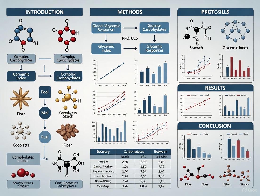
This article provides a comprehensive guide for researchers and drug development professionals on the protocols for determining the glycemic index (GI) of complex carbohydrates.
Measuring Glycemic Index in Complex Carbohydrates: Standard Protocols, Methodological Challenges, and Clinical Validation
Abstract
This article provides a comprehensive guide for researchers and drug development professionals on the protocols for determining the glycemic index (GI) of complex carbohydrates. It covers the foundational principles of GI and its physiological significance, details the standardized in vivo testing methodology as defined by leading research services, and addresses key methodological challenges and optimization strategies, including the impact of food processing and meal composition. Furthermore, it critically examines the validation of these methods through comparative analysis with direct meal testing and explores the significant role of inter-individual metabolic variability, a frontier area reinforced by recent 2025 research. The synthesis offers a critical perspective on the applicability and limitations of current GI measurement protocols in biomedical research and clinical practice.
Glycemic Index Fundamentals: From Physiological Basis to Public Health Relevance
The Glycemic Index (GI) and Glycemic Load (GL) are quantitative metrics used to classify carbohydrate-containing foods based on their postprandial blood glucose response. The GI represents the relative quality of a food's carbohydrate, indicating its potential to raise blood glucose compared to a reference food, typically pure glucose [1]. This concept was developed to address the limitation of the traditional "simple" versus "complex" carbohydrate classification, which proved too simplistic as different complex carbohydrates elicit considerably varied glycemic responses [1]. The GI is defined mathematically as the incremental area under the blood glucose response curve (iAUC) after consuming a test food containing 50 grams of available carbohydrate, divided by the iAUC after consuming a control food (glucose or white bread) containing the same amount of carbohydrate, multiplied by 100 [1] [2].
The Glycemic Load (GL) was subsequently developed to provide a more comprehensive picture by considering both the quality (GI) and quantity of carbohydrate in a typical food serving [1] [3]. A food's GL is calculated by multiplying its GI by the amount of available carbohydrate in grams per serving and dividing by 100 [1]. This distinction is crucial because while a food may have a high GI, its GL might be low if it contains minimal carbohydrate per serving, as exemplified by watermelon which has a high GI of 76-80 but a low GL of 5-8 due to its high water content [1] [3] [4].
Table 1: Classification Standards for Glycemic Index and Glycemic Load
| Metric | Low | Medium | High |
|---|---|---|---|
| Glycemic Index (GI) | ≤ 55 | 56 - 69 | ≥ 70 [1] [4] |
| Glycemic Load (GL) | ≤ 10 | 11 - 19 | ≥ 20 [1] [4] |
Core Concepts and Calculations
Fundamental Formulas and Physiological Basis
The mathematical foundation for GI and GL calculations is standardized, though variations exist in reference foods. The core formulas are:
Glycemic Index Calculation:
GI = (iAUC_test food / iAUC_glucose) × 100 [1]
Where iAUC represents the incremental area under the blood glucose response curve over 2 hours following consumption.
Glycemic Load Calculation:
GL = (GI × grams of available carbohydrate per serving) / 100 [1]
Available carbohydrate is typically calculated as total carbohydrate minus dietary fiber [2].
Physiologically, consumption of high-GI foods causes a sharp, rapid increase in postprandial blood glucose concentration that declines quickly. In contrast, low-GI foods result in a lower, more gradual blood glucose elevation that declines slowly [1]. This differential response significantly impacts insulin demand, with high-GI foods provoking stronger insulin secretion from pancreatic β-cells, potentially leading to reactive hypoglycemia [1].
Experimental Protocol for GI Determination
The International Organization for Standardization (ISO) provides the definitive protocol for measuring GI [5] [6]. The standard methodology requires healthy human volunteers to consume test foods and reference foods (glucose or white bread) on separate days after an overnight fast [1].
Key Experimental Parameters:
- Carbohydrate Load: Test foods must provide exactly 50 grams of available carbohydrate [1] [6]
- Reference Foods: Pure glucose (GI=100) or white bread; minimum of two tests per reference [1] [6]
- Blood Sampling: Fingertip capillary blood collected at baseline (0 min) and at 15, 30, 45, 60, 90, and 120 minutes after starting to eat [5] [6]
- Subject Number: Minimum of 10 healthy participants per test food [6]
- Testing Conditions: Standardized physical activity, no intense exercise before testing, no alcohol consumption previous day [6]
Diagram Title: GI Determination Workflow
Factors Influencing Glycemic Responses
Multiple factors beyond carbohydrate content influence a food's GI and the subsequent glycemic response. Starch composition significantly affects digestibility, with rapidly digestible starch (RDS) causing rapid glucose absorption, slowly digestible starch (SDS) providing sustained glucose release, and resistant starch (RS) escaping digestion in the small intestine [2]. Food processing and cooking methods alter starch gelatinization, while physical and chemical characteristics including acidity, particle size, and variety selection further modify GI [7] [2].
Recent research emphasizes considerable interindividual variability in postprandial glycemic responses (PPGRs) to identical meals [8]. A 2025 study demonstrated that responses to standardized carbohydrate meals varied significantly based on underlying metabolic physiology, with "rice-spikers" more likely to be of Asian ethnicity and "potato-spikers" exhibiting greater insulin resistance [8]. This variability challenges the concept of fixed GI values and highlights the importance of personalized nutritional approaches.
Table 2: Glycemic Index Values of Common Foods
| Food Item | GI Value | Classification |
|---|---|---|
| Peanuts | 7 | Low |
| Skim Milk | 32 | Low |
| Carrots | 35 | Low |
| Apple | 39 | Low |
| Baked Beans | 40 | Low |
| Whole Grain Bread | 51 | Low |
| Brown Rice | 50 | Low |
| Sweet Potato | 70 | High |
| Watermelon | 72 | High |
| White Bagel | 72 | High |
| Instant Oatmeal | 83 | High |
| White Rice | 89 | High |
| Baked Russet Potato | 111 | High [4] |
Research Reagents and Methodological Toolkit
Table 3: Essential Research Materials for GI/GL Determination
| Item | Specification/Function |
|---|---|
| Reference Carbohydrate | Pure glucose (dextrose, anhydrous) or standardized white bread [5] [6] |
| Blood Glucose Meter | ACCU-CHEK Performa or equivalent; calibrated for capillary blood [5] [6] |
| Continuous Glucose Monitor (CGM) | Abbott Freestyle Libre or equivalent for intensive PPGR monitoring [9] [8] |
| Body Composition Analyzer | InBody 270 or equivalent for participant screening [6] |
| Standardized Test Meals | Precisely formulated to deliver 50g available carbohydrate [5] [8] |
| Dietary Analysis Software | For calculating available carbohydrate (total carbohydrate - dietary fiber) [2] |
Advanced Methodological Considerations
Mixed Meal Glycemic Response Prediction
Predicting glycemic responses to mixed meals presents significant methodological challenges. Traditional calculation methods summing weighted GI values of individual components often overestimate actual responses by 22-50% compared to direct measurement [1]. Recent research has developed prediction models incorporating multiple nutrient parameters. One validated formula for ready-to-eat meals is:
GL = 19.27 + (0.39 × available carbohydrate) - (0.21 × fat) - (0.01 × protein²) - (0.01 × fiber²) [5]
This model demonstrates the complex interplay between macronutrients in determining postprandial glycemia, with fat and fiber exerting moderating effects on the carbohydrate-driven glycemic response [5] [6].
Limitations and Research Gaps
Current GI methodology faces several limitations: poor reproducibility for mixed meals, interindividual variability based on ethnicity and metabolic phenotype, and insufficient characterization of food processing effects [7] [8]. Research gaps include limited data on glycemic responses in diverse populations, particularly those with high carbohydrate intakes, and insufficient understanding of how individual metabolic factors (insulin resistance, beta cell function) modify PPGRs [9] [8]. Future protocols should incorporate continuous glucose monitoring, standardized mixed meals, and comprehensive metabolic phenotyping to address these limitations [9] [8].
Carbohydrate digestion is a fundamental physiological process that provides essential energy for the human body while playing a critical role in metabolic health. The Glycemic Index (GI) has emerged as a key tool for quantifying how different carbohydrate-containing foods affect blood glucose levels, making it particularly valuable for research on diabetes, cardiovascular disease, and metabolic disorders [7]. This article examines the physiology of carbohydrate digestion, explores the research methodologies for determining GI, and addresses both the applications and limitations of GI in nutritional science and drug development.
Biochemical Classification of Carbohydrates
Carbohydrates are classified based on their chemical structure, which directly influences their digestion rate and metabolic effects [10].
Table 1: Biochemical Classification of Dietary Carbohydrates
| Category | Subcategory | Chemical Structure | Examples | Primary Food Sources |
|---|---|---|---|---|
| Simple Carbohydrates | Monosaccharides | Single sugar unit (C₆H₁₂O₆) | Glucose, Galactose, Fructose | Honey, Fruits, Sweeteners |
| Disaccharides | Two monosaccharide units (C₁₂H₂₂O₁₁) | Sucrose, Lactose, Maltose | Table sugar, Milk, Malt products | |
| Complex Carbohydrates | Oligosaccharides | 3-10 monosaccharide units | Maltodextrins, Raffinose | Legumes, Onions, Whole Grains |
| Polysaccharides | Long chains of monosaccharides | Starch (Amylose), Cellulose, Glycogen | Potatoes, Grains, Cell walls of plants | |
| Fiber | Soluble Fiber | Non-digestible; forms gel with water | Pectin, Beta-glucans | Oats, Broccoli, Dried Beans |
| Insoluble Fiber | Non-digestible; adds bulk to stool | Cellulose, Hemicellulose, Lignin | Brans, Seeds, Vegetable Skins |
Physiological Pathway of Carbohydrate Digestion
The process of carbohydrate digestion and absorption is systematic, beginning in the mouth and concluding with glucose utilization throughout the body.
Figure 1: Physiological Pathway of Carbohydrate Digestion and Absorption. The process converts complex carbohydrates into absorbable monosaccharides through sequential enzymatic action.
Digestion begins in the oral cavity with mechanical breakdown and the action of salivary amylase, though this activity is largely inhibited by the acidic environment of the stomach [10]. The primary site of carbohydrate digestion is the small intestine, where pancreatic α-amylase continues the breakdown of starches into disaccharides and oligosaccharides. The final digestive step occurs at the brush border of the intestinal mucosa, where the enzymes maltase, lactase, and sucrase hydrolyze disaccharides into their constituent monosaccharides: glucose, galactose, and fructose [10].
These monosaccharides are then absorbed into the bloodstream, triggering the pancreas to secrete insulin. Insulin signals the body's cells to absorb glucose for energy production or storage as glycogen in the liver and muscles. This intricate regulatory system maintains blood glucose homeostasis, with fiber remaining largely undigested and providing bulk that supports digestive health [10].
Research Protocols for Glycemic Index Determination
The standardized methodology for determining a food's Glycemic Index (GI) is defined by the International Standards Organization (ISO 26642:2010). The GI is a measure of the ability of the available carbohydrate in a food to increase blood glucose, calculated by comparing the area under the blood glucose response curve (AUC) of a test food to a reference food (typically pure glucose or white bread) [11].
Standardized GI Testing Protocol
Table 2: Key Phases of the ISO Glycemic Index Testing Protocol
| Phase | Duration | Procedures & Measurements | Quality Control Parameters |
|---|---|---|---|
| Pre-Test Preparation | 12 hours | Participant fasting; health screening; standardized pre-test meal and activity. | Confirmed fasting blood glucose: 4.0-5.5 mmol/L (72-99 mg/dL). |
| Test Food Administration | 10-12 minutes | Serve test food containing 50g (or 25g) available carbohydrate; timed consumption. | Precise carbohydrate quantification via proximate analysis. |
| Blood Glucose Monitoring | 2 hours | Capillary blood sampling at t=0 (fasting), 15, 30, 45, 60, 90, and 120 minutes. | Minimal SEM; samples analyzed in duplicate via glucose oxidase method. |
| Data Analysis & GI Calculation | Post-monitoring | Plot AUC for test and reference foods; calculate GI = (AUCtest / AUCreference) × 100. | Report mean GI value with standard error of the mean (SEM); n ≥ 10 healthy subjects. |
The Scientist's Toolkit: Essential Reagents and Materials
Table 3: Essential Research Reagents and Equipment for GI Determination
| Item | Specification/Function | Application Notes |
|---|---|---|
| Reference Standard | Anhydrous Glucose or White Bread (providing 50g available carbohydrate) | Serves as the biological benchmark (GI = 100); must be consistent across all trials. |
| Test Foods | Foods containing 50g or 25g of available carbohydrate (calculated as total carbohydrate minus dietary fiber) | Requires precise macronutrient analysis prior to testing; portion sizes must be accurately measured. |
| Blood Glucose Analyzer | Automated clinical analyzer using glucose oxidase method | Preferred over glucose dehydrogenase methods for superior accuracy; must be calibrated regularly. |
| Blood Collection Supplies | Sterile lancets and capillary tubes for serial blood sampling | Minimize participant discomfort while ensuring sufficient plasma/serum volume for analysis. |
| Statistical Analysis Software | Software capable of calculating incremental AUC (iAUC) and statistical comparisons (e.g., SPSS, R) | Used to compute mean GI values and SEM; excludes area below the fasting baseline. |
Methodological Challenges and Research Considerations
Despite standardization, significant methodological challenges persist in GI research, particularly in epidemiological studies. A primary issue is the considerable variation in GI values assigned to the same food across different studies [12]. For instance, inter-rater and inter-method comparisons show considerable variation for GI (with weighted κ coefficients as low as 0.25), though agreement is better for Glycemic Load (GL) [12].
Multiple factors contribute to this variability in measured GI values:
- Food-related factors: Processing methods, cooking time and temperature, ripeness, and variety can dramatically alter a food's GI [7] [13]. For example, different rice varieties can yield GI values ranging from 37 to 116 [11].
- Subject-related factors: Age, health status, baseline metabolism, and gut microbiota composition create substantial inter-individual variability in glycemic responses [13].
- Methodological factors: Differences in the number of healthy subjects, sampling protocols, and analytical techniques between laboratories can influence results [12].
The complexity of real-world eating patterns further complicates GI application. The GI is typically determined for individual foods consumed alone, but when foods are combined in a meal containing fat, protein, and fiber, the overall glycemic response changes significantly [13].
Data Interpretation and Research Applications
International GI and GL Classifications
Table 4: International GI and GL Classifications for Research and Clinical Application
| Glycemic Index (GI) Value | Classification | Representative Foods | Research Implications |
|---|---|---|---|
| ≤ 55 | Low GI | Dairy, legumes, pasta, non-starchy fruits, specific whole grains. | Associated with reduced risk for T2DM and CHD in some cohort studies [7]. |
| 56 - 69 | Medium GI | Quick oats, brown rice, whole-wheat bread, some regional foods. | Wide variation within categories necessitates careful food selection for clinical trials. |
| ≥ 70 | High GI | White bread, cornflakes, most potato varieties, rice cakes. | Associated with increased risk of T2DM, heart disease, and obesity [10]. |
| Glycemic Load (GL) Value | Classification | Calculation Formula | Research Utility |
| ≤ 10 | Low GL | GL = (GI × grams of carbohydrate per serving) ÷ 100 | Accounts for portion size; may better reflect real-world intake than GI alone. |
| 11 - 19 | Medium GL | Adjusts for carbohydrate density; watermelon (high GI, low GL) exemplifies its utility. | |
| ≥ 20 | High GL | Complements GI by quantifying both quality and quantity of carbohydrates consumed. |
Composite Metrics for Carbohydrate Quality Assessment
Researchers have developed more comprehensive metrics to address the limitations of GI. The Carbohydrate Quality Index (CQI) incorporates multiple dimensions of carbohydrate quality: dietary fiber, GI, the ratio of whole grains to total grains, and the ratio of solid to liquid carbohydrates [7]. Higher CQI scores have been associated with lower risks of obesity, cardiovascular disease, and related risk factors in observational studies [7].
The carbohydrate-to-fiber ratio has emerged as another valuable indicator, with some studies suggesting it may be a better predictor of health outcomes like waist circumference change than GI alone [7]. These composite metrics provide a more nuanced approach to evaluating carbohydrate-containing foods in research contexts.
The physiology of carbohydrate digestion reveals a complex system with significant implications for metabolic health and disease prevention. While the Glycemic Index provides a valuable framework for understanding how different carbohydrates impact blood glucose, researchers must acknowledge its limitations, including methodological variability and the challenge of applying single-food metrics to mixed meals. The standardized ISO protocol for GI determination provides a critical foundation for comparative research, but emerging approaches that account for individual variability and composite carbohydrate quality metrics represent the future of this field. For drug development and clinical research, understanding these nuances is essential for designing effective nutritional interventions and therapeutics targeting metabolic disorders.
The Glycemic Index (GI) and Glycemic Load (GL) represent critical tools in nutritional epidemiology for quantifying the impact of carbohydrate-containing foods on postprandial glycemia. GI measures the quality of carbohydrates by ranking foods according to their postprandial blood glucose response compared to a reference food, while GL incorporates both the quality and quantity of carbohydrates consumed, providing a more comprehensive assessment of glycemic impact. Within epidemiological research, these indices have emerged as significant biomarkers for investigating associations between dietary patterns and chronic disease etiology, particularly as postprandial hyperglycemia has been identified as a universal mechanism in disease progression pathways [14].
The application of GI and GL in chronic disease risk assessment has gained substantial methodological sophistication through advances in nutritional epidemiology. These tools allow researchers to move beyond simplistic nutrient-based analyses to capture the complex, cumulative effects of overall dietary patterns on physiological responses. As the field has evolved, prospective cohort studies have utilized GI/GL assessments to elucidate relationships with various chronic conditions, enabling more targeted dietary recommendations for disease prevention at both population and individualized levels [14] [8].
Quantitative Associations Between GI/GL and Chronic Disease Risk
Meta-Analysis of Observational Studies
A comprehensive meta-analysis of 37 prospective cohort studies has quantified the association between dietary GI/GL and chronic disease risk, providing robust epidemiological evidence for public health recommendations. The analysis included studies with follow-up periods ranging from 4 to 20 years, capturing 40,129 incident cases across multiple disease endpoints. The findings demonstrated significant positive associations between high GI/GL diets and several chronic conditions in fully adjusted models, with particularly strong effect sizes observed for metabolic diseases [14].
Table 1: Chronic Disease Risk Associated with High GI/GL Diets from Meta-Analysis of Prospective Cohort Studies
| Disease Outcome | GI Risk Ratio (Highest vs. Lowest Quantile) | GL Risk Ratio (Highest vs. Lowest Quantile) | 95% Confidence Intervals |
|---|---|---|---|
| Type 2 Diabetes | 1.40 | 1.27 | GI: 1.23-1.59; GL: 1.12-1.45 |
| Coronary Heart Disease | 1.25 | Not Significant | 1.00-1.56 |
| Gallbladder Disease | 1.26 | 1.41 | GI: 1.13-1.40; GL: 1.25-1.60 |
| Breast Cancer | 1.08 | Not Significant | 1.02-1.16 |
| All Diseases Combined | 1.14 | 1.09 | GI: 1.09-1.19; GL: 1.04-1.15 |
The protection offered by low-GI and/or low-GL diets against chronic diseases appears comparable to that observed for whole grain and high fiber intakes, supporting the integration of GI/GL concepts into dietary guidance for chronic disease prevention. The consistency of findings across multiple studies, particularly for type 2 diabetes and cardiovascular outcomes, strengthens the evidence base for considering postprandial glycemia as a modifiable risk factor in clinical and public health contexts [14].
Interindividual Variability in Glycemic Responses
Recent research has revealed substantial interindividual variability in postprandial glycemic responses (PPGRs) to the same carbohydrate foods, challenging the concept of fixed GI values and highlighting the need for personalized nutritional approaches. A comprehensive study involving 55 well-phenotyped participants assessed PPGRs to seven different standardized carbohydrate meals using continuous glucose monitoring, with each meal containing 50g of total carbohydrates [8].
Table 2: Interindividual Variability in Glycemic Responses to Standardized Carbohydrate Meals
| Carbohydrate Source | Mean PPGR Profile | Interindividual Variability | Metabolic Phenotypes Associated with High Response |
|---|---|---|---|
| Jasmine Rice | Highest overall | 19 participants had highest response | More prevalent in Asian individuals (Rice-spikers) |
| Buttermilk Bread | High | Considerable variation | Associated with higher blood pressure (Bread-spikers) |
| Potatoes | High | Distinct response patterns | Insulin resistance and lower beta cell function (Potato-spikers) |
| Grapes | Early, high peak | Marked variability | Insulin sensitivity (Grape-spikers) |
| Pasta | Moderate | Moderate variability | Not specifically characterized |
| Mixed Berries | Low | Lower variability | Not specifically characterized |
| Black Beans | Lowest overall | Lower variability | Not specifically characterized |
The study demonstrated that while certain carbohydrates consistently produced higher glycemic responses across the cohort (rice, bread, potatoes), individuals exhibited unique response patterns based on underlying metabolic physiology. This variability was quantitatively assessed through intraindividual correlation coefficients (ICCs) of area under the curve (AUC) measurements, which ranged from 0.26 for beans to 0.73 for pasta, indicating generally reproducible responses within individuals across test occasions [8].
The food's fiber content emerged as a significant modifier of PPGRs, with total dietary fiber content negatively correlating with both AUC (>baseline) (-0.71) and delta glucose peak (-0.75). These findings underscore the importance of considering both food composition and individual physiological differences when applying GI concepts in nutritional epidemiology and clinical practice [8].
Methodological Protocols for GI Research in Nutritional Epidemiology
Standardized Carbohydrate Challenge Protocol
The assessment of glycemic responses to carbohydrates requires rigorous standardization to ensure methodological consistency and reproducible results. The following protocol details the procedures for conducting controlled carbohydrate challenges in epidemiological and clinical research settings, based on established methodologies from recent investigations [8].
Protocol 1: Standardized Carbohydrate Meal Challenge for PPGR Assessment
Objective: To quantify postprandial glycemic responses to standardized carbohydrate meals under controlled conditions while accounting for interindividual variability.
Pre-Test Requirements:
- Participants fast overnight for 10-12 hours
- Abstain from alcohol, caffeine, and strenuous exercise for 24 hours prior to testing
- Continuous glucose monitors (CGMs) calibrated and inserted 24 hours before first test
- Baseline blood samples collected for fasting glucose and insulin measurements
Test Meal Preparation:
- Prepare test meals containing exactly 50g of total carbohydrates
- Use standardized cooking methods for all prepared foods:
- Rice (jasmine): cooked according to standardized package instructions
- Pasta (macaroni): precooked per instructions, cooled, and frozen until use
- Potatoes: shredded and prepared using standardized methods
- Bread (buttermilk): served fresh from standardized source
- Legumes: canned black beans, rinsed and drained
- Fruits: fresh grapes and mixed berries (blackberries, strawberries, blueberries) portioned to contain 50g carbohydrates
- Record precise nutrient composition for each meal, with emphasis on:
- Total dietary fiber content
- Resistant starch content
- Simple vs. complex carbohydrate ratio
- Serve all meals with 250mL water at room temperature
Testing Procedure:
- Conduct tests in a quiet, controlled environment
- Administer test meal within 10-minute consumption window
- Record baseline glucose value immediately before meal consumption (t=0)
- Monitor glucose responses continuously for 3 hours postprandially using CGM
- Ensure each participant completes at least two replicates of each test meal on separate days
- Maintain consistent timing of tests across participants (typically morning sessions)
Data Collection and Processing:
- Extract glucose values at 5-minute intervals from CGM devices
- Calculate the following PPGR parameters:
- Area under the curve above baseline (AUC(>baseline))
- Delta glucose peak (maximum increase from baseline)
- Time from baseline to peak glucose
- Time to return to baseline glucose
- Incremental area under the curve (iAUC)
- Average replicate tests for each participant-meal combination
- Apply appropriate statistical models to account for within-subject correlations
This protocol enables the systematic characterization of both between-food and between-individual differences in glycemic responses, providing a robust methodology for nutritional epidemiological investigations of carbohydrate quality [8].
Metabolic Phenotyping Protocol for Response Stratification
Comprehensive metabolic phenotyping is essential for understanding the physiological basis of interindividual variability in glycemic responses. The following protocol details the gold-standard methods for characterizing metabolic parameters that modify PPGRs to carbohydrate ingestion.
Protocol 2: Comprehensive Metabolic Phenotyping for Carbohydrate Response Stratification
Objective: To assess key metabolic parameters that influence individual glycemic responses to carbohydrate ingestion, enabling stratification of participants into relevant physiological subgroups.
Insulin Resistance Assessment (Steady-State Plasma Glucose Method):
- Administer intravenous octreotide (30μg/m²/min), insulin (25mU/m²/min), and glucose (240mg/m²/min) simultaneously over 150 minutes
- Collect blood samples at 10-minute intervals from 120 to 150 minutes for plasma glucose measurements
- Calculate steady-state plasma glucose (SSPG) as the mean of last four measurements
- Classify participants as insulin resistant (SSPG ≥120mg/dL) or insulin sensitive (SSPG <120mg/dL)
Beta Cell Function Assessment (Disposition Index):
- Perform frequently sampled intravenous glucose tolerance test (FSIVGTT)
- Administer glucose dose (0.3g/kg) intravenously over 1 minute
- Collect blood samples at -10, -5, -1, 2, 3, 4, 5, 6, 7, 8, 10, 12, 14, 16, 19, 22, 24, 25, 27, 30, 40, 50, 60, 70, 80, 90, 100, 120, 140, 160, and 180 minutes
- Calculate acute insulin response to glucose (AIRg) and insulin sensitivity index (SI)
- Compute disposition index as DI = AIRg × SI
Additional Metabolic Measurements:
- Hepatic insulin resistance assessment via stable isotope methods
- Adipocyte insulin resistance through adipose tissue microdialysis
- Body composition analysis using DXA scanning
- Basic clinical parameters: blood pressure, lipid profile, HbA1c
Multi-Omics Profiling:
- Collect fasting blood samples for:
- Metabolomics (GC-MS and LC-MS platforms)
- Lipidomics (targeted and untargeted approaches)
- Proteomics (high-throughput immunoassays)
- Collect stool samples for microbiome analysis:
- 16S rRNA sequencing for community profiling
- Shotgun metagenomics for functional potential
- Metabolomic analysis of fecal samples
This comprehensive phenotyping protocol enables researchers to identify metabolic subtypes associated with distinctive glycemic response patterns, facilitating a more personalized approach to nutritional epidemiology and dietary recommendation development [8].
Advanced Methodologies in Nutritional Epidemiology
Dietary Pattern Analysis Using Machine Learning Approaches
Nutritional epidemiology has increasingly adopted advanced statistical methods to enhance the prediction of disease risk factors from dietary intake data. The LASSO (Least Absolute Shrinkage and Selection Operator) model represents a significant methodological advancement over traditional dietary pattern analysis techniques such as principal component analysis (PCA).
Protocol 3: LASSO-Based Dietary Pattern Analysis for Chronic Disease Risk Prediction
Objective: To identify dietary patterns predictive of chronic disease risk factors using penalized regression approaches that improve variable selection and prediction accuracy.
Data Preparation:
- Collect dietary intake data using validated Food Frequency Questionnaires (FFQs)
- Combine individual food items into logical food groups (typically 35-40 groups)
- Apply appropriate sampling weights to account for complex survey design
- Log-transform and standardize intake variables to address skewness
- Partition data into training and test sets (typically 70/30 split)
Model Implementation:
- Apply LASSO regression with ten-fold cross-validation to select optimal lambda value
- Use cardiovascular disease risk factors (triglycerides, LDL-C, HDL-C, total cholesterol) as outcome variables
- Include relevant confounders (age, sex, BMI, physical activity) in the model
- Evaluate model performance using adjusted R² values on test set
- Compare results with traditional PCA-based dietary pattern analysis
Interpretation and Validation:
- Identify food groups with non-zero coefficients as components of predictive dietary patterns
- Validate identified patterns in independent cohorts when possible
- Assess robustness through bootstrap resampling methods
- Examine biological plausibility of identified patterns
This advanced methodology has demonstrated superior performance compared to traditional approaches, with LASSO achieving adjusted R² values of 0.861, 0.899, 0.890, and 0.935 for triglycerides, LDL cholesterol, HDL cholesterol, and total cholesterol respectively, substantially outperforming PCA-based methods [15].
Gastrointestinal Tolerance Assessment for NDC Interventions
The evaluation of gastrointestinal effects and tolerance is essential when investigating the health impacts of nondigestible carbohydrates (NDCs), many of which function as dietary fibers with potential benefits for glycemic control and chronic disease risk reduction.
Protocol 4: Assessment of Gastrointestinal Tolerance to Nondigestible Carbohydrates
Objective: To quantitatively evaluate gastrointestinal tolerance and functional effects of NDC interventions in clinical and epidemiological studies.
Study Design Considerations:
- Utilize double-masked, placebo-controlled, parallel-group designs when possible
- Implement appropriate randomization and concealed allocation methods
- Include run-in periods to establish baseline symptoms
- Define primary endpoints related to gastrointestinal symptoms and function
Tolerance Assessment Methods:
- Daily symptom diaries using validated scales (e.g., visual analog scales)
- Assessment of specific symptoms:
- Abdominal pain/cramping
- Bloating/distension
- Flatulence
- Borborygmi (rumbling)
- Nausea
- Categorize symptom severity as none, mild, moderate, or severe
- Record symptom frequency and timing relative to interventions
Functional Outcome Measures:
- Gastrointestinal transit time assessment:
- Radio-opaque markers with abdominal radiography
- Smart pill technologies
- Breath hydrogen testing
- Stool characterization:
- Frequency of bowel movements
- Stool consistency (Bristol Stool Form Scale)
- Stool weight and moisture content
- Define normal ranges and clinically significant changes
Tolerable Intake Level Determination:
- Establish dose-response relationships for specific NDCs
- Identify intake levels associated with no more than mild symptoms in majority of participants
- Develop NDC-specific recommendations based on available evidence
This methodological approach supports the development of evidence-based intake recommendations for various NDCs, with tolerable intake levels ranging from 3.75 g/d for alginate to 25 g/d for soy fiber, depending on the specific carbohydrate's physicochemical properties [16].
Research Reagent Solutions for GI and Chronic Disease Studies
Table 3: Essential Research Reagents and Materials for GI and Chronic Disease Epidemiology
| Reagent/Material | Specification | Application in GI Research | Example Vendor/Source |
|---|---|---|---|
| Continuous Glucose Monitoring System | Factory-calibrated, research-grade | Continuous measurement of interstitial glucose levels in free-living conditions | Dexcom G6, Abbott Freestyle Libre |
| Standardized Carbohydrate Meals | Precisely formulated to contain 50g available carbohydrates | Controlled challenge tests for PPGR assessment | Research kitchen preparation |
| Octreotide Acetate | Pharmaceutical grade, sterile | Suppression of endogenous insulin secretion during SSPG tests | Novartis (Sandostatin) |
| Human Insulin | Recombinant, 100U/mL | Insulin infusion during metabolic phenotyping | Eli Lilly (Humulin R) |
| Stable Isotope Tracers | [6,6-²H₂]glucose, [U-¹³C]glucose | Assessment of hepatic glucose production and carbohydrate metabolism | Cambridge Isotope Laboratories |
| DNA/RNA Extraction Kits | High-throughput, standardized | Isolation of genetic material from blood and stool samples | Qiagen DNeasy Blood & Tissue Kit |
| Metabolomics/Lipidomics Platforms | LC-MS/MS, GC-MS systems | Comprehensive profiling of metabolites and lipids | Waters, Agilent, Sciex |
| 16S rRNA Sequencing Reagents | V4 region primers, sequencing standards | Microbiome community profiling | Illumina MiSeq system |
| Food Frequency Questionnaire | Validated, comprehensive | Assessment of habitual dietary intake for GI/GL calculation | NHANES DietHHQ, Block FFQ |
| Glycated Hemoglobin Assay | NGSP certified, standardized | Assessment of long-term glycemic control | Bio-Rad D-100, Tosoh G8 |
Workflow Diagrams for GI Research Protocols
Comprehensive Metabolic Phenotyping and PPGR Assessment Workflow
Nutritional Epidemiology Study Design for GI-Chronic Disease Associations
Advanced Dietary Pattern Analysis Methodology
For decades, the classification of carbohydrates as either "simple" or "complex" has served as a foundational concept in nutritional science. This system, largely based on chemical structure and degree of polymerization (DP), categorizes sugars (DP 1-2) as simple and oligosaccharides (DP 3-9) and polysaccharides (DP≥10) as complex [17]. While chemically intuitive, this classification provides limited predictive value regarding a carbohydrate's physiological impact, particularly its effect on postprandial blood glucose levels [18]. The understanding of carbohydrate digestion has evolved to recognize that multiple factors beyond molecular size—including food structure, cooking method, and interactions with other food components—collectively determine the glycemic response [19] [20].
The concept of Glycemic Index (GI) emerged to address this gap, offering a physiological measure to classify carbohydrates based on their actual blood glucose elevation potential [21]. However, accurately measuring GI and understanding the factors that modulate it requires moving beyond outdated classifications and embracing sophisticated experimental models that simulate the complex process of human digestion. This document provides detailed application notes and protocols to support research on the glycemic impact of carbohydrates, with a focus on physiologically relevant in vitro methodologies.
Advanced Experimental Models for GI Prediction
Accurate prediction of a food's GI requires in vitro models that closely mimic the dynamic physiological conditions of the human gastrointestinal tract. Research demonstrates that dynamic digestion systems offer significant advantages over traditional static models by better replicating key parameters such as gastric peristalsis, gradual fluid secretion, and controlled emptying [19].
Table 1: Comparison of Static vs. DynamicIn VitroDigestion Models for GI Research
| Feature | Static Model | Dynamic Model (DIVHS Example) |
|---|---|---|
| Physical Process | Passive mixing in a glass vessel [19] | Simulated peristalsis via motor-driven walls; controlled intragastric pressure (~25 kPa) [19] |
| Chyme-Enzyme Contact Area | 160.4 ± 6.0 cm² [19] | 451.2 ± 4.4 cm² [19] |
| Particle Size Reduction | Less effective, larger fragments [19] | More effective, smaller fragments [19] |
| Digestive Juice Addition | Single, bolus addition [19] | Gradual, controlled infusion mimicking physiological secretion [19] |
| Glycemic Index (GI) Prediction | Less accurate correlation with human data [19] | Improved agreement with reported human GI values [19] |
| Biological Relevance (Caco-2 Transcriptome) | Induces weaker transcriptional response [19] | Induces stronger transcriptional response (421 genes up-regulated) [19] |
The following workflow diagram illustrates the key stages of a dynamic in vitro protocol for assessing carbohydrate digestibility and estimating GI.
Detailed Experimental Protocol: DynamicIn VitroDigestion of Carbohydrates
This protocol is adapted from a study comparing the digestion of cereals using a Dynamic In Vitro Human Stomach (DIVHS) system [19].
Materials and Reagent Solutions
Table 2: Research Reagent Solutions for DynamicIn VitroCarbohydrate Digestion
| Reagent / Material | Function / Specification | Example Source / Preparation |
|---|---|---|
| Dynamic In Vitro Human Stomach (DIVHS) | Silicone-based system simulating esophagus, stomach, and duodenum with motor-driven peristalsis [19] | Custom-built system with 3D-printed molds [19] |
| Salivary α-Amylase (A6255) | Initiates starch hydrolysis in the oral phase [19] | Sigma-Aldrich [19] |
| Pepsin (P7000) | Gastric protease for protein digestion [19] | Sigma-Aldrich [19] |
| Pancreatin (P7545) | Source of pancreatic enzymes, including α-amylase, for intestinal digestion [19] | Sigma-Aldrich [19] |
| Amyloglucosidase (A7095) | Hydrolyzes maltose and other dextrins to glucose for final quantification [19] | Sigma-Aldrich [19] |
| Bile Salts (48305) | Emulsifies lipids, simulating intestinal conditions [19] | Sigma-Aldrich [19] |
| Simulated Gastric Fluid (SGF) | Acidic environment for gastric digestion [19] | According to INFOGEST protocol: KCl, KH₂PO₄, NaHCO₃, NaCl, HCl, etc. [19] |
| Simulated Intestinal Fluid (SIF) | Buffered environment for intestinal digestion [19] | According to INFOGEST protocol: KCl, KH₂PO₄, NaHCO₃, NaCl, etc., with bile salts and enzymes [19] |
| Phosphate Buffer (pH 6.9, 0.1 M) | Mimics salivary and initial gastric conditions for starch hydrolysis studies [19] [20] | Standard laboratory preparation |
| Caco-2 Cell Line | Human colon adenocarcinoma cell line; model for intestinal epithelium to study glucose transport and transcriptomic responses [19] | Cell Bank of the Chinese Academy of Sciences [19] |
Step-by-Step Methodology
- Sample Preparation: Mill or prepare the carbohydrate-rich food (e.g., rice, corn, millet) to a defined particle size. Weigh a precise amount (e.g., equivalent to 50g available carbohydrate) for digestion.
- Oral Phase (Pre-digestion): Mix the food sample with simulated salivary fluid containing salivary α-amylase (e.g., 75 U/mL per gram of food) and incubate for a short, defined period (e.g., 2 minutes) at 37°C with gentle agitation, mimicking mastication.
- Dynamic Gastric Phase:
- Transfer the bolus to the pre-warmed (37°C) DIVHS stomach compartment.
- Initiate the dynamic peristalsis program, which applies rhythmic contractions to the stomach walls.
- Start the gradual infusion of simulated gastric fluid (SGF) containing pepsin. The infusion rate should be controlled (e.g., over 30-60 minutes) to simulate physiological gastric secretion. The final gastric pH should be adjusted to ~3.
- Gastric emptying is regulated by the simulated pyloric valve, typically allowing chyme to pass into the duodenum compartment in a gradual, linear manner over a 1-2 hour period.
- Intestinal Phase:
- Collect the gastric chyme as it empties from the stomach compartment and mix it with an equal volume of simulated intestinal fluid (SIF) containing pancreatin (e.g., 100 U/mL of α-amylase activity based on food weight) and bile salts (e.g., 10 mM).
- Incubate this mixture at 37°C with constant agitation (e.g., in a shaking water bath) for a set period (e.g., 2-4 hours) to simulate small intestinal digestion.
- To complete hydrolysis for glucose measurement, add amyloglucosidase (e.g., 30 U/mL) to the intestinal chyme and incubate further (e.g., 30-60 minutes).
- Sampling and Analysis:
- At regular intervals throughout the intestinal phase (e.g., 0, 10, 20, 30, 60, 90, 120 minutes), withdraw small aliquots (e.g., 0.5 mL) of the digest.
- Immediately inactivate enzymes in the aliquots, typically by heating (e.g., 100°C for 5 minutes) or adding an inhibitor.
- Centrifuge the samples and analyze the supernatant for reducing sugar content using standard colorimetric methods (e.g., DNS assay) or a glucose meter [19] [20].
- Data Modeling and eGI Calculation:
- Plot the kinetics of sugar release. The hydrolysis curve can be fitted to a first-order equation: ( Gt = G\infty (1 - e^{-kt}) ), where ( Gt ) is the concentration at time ( t ), ( G\infty ) is the equilibrium concentration, and ( k ) is the rate constant.
- Calculate the Hydrolysis Index (HI) by dividing the area under the hydrolysis curve (AUC) for the test food by the AUC of a reference food (typically white bread or glucose) digested under the same conditions.
- Estimate the Glycemic Index (eGI) using an empirical formula, for example: ( eGI = 17.24 + 0.94 \times HI ) [19].
Critical Factors Modulating Carbohydrate Digestibility and GI
The following diagram synthesizes the multifaceted factors, as revealed by modern research, that influence the glycemic response to carbohydrate-rich foods, moving far beyond the simple/complex dichotomy.
The Role of Advanced Glycation End Products (AGEs)
Modern cooking methods, particularly grilling, broiling, roasting, and frying, expose protein- and fat-rich foods to high dry heat. This promotes the formation of Advanced Glycation End Products (AGEs) [22] [23]. Diets high in AGEs are associated with increased oxidative stress and inflammation, which are linked to insulin resistance and impaired glucose metabolism [22] [23]. The choice of cooking method thus not only affects starch accessibility but also the metabolic milieu that handles glucose.
Table 3: Impact of Cooking Method on AGE Content and Potential Metabolic Load
| Food Item | Cooking Method | AGE Content (kU/serving) | Comparison & Metabolic Implication |
|---|---|---|---|
| Chicken (3 oz) | Grilled | 5,200 kU [23] | Poaching reduces AGE load by ~80%, potentially lowering inflammatory burden [23]. |
| Poached | 1,000 kU [23] | ||
| Beef Steak (3 oz) | Broiled | 6,600 kU [23] | Braising with moist heat reduces AGE formation by ~67% [23]. |
| Braised | 2,200 kU [23] | ||
| Potato (3 oz) | French Fries | 690 kU [23] | Baking results in a 90% reduction in AGEs compared to frying [23]. |
| Baked | 70 kU [23] | ||
| Egg (1) | Fried | 1,240 kU [23] | Scrambling (using lower heat and potentially added moisture) drastically reduces AGE formation [23]. |
| Scrambled | 75 kU [23] |
The evolution of GI research necessitates a definitive move beyond the simplistic binary classification of carbohydrates. Predicting a food's true glycemic impact requires a integrated approach that considers:
- Physiologically Relevant Models: Prioritizing dynamic in vitro systems that replicate mechanical and biochemical digestion dynamics.
- Food Matrix Effects: Accounting for the profound effects of fiber, protein, fat, and other components on starch digestibility.
- Processing and Culinary History: Acknowledging that cooking methods and the formation of compounds like AGEs can significantly alter metabolic outcomes.
The protocols and data presented herein provide a framework for researchers to investigate the glycemic response with the sophistication it demands, enabling the development of foods and dietary recommendations that better support metabolic health.
GI Measurement in Practice: Standardized Protocols and Laboratory Procedures
The glycemic index (GI) is a physiological classification system for carbohydrate-rich foods based on their postprandial blood glucose response. The gold-standard method for its determination is an in vivo human study, as defined by joint Food and Agriculture Organization (FAO) and World Health Organization (WHO) recommendations [24] [25]. The reliability of glycemic response data for research, clinical practice, and public health guidance is fundamentally dependent on the rigor of the experimental protocol used to generate it. Variations in study population, test procedures, or data analysis can introduce significant inter-study variability, highlighting the necessity for a standardized approach [24]. This document details the essential components of the gold-standard in vivo protocol, with a specific focus on study design and volunteer selection, providing a framework for generating high-quality, reproducible GI data for research on complex carbohydrates.
Volunteer Selection Criteria
The selection of an appropriate subject cohort is critical to the validity of GI measurements. The following criteria ensure a standardized, healthy population that minimizes confounding physiological variables.
Table 1: Volunteer Inclusion and Exclusion Criteria
| Category | Inclusion Criteria | Exclusion Criteria |
|---|---|---|
| Health Status | Healthy adult volunteers [24] | History of gastrointestinal disorders, diabetes, metabolic disease [24] [25] |
| Pharmacological | Not taking medication for chronic disease conditions [24] | Use of medication influencing blood glucose levels [26] [25] |
| Physiological | Normal body mass index (BMI) and vital signs [25] | Pregnancy, lactation [24] |
| Other | No food allergies or intolerances to test foods [24] | History of eating disorders [26] |
Cohort Size and Characteristics
The FAO/WHO protocol recommends that each test food be consumed by a minimum of 10 healthy subjects [24] [25]. Studies have successfully followed this guidance, employing cohorts of 10 participants to determine the GI of various staple foods [25]. Larger cohorts, such as 42 volunteers, may be used when testing multiple foods to ensure statistical power across all test items [24]. Participants should have a mean age representative of the adult population, typically ranging from young adults (e.g., mean 23 years) [25] to older individuals (e.g., mean 64 years in diabetic cohorts for meal testing) [27].
Study Design Parameters
The core of the GI testing protocol is a controlled, acute feeding trial comparing the glycemic response to a test food against a standard reference.
Reference and Test Food Administration
The protocol is a randomized, crossover design where each subject serves as their own control.
Table 2: Food Administration Protocol
| Parameter | Specification | Rationale & Context |
|---|---|---|
| Reference Food | Glucose (Dextrose monohydrate) [24] | Standard for calculating the relative GI (100%) [25] |
| Reference Dosing | 50 g or 25 g available glucose [24] | 50g is standard; 25g may be used if portion size is too large [24] |
| Test Food Portion | Contains 50 g (or 25 g) available carbohydrate [24] [25] | Ensures iso-carbohydrate comparison with the reference |
| Number of Reference Tests | Three separate sessions [24] [25] | Accounts for intra-individual variation in glucose response |
| Test Food Sessions | Once per test food, in random order [24] | Minimizes sequence effects |
| Consumption Time | Within 15 minutes [24] [25] | Standardizes the rate of food intake |
Pre-Test Standardization
To minimize pre-test variability, participants must adhere to strict pre-test conditions on the day before and the morning of each test session:
- Overnight Fast: A 10-hour minimum fast is required [24] [25].
- Diet & Activity: Avoid unusually large meals, alcohol, vigorous exercise, and specific foods (e.g., legumes, high-fat foods) [24] [26].
- Glucose Criteria: Fasting blood glucose must be within a predefined range (e.g., 4–11 mmol/L or 4–10 mmol/L) for the session to commence [26].
Experimental Protocol
The following workflow outlines the step-by-step procedures for a single test session.
Blood Glucose Monitoring & Analysis
- Sampling Method: Capillary blood is obtained via finger prick using a lancet [24] [25].
- Sampling Timepoints: Measurements are taken in the fasted state and at 15, 30, 45, 60, 90, and 120 minutes after starting to eat the test or reference food [24] [25].
- Glucose Measurement: Blood glucose level (BGL) is measured immediately using a calibrated glucometer and test strips [24] [25].
Data Analysis and GI Calculation
The GI value is calculated from the incremental area under the blood glucose response curve (IAUC).
Calculation of Incremental Area Under the Curve (IAUC)
The IAUC for each test food and the mean IAUC for the reference food are calculated geometrically using the trapezoid rule, ignoring the area below the fasting baseline [24] [25]. The formula for the IAUC between two timepoints is:
[ IAUC = \frac{(BGt + BG{t+1})}{2} \times (Time{t+1} - Timet) ]
where ( BG_t ) is the blood glucose concentration at time ( t ).
Final GI Determination
For each subject and test food, the ratio of the IAUC for the test food to the mean IAUC for their three reference glucose tests is calculated. The GI value for the test food is the mean of these ratios across all subjects expressed as a percentage [24] [25].
[ \text{GI (for a single subject)} = \frac{\text{IAUC}{\text{Test Food}}}{\text{Mean IAUC}{\text{Reference Glucose}}} \times 100 ]
[ \text{Final GI} = \text{Mean of all individual subject GIs} ]
Table 3: Glycemic Index and Load Classification
| Category | Glycemic Index (GI) Range | Glycemic Load (GL) Range | Example from Literature |
|---|---|---|---|
| Low | 55 or less [25] | 10 or less [25] | Teff Injera (GI: 36, GL: 7) [25] |
| Medium | 56 - 69 [25] | 11 - 19 [25] | White Wheat Bread (GI: 46*, GL: 14) [25] *Note: GI 46 is low, GL 14 is medium |
| High | 70 or more [25] | 20 or more [25] | Corn Injera (GI: 97, GL: 22) [25] |
The Scientist's Toolkit
Table 4: Essential Reagents and Materials for GI Clinical Testing
| Item | Specification / Example | Function in Protocol |
|---|---|---|
| Reference Food | Glucose (Dextrose monohydrate) [24] | Standard for calculating the relative GI (100%) [25] |
| Blood Glucose Meter | Calibrated glucometer (e.g., Accu-Chek series) [24] [25] | Immediate analysis of capillary blood glucose levels |
| Blood Sampling Kit | Lancets, test strips [25] | Sterile collection and measurement of capillary blood |
| Balances & Scales | High-precision digital scales | Accurate portioning of test and reference foods to provide exact available carbohydrate |
| Data Analysis Software | SPSS, SigmaPlot, R [24] | Statistical analysis and calculation of IAUC and GI values |
Methodological Considerations and Advanced Applications
The gold-standard in vivo method is resource-intensive, prompting research into predictive in vitro models. These models simulate gastrointestinal digestion and measure glucose release, showing significant correlation with in vivo GI [28] [27]. Recent advancements focus on integrating non-enzymatic electrochemical sensors for rapid glucose detection in complex food matrices and artificial intelligence/machine learning (AI/ML) models to improve GI prediction accuracy by analyzing complex digestion data [29].
Furthermore, the application of Continuous Glucose Monitoring (CGM) in research provides dense data on interstitial glucose levels. The CGM-derived metric Time in Range (TIR), the percentage of time glucose is within a target range (e.g., 3.9–10.0 mmol/L), is emerging as a valuable endpoint in clinical trials for diabetes management, potentially complementing single-meal GI studies by offering a dynamic picture of glycemic control [30].
Calculating the Incremental Area Under the Curve (iAUC) for GI Determination
The Glycemic Index (GI) is a physiological metric that classifies carbohydrate-containing foods based on their potential to raise blood glucose levels postprandially [7]. A cornerstone of GI determination is the calculation of the Incremental Area Under the Curve (iAUC), which quantifies the change in blood glucose concentration after consuming a test food compared to a reference food, typically glucose [31]. This protocol details the application of iAUC for GI determination within research on complex carbohydrates, providing a standardized methodology for researchers and scientists in nutritional science and drug development.
The iAUC is designed to minimize the influence of variations in baseline (fasting) blood glucose between individuals by focusing on the postprandial increase [31]. Accurate GI determination relies on precise iAUC calculation, as the GI value is expressed as a percentage of the iAUC of the test food relative to the iAUC of the reference food [31].
Core Concepts and Empirical Data
iAUC in Context: Comparison of AUC Types
The table below summarizes the key types of Area Under the Curve used in glycemic response research.
Table 1: Types of Area Under the Curve (AUC) in Diabetes and Nutrition Research
| Acronym | Full Name | Description | Primary Application Context |
|---|---|---|---|
| tAUC | Total Area Under the Curve | Calculates the total area under the glucose curve from time zero, including the baseline fasting glucose level [31]. | Considered more reliable for animal studies involving chronic treatments; correlates well with 2-hour OGTT glucose levels [31]. |
| iAUC | Incremental Area Under the Curve | Calculates the area under the glucose curve above the baseline fasting level; the baseline is subtracted, and only the incremental change is measured [32] [31]. | Standard for Glycemic Index (GI) determination in healthy human subjects [31]. |
| pAUC | Positive Incremental Area Under the Curve | A variant of iAUC where only the values above the baseline are considered, and any negative areas (dips below baseline) are ignored [31]. | Applied in some nutritional studies to focus exclusively on the glucose increase. |
Limitations and Considerations for iAUC Application
The use of iAUC, while standard for GI calculation, presents specific limitations that researchers must consider:
- Mathematical and Clinical Challenges: Subtracting the baseline value can produce negative values, which is considered problematic by some researchers. The suitability of iAUC has been questioned both mathematically and in clinical reports [31].
- Context-Dependent Suitability: iAUC is most suitable for acute interventions in healthy volunteers, such as GI determination. It is not recommended for animal studies involving chronic treatments, where variations in fasting glucose are part of the measured effect, and tAUC is preferred [31].
- Predictive Limitations for Mixed Meals: Importantly, recent research indicates that the GI of a meal, often calculated from the weighted sum of its individual food components' iAUC, may not accurately predict the actual, directly measured glycemic response. One study found that predicted meal GIs overestimated the directly measured GIs by 22% to 50% [33]. This highlights a significant limitation in extrapolating iAUC and GI values from single foods to complex meals.
Experimental Protocol: iAUC Determination for Glycemic Index
The following diagram outlines the comprehensive workflow for determining the GI of a food using iAUC, from initial participant recruitment to final data analysis.
Detailed Methodology
Objective: To determine the Glycemic Index (GI) of a test food by calculating the Incremental Area Under the Blood Glucose Response Curve (iAUC) in healthy human subjects.
Principle: The GI is defined as the percentage of the iAUC for the test food (containing 50g of available carbohydrate) relative to the iAUC of a reference food (the same amount of available carbohydrate from pure glucose), consumed by the same individual on a different day [31].
Pre-Test Procedures
Participant Recruitment:
- Recruit a minimum of 10 healthy, non-diabetic adult participants [31].
- Obtain informed consent. The study must be approved by an institutional ethics committee.
Pre-Test Standardization:
- Participants should fast for 10-12 hours overnight prior to each test day.
- They should avoid alcohol and strenuous physical activity for 24 hours before testing.
- Tests should be conducted in the morning under standardized, calm conditions.
Test Day Procedure
- Baseline Blood Sample (t=0 min): A fasting blood sample is collected by finger-prick or venipuncture.
- Food Consumption: The participant consumes the test food or reference food (50g available carbohydrate) within a 10-15 minute period.
- Serial Blood Sampling: Further blood samples are collected at 15, 30, 45, 60, 90, and 120 minutes after the start of food consumption.
- Blood Glucose Measurement: Blood glucose concentration (in mmol/L or mg/dL) is measured immediately for each sample.
- Replication: The entire procedure is repeated with the same participant on separate days for the reference food and for each test food. The test order should be randomized.
Data Analysis and iAUC Calculation
- Plotting: Plot a graph of blood glucose concentration (y-axis) against time (x-axis) for each food and each participant.
- iAUC Calculation: Calculate the iAUC for each curve using the trapezoidal rule, considering only the area above the fasting baseline concentration. Negative areas, when the curve falls below the baseline, are ignored [31].
- The formula for iAUC between two time points is:
((C1 + C2)/2 - C0) * (t2 - t1)- Where
C1andC2are blood glucose concentrations at timest1andt2. C0is the baseline fasting glucose concentration.
- Where
- The total iAUC is the sum of all such incremental areas between consecutive time points over the 2-hour period.
- The formula for iAUC between two time points is:
- GI Calculation:
- For each participant, calculate the GI of the test food:
GI = (iAUC_test food / iAUC_reference food) * 100. - The final GI value for the food is the average GI across all participants.
- For each participant, calculate the GI of the test food:
The Scientist's Toolkit: Research Reagent Solutions
Table 2: Essential Materials and Reagents for iAUC and GI Determination
| Item | Function/Description | Example/Specification |
|---|---|---|
| Reference Food | Serves as the standard (GI = 100) for comparison. Contains 50g of available carbohydrate [31]. | Anhydrous Glucose or Dextrose. |
| Test Foods | The carbohydrate-containing foods to be evaluated. Portion size is adjusted to provide 50g of available carbohydrate. | Complex carbohydrates (e.g., rice, pasta, bread). |
| Blood Glucose Analyzer | For precise and immediate measurement of glucose levels in blood samples. | YSI Analyzer, Glucose Oxidase Method; or high-quality handheld glucometers validated for clinical research. |
| Lancets & Microcuvettes | For safe and efficient collection of capillary blood samples via finger-prick. | Single-use, sterile devices. |
| Standardized Meal Protocol | Ensures consistency in meal preparation, as cooking method (e.g., cooling and reheating pasta) can significantly alter starch structure and GI [8]. | Documented and reproducible cooking instructions. |
| Continuous Glucose Monitor (CGM) | An alternative to serial finger-pricks; measures interstitial glucose every 5-15 minutes, providing a higher-resolution curve [32] [8]. | Devices like FreeStyle Libre (Abbott) or Dexcom G-series. Requires validation against plasma glucose. |
| iAUC Calculation Software | To automate the calculation of iAUC from the series of blood glucose values. | Custom scripts in R or Python implementing the trapezoidal rule, or specialized pharmacokinetic software. |
The Glycemic Index (GI) is a physiological classification system for carbohydrate-containing foods based on their postprandial blood glucose response relative to a standard reference food. The foundational principle of GI methodology requires that both test and reference foods contain equivalent available carbohydrate quantities (typically 50 g) to enable valid comparative analysis. The selection of an appropriate reference standard is critical for methodological consistency and biological relevance. International standards (ISO 26642:2010) permit either glucose (the definitive chemical standard) or white bread (a physiological reference) as primary reference foods, with established conversion factors enabling cross-comparison between different reference scales. This protocol examines the technical specifications, experimental applications, and calibration methodologies for these two principal reference standards in GI research, with particular emphasis on their applicability to complex carbohydrate analysis.
Reference Food Specifications and Standards
Chemical and Physiological Reference Options
GI testing methodology recognizes two primary categories of reference foods, each with distinct properties and applications in research settings.
Pure Glucose Standards: Represent the definitive chemical reference for glycemia.
- Anhydrous Glucose: Pharmaceutical-grade D-glucose powder containing 100% glucose (50 g dose provides 50 g available carbohydrate).
- Dextrose Monohydrate: Glucose monohydrate containing approximately 90% glucose and 10% water (requires 55 g to provide 50 g available carbohydrate).
- Commercial OGTT Solutions: Pre-mixed oral glucose tolerance test solutions standardized to contain 50 g glucose per serving.
White Bread Standard: Represents a physiologically relevant starchy reference.
- Composition: Refined wheat flour (typically ≥95% extraction rate), water, yeast, salt.
- Carbohydrate Content: Must be analytically verified to determine portion size providing exactly 50 g available carbohydrate.
- Preparation: Standardized baking protocol with specified ingredients and thermal parameters.
Table 1: Reference Food Specifications and Preparation Protocols
| Reference Food | Chemical Form | Dosage for 50g Available CHO | Preparation Protocol | Key Characteristics |
|---|---|---|---|---|
| Glucose | Anhydrous glucose | 50 g | Dissolved in 250-300 mL water; served at room temperature | Rapid absorption; maximal glycemic response; chemical purity |
| Glucose | Dextrose monohydrate | 55 g | Dissolved in 250-300 mL water; served at room temperature | Contains 10% water by weight; requires dosage correction |
| White Bread | Refined wheat flour | Variable (analytically determined) | Standardized recipe; fixed baking time/temperature | Physiologically relevant; contains protein/fiber; complex matrix |
Critical Methodological Considerations
Research indicates that reference food selection significantly influences observed GI values, particularly for starchy foods. A comparative study demonstrated that ethnic differences in GI values observed when using glucose as a reference (e.g., Chinese vs. European participants showing 12-15 GI unit differences for rice varieties) were eliminated when a starchy reference (jasmine rice) was employed [34]. This finding suggests that starchy references may be more appropriate than glucose beverages when attempting to derive universally applicable GI values for starchy foods, as they potentially normalize for differences in digestive physiology across populations [34].
A pervasive methodological issue in the literature involves incorrect dosage of monohydrated glucose formulations. Analysis of published GI studies reveals that trademarked products like Glucodin, Glucolin, and Clintose (all monohydrated forms) have frequently been administered at 50 g rather than the required 55 g, introducing a systematic 10% error in carbohydrate load between reference and test foods [35]. This dosage inaccuracy fundamentally violates the GI principle of equivalent carbohydrate comparison and may lead to misclassification of foods near GI category thresholds.
Experimental Protocol for GI Determination
Participant Preparation and Selection
- Sample Size: Minimum of 10 participants (ISO standard); research-grade determinations typically utilize 10-15 healthy individuals to ensure statistical power accounting for expected intra-individual variability (CV ≈20%) [36].
- Health Status: Participants should be normoglycemic, free from gastrointestinal disorders, and not using medications known to affect carbohydrate metabolism.
- Pre-test Conditions:
- 10-12 hour overnight fast preceding test sessions.
- Abstention from alcohol and strenuous exercise for 24 hours prior.
- Standardized evening meal (composition and timing) before test days.
- Ethical Considerations: Study protocol approval by institutional ethics committee; written informed consent from all participants.
Test Session Protocol
- Study Design: Randomized controlled trial with repeated measures.
- Each participant tests all reference foods (minimum 3 administrations) and test foods (minimum 2 administrations) in random order.
- Test sessions separated by ≥1 day washout period.
- Test Meal Administration:
- Reference or test food consumed within 10-15 minutes.
- 250-300 mL water served concurrently with test meal.
- Additional water permitted after first 30 minutes.
- Blood Sampling Schedule:
- Fasting sample collected immediately before meal consumption (-5 and 0 minutes).
- Postprandial samples at 15, 30, 45, 60, 90, and 120 minutes after meal commencement.
- Capillary (fingerprick) or venous blood collection following standardized procedures.
- Blood Glucose Analysis: Glucose oxidase or hexokinase methods preferred; point-of-care glucose meters requiring rigorous quality control [35].
Figure 1: Experimental workflow for GI determination showing participant preparation, blood sampling schedule, and analytical procedures.
Data Analysis and GI Calculation
- Incremental Area Under Curve (iAUC) Calculation:
- Calculate using trapezoidal rule while ignoring area below fasting baseline.
- Formula: iAUC = Σ[((Gᵢ - G₀) + (Gᵢ₋₁ - G₀))/2 × Δt] for all time intervals, where Gᵢ = glucose concentration at time i, G₀ = fasting glucose, Δt = time interval.
- For glucose values below fasting, contribution to iAUC = 0 [37].
- Individual GI Calculation:
- GI = (iAUCₜₑₛₜ food / mean iAUCᵣₑfₑᵣₑₙcₑ food) × 100
- Mean reference iAUC calculated from all reference food tests completed by each individual.
- Group GI Determination:
- Final GI value = arithmetic mean of individual GI values.
- Report with measure of variability (standard deviation or standard error).
Table 2: Blood Sampling Protocol for GI Determination
| Time Point (minutes) | Sample Type | Analytical Method | Critical Procedures |
|---|---|---|---|
| -5, 0 (fasting) | Capillary/venous | Glucose oxidase/hexokinase | Duplicate baseline measurement |
| 15, 30, 45 | Capillary/venous | Glucose oxidase/hexokinase | Precise timing from meal commencement |
| 60, 90, 120 | Capillary/venous | Glucose oxidase/hexokinase | Maintain participant in seated position |
| Processing | Plasma/serum separation | Standardized protocol | Immediate centrifugation; frozen storage at -80°C |
Calibration Between Reference Scales
Mathematical Conversion Principles
The GI scale was originally defined with glucose as the anchor point (GI = 100). When using white bread as a reference, a conversion factor is required to express GI values on the standard glucose scale. The fundamental conversion relationship follows the formula:
GIᵢ (glucose scale) = GIᵢ (bread scale) × [mean iAUCᵦᵣₑₐd / mean iAUCgₗᵤcₒₛₑ]
Where the ratio [mean iAUCᵦᵣₑₐd / mean iAUCgₗᵤcₒₛₑ] represents the conversion factor between reference systems.
Empirical studies indicate the typical conversion factor from white bread to glucose ranges between 0.7-0.75, meaning white bread typically elicits approximately 70-75% of the glycemic response of an equivalent carbohydrate load of glucose [34]. However, this factor demonstrates significant inter-individual and inter-population variability. Research comparing Chinese and European participants found significantly different conversion factors (0.8 vs. 0.7, respectively; p=0.038) when using jasmine rice as an alternative starchy reference [34].
Figure 2: Relationship and conversion between white bread and glucose reference scales showing the mathematical transformation pathway.
Laboratory Determination of Conversion Factors
For precise research applications, laboratory-specific determination of the glucose-to-bread conversion factor is recommended:
- Experimental Design: All participants complete multiple tests with both glucose and white bread references using identical methodological conditions.
- Calculation Method:
- Individual conversion factor = mean iAUCᵦᵣₑₐd / mean iAUCgₗᵤcₒₛₑ for each participant.
- Laboratory conversion factor = arithmetic mean of all individual conversion factors.
- Validation: Statistical comparison of iAUC values for both references; coefficient of variation assessment for precision evaluation.
The Researcher's Toolkit: Essential Reagents and Materials
Table 3: Essential Research Reagents and Equipment for GI Determination
| Category | Specific Items | Technical Specifications | Application Notes |
|---|---|---|---|
| Reference Standards | Anhydrous glucose (USP grade) | ≥99.5% purity; moisture-controlled | Verify certificate of analysis; store in desiccator |
| White bread flour | Standardized protein content (10-12%) | Consistent supplier; monitor lot variability | |
| Blood Collection | Capillary blood tubes | Lithium heparin or fluoride/oxalate | Maintain cold chain; validated collection devices |
| Lancets | Single-use, controlled depth | Standardize sampling site; minimize discomfort | |
| Glucose Analysis | Glucose oxidase reagent | Validated linear range (1-30 mmol/L) | Daily calibration; quality control samples |
| Portable glucose meters | CV <5%; ISO 15197:2013 compliant | Research-grade models preferred | |
| Software & Computation | iAUC calculation scripts | Trapezoidal rule implementation | Automated data processing; outlier detection |
| Statistical packages | R, SPSS, or equivalent | Mixed models for repeated measures |
Methodological Variability and Quality Control
The reliability of GI values is influenced by numerous sources of biological and methodological variability. Studies in healthy volunteers demonstrate substantial intra-individual (CV = 20%) and inter-individual (CV = 25%) variability in GI values, which is not substantially reduced by increasing sample size, replication of reference foods, or extended blood sampling protocols [36]. Key biological factors including insulin index and glycated hemoglobin values explain approximately 15-16% of the variability in GI values [36].
Quality control measures should include:
- Reference Food Verification: Analytical confirmation of available carbohydrate content via AOAC methods or starch analysis kits [34].
- Blinded Replication: Incorporate duplicate test meals with different identifiers to assess within-laboratory reproducibility.
- Positive Controls: Include previously characterized reference foods in each test batch to monitor assay performance.
- Data Quality Monitoring: Implement pre-defined criteria for test validity (e.g., fasting glucose stability, complete blood sampling series).
Automated computational approaches such as the DegifXL software platform can significantly enhance reproducibility by standardizing iAUC calculations and reducing manual processing time from 2000 to 160 minutes for typical study datasets [37].
The accurate measurement of the glycemic index (GI) of complex carbohydrates is fundamental to nutritional science and the development of foods for health. The execution of these protocols, however, is highly sensitive to specific operational factors. Variations in dosage calculation, blood sampling procedures, and environmental controls can significantly impact the reproducibility and reliability of results. This application note details the critical methodologies and controls required for rigorous GI research, providing a standardized framework for researchers and drug development professionals engaged in metabolic studies.
Systematic characterization of physiological responses is crucial for understanding carbohydrate quality. The following table summarizes quantitative data from a study investigating postprandial glycemic responses (PPGRs) to different carbohydrate meals, highlighting the significant interindividual variability that rigorous protocols must account for.
Table 1: Postprandial Glycemic Responses to Standardized Carbohydrate Meals (50g available carbohydrate) [8]
| Carbohydrate Meal | Mean Delta Glucose Peak (mg/dL) | Time to Peak (Minutes) | Key Associated Metabolic Phenotypes in "Spikers" |
|---|---|---|---|
| Rice (Jasmine) | Highest among tested meals | ~60 | More likely in individuals of Asian ethnicity |
| Bread (Buttermilk) | High | ~60 | Higher blood pressure |
| Potatoes (Shredded) | High | ~60 | Higher insulin resistance, lower beta cell function |
| Pasta (Macaroni) | Moderate | ~60 | - |
| Grapes | High | Earlier than starchy meals | Higher insulin sensitivity |
| Mixed Berries | Low | Similar to grapes | - |
| Beans (Canned Black) | Lowest among tested meals | ~60 | - |
Experimental Protocols for GI Assessment
Participant Preparation and Standardized Meal Administration
The foundation of a valid GI test is the standardization of both the participant and the test food [8].
- Participant Screening: Recruit participants with no history of diabetes. Prior to test days, phenotype participants using gold-standard tests for metabolic traits, including insulin resistance (e.g., Steady-State Plasma Glucose, SSPG) and beta cell function (e.g., Disposition Index) [8].
- Pre-Test Standardization: Instruct participants to fast overnight (typically 10-12 hours) and avoid strenuous physical activity and alcohol for 24 hours prior to testing.
- Meal Preparation: Precisely calculate and prepare the test meal to contain a fixed amount of available carbohydrate (commonly 50g or 25g). The meal's weight should be based on its carbohydrate content determined by proximate analysis. The same lot of food should be used for all tests of a given food [8].
- Meal Administration: The test meal should be consumed within a fixed, short period (e.g., 10-15 minutes) under supervision. Water may be allowed ad libitum.
Blood Sampling and Glycemic Response Measurement
Accurate and timely blood sampling is the most critical factor in defining the glycemic response curve.
- Baseline Sampling: Collect a fasting blood sample immediately before meal consumption (t=0).
- Postprandial Sampling: Collect subsequent blood samples at pre-specified intervals. For a 50g carbohydrate challenge, a typical sampling schedule is at 15, 30, 45, 60, 90, and 120 minutes after the start of meal consumption. Using Continuous Glucose Monitoring (CGM) devices allows for a more detailed evaluation of PPGRs without repeated finger pricks [8].
- Sample Handling: Centrifuge blood samples promptly to separate plasma or serum. Analyze glucose concentration using a reliable, validated method (e.g., glucose oxidase). All samples from a single participant should be analyzed in the same batch to minimize inter-assay variation.
Data Processing and GI Calculation
- Response Curve plotting: Plot glucose concentration against time for each test food and the reference food (glucose or white bread).
- Incremental Area Under the Curve (iAUC) Calculation: Calculate the iAUC for each curve, ignoring the area beneath the fasting concentration.
- Glycemic Index Calculation: The GI value is calculated as the ratio of the iAUC of the test food to the iAUC of the reference food (pure glucose), expressed as a percentage. The final GI value should be the average of results from a minimum of 10 healthy subjects [7].
Visualization of Experimental Workflow
The following diagram outlines the key stages in a robust glycemic response study, from participant screening to data analysis.
Environmental Controls in a Research Setting
Environmental sampling and controls are generally not recommended for routine monitoring but are indicated during outbreak investigations or for validating hazardous conditions or equipment performance [38].
Table 2: Situations Warranting Microbiologic Environmental Sampling in Research [38]
| Situation | Purpose | Protocol Considerations |
|---|---|---|
| Outbreak Investigation | To support an investigation when environmental reservoirs are implicated in disease transmission. | Sampling must be supported by epidemiological data. Linking environmental isolates with clinical isolates via molecular epidemiology is crucial. |
| Research | To provide new information on the spread of disease using well-designed and controlled experimental methods. | Requires a written, defined, multidisciplinary protocol for sample collection, culture, and interpretation. |
| Hazard Monitoring | To detect and confirm a hazardous biological agent and validate its successful abatement. | Can be used to detect bioaerosols from equipment or for industrial hygiene/safety purposes (e.g., "sick building" investigations). |
| Quality Assurance | To evaluate the effects of a change in infection-control practice or to ensure equipment performs to specification. | Must follow sound sampling protocols and use properly selected controls. Examples include commissioning new special care areas or testing air handling system performance. |
Air Sampling Protocol for Quality Assurance
If air sampling is conducted to validate air handling system integrity during a study, the following preliminary concerns must be addressed.
Table 3: Preliminary Concerns for Conducting Microbiologic Air Sampling [38]
| Consideration | Application to GI/Metabolic Research Facility |
|---|---|
| Aerosol Characteristics | Consider the size range of particles, concentration of microorganisms, and environmental factors like temperature and relative humidity. |
| Sampling Strategy | Determine the type of sampling instrument (e.g., impaction, impingement), sampling time, and duration of the sampling program. |
| Sample Number | Determine the number of samples to be taken to achieve statistical relevance. |
| Assay Method | Determine the method of assay that will ensure optimal recovery of microorganisms. Ensure adequate equipment and supplies are available. |
| Laboratory Support | Select a laboratory that can provide proper microbiologic support. Ensure samples can be refrigerated if they cannot be assayed promptly. |
The Scientist's Toolkit: Research Reagent Solutions
Table 4: Essential Materials and Reagents for Glycemic Response Studies [8]
| Item | Function/Application |
|---|---|
| Standardized Carbohydrate Meals | Test substances (e.g., jasmine rice, buttermilk bread, shredded potatoes) precisely prepared to contain a fixed amount (e.g., 50g) of available carbohydrate. |
| Continuous Glucose Monitoring (CGM) System | Devices that measure and record interstitial glucose concentrations at frequent intervals, providing high-resolution data for PPGR curve analysis. |
| Gold-Standard Metabolic Tests | Tests such as the Insulin Suppression Test for measuring Steady-State Plasma Glucose (SSPG, a direct measure of insulin resistance) and the Disposition Index (for beta cell function). |
| Preload 'Mitigators' | Substances like pea fiber, egg white (protein), and cream (fat) used to study the effect of macronutrient preloading on the PPGR to a subsequent carbohydrate meal. |
| Multi-Omics Profiling Platforms | Technologies for metabolomics, lipidomics, proteomics, and microbiome analysis used to discover molecular signatures associated with PPGRs and metabolic phenotypes. |
Navigating Methodological Complexities: From Food Variables to Data Interpretation
Application Notes
This document provides detailed protocols and application notes for researchers investigating the glycemic index (GI) of complex carbohydrates. A critical challenge in this field is the significant variability introduced by intrinsic factors (e.g., food variety, ripeness) and extrinsic factors (e.g., processing methods). Standardizing experimental procedures is essential to generate reliable, reproducible data that can inform dietary guidelines and therapeutic nutritional strategies [7] [13]. The following sections summarize key sources of variability, provide structured experimental protocols, and outline essential reagents and workflows for conducting this research.
The following tables consolidate empirical data on how ripeness, processing, and food type influence glycemic response, providing a reference for experimental planning and data interpretation.
Table 1: Impact of Fruit Ripeness Stage on Glycemic Index (GI) and Glycemic Load (GL)
| Fruit Type | Ripeness Stage | Total Sugar (%) | Glycemic Index (GI) | Glycemic Load (GL) | Classification |
|---|---|---|---|---|---|
| Sweet Banana [39] | Ripe | 6.93 | 35.91 | Low GI | Low GI |
| Very Ripe | 16.49 | 58.18 | Moderate GI | Moderate GI | |
| Papaya [39] | Ripe | 12.98 | Low GI | Low GI | Low GI |
| Very Ripe | 52.31 | Moderate GI | Moderate GI | Moderate GI | |
| Mango [39] | Ripe | 28.84 | Low GI | Low GI | Low GI |
| Very Ripe | 42.28 | Low GI | Low GI | Low GI | |
| Apple [39] | Ripe | 29.19 | Low GI | Low GI | Low GI |
| Very Ripe | 32.33 | Low GI | Low GI | Low GI | |
| Orange [39] | Ripe | 31.59 | Low GI | Low GI | Low GI |
| Very Ripe | 37.67 | Low GI | Low GI | Low GI | |
| Khalas Date [40] | Khalal | 69.14 | High GI | High GI | High GI |
| Rutab | 62.49 | Medium GI | Medium GI | Medium GI | |
| Tamer | 53.09 | Low GI | Low GI | Low GI | |
| Barhi Date [40] | Khalal | 71.06 | High GI | High GI | High GI |
| Rutab | 65.99 | Medium GI | Medium GI | Medium GI | |
| Tamer | 51.58 | Low GI | Low GI | Low GI |
Table 2: Effect of Food Processing Method on Glycemic Index
| Food Item | Processing Method | Mean Glycemic Index (GI) | Reference |
|---|---|---|---|
| Common Staples [41] | Boiling | 35 (SE 3) - 63 (SE 7) | Boiling of foods may contribute to a lower GI diet. |
| Frying | 45 (SE 5) - 65 (SE 5) | ||
| Roasting | 72 (SE 7) - 89 (SE 9) | Foods processed by roasting or baking may result in higher GI. | |
| Baking | 64 (SE 5) - 94 (SE 8) | ||
| Dough Products [42] | Baking (Control) | No significant difference in vivo | In vivo glucose response did not differ significantly between processing methods. |
| Extrusion at 150°C | No significant difference in vivo | ||
| Extrusion at 180°C | No significant difference in vivo |
Experimental Protocols
Protocol: Determining GI as a Function of Fruit Ripeness
This protocol is adapted from clinical trials investigating dates and other fruits [40] [39].
1. Objective: To quantify the effect of fruit ripeness stage on the glycemic index (GI) and glycemic load (GL).
2. Reagents and Materials:
- Fruit samples at distinct, predefined ripeness stages (e.g., Khalal, Rutab, Tamer for dates; Ripe, Very Ripe for other fruits).
- Reference food (Anhydrous glucose or white bread).
- Capillary blood glucose meters and test strips.
- Lancet devices.
- Timer.
3. Subject Selection:
- Inclusion Criteria: Healthy adults (e.g., aged 18–60); normal body mass index (BMI < 30); normal fasting blood glucose and HbA1c [40].
- Exclusion Criteria: History of diabetes, prediabetes, pregnancy, gastrointestinal disorders, or use of medications affecting metabolism [40].
4. Procedure:
- Day 1 - Reference Food: After an overnight fast (10-12 hours), obtain baseline capillary blood glucose (t=0). Administer a reference solution containing 50 g of available glucose. Measure blood glucose at t=15, 30, 45, 60, 90, and 120 minutes [40] [39].
- Subsequent Days - Test Foods: On separate days, repeat the procedure, administering a portion of the test fruit that contains 50 g of available carbohydrates. The ripeness stage of the test fruit must be documented and consistent for all participants on that test day.
- Chemical Analysis: Analyze the chemical composition of each ripeness stage, including total sugars, dietary fiber, protein, and lipid content, to correlate with glycemic responses [39].
5. Data Analysis:
- Calculate the incremental area under the curve (iAUC) for the reference and test foods, ignoring the area beneath the baseline.
- Glycemic Index (GI): GI = (iAUC of test food / iAUC of reference food) × 100. Report the mean GI for each ripeness stage.
- Glycemic Load (GL): GL = (GI × grams of carbohydrate per serving) / 100.
Protocol: Evaluating the Impact of Food Processing on GI
This protocol is based on studies comparing boiling, frying, baking, and roasting [41].
1. Objective: To assess how different food processing methods alter the glycemic response of a single food source.
2. Reagents and Materials:
- Uniform batch of the food to be tested (e.g., yam, potato, wheat flour).
- Equipment for various processing methods (e.g., boiling pot, oven, fryer).
3. Subject Selection: As per Protocol 2.1.
4. Procedure:
- Food Preparation: Process the uniform raw food material using standardized methods (boiling, frying, baking, roasting). Control for variables such as cooking time, temperature, and sample size.
- GI Determination: Follow the same blood glucose measurement protocol as in 2.1 for each processed food type, ensuring the served portions contain 50 g of available carbohydrates.
- In Vitro Analysis (Optional): Complementary in vitro analysis can include assessment of starch digestibility, dietary fiber composition, and resistant starch content to elucidate mechanisms behind GI changes [42].
5. Data Analysis:
- Compare the iAUC and calculated GI values across the different processing methods using appropriate statistical tests (e.g., ANOVA) [41].
Workflow and Signaling Pathway Visualization
Experimental Workflow for GI Variability Research
Inter-Individual Glycemic Response Variability
The Scientist's Toolkit: Research Reagent Solutions
Table 3: Essential Materials and Reagents for GI Variability Research
| Item | Function/Application in Research | Specification & Notes |
|---|---|---|
| Reference Food | Serves as the standard (GI=100) for calculating the GI of test foods. | Typically anhydrous glucose or white bread. Must be pharmaceutical or high analytical grade [40] [39]. |
| Capillary Blood Glucose Monitoring System | For frequent measurement of postprandial blood glucose to determine the iAUC. | Must provide rapid, reliable results. Continuous Glucose Monitors (CGMs) can provide higher-resolution data [13]. |
| Standardized Food Preparation Equipment | To ensure consistent application of processing variables (e.g., ripeness, cooking). | Requires controlled temperature water baths, ovens, fryers, and standardized preparation protocols [41]. |
| Chemical Analysis Kits | To quantify macronutrient composition of test foods, correlating composition with function. | Kits for determining dietary fiber, resistant starch, total sugars, and other relevant components are essential [39] [42]. |
| Data Analysis Software | For calculating iAUC, GI, GL, and performing statistical analysis to compare treatments. | Standard statistical software (e.g., R, SPSS) and tools for modeling dose-response curves are necessary [43]. |
The glycemic index (GI) serves as a fundamental metric in nutritional science for evaluating the blood glucose-raising potential of carbohydrate-rich foods. However, a food's GI is not an immutable property. A growing body of evidence demonstrates that post-harvest processing techniques—particularly cooking and cooling protocols—significantly alter starch digestibility and consequent glycemic response through structural transformation of starch molecules [44] [45]. This application note examines the phenomenon of starch retrogradation and its implications for GI testing methodologies, providing researchers with standardized protocols to account for these preparative variables in metabolic studies.
The process of starch retrogradation describes the molecular recrystallization of starch polymers following gelatinization, fundamentally altering their susceptibility to enzymatic digestion [46]. When starch-rich foods are cooked and subsequently cooled, linear amylose chains reassociate into structured crystalline arrangements that resist hydrolysis by pancreatic amylase, thereby increasing the resistant starch (RS) content, specifically classified as RS3 [45]. This structural transformation has meaningful implications for predicting the metabolic effects of carbohydrate-rich foods in both research and clinical settings.
Starch Transformation Mechanisms
Molecular Basis of Starch Digestibility
Starch comprises two primary glucose polymers: the largely linear amylose and the highly branched amylopectin. The ratio of these polymers significantly influences both the functional properties and metabolic fate of starch-containing foods [44].
Table 1: Starch Classification by Digestibility
| Classification | Digestion Rate | Physiological Impact | Representative Foods |
|---|---|---|---|
| Rapidly Digestible Starch (RDS) | Rapid (within 20-30 minutes) | Sharp postprandial glucose spike | Freshly cooked white bread, processed snacks |
| Slowly Digestible Starch (SDS) | Gradual (over ~2 hours) | Sustained glucose release | Legumes, whole grains |
| Resistant Starch (RS) | Resists digestion in small intestine | Fermented in colon to SCFAs; minimal glucose impact | Cooked-and-cooled potatoes, pasta, rice |
During thermal processing with water, starch undergoes gelatinization—the disruption of molecular order within granules—which typically increases its accessibility to digestive enzymes [47]. However, upon cooling, starch chains reassociate through hydrogen bonding in a process termed retrogradation, which is primarily responsible for the formation of RS3 [46] [45]. The amylose fraction, being predominantly linear, retrogrades rapidly within hours, while the branched amylopectin fraction retrogrades more slowly over several days [44].
Experimental Evidence for Retrogradation Effects
Research across multiple starch sources confirms the significant impact of cooking and cooling protocols on starch digestibility:
Rice Varieties: The GI of different rice genotypes can vary substantially (48-92), with higher amylose content correlating negatively with GI (r = -0.528) [44]. Parboiling and post-cooking cooling effectively reduce glycemic response in healthy subjects [48].
Processing Methods: Extrusion processing at 180°C increased resistant starch content by 60% in dough products, impairing in vitro glucose release [42]. Similarly, cooking and cooling treatments in mung beans and wheat products increased RS content and favorable metabolic outcomes in vivo [45].
Structural Analysis: Studies on kudzu starch demonstrate that the degree of gelatinization (DG) serves as a primary factor governing starch functionality and digestibility, with increasing DG enhancing iodine complexation capacity and altering hydrolysis kinetics [47].
Research Reagent Solutions
Table 2: Essential Materials for Starch Digestibility Research
| Reagent/Material | Specifications | Research Application |
|---|---|---|
| High-Amylose Rice Varieties | Amylose content >20% (e.g., Basmati) | Study of inherent resistant starch effects on glycemia |
| α-Amylase (Porcine Pancreatic) | ≥5 units/mg | Simulation of human salivary and pancreatic digestion |
| Amyloglucosidase (A. niger) | 100,000 units/mL | Complete hydrolysis to D-glucose for quantification |
| D-Glucose Assay Kit (GOPOD) | Enzymatic colorimetric method | Accurate glucose measurement for GI determination |
| Iodine-Potassium Iodide Solution | Analytical grade | Determination of apparent amylose content |
| Differential Scanning Calorimeter | Controlled rate instrumentation | Thermal analysis of starch transitions and crystallinity |
| In Vitro Digestion Model | Multi-enzyme, timed incubation | Prediction of starch digestibility and glycemic response |
Experimental Protocols
Standardized Starch Retrogradation Protocol
This protocol outlines the preparation of retrograded starch samples for glycemic response studies, adapted from methodologies described in multiple sources [46] [45] [49].
Materials:
- Starch source (rice, potatoes, pasta, etc.)
- Deionized water
- Refrigeration equipment (maintaining 4°C)
- Food-grade thermometer
- Airtight storage containers
Procedure:
- Cooking/Gelatinization: Prepare starch sample according to standardized cooking procedures (e.g., rice with 1.5:1 water-to-rice ratio, boiling for 15 minutes).
- Initial Cooling: Allow cooked starch to reach ambient temperature (approximately 25°C) for 30-60 minutes.
- Retrogradation Incubation: Transfer sample to airtight container and refrigerate at 4°C for 12-24 hours to facilitate starch crystallization.
- Reheating (Optional): For consumption or testing of heated samples, reheat to internal temperature of 75°C while monitoring to prevent further gelatinization.
- Sample Analysis: Proceed with in vitro digestibility assays or in vivo glycemic response testing.
Technical Notes:
- Retrogradation is most pronounced when foods are cooled to 40°F (4°C) or cooler for at least 24 hours [46].
- For in vitro analyses, determine resistant starch content using enzymic digestion procedures (e.g., Megazyme Resistant Starch Assay Kit).
- The cooling rate affects retrogradation kinetics; standardized cooling protocols are essential for reproducibility.
In Vitro Starch Digestibility Assessment
This protocol provides a methodology for predicting glycemic response through multi-enzyme digestion simulation [44] [47].
Materials:
- Prepared starch samples (freshly cooked and retrograded)
- α-Amylase (porcine pancreatic, ≥5 units/mg)
- Amyloglucosidase (from A. niger, 100,000 units/mL)
- D-Glucose Assay Kit (GOPOD format)
- Incubation water bath (37°C)
- pH meter and buffers
Procedure:
- Sample Preparation: Grind test samples to particle size of 0.5-1.0mm to simulate mastication.
- Oral Phase Simulation: Incubate 1g sample with 5mL α-amylase solution (10-20 U/mL in appropriate buffer) at 37°C for 5 minutes with continuous agitation.
- Gastric Phase: Adjust to pH 3.0 with HCl, add pepsin if desired, and incubate for 30 minutes at 37°C.
- Intestinal Phase: Adjust to pH 6.0, add pancreatic α-amylase (100-200 U/mL) and amyloglucosidase (10-20 U/mL), incubate at 37°C with aliquots taken at 0, 20, 60, 90, and 120 minutes.
- Glucose Quantification: Analyze aliquots using GOPOD method to determine glucose release kinetics.
- Data Analysis: Calculate hydrolysis index (HI) by comparing area under curve (AUC) to reference material (white bread or glucose). Compute predicted GI using established formulae.
Technical Notes:
- Run internal controls with known GI values to validate assay performance.
- The degree of gelatinization (DG) should be quantified via DSC, enzymatic assay, or iodine-binding methods for correlation with digestibility [47].
- Inter-laboratory validation studies show significant variability; internal standardization is critical.
Visualization of Starch Transformation Pathways
Molecular Transformation Pathway
Experimental Workflow for GI Assessment
Implications for Glycemic Index Research
The cooking and cooling effect presents both methodological challenges and opportunities for GI research. The substantial heterogeneity in individual glycemic responses to the same food—with studies showing a five-fold difference in post-meal glucose levels between individuals consuming identical foods—is further complicated by preparative variables [13]. This variability underscores the limitation of fixed GI values and emphasizes the need for standardized preparation protocols in study designs.
Future research should prioritize the development of preparative standards that account for starch retrogradation effects in GI testing methodologies. The integration of in vitro digestibility models that simulate both cooking and cooling treatments shows promise for predicting glycemic responses while reducing the resource intensity of human trials [44] [47]. Furthermore, personalized nutrition approaches may benefit from considering individual differences in response to retrograded starches, potentially mediated by gut microbiota composition [45].
As carbohydrate quality assessment evolves beyond simplistic GI classification, the cooking and cooling effect represents a critical variable that must be controlled in research settings and communicated in clinical applications. By standardizing preparative methodologies and acknowledging the dynamic nature of starch digestibility, researchers can enhance the predictive value of GI testing and develop more accurate models of carbohydrate metabolism.
The Glycemic Index (GI) is a physiological classification system that ranks carbohydrate-containing foods based on their postprandial blood glucose response compared to a reference food, typically glucose or white bread [50]. Originally developed for individual foods, the GI concept has been extended to mixed meals and entire diets through a formula that calculates a weighted average of the constituent food items' GI values. This calculated meal GI is derived by summing the products of each food's GI and its proportional contribution to the meal's total available carbohydrate [33]. However, this methodology makes a fundamental assumption that the glycemic response to a mixed meal is predictable from its individual components, an assumption that growing evidence suggests may be flawed.
The extension of GI methodology from single foods to mixed meals presents significant challenges for research and clinical practice. Despite international standardization efforts such as ISO 26642:2010, substantial methodological variability persists in GI testing protocols, including choices in reference foods, blood sampling sites, and analytical methods [35]. This review examines the limitations of weighted sum calculations for mixed meal GI, presents quantitative evidence of its shortcomings, details experimental protocols for direct measurement, and proposes standardized approaches for advancing research in this field. Understanding these limitations is crucial for researchers, scientists, and drug development professionals working on metabolic diseases, nutritional interventions, and personalized nutrition strategies.
Quantitative Evidence: Calculated vs. Measured Meal GI
Empirical evidence demonstrates significant discrepancies between calculated and directly measured glycemic responses to mixed meals. The following tables summarize key findings from controlled trials that compared these two methodologies.
Table 1: Discrepancies Between Predicted and Measured Meal Glycemic Index Values
| Meal Type | Predicted GI (Mean, 95% CI) | Measured GI (Mean, 95% CI) | Absolute Difference (GI units) | Relative Overestimation (%) | Citation |
|---|---|---|---|---|---|
| Potato meal | 63 (56, 70) | 53 (46, 62) | 10 | 22% | [33] |
| Rice meal | 51 (45, 56) | 38 (33, 45) | 13 | 40% | [33] |
| Spaghetti meal | 54 (49, 60) | 38 (33, 44) | 16 | 50% | [33] |
| Rice + Egg White + Bean Sprouts + Oil (RESO) | Calculated: 73.4 | Actual: 53.8 | 19.6 | 36% | [51] |
Table 2: Glycemic Response to High vs. Low GI Mixed Meals in Obese Youth
| Parameter | High GI Meal | Low GI Meal | P-value | Citation |
|---|---|---|---|---|
| Daytime Mean Blood Glucose (mg/dL) | 127.5 ± 51.1 | 105.5 ± 41.9 | 0.003 | [52] |
| Blood Glucose Variability (CONGA4) | 23.5 ± 27.0 | 16.8 ± 9.3 | 0.07 (trend) | [52] |
| Fat Oxidation During Exercise | Lower | Higher | <0.05 | [53] |
The consistent overestimation of meal GI by weighted sum calculations reveals fundamental limitations in this approach. The extent of overestimation varies considerably (22-50%) depending on meal composition, suggesting that food interactions differentially affect glycemic responses [33]. These discrepancies remain significant whether using measured food GI values or published values for calculations [33].
Experimental Protocols for Direct Meal GI Assessment
Standardized Protocol for Direct Meal GI Measurement
Objective: To directly determine the glycemic response to mixed meals and compare with values calculated using the weighted sum method.
Subjects: Recruit 30 healthy participants (or sample size as justified by power calculation). Exclusion criteria should include diabetes, gastrointestinal disorders, medications affecting glucose metabolism, and strenuous physical activity prior to testing [33] [51].
Test Meals: Design mixed meals each providing 50 g of available carbohydrate. Example composition: staple (potato, rice, or spaghetti) + vegetables + sauce + protein source (e.g., pan-fried chicken) [33].
Reference Food: Administer 50 g anhydrous glucose dissolved in water or 55 g dextrose monohydrate on separate occasions [35].
Procedure:
- Participants fast for 10-12 hours overnight prior to each test session
- Conduct tests in the morning (between 8:00-8:30 AM) in a controlled setting
- Obtain fasting blood sample (time 0)
- Participants consume test meal or reference food within 10-15 minutes
- Collect capillary or venous blood samples at 15, 30, 45, 60, 90, and 120 minutes postprandially
- Maintain participants in a seated position with minimal activity during testing
- Washout period of ≥1 day between tests [33] [51]
Blood Glucose Analysis: Measure glucose concentrations using validated methods (glucose oxidase or hexokinase). Using continuous glucose monitors (CGM) enables higher-resolution data collection [52] [8].
Calculations:
- Calculate incremental area under the curve (iAUC) for each test meal and reference food using the trapezoidal rule, ignoring area below baseline
- Compute directly measured meal GI = (iAUCtest meal / iAUCreference food) × 100
- Calculate predicted meal GI using weighted sum of individual food GI values: Σ(GIfood × carbohydrate contentfood) / total carbohydratecontentmeal [33] [51]
Statistical Analysis: Compare measured vs. predicted GI values using paired t-tests. Express data as means with 95% confidence intervals. Consider P < 0.05 statistically significant [33].
Protocol for Assessing Macronutrient Mitigation Effects
Objective: To evaluate how different macronutrients added to a carbohydrate base affect postprandial glycemic responses.
Design: Randomized controlled crossover trial with six test conditions [51]:
- Oral glucose (68 g) in water - reference
- Cooked white rice (210 g) - carbohydrate control
- Rice + 170 g egg white - protein mitigation
- Rice + 200 g bean sprouts - fiber mitigation
- Rice + 10 g oil - fat mitigation
- Rice + egg white + bean sprouts + oil - combined mitigation
Procedure:
- Standardized preparation methods for all test meals
- Serial blood sampling at 0, 15, 30, 45, 60, 90, and 120 minutes postprandial
- iAUC calculation for each condition
- Statistical comparison of iAUC values across conditions using ANOVA with post-hoc tests [51]
Methodological Challenges and Variability
The accurate determination of meal GI faces numerous methodological challenges that contribute to variability in research findings.
Table 3: Key Methodological Variables in GI Testing
| Variable | Options in Literature | Impact on GI Values |
|---|---|---|
| Reference Food | Anhydrous glucose (50 g), Dextrose monohydrate (55 g), Commercial OGTT solution | Improper correction for monohydrate forms can introduce 10% error in reference carbohydrate [35] |
| Blood Sampling Site | Capillary, Venous | Capillary blood typically shows higher postprandial glucose peaks than venous blood [35] |
| Analytical Method | Glucose oxidase, Hexokinase | Different specificities may yield varying glucose readings [35] |
| Participant Factors | Insulin sensitivity, Beta-cell function, Microbiome composition | Interindividual variation in PPGRs to identical meals can be substantial [8] |
| Food Preparation | Cooking method, Cooling/reheating, Particle size | Processing can significantly alter starch structure and digestibility [50] |
Interindividual variability represents a particularly significant challenge. Recent research demonstrates that glycemic responses to identical carbohydrate meals vary substantially between individuals, influenced by factors including insulin resistance, beta-cell function, and microbiome composition [8]. For example, individuals with higher insulin resistance show different response patterns to carbohydrate challenges compared to insulin-sensitive individuals [8]. This variability contradicts the concept of fixed GI values for foods and complicates the prediction of meal glycemic responses.
Visualization of Methodological Limitations
The following diagram illustrates the key factors contributing to discrepancies between calculated and measured mixed meal GI values:
The Scientist's Toolkit: Research Reagent Solutions
Table 4: Essential Research Materials for Mixed Meal GI Studies
| Item | Specification | Application/Function | Protocol Considerations |
|---|---|---|---|
| Reference Carbohydrates | Anhydrous glucose (50 g) or dextrose monohydrate (55 g), Pharmaceutical grade | Serves as standard for GI calculation; Enables comparison across studies | Must account for hydration state; Use consistent source between tests [35] |
| Continuous Glucose Monitoring (CGM) | Abbott Freestyle Libre, Medtronic MiniMed | Provides high-resolution glucose data without repeated blood draws; Captures glucose variability | Sensor placement according to manufacturer; Proper calibration [52] [8] |
| Blood Glucose Meters | Accu-Chek Performa, Glucose oxidase/hexokinase methods | Point-of-care glucose measurement; Backup for CGM data | Standardized across study sites; Regular quality control [51] [9] |
| Standardized Meal Components | Pre-weighed, uniform preparation protocols | Controls for food variability; Ensures replicability | Document cooking methods, temperatures, storage conditions [33] [8] |
| Data Collection Platform | Customized apps, Electronic diaries, EPIC-SOFT | Captures dietary intake, activity, medication use | Ensure compatibility with CGM data export [9] [54] |
| Biological Sample Collection | EDTA tubes, Serum separator tubes, Stabilization solutions | Biobanking for multi-omics analyses (metabolomics, lipidomics, proteomics) | Standardize processing protocols; Immediate freezing at -80°C [8] |
The evidence presented demonstrates significant limitations in using weighted sum calculations to predict mixed meal glycemic responses. The consistent overestimation of meal GI by 22-50% across studies indicates that this methodology fails to account for complex food interactions and individual physiological factors. These findings have important implications for nutritional epidemiology, clinical nutrition advice, and drug development studies where accurate assessment of glycemic responses is crucial.
Future research should prioritize the development of more sophisticated prediction models that incorporate food matrix effects, meal preparation methods, and individual physiological characteristics. The integration of multi-omics approaches (metabolomics, lipidomics, proteomics, microbiome analysis) with continuous glucose monitoring data offers promising avenues for developing personalized nutrition approaches that account for interindividual variability in glycemic responses [8]. Furthermore, standardization of GI testing protocols, particularly regarding reference foods and analytical methods, is essential to improve comparability across studies [35] [54].
Until more accurate prediction models are developed and validated, researchers should consider direct measurement of meal glycemic responses rather than reliance on calculated values, particularly for mixed meals containing protein, fat, and fiber components that significantly modify postprandial glucose metabolism. This approach will enhance the validity of research findings and strengthen the evidence base for dietary recommendations aimed at glycemic control.
The Glycemic Index (GI) is a physiological classification of carbohydrate foods based on their postprandial blood glucose response compared to a reference food, typically glucose. Since its inception, the GI has become a significant tool in nutritional science, informing dietary guidelines for diabetes management, cardiovascular health, and weight control. However, the utility of GI values hinges on their accuracy and reproducibility across different testing environments and populations. A fundamental challenge in GI research is the inherent biological variability of human glycemic responses coupled with methodological choices permitted within standardized protocols. This application note examines the sources and magnitude of intra- and inter-laboratory variability in GI testing and delineates the standardized protocols essential for ensuring reproducible and reliable results. The content is framed within a broader thesis on methodologies for measuring the GI of complex carbohydrates, providing researchers and drug development professionals with a critical resource for designing and evaluating GI-related studies.
Quantitative Data on Variability in GI Testing
Understanding the magnitude of variability is crucial for interpreting GI values and designing robust experiments. The data below summarize key findings on intra-individual, intra-laboratory, and inter-laboratory variability.
Table 1: Summary of Intra-Individual and Intra-Laboratory Variability in GI Testing
| Variability Type | Description | Magnitude / Coefficient of Variation (CV) | Key Influencing Factor | Citation |
|---|---|---|---|---|
| Intra-Individual | Reproducibility of GI value for white bread in the same individual over multiple tests. | CV ranged from 28% to 50% for individual test sets; mean CV was 30%. | Within-individual variation was a greater contributor than among-individual variability. | [55] |
| Intra-Laboratory | Standard Deviation (SD) of GI values within a single laboratory. | SD ranged from 17.8 to 22.5 for various foods. | Specific laboratory practices (e.g., glucose analysis method). | [56] |
| Within-Subject Reference Variation | CV of the incremental Area Under the Curve (AUC) for repeated reference food tests (refCV). | A refCV of < 30% is required for accuracy. | High refCV negatively impacts the accuracy of individual subject GI values. | [57] |
Table 2: Summary of Inter-Laboratory Variability in GI Testing
| Study Description | Number of Laboratories | Test Foods | Between-Laboratory SD of GI | Key Findings | Citation |
|---|---|---|---|---|---|
| Interlaboratory study using the FAO/WHO method. | 28 | Cheese-puffs, Fruit-leather | ≈ 9 units (e.g., 74.3 ± 10.5 and 33.2 ± 7.2) | 19% of reported GI values differed by >5 units from central calculation. | [57] [58] |
| Validation of the ISO 26642:2010 method. | 3 | Six cereal products | 5.1 units (mean) | The ISO method is precise enough to distinguish a mean GI of 55 from ≥70. | [56] |
Detailed Experimental Protocols for GI Determination
Adherence to a standardized protocol is paramount for minimizing variability. The following details the methodology as prescribed by the international standard ISO 26642:2010.
Participant Selection and Preparation
- Cohort Size: The average GI value is derived from data obtained from at least 10 healthy subjects. Typically, a minimum of 15 eligible participants are enrolled to allow for drop-outs and ensure statistical power, with at least 15 evaluable subjects completing the trial [59].
- Health Status: Participants must be healthy individuals without diabetes or glucose intolerance [59].
- Pre-test Conditions: Participants must fast for at least 10-12 hours overnight prior to testing [56] [59]. They should avoid strenuous physical activity, alcohol, and caffeine before the test [59]. Consistency in dietary patterns leading up to the study is also recommended [59].
Reference and Test Food Administration
- Reference Food Standards: The protocol mandates the use of specific reference foods [59]:
- Anhydrous glucose powder (50 g)
- Dextrose (glucose monohydrate, 55 g)
- Commercial oral glucose tolerance test (OGTT) solution containing glucose (50 g)
- Critical Note: A common source of error is using 50 g of monohydrate glucose powders (e.g., Glucolin, Glucodin) instead of the correct 55 g, leading to a 10% error in carbohydrate dosage [35].
- Test Food: The test food is administered in a portion containing 50 g of available carbohydrates. The available carbohydrate content is calculated as: 1.1 × (Available starch) + 1.05 × (Disaccharides) + Monosaccharides [56].
- Consumption: Participants consume the test or reference food together with 250 mL of water within 12 minutes [56].
Blood Sampling and Analysis
- Blood Sampling Site: Capillary blood is collected by finger-pricking. The hand is often heated in warm water or with heating pads to increase blood circulation [56].
- Sampling Schedule: Two baseline samples are collected at -10 and -5 minutes. After starting to eat, postprandial samples are taken at 15, 30, 45, 60, 90, and 120 minutes [56] [59].
- Blood Glucose Analysis: Blood samples are collected in microtubes containing anticoagulant and preservative. Plasma glucose is typically measured in duplicate using enzymatic methods (e.g., glucose hexokinase assay) on automated analyzers [56]. Using the same assay across tests improves precision.
Data Processing and GI Calculation
- Incremental Area Under the Curve (AUC): The incremental AUC for the 2-hour blood glucose response is calculated for both the test food and the reference food for each subject. The baseline glucose value is subtracted, and the area below the baseline is ignored [57].
- GI Calculation for an Individual: The GI value for a single subject is calculated as:
GI_i = (iAUC_test food / iAUC_reference food) × 100
- Final GI Value: The final GI value for the food is the mean of the individual GI values from all subjects [59].
- Critical Step: Standardized data analysis is crucial. A primary interlaboratory study found that 54% of laboratories reported AUC values that differed from a central calculation, leading to significant discrepancies in the final GI value [57] [58].
Diagram 1: GI testing workflow
The Scientist's Toolkit: Research Reagent Solutions
Table 3: Essential Materials and Reagents for GI Testing
| Item | Function / Specification | Key Considerations |
|---|---|---|
| Reference Carbohydrate | Provides the standard (GI=100) for comparison. | Use 50g anhydrous glucose, 55g dextrose monohydrate, or 50g OGTT solution. Verify chemical form to avoid dosage errors [35] [59]. |
| Blood Collection System | For serial capillary blood sampling. | Lancets and micro-containers with appropriate anticoagulants (e.g., heparin for insulin; fluoride/oxalate for glucose) [56]. |
| Glucose Analyzer & Assay | Precise measurement of blood glucose concentrations. | Use validated enzymatic methods (e.g., glucose hexokinase/GOD-PAP). Automated analyzers (e.g., Roche Hitachi, YSI STAT) are preferred. Duplicate measurements improve precision [57] [56]. |
| Insulin Assay Kit | For determining Insulinemic Index (II). | Using the same commercial kit across laboratories minimizes inter-assay variation in insulin measurements [56]. |
| Standardized Test Foods | The food product being evaluated. | Must be from a single, homogenous batch when comparing across labs. The portion is calculated to deliver exactly 50g available carbohydrate [56]. |
Critical Factors Influencing Reproducibility and Mitigation Strategies
The following diagram synthesizes the primary sources of variability identified in the literature and the corresponding mitigation strategies derived from the ISO standard and interlaboratory studies.
Diagram 2: Variability sources and mitigation
Strategies Explained
- Control Pre-Analytical Variables: The mean laboratory GI and its within-laboratory standard deviation are significantly related to pre-test conditions. Enforcing restrictions on alcohol consumption and the previous night's dinner, as stipulated in the ISO protocol, is essential for improving precision [57] [58] [59].
- Standardize Analytical Methods: The method of glucose analysis and whether glucose measures are duplicated impact the within-laboratory SD of GI. Using consistent, high-precision methods across tests and laboratories is critical [57].
- Ensure Proper Reference Food Dosage: As highlighted in the toolkit, a prevalent issue is the miscalculation of reference food dosage, particularly with monohydrated glucose. Rigorous verification of the carbohydrate amount administered is a simple yet effective step to reduce systematic error [35].
Reproducible GI measurement is achievable but requires meticulous attention to a standardized protocol. The international standard ISO 26642:2010 provides a robust framework, yet its precision depends on strict adherence to its components, from participant preparation and reference food dosage to blood sampling and data analysis. The inter-laboratory variability (SD ≈ 5-9) and significant intra-individual variability (CV ≈ 30%) are inherent to the biological system but can be managed through rigorous methodology. For researchers investigating the GI of complex carbohydrates, this document serves as a critical application note, emphasizing that the reliability of GI data for clinical or commercial use is directly proportional to the rigor applied in controlling these documented sources of variation. Future work should continue to refine the protocol, exploring the cost-benefit of various controls to further optimize precision and accessibility.
Assessing GI Protocol Validity: Direct Measurement vs. Prediction and Individual Variability
Quantitative Data on Predictive Inaccuracy
Table 1: Comparison of Predicted vs. Directly Measured Meal Glycemic Index (GI) [33]
| Meal Type | Predicted Meal GI (Mean, 95% CI) | Directly Measured Meal GI (Mean, 95% CI) | Overestimation (GI Units) | Overestimation (%) |
|---|---|---|---|---|
| Potato Meal | 63 (56, 70) | 53 (46, 62) | 12 | 22% |
| Rice Meal | 51 (45, 56) | 38 (33, 45) | 15 | 40% |
| Spaghetti Meal | 54 (49, 60) | 38 (33, 44) | 19 | 50% |
The data demonstrate that the formula-based calculation consistently overestimated the meal GI, with the magnitude of inaccuracy varying significantly (22% to 50%) and depending on the specific staple food in the meal [33]. This overestimation occurs because the standard formula does not fully account for food-specific interactions, preparation methods, and the GI-lowering effects of other meal components like protein and fat [60] [1].
Experimental Protocols for Direct Meal GI Measurement
Core Protocol for Direct Meal GI Measurement
This protocol outlines the standardized methodology for directly determining the glycemic index of a complete meal, based on international standards and applied research [33] [61] [1].
Objective: To directly measure the glycemic response elicited by a test meal containing 50 g of available carbohydrate and calculate its GI relative to a reference food (glucose or white bread).
Materials:
- Reference Food: Anhydrous glucose (50 g) dissolved in 250-500 mL of water, or 50 g of available carbohydrate from white bread (e.g., 96.25 g of Pepperidge Farm Original White Bread) with 500 mL water [61].
- Test Meal: A mixed meal providing exactly 50 g of available carbohydrate.
- Participants: Healthy volunteers (typically n=10 or more). Exclusion criteria include diabetes, gastrointestinal diseases, and use of medications affecting glucose metabolism [61].
- Equipment: Retrograde intravenous catheter for arterialized venous blood sampling, heated box (44–46°C), serum separation centrifuge, clinical chemistry analyzer [61].
Procedure:
- Screening & Preparation: Obtain ethical approval and informed consent. Screen participants for health status. Instruct volunteers to fast for 12 hours overnight, refrain from alcohol and strenuous exercise for 72 hours prior to each test session [61].
- Study Design: A randomized, controlled crossover design is employed. Each participant undergoes both the reference food and test meal challenges on separate days, in random order [33] [61].
- Blood Sampling Baseline: Insert a retrograde intravenous catheter. Place the participant's hand in a heated box 15 minutes before baseline blood draw (t=0 min) to arterialize venous blood [61].
- Food Consumption: Participants consume the test meal or reference food within a 10-minute period [61].
- Postprandial Blood Sampling: Collect blood samples at 15, 30, 45, 60, 90, 120, 150, 180, 210, 240, 270, and 300 minutes after starting to eat. For each time point, place the hand in the heated box 15 minutes prior to sampling [61].
- Sample Analysis: Immediately centrifuge blood samples to separate serum. Analyze serum glucose concentrations using a standardized method (e.g., commercial kit on a clinical analyzer like Cobas MIRA) [61].
- Data Processing: For each individual and session, calculate the incremental Area Under the Curve (iAUC) for blood glucose, ignoring the area beneath the baseline.
Calculation:
The GI of the test meal is calculated for each participant using the formula:
GI = (iAUC_test meal / iAUC_reference food) × 100
The final reported meal GI is the mean value across all participants [1].
Protocol for Validating Predictive Models
Objective: To assess the accuracy of a formula-predicted meal GI against a directly measured meal GI.
Procedure:
- Direct Measurement: Follow the core protocol (Section 2.1) to determine the directly measured GI of the test meal.
- Predicted Calculation: Calculate the predicted GI for the same test meal using the standard formula:
Predicted Meal GI = [Σ(GI_i × AvCarb_i)] / Total AvCarbwhereGI_iis the published GI of the i-th food component andAvCarb_iis the grams of available carbohydrate from that component [33] [1]. - Statistical Comparison: Use paired t-tests to compare the directly measured and predicted GI values. Report the mean difference (bias), 95% limits of agreement, and correlation coefficients [33].
Diagram 1: Direct Meal GI Measurement Workflow
The Scientist's Toolkit: Research Reagent Solutions
Table 2: Essential Materials and Reagents for Meal GI Research
| Item | Function/Justification | Example/Specification |
|---|---|---|
| Reference Carbohydrate | Serves as the baseline (GI=100) for calculating the iAUC ratio. Pure glucose is the gold standard [1]. | Anhydrous glucose (50 g), USP grade, dissolved in water [61]. |
| Standardized Test Meals | Ensures consistency and reproducibility in carbohydrate content across all participant tests. | Meals must be precisely formulated to deliver exactly 50 g of available carbohydrate (Total Carbohydrates - Dietary Fiber) [33] [1]. |
| Arterialized Venous Blood Sampling Kit | Provides more accurate reflection of arterial glucose levels than standard venous sampling, improving data reliability [61]. | Retrograde IV catheter, heated box or pad (44–46°C), normal saline flush [61]. |
| Serum Separation Tubes | Enables processing of blood samples to obtain serum for glucose analysis. | Clot activator tubes. |
| Clinical Chemistry Analyzer | For precise and high-throughput measurement of serum glucose concentrations. | Platforms like Cobas MIRA or equivalent with low interassay CV (<2%) for glucose [61]. |
| Continuous Glucose Monitoring (CGM) Systems | (Advanced Application) Allows for dense, ambulatory glucose monitoring with minimal discomfort, capturing detailed PPGR dynamics [8]. | Commercial CGM devices approved for research. |
Advanced Considerations & Signaling Pathways
The physiological response to high- versus low-GI foods involves key metabolic signaling pathways that explain the clinical importance of accurate GI measurement.
Diagram 2: Signaling Cascade Following High-GI Meal
Metabolic Workflow and Consequences:
- High-GI Meal → Rapid ↑ Blood Glucose: Consumption of a high-GI meal causes a sharp increase in postprandial blood glucose concentration (hyperglycemia) [1].
- Glucose Sensing & Insulin Secretion: This acute hyperglycemia is a potent signal to the β-cells of the pancreas, triggering a significant increase in insulin secretion (hyperinsulinemia) [1].
- Glucose Uptake & Reactive Hypoglycemia: The high insulin levels drive rapid glucose uptake into peripheral tissues, often leading to a sharp decrease in blood glucose concentration, sometimes dipping below baseline (reactive hypoglycemia) [1].
- Long-Term Consequences: Repeated cycles of this metabolic stress are hypothesized to contribute to β-cell exhaustion and increased risk of insulin resistance, forming a pathophysiological link to Type 2 Diabetes Mellitus [1].
This physiological understanding underscores the necessity of precise meal GI quantification, as inaccurate predictions from formula-based methods could lead to incorrect assessments of a meal's metabolic impact.
The management of postprandial glycemic responses (PPGRs) represents a critical frontier in nutritional science and metabolic disease prevention. Traditional approaches to carbohydrate classification, notably the glycemic index (GI), have provided a foundational understanding of how different foods affect blood glucose levels. The GI is a measure of the blood glucose-raising potential of a food's carbohydrate content compared to a reference food, generally pure glucose, and is classified as high (≥70), moderate (56-69), or low (≤55) [1]. However, emerging research reveals substantial interindividual variability in glycemic responses to identical foods, challenging the universal application of standardized GI values [8] [62]. This application note examines the physiological determinants of this variability and presents standardized protocols for its investigation, providing researchers with methodologies to advance personalized nutrition strategies.
Elevated PPGRs are associated with significant health risks, including type 2 diabetes and cardiovascular disease [8]. While dietary carbohydrates are the primary drivers of PPGRs, the magnitude and pattern of these responses vary considerably between individuals consuming the same standardized meal [8] [62]. A 2025 study demonstrated that even with equivalent carbohydrate loads, different foods elicit dramatically different PPGRs, with rice producing the highest responses, followed by bread and potatoes, while beans and pasta elicited significantly lower peaks [8]. Understanding the metabolic physiology underlying these variations is essential for developing targeted dietary interventions that account for individual differences in insulin sensitivity, beta cell function, and other physiological parameters.
Physiological Determinants of Glycemic Variability
Metabolic Phenotypes and Carbohydrate Response Patterns
Recent research utilizing continuous glucose monitoring (CGM) and gold-standard metabolic tests has revealed distinct metabolic profiles associated with different glycemic response patterns to specific carbohydrates.
Table 1: Metabolic Associations with Carbohydrate-Specific Glycemic Responses
| Response Pattern | Metabolic Characteristics | Additional Associations |
|---|---|---|
| Potato-Spikers | Greater insulin resistance, lower beta cell function [8] | - |
| Grape-Spikers | Higher insulin sensitivity [8] | - |
| Rice-Spikers | - | More prevalent among Asian individuals [8] |
| Bread-Spikers | - | Higher blood pressure [8] |
The potato versus grape ratio (PG-ratio), calculated as the ratio between the delta glucose peak of potatoes and grapes, has emerged as a particularly informative metric that highlights differences between a starchy carbohydrate with resistant starch and a simple-carbohydrate meal [8]. This ratio captures individual physiological differences in processing different carbohydrate types, potentially reflecting underlying metabolic phenotypes.
Impact of Meal Composition and Mitigation Strategies
Food composition significantly influences PPGRs, with dietary fiber content demonstrating a strong negative correlation with both area under the curve above baseline (AUC(>baseline)) and delta glucose peak [8]. This relationship underscores the importance of considering food matrix effects beyond simply quantifying total carbohydrates.
Preloading strategies employing "mitigators" such as fiber, protein, or fat before carbohydrate consumption show variable effectiveness dependent on metabolic status. Notably, these mitigators are less effective in reducing PPGRs in insulin-resistant individuals compared to their insulin-sensitive counterparts [8]. This finding has profound implications for personalized nutritional recommendations, suggesting that uniform dietary advice may have disparate efficacy across different metabolic phenotypes.
Experimental Protocols for Assessing Glycemic Responses
Standardized Glycemic Index Determination Protocol
The measurement of glycemic index follows internationally standardized methodologies to ensure consistency and comparability across studies [24] [57].
Table 2: Key Reagents and Equipment for GI Determination
| Category | Specific Items | Function/Application |
|---|---|---|
| Reference Solutions | Glucose (dextrose monohydrate), White wheat bread | Standard for comparison [24] [57] |
| Test Meals | Various carbohydrate-rich foods (breads, cereals, rice, pasta, potatoes) | GI determination of specific foods [24] |
| Blood Glucose Monitoring | Capillary blood glucose meters (e.g., Accu-Chek Advantage), Continuous Glucose Monitors (CGM) | Glucose measurement at prescribed intervals [8] [24] |
| Food Preparation | Standardized cooking equipment, Kitchen scales | Consistent food preparation [24] |
Protocol Steps:
Subject Preparation: Recruit healthy volunteers following inclusion/exclusion criteria. Exclude individuals with diabetes, gastrointestinal disorders, or those taking medications affecting glucose metabolism [24]. Instruct participants to fast for 10-12 hours overnight, avoid alcohol and strenuous exercise for 24 hours prior to testing, and avoid walking or cycling to the testing facility [24].
Reference Food Administration: On three separate occasions, administer 50g or 25g of available carbohydrate as glucose reference solution [24]. For some foods with large portion sizes, 25g available carbohydrate portions may be used to ensure comfortable consumption within 15 minutes [24].
Test Meal Administration: Provide test foods in random order on separate days, with portion sizes calculated to provide 50g available carbohydrate (or 25g for certain foods) [24]. Serve foods as typically consumed, with standardized additions such as 150ml semi-skimmed milk for breakfast cereals or 10g margarine for breads, potatoes, rice, and pasta [24].
Blood Sampling: Collect capillary blood samples via finger prick in the fasted state and at 15, 30, 45, 60, 90, and 120 minutes after commencing food consumption [24]. For CGM studies, ensure continuous monitoring throughout the postprandial period [8].
Data Analysis: Calculate the incremental area under the curve (iAUC) for both test and reference foods, ignoring the area below the fasting baseline [24] [1]. Compute GI values using the formula: GI = (iAUCtest food/iAUCglucose) × 100 [1].
Comprehensive Metabolic Phenotyping Protocol
To investigate the physiological basis of interindividual variability, deep metabolic phenotyping is essential alongside standard GI determination.
Protocol Steps:
Gold-Standard Metabolic Tests:
- Insulin Resistance: Assess using steady-state plasma glucose (SSPG) during insulin suppression test [8].
- Beta Cell Function: Measure via disposition index derived from intravenous glucose tolerance tests [8].
- Additional Measures: Include hepatic insulin resistance and adipocyte insulin resistance assessments [8].
Multi-Omics Profiling:
- Collect blood and stool samples for metabolomics, lipidomics, proteomics, and microbiome analysis [8].
- Identify molecular signatures associated with PPGR patterns, including insulin-resistance-associated triglycerides, hypertension-associated metabolites, and PPGR-associated microbiome pathways [8].
Standardized Meal Challenges with CGM:
- Administer seven different standardized carbohydrate meals (50g available carbohydrate each) in replicate, including both starchy (rice, bread, potatoes, pasta, beans) and simple-carbohydrate meals (berries, grapes) [8].
- Evaluate mitigation strategies by preloading rice meals with fiber, protein, or fat 10 minutes before carbohydrate consumption [8].
- Extract multiple features from CGM curves, including scale parameters (AUC(>baseline), delta glucose peak) and rate parameters (time baseline to peak, time return to baseline) [8].
Diagram 1: Experimental workflow for assessing interindividual glycemic variability
Data Analysis and Interpretation
Analytical Framework for Glycemic Response Data
Robust analysis of glycemic response data requires both traditional and novel analytical approaches:
Response Curve Analysis: Calculate standard parameters including iAUC, delta glucose peak (difference between peak and baseline glucose), time to peak, and time to return to baseline [8].
Reproducibility Assessment: Evaluate within-individual reproducibility between meal replicates using intraindividual correlation coefficients (ICCs) for AUC(>baseline) [8].
Response Pattern Classification: Identify individual-specific response patterns (e.g., "potato-spikers," "rice-spikers") based on which carbohydrates elicit the highest PPGRs for each participant [8].
Multi-Omics Integration: Employ multivariate statistical models to identify associations between metabolic parameters, molecular signatures, and specific glycemic response patterns [8].
Methodological Considerations and Limitations
Several factors must be considered when interpreting glycemic response data:
Within-Individual Variation: Glycemic responses to duplicate meals consumed days apart show only moderate correlation (R = 0.43-0.47), highlighting significant intraindividual variability that must be accounted for in study design [62].
Temporal Influences: Time of day significantly affects PPGRs, with different responses observed at lunch and dinner compared to breakfast [62].
Biological Rhythms: In premenopausal women, menstrual cycle phase influences glycemic responses, with significant differences observed in the perimenstrual period [62].
Laboratory Variability: Interlaboratory studies demonstrate that GI values can vary significantly (SD ≈9) between facilities, emphasizing the need for standardized methodologies and low within-subject variation (refCV < 30%) for accurate results [57].
The investigation of interindividual variability in glycemic responses represents a paradigm shift in nutritional science, moving from population-based dietary recommendations toward personalized nutrition strategies. The protocols outlined in this application note provide researchers with comprehensive methodologies to explore the physiological determinants of this variability, incorporating gold-standard metabolic tests, multi-omics profiling, and continuous glucose monitoring. By adopting these standardized approaches, the scientific community can advance our understanding of how individual physiology dictates glycemic responses to carbohydrates, ultimately enabling more effective, personalized dietary interventions for metabolic disease prevention and management.
The precise measurement of the glycemic index (GI) of complex carbohydrates is a cornerstone of metabolic research, with direct implications for understanding and managing conditions like type 2 diabetes and obesity. Traditional GI assessment methods, while valuable, often fail to account for the significant inter-individual variability in postprandial glycemic responses (PPGRs) observed in clinical practice [8]. This variability is influenced by a complex interplay of host physiology, including insulin resistance and beta-cell function, as well as molecular factors such as the gut microbiome and the host's metabolomic profile [8]. The integration of multi-omics technologies represents a paradigm shift, moving beyond static food composition tables to a dynamic, personalized understanding of carbohydrate metabolism.
Emerging evidence firmly establishes that the gut microbiome and the plasma metabolome are critical modulators of glycemic health. Studies have demonstrated that diabetes is associated with significant alterations in gut microbial composition, characterized by increased diversity and distinct enrichment of taxa such as Bacteroides, Blautia, and members of the Lachnospiraceae family [63]. Concurrently, metabolomic analyses reveal profound disruptions in pathways related to fatty acid metabolism, branched-chain amino acid (BCAA) biosynthesis, and bile acid metabolism in diabetic individuals [63]. Furthermore, specific plasma metabolomic profiles have been robustly linked to dietary carbohydrate quality indices, offering a functional readout of dietary intake and its metabolic impact [64]. The convergence of these data streams—microbiome and metabolome—provides an unprecedented opportunity to develop more accurate predictive models of glycemic responses and to uncover novel mechanistic insights into metabolic dysfunction. This protocol outlines the application of these integrated technologies within the specific context of GI research.
Key Experimental Findings and Data Synthesis
The following tables synthesize quantitative findings from recent studies that underpin the integration of omics data for predicting glycemic responses.
Table 1: Gut Microbial Taxa Associated with Glycemic and Metabolic Phenotypes
| Taxon | Association | Phenotypic Context | Statistical Notes | Source |
|---|---|---|---|---|
| Bacteroides & Blautia | Enriched | Type 2 Diabetic Individuals | Identified via LEfSe analysis | [63] |
| Lachnospiraceae (e.g., FCS020 group) | Enriched | Type 2 Diabetic Individuals | Identified via LEfSe analysis | [63] |
| Lachnospiraceae UCG-010 | Positively associated | Metabolic Dysfunction-Associated Steatotic Liver Disease (MASLD) | Odds Ratio = 1.14 (1.06–1.23); q-value < 0.05 | [65] |
| Lachnospiraceae (various) | Increased abundance | High Glycemic Diet & Aging (Mouse Model) | Diet-aging interaction | [66] |
| BacteroidesH genus | Increased abundance | Low Glycemic Diet (Mouse Model) | Diet-aging interaction | [66] |
| Akkermansia muciniphila | Decreased abundance | Aging (Mouse Model) | Diet-aging interaction | [66] |
Table 2: Metabolomic Pathways and Biomarkers in Glycemic Control and Diabetes
| Metabolite/Pathway | Association | Context | Significance/Statistics | Source |
|---|---|---|---|---|
| Branched-Chain Amino Acid (BCAA) Pathways | Elevated | Type 2 Diabetes | Pathway enrichment analysis | [63] |
| Bile Acid Metabolism | Elevated | Type 2 Diabetes | Pathway enrichment analysis | [63] |
| Fatty Acid Elongation & β-Oxidation | Enriched | Type 2 Diabetes | Pathway enrichment analysis | [63] |
| Choline, γ-butyrobetaine | Positively with GI/GL; Negatively with CQI | Dietary Carbohydrate Indices | Identified via elastic net regression | [64] |
| Fructose-Glucose-Galactose | Negatively with GI/GL; Positively with CQI | Dietary Carbohydrate Indices | Identified via elastic net regression | [64] |
| Kynurenic acid, 22:1 sphingomyelin | Negatively with GI and GL | Dietary Carbohydrate Indices | Identified via elastic net regression | [64] |
| Plasma Metabolomic Profile | Classified test diet | Low Fat vs. Low GI vs. Very-Low Carb | 95% accuracy (60/63 cases) | [67] |
Detailed Experimental Protocols
Protocol 1: Standardized Carbohydrate Challenge and Continuous Glucose Monitoring
This protocol is designed to capture inter-individual variability in PPGRs to controlled carbohydrate meals [8].
- Objective: To measure PPGRs to standardized carbohydrate meals and link them to underlying host physiology and omics profiles.
- Materials:
- Continuous Glucose Monitor (CGM) sensors (e.g., measuring range 40–500 mg/dL) [8].
- Standardized meals each containing 50 g of available carbohydrates. Examples include: jasmine rice (low fiber), buttermilk bread, shredded potatoes, pasta (cooked, cooled, frozen to increase resistant starch), canned black beans (high fiber), mixed berries (high fiber), and grapes (low fiber) [8].
- Anthropometric measurement tools (e.g., for weight, height, blood pressure).
- Gold-standard metabolic phenotyping facilities (for insulin resistance via SSPG test, beta-cell function via disposition index) [8].
- Procedure:
- Participant Preparation: Recruit a cohort of well-phenotyped participants, ensuring diversity in ethnicity and metabolic health (euglycemic, prediabetic). Obtain informed consent.
- Baseline Phenotyping: Collect fasting blood for HbA1c, metabolomics, lipidomics, and proteomics. Perform gold-standard tests for insulin resistance (e.g., SSPG) and beta-cell function. Collect stool sample for microbiome analysis [8].
- CGM Calibration and Meals: Apply CGM sensors according to manufacturer instructions. Participants consume each of the seven standardized carbohydrate meals on separate days, in a randomized order, after an overnight fast. Each meal should be administered in replicate (at least twice) to assess reproducibility [8].
- Mitigator Tests (Optional): In a separate session, administer preload "mitigators" (e.g., pea fiber, egg white protein, cream) 10 minutes before a standardized rice meal to assess their PPGR-lowering effects [8].
- Data Collection: Collect CGM data for at least 3 hours postprandially. Extract key PPGR features from the CGM curve, including:
- Area Under the Curve above baseline (AUC>baseline)
- Delta Glucose Peak (peak minus baseline glucose)
- Time from baseline to peak
- Time to return to baseline [8].
The workflow for this integrated phenotyping approach is summarized in the diagram below.
Protocol 2: Integrated 16S rRNA Microbiome and LC-MS Metabolomics Profiling
This protocol details the simultaneous characterization of the gut microbiome and the plasma metabolome to identify functional biomarkers associated with glycemic phenotypes [63].
- Objective: To assess gut microbial diversity, composition, and associated metabolomic pathway alterations in relation to diabetes or glycemic control.
- Materials:
- Stool Sample Collection: Sterile collection tubes, stored at -80°C.
- DNA Extraction: QIAamp Fast DNA Stool Mini Kit (or equivalent), NanoDrop spectrophotometer.
- 16S rRNA Sequencing: Illumina MiSeq platform, primers for V3-V4 region (e.g., 341F and 806R) [63].
- Blood Sample Collection: EDTA plasma tubes, stored at -80°C.
- Metabolite Extraction: Acetonitrile, methanol, formic acid, internal standards (e.g., valine-d8).
- LC-MS Platform: High-resolution mass spectrometer coupled to U-HPLC (e.g., Q Exactive, Exactive Plus). HILIC column for polar metabolites; C8 column for lipids [63] [64].
- Procedure:
- Microbiome Analysis:
- DNA Extraction & QC: Extract microbial DNA from ~200 mg stool. Check concentration and purity (A260/280) [63].
- 16S rRNA Library Prep & Sequencing: Amplify the V3-V4 hypervariable region using barcoded primers. Perform Illumina MiSeq sequencing [63].
- Bioinformatic Processing: Process raw sequences using QIIME2. Denoise with DADA2 to generate Amplicon Sequence Variants (ASVs). Assign taxonomy using a pre-trained classifier (e.g., Silva database) [63].
- Statistical Analysis: Calculate alpha-diversity (Shannon index, Pielou's evenness) and beta-diversity (Bray-Curtis, UniFrac) metrics. Use PCoA for visualization. Perform differential abundance analysis (LEfSe, Wilcoxon test) [63].
- Metabolomics Analysis:
- Metabolite Extraction: For plasma, use a 3:1 (v/v) acetonitrile:methanol buffer for protein precipitation and metabolite extraction. For stool, homogenize ~100 mg in a similar reconstitution buffer [63] [64].
- LC-MS Profiling: Inject extracts onto LC-MS system. For polar metabolites, use a HILIC column with a gradient of aqueous ammonium formate/formic acid and acetonitrile/formic acid. For lipids, use a C8 column with a gradient of methanol/acetonitrile/water [64].
- Data Processing: Use software like MS-DIAL or Progenesis QI for peak picking, alignment, and annotation against databases (e.g., HMDB) [63] [64].
- Statistical Analysis: Perform multivariate statistics, pathway enrichment analysis (e.g., via MetaboAnalyst), and correlation analysis with microbial taxa [63].
- Integration: Use tools like PICRUSt2 for predicting metagenomic functions from 16S data. Perform correlation network analysis (e.g., Sankey plots) to link significant microbial genera with altered metabolic pathways [63].
- Microbiome Analysis:
Protocol 3: Metatranscriptomic Analysis of Functional Microbiome Activity
This protocol goes beyond taxonomic composition to assess the actively transcribed genes in the gut microbiome, providing a direct link to function in glycemic control [68].
- Objective: To characterize the active functional pathways of the gut microbiome that correlate with PPGR.
- Materials:
- Stool Sample Collection for RNA: RNA-later stabilization solution or immediate freezing at -80°C to preserve RNA.
- RNA Extraction Kit: Designed for complex samples like stool, with protocols to remove inhibitors.
- Ribosomal RNA Depletion Kit: To enrich for messenger RNA.
- Library Prep Kit: For shotgun metatranscriptomic sequencing.
- High-Throughput Sequencer: Illumina or similar platform.
- Procedure:
- RNA Extraction & QC: Extract total RNA from stool. Assess RNA integrity (RIN).
- rRNA Depletion & Library Prep: Deplete ribosomal RNA to enrich for microbial mRNA. Prepare sequencing libraries.
- Shotgun Metatranscriptomic Sequencing: Sequence on an Illumina platform to generate high-depth, paired-end reads.
- Bioinformatic Analysis:
- Quality Control: Trim adapters and low-quality bases.
- Taxonomic Profiling: Align non-rRNA reads to a curated genomic database for strain-level classification.
- Functional Profiling: Map reads to functional databases (e.g., KEGG) to quantify gene orthologs (KOs) and pathway abundances.
- Modeling: Integrate curated microbiome activity scores (e.g., for fucose metabolism, indoleacetate production) into machine learning models (e.g., mixed-effects linear regression, gradient boosting) to predict PPGR [68].
The Scientist's Toolkit: Essential Research Reagents and Materials
Table 3: Key Reagents and Equipment for Integrated Omics Studies
| Item | Function/Application | Example Specifications/Notes | Source |
|---|---|---|---|
| QIAamp Fast DNA Stool Mini Kit | Microbial DNA extraction from complex stool samples | Ensures high-quality DNA for sequencing; includes inhibitors removal | [63] |
| Illumina MiSeq System | 16S rRNA gene amplicon sequencing | V2, 2x250 bp kit for V3-V4 region; standard for microbial community profiling | [63] |
| Nextera XT DNA Library Prep Kit | Preparation of sequencing libraries for Illumina platforms | Used for 16S rRNA amplicon library construction | [63] |
| Liquid Chromatography-Mass Spectrometry (LC-MS) | Untargeted profiling of polar metabolites and lipids | Q Exactive or Exactive Plus Orbitrap mass spectrometer; HILIC and C8 columns | [64] [67] |
| Continuous Glucose Monitor (CGM) | Dense, real-time measurement of interstitial glucose | Measures range 40-500 mg/dL; data every 5-15 mins for 1-2 weeks | [8] [68] |
| RNA Stabilization Reagent (e.g., RNA-later) | Preserves RNA integrity in stool samples for metatranscriptomics | Critical for preventing degradation of labile mRNA | [68] |
| Ribo-Zero rRNA Removal Kit | Depletion of ribosomal RNA from total RNA extracts | Enriches for messenger RNA for metatranscriptomic sequencing | [68] |
| Standardized Meal Components | Controlled dietary challenges for PPGR assessment | Jasmine rice, black beans, grapes, etc., portioned to 50g available carbs | [8] |
Visualization of Integrated Workflow and Pathway Relationships
The integration of multi-omics data generates a systems-level view of the interactions between diet, gut microbiome activity, host metabolism, and glycemic response. The diagram below illustrates the core conceptual workflow and the key functional relationships identified in the cited research.
The glycemic index (GI), which ranks carbohydrate-containing foods by their postprandial blood glucose response, has been a foundational tool in carbohydrate research since its development in 1981 [69] [13]. While it advanced understanding beyond simple versus complex carbohydrate distinctions, its role in guiding food choices within healthy dietary patterns is now critically reevaluated [7]. A one-dimensional indicator like the GI risks categorizing foods as "good" or "bad" without characterizing their multiple contributions to overall diet quality, including nutrient density - a core concept in modern dietary guidance [7].
This Application Note argues for a paradigm shift from isolated GI measurement to integrated assessment within broader dietary patterns. We provide researchers with protocols to evaluate carbohydrate quality through multifactorial indices and methodologies that account for individual variability, moving beyond the limitations of traditional GI frameworks that ignore food processing, preparation methods, serving size, and combination with other foods [13].
Defining Diet Quality: Beyond Single Metrics
Conceptual Framework for Diet Quality
Diet quality encompasses multiple dimensions that extend beyond the glycemic response to a single food. The Healthy Eating Index (HEI) operationalizes diet quality as adherence to the Dietary Guidelines for Americans, focusing on adequacy, moderation, balance, and variety across entire dietary patterns [70] [71]. As illustrated in Figure 1, this multidimensional framework provides a more comprehensive approach to evaluating carbohydrates than the singular lens of GI.
Figure 1. Conceptual framework comparing comprehensive diet quality assessment versus the reductionist glycemic index approach.
Classification of Diet Quality Indices
Research has identified five major groups of indices for evaluating diet quality, each with distinct characteristics and applications as summarized in Table 1 [71]. These indices provide researchers with validated alternatives to GI for assessing the nutritional contribution of carbohydrate foods within broader dietary contexts.
Table 1: Classification of Diet Quality Indices for Carbohydrate Food Assessment
| Group | Index Type | Characteristics | Examples | Research Applications |
|---|---|---|---|---|
| A | Nutrient/Food Quantity-Based | Ratio between nutrient/food content and reference amounts for qualifying/disqualifying nutrients | Nutrient Rich Food (NRF) indices, Nutri-Score | Food product development, nutritional profiling |
| B | Guideline-Based | Adherence to specific dietary guidelines | Healthy Eating Index (HEI), Mediterranean Eating Index (MEI) | Epidemiological studies, dietary pattern analysis |
| C | Diversity-Based | Measures variety across food groups | Dietary Diversity Score (DDS) | Population-level diet quality monitoring |
| D | Nutrient Quality-Based | Considers nutrient bioavailability, digestibility | Digestible Indispensable Amino Acid Score (DIAAS) | Bioavailability studies, protein quality assessment |
| E | Health-Based | Accounts for health impacts based on dietary risk factors | Health Nutritional Index (HENI) | Health outcome research, risk assessment |
Experimental Protocols: Measuring Individual Glycemic Responses
Standardized Carbohydrate Meal Challenge Protocol
Recent research reveals substantial interindividual variability in postprandial glycemic responses (PPGRs) to the same carbohydrate foods, challenging the concept of fixed GI values [8]. The following protocol details methodology for characterizing this variability.
Protocol 1: Individual PPGR Assessment to Carbohydrate Meals
- Objective: To quantify interindividual variability in PPGRs to standardized carbohydrate meals and identify metabolic phenotypes associated with specific response patterns.
- Participants: Recruit well-phenotyped cohort (n=55 recommended) including varied metabolic health states (euglycemic, prediabetic), ethnicities, and BMI categories [8].
- Standardized Meals: Prepare seven different 50g available carbohydrate meals:
- Starchy carbohydrates: Jasmine rice, buttermilk bread, shredded potatoes, precooked/frozen/cooled macaroni, canned black beans
- Simple carbohydrates: Mixed berries (high fiber), grapes (low fiber)
- Experimental Procedure:
- Participants undergo deep metabolic phenotyping using gold-standard tests (SSPG for insulin resistance, disposition index for beta cell function) [8]
- Collect baseline multi-omics samples (metabolomics, lipidomics, proteomics, microbiome)
- Administer carbohydrate meals in randomized order with replicate tests (≥2 repetitions per meal type)
- Monitor PPGRs using continuous glucose monitors (CGMs)
- Extract 11 CGM curve features including AUC(>baseline), delta glucose peak, time to peak, and time to return to baseline
- Data Analysis:
- Calculate intraindividual correlation coefficients between replicates
- Cluster participants by response patterns (e.g., "rice-spikers," "potato-spikers")
- Correlate PPGRs with metabolic phenotypes and omics profiles
Mitigator Preload Intervention Protocol
Protocol 2: Assessing Macronutrient Mitigation of PPGRs
- Objective: To evaluate efficacy of fiber, protein, and fat preloads in modulating PPGRs to a standard carbohydrate meal.
- Design: Randomized crossover trial with preload interventions administered 10 minutes before standardized 50g carbohydrate (rice) meal [8].
- Mitigator Conditions:
- Fiber: Pea fiber supplement
- Protein: Egg white preparation
- Fat: Cream supplement
- Outcome Measures: CGM-derived PPGR parameters (AUC, peak glucose, time to peak) compared to control (rice alone).
- Subgroup Analysis: Stratify results by insulin resistance status (SSPG ≥120 mg/dL vs <120 mg/dL).
Research Findings: Key Data on Individual Variability
Interindividual Response Patterns
The standardized meal challenge reveals substantial variability in individual responses to different carbohydrate sources, as summarized in Table 2 [8].
Table 2: Interindividual Variability in Postprandial Glycemic Responses to Standardized Carbohydrate Meals (50g available carbohydrate)
| Carbohydrate Meal | Mean Delta Glucose Peak (mg/dL) | Interindividual Coefficient of Variation (%) | Key Associated Metabolic Phenotypes | Fiber Content (g) |
|---|---|---|---|---|
| Jasmine Rice | 52.3 | 38.2% | Higher in Asian individuals | 0.6 |
| Buttermilk Bread | 49.1 | 35.7% | Associated with higher blood pressure | 2.2 |
| Shredded Potatoes | 47.8 | 42.1% | Higher in insulin-resistant individuals | 3.4 |
| Grapes | 45.2 | 39.5% | Higher in insulin-sensitive individuals | 1.0 |
| Precooked/Frozen Pasta | 32.6 | 36.9% | No specific phenotype association | 3.8 |
| Mixed Berries | 25.4 | 41.3% | No specific phenotype association | 10.2 |
| Canned Black Beans | 19.1 | 44.7% | No specific phenotype association | 14.5 |
Metabolic Signatures of Response Patterns
Distinct metabolic profiles characterize individuals with heightened responses to specific carbohydrates [8]:
- Potato-spikers: Demonstrate greater insulin resistance and lower beta cell function
- Grape-spikers: Tend toward higher insulin sensitivity
- Rice-spikers: More likely to be of Asian ethnicity, suggesting potential genetic or microbiome adaptations
- Bread-spikers: Exhibit higher blood pressure readings
These associations remain significant after adjusting for age, sex, and BMI, indicating that underlying physiology significantly influences PPGRs beyond food composition alone.
The Researcher's Toolkit: Methodologies and Reagents
Table 3: Essential Research Reagent Solutions for Carbohydrate Quality Assessment
| Reagent/Technology | Function/Application | Research Considerations |
|---|---|---|
| Continuous Glucose Monitoring (CGM) Systems | Continuous measurement of interstitial glucose for detailed PPGR characterization | Enables high-frequency sampling without repeated blood draws; superior to single timepoint measurements |
| Standardized Carbohydrate Meals | Controlled challenge meals with precise 50g available carbohydrate | Requires verification of carbohydrate content; standardization of preparation methods critical |
| Steady-State Plasma Glucose (SSPG) Methodology | Gold-standard assessment of insulin resistance | Labor-intensive but provides definitive metabolic phenotyping |
| Multi-omics Profiling Platforms (Metabolomics, Lipidomics, Proteomics) | Molecular characterization of response phenotypes | Requires specialized bioinformatics support; reveals mechanistic insights |
| Microbiome Sequencing (16S rRNA, metagenomics) | Characterization of gut microbiota composition and function | Sample stabilization critical; analysis of functional pathways versus mere taxonomy |
| Preload Mitigators (pea fiber, egg white, cream) | Intervention to modify PPGRs | Timing (10 minutes pre-meal) and dosing must be standardized |
Integrated Workflow for Comprehensive Assessment
The following workflow diagram (Figure 2) illustrates the integrated methodology for evaluating carbohydrate quality within the context of broader diet quality, incorporating the multi-dimensional assessment approaches detailed in this Application Note.
Figure 2. Integrated research workflow for comprehensive carbohydrate quality assessment, combining physiological response monitoring with multi-omics and diet quality frameworks.
The protocols and data presented support a fundamental shift from reductionist GI measurement toward multidimensional assessment of carbohydrate quality. Researchers should prioritize these key approaches:
- Implement standardized meal challenges with replication to capture true interindividual variability in PPGRs
- Integrate gold-standard metabolic phenotyping with PPGR assessment to identify physiological bases of response differences
- Apply comprehensive diet quality indices (Table 1) rather than single metrics like GI to evaluate carbohydrate foods
- Explore personalized mitigation strategies using protein, fat, and fiber preloads tailored to individual metabolic phenotypes
This multidimensional framework more effectively addresses the complexity of how carbohydrate foods influence health outcomes through both glycemic response and broader nutritional contributions, supporting development of more personalized nutritional recommendations and interventions.
Conclusion
The established protocols for measuring the glycemic index of complex carbohydrates provide a vital, yet imperfect, tool for understanding carbohydrate quality. While the standardized methodology offers a reproducible framework for ranking foods, significant challenges remain. These include the poor predictive value for mixed meals, substantial influence of food processing and preparation, and, most critically, the profound inter-individual variability in glycemic responses driven by underlying metabolic phenotypes, gut microbiome, and other personal factors, as highlighted by recent 2025 research. For biomedical and clinical research, the future lies not in discarding the GI but in refining its application. This involves acknowledging its limitations for personalized nutrition and moving towards integrated models that combine direct measurement with deep metabolic phenotyping and multi-omics data. Such a holistic approach is essential for developing truly effective, personalized dietary strategies for preventing and managing chronic diseases in diverse populations.
References
- 1. Glycemic Index and Glycemic Load | Linus Pauling Institute [https://lpi.oregonstate.edu/mic/food-beverages/glycemic-index-glycemic-load]
- 2. The concept of low glycemic index and glycemic load foods ... [https://pmc.ncbi.nlm.nih.gov/articles/PMC4994556/]
- 3. The lowdown on glycemic index and glycemic load [https://www.health.harvard.edu/diseases-and-conditions/the-lowdown-on-glycemic-index-and-glycemic-load]
- 4. Glycemic Index and Glycemic Load | CAES Field Report - UGA [https://fieldreport.caes.uga.edu/publications/C1323-03/glycemic-index-and-glycemic-load/]
- 5. Development of a Prediction Model to Estimate the ... [https://pmc.ncbi.nlm.nih.gov/articles/PMC8625555/]
- 6. Validation Study of the Estimated Glycemic Load Model ... [https://www.frontiersin.org/journals/nutrition/articles/10.3389/fnut.2022.892403/full]
- 7. Perspective: The Glycemic Index Falls Short as a ... [https://pmc.ncbi.nlm.nih.gov/articles/PMC9067577/]
- 8. Individual variations in glycemic responses to ... [https://www.nature.com/articles/s41591-025-03719-2]
- 9. Use of Machine Learning to Predict Individual Postprandial ... [https://www.researchprotocols.org/2025/1/e59308]
- 10. Physiology, Carbohydrates - StatPearls - NCBI Bookshelf - NIH [https://www.ncbi.nlm.nih.gov/books/NBK459280/]
- 11. International tables of glycemic index and glycemic load ... [https://pubmed.ncbi.nlm.nih.gov/34258626/]
- 12. Methodological Challenges in the Application of ... [https://www.sciencedirect.com/science/article/pii/S0022316622066901]
- 13. Is it time to rethink the glycemic index? [https://www.nature.com/articles/d42473-022-00018-z]
- 14. Glycemic index, glycemic load, and chronic disease risk—a ... [https://www.sciencedirect.com/science/article/pii/S0002916523235315]
- 15. Application of a new dietary pattern analysis method in ... [https://bmcmedresmethodol.biomedcentral.com/articles/10.1186/s12874-018-0585-8]
- 16. Gastrointestinal Effects and Tolerance of Nondigestible ... [https://pmc.ncbi.nlm.nih.gov/articles/PMC9776669/]
- 17. Carbohydrate terminology and classification [https://pubmed.ncbi.nlm.nih.gov/17992187/]
- 18. Classification and methodology of food carbohydrates as ... [https://www.sciencedirect.com/science/article/pii/S0002916523187533]
- 19. Physiologically Relevant Simulation of Carbohydrate ... [https://pmc.ncbi.nlm.nih.gov/articles/PMC12651793/]
- 20. Mass Transfer Resistance and Reaction Rate Kinetics for ... [https://www.mdpi.com/2304-8158/13/18/2881]
- 21. A Review of the Methods for Measuring Glycemic Index (GI ... [https://www.sciopen.com/article/10.7506/spkx1002-6630-20220719-225]
- 22. Advanced Glycation End Products in Foods and a Practical ... [https://pmc.ncbi.nlm.nih.gov/articles/PMC3704564/]
- 23. Advanced Glycation End Products (AGEs) [https://www.healthline.com/nutrition/advanced-glycation-end-products]
- 24. Determination of the glycaemic index of various staple ... [https://pmc.ncbi.nlm.nih.gov/articles/PMC2699495/]
- 25. Glycemic Index and Load of Selected Ethiopian Foods [https://pmc.ncbi.nlm.nih.gov/articles/PMC6948299/]
- 26. Do the Types of Dietary Carbohydrate and Protein Affect ... [https://www.mdpi.com/2072-6643/17/11/1868]
- 27. The Potential of an in Vitro Digestion Method for Predicting ... [https://www.mdpi.com/2072-6643/8/4/209]
- 28. an in vitro measurement that reflects the glycemic response [https://www.sciencedirect.com/science/article/pii/S000291652204299X]
- 29. Biomimic models for in vitro glycemic index: Scope of ... [https://www.sciencedirect.com/science/article/pii/S2590157524010204]
- 30. Time in range—A new gold standard in type 2 diabetes ... [https://pmc.ncbi.nlm.nih.gov/articles/PMC11965008/]
- 31. The Areas Under Curves (AUC) used in diabetes research [https://www.oatext.com/the-areas-under-curves-auc-used-in-diabetes-research-update-view.php]
- 32. Personalized glucose prediction using in situ data only - PMC [https://pmc.ncbi.nlm.nih.gov/articles/PMC12183026/]
- 33. Calculating meal glycemic index by using measured and ... [https://www.sciencedirect.com/science/article/pii/S0002916523024711]
- 34. The use of different reference foods in determining the ... [https://pmc.ncbi.nlm.nih.gov/articles/PMC4047427/]
- 35. Potential contributions of the methodology to the variability of ... [https://pmc.ncbi.nlm.nih.gov/articles/PMC7839170/]
- 36. Estimating the reliability of glycemic index values and ... [https://www.sciencedirect.com/science/article/pii/S0002916522046251]
- 37. Automated Computation of Glycemic Index for Foodstuffs ... [https://pmc.ncbi.nlm.nih.gov/articles/PMC2769714/]
- 38. F. Environmental Sampling | Infection Control [https://www.cdc.gov/infection-control/hcp/environmental-control/environmental-sampling.html]
- 39. Effect of fruit ripening stage on glycemic index ... [https://www.sciencedirect.com/science/article/pii/S2772502225004822]
- 40. Influence of Date Ripeness on Glycemic Index, ... [https://pmc.ncbi.nlm.nih.gov/articles/PMC10702250/]
- 41. Food processing methods influence the glycaemic indices ... [https://pubmed.ncbi.nlm.nih.gov/16925852/]
- 42. Impact of food processing on the in vitro and in vivo ... [https://www.sciencedirect.com/science/article/pii/S1756464624002329]
- 43. Effect of dietary glycemic index on insulin resistance in ... [https://pmc.ncbi.nlm.nih.gov/articles/PMC11864931/]
- 44. Starches in Rice: Effects of Rice Variety and Processing ... [https://pmc.ncbi.nlm.nih.gov/articles/PMC12192320/]
- 45. Editorial: Resistant starch: advances and applications in ... [https://www.frontiersin.org/journals/nutrition/articles/10.3389/fnut.2025.1636551/full]
- 46. Starch Retrogradation: A Method for Post-Mealtime Blood ... [https://hgic.clemson.edu/factsheet/starch-retrogradation-a-method-for-post-mealtime-blood-sugar-management-in-individuals-with-diabetes/]
- 47. Effects of Different Degrees of Gelatinization on Structural, ... [https://www.mdpi.com/2304-8158/14/21/3614]
- 48. Effects of Cooking Techniques on the Nutritional Profile ... [https://pmc.ncbi.nlm.nih.gov/articles/PMC12567974/]
- 49. Culinary strategies to manage glycemic response in ... [https://www.frontiersin.org/journals/nutrition/articles/10.3389/fnut.2022.1025993/full]
- 50. Carbohydrates and the glycaemic index [https://www.betterhealth.vic.gov.au/health/healthyliving/carbohydrates-and-the-glycaemic-index]
- 51. Effect of nutrient composition in a mixed meal on the ... - PMC [https://pmc.ncbi.nlm.nih.gov/articles/PMC6449539/]
- 52. High and Low Glycemic Index Mixed Meals and Blood ... [https://pmc.ncbi.nlm.nih.gov/articles/PMC2771116/]
- 53. Influence of high-carbohydrate mixed meals with different ... [https://www.sciencedirect.com/science/article/pii/S0002916523290176]
- 54. Methodological Challenges in the Application of the Glycemic ... [https://pmc.ncbi.nlm.nih.gov/articles/PMC2714379/]
- 55. Interindividual variability and intra-individual reproducibility ... [https://pubmed.ncbi.nlm.nih.gov/17384339/]
- 56. Glycemic Index and Insulinemic Index of Foods [https://pmc.ncbi.nlm.nih.gov/articles/PMC6770275/]
- 57. Measuring the glycemic index of foods: interlaboratory study [https://www.sciencedirect.com/science/article/pii/S000291652323467X]
- 58. Measuring the glycemic index of foods: interlaboratory study [https://pubmed.ncbi.nlm.nih.gov/18175765/]
- 59. ISO 26642:2010: a scientific and legal guide to the ... [https://www.foodtimes.eu/consumers-and-health/iso-26642-2010-glycemic-index-scientific-legal-guide/]
- 60. Predicting Glycemic Index and Glycemic Load from ... [https://pmc.ncbi.nlm.nih.gov/articles/PMC6566235/]
- 61. Estimating the reliability of glycemic index values and ... [https://pmc.ncbi.nlm.nih.gov/articles/PMC5039811/]
- 62. Predicting Postprandial Glycemic Responses With Limited ... [https://pmc.ncbi.nlm.nih.gov/articles/PMC11883769/]
- 63. Integrative analysis of gut microbiota and metabolic ... [https://www.nature.com/articles/s41598-025-09328-w]
- 64. Plasma Metabolomic Profiles of Glycemic Index ... [https://pmc.ncbi.nlm.nih.gov/articles/PMC7779218/]
- 65. Integrating Gut Microbiome and Metabolome to Gain ... [https://www.sciencedirect.com/science/article/pii/S2772572325001700]
- 66. Aging Modulates the Effect of Dietary Glycemic Index on ... [https://www.sciencedirect.com/science/article/pii/S002231662400395X]
- 67. Metabolomic profiles as reliable biomarkers of dietary ... [https://www.sciencedirect.com/science/article/pii/S0002916522047906]
- 68. Gut Microbiome Activity Contributes to Prediction ... [https://pmc.ncbi.nlm.nih.gov/articles/PMC8776936/]
- 69. Low-glycemic index diet: What's behind the claims? [https://www.mayoclinic.org/healthy-lifestyle/nutrition-and-healthy-eating/in-depth/low-glycemic-index-diet/art-20048478]
- 70. Food Consumption, Nutrient Intakes, and Diet Quality [http://www.ers.usda.gov/data-products/food-consumption-nutrient-intakes-and-diet-quality]
- 71. Review of Diet Quality Indices that can be Applied to the ... [https://pmc.ncbi.nlm.nih.gov/articles/PMC11133024/]