In Vitro Dialyzability Methods for Mineral Absorption: Protocols, Applications, and Advancements for Researchers
This article provides a comprehensive resource for researchers and scientists on the use of in vitro dialyzability methods to estimate mineral bioavailability.
In Vitro Dialyzability Methods for Mineral Absorption: Protocols, Applications, and Advancements for Researchers
Abstract
This article provides a comprehensive resource for researchers and scientists on the use of in vitro dialyzability methods to estimate mineral bioavailability. It covers the foundational principles of the technique, which simulates human gastrointestinal digestion and dialysis to estimate the fraction of minerals available for absorption. The scope includes detailed methodological protocols, critical factors for standardization, common troubleshooting scenarios, and optimization strategies. Furthermore, the article presents a critical analysis of how dialyzability data correlates with in vivo absorption and compares it with other established in vitro models, such as solubility assays, Caco-2 cell cultures, and sophisticated dynamic systems like TIM. The intent is to serve as a practical guide for the effective application and validation of this screening tool in nutritional science, food development, and pharmaceutical research.
The Principles of In Vitro Dialyzability: Estimating Mineral Bioaccessibility
Defining Bioaccessibility vs. Bioavailability in Mineral Analysis
In the field of mineral analysis, particularly within nutritional science and toxicology, accurately predicting the physiological impact of ingested minerals requires distinguishing between two fundamental concepts: bioaccessibility and bioavailability. Understanding this distinction is critical for researchers and drug development professionals employing in vitro methods, such as the dialyzability assay, to estimate mineral absorption. Bioaccessibility describes the fraction of a mineral that is released from its food matrix during digestion and becomes available for potential intestinal absorption [1]. It encompasses processes of liberation from the food and solubility within the gastrointestinal chyme. In contrast, bioavailability refers to the proportion of an ingested mineral that is absorbed, passes into the systemic circulation, and is utilized for physiological functions [1] [2]. This broader term includes not only digestion and absorption but also metabolism, tissue distribution, and bioactivity.
The relationship between these concepts is sequential: a mineral must first be bioaccessible before it can become bioavailable. This hierarchy is particularly relevant when employing in vitro dialyzability methods, which primarily measure bioaccessibility as a proxy for estimating potential bioavailability [3] [1]. The following conceptual diagram illustrates this relationship and the primary factors influencing each stage, from ingestion to physiological efficacy.
In Vitro Methodologies: From Dialyzability to Bioavailability Assessment
Various in vitro approaches have been developed to measure bioaccessibility and components of bioavailability, each with distinct endpoints and applications. The selection of an appropriate method depends on the research question, with dialyzability representing one specific technique for estimating bioaccessibility [1].
Table 1: Comparison of Primary In Vitro Methods Used in Mineral Analysis
| Method | Primary Endpoint | Key Advantages | Inherent Limitations |
|---|---|---|---|
| Solubility | Bioaccessibility | Simple to perform; relatively inexpensive; requires basic laboratory equipment [1] | Unreliable indicator of full bioavailability; cannot assess uptake kinetics or nutrient competition [1] |
| Dialyzability | Bioaccessibility | Simple and inexpensive to perform; uses standard laboratory equipment; correlates with human absorption for ranking mineral availability from different meals [3] [1] | Cannot assess rate of uptake or absorption kinetics; excludes some bound forms that may be available; includes some small molecules not always available [3] [1] |
| Gastrointestinal Models (TIM) | Bioaccessibility (can be coupled with cells for bioavailability) | Incorporates dynamic digestion parameters (peristalsis, churning, body temperature); allows collection of digest at any digestive system step [1] | Expensive equipment; requires specialized technical expertise; limited validation studies [1] |
| Caco-2 Cell Model | Bioavailability (uptake/transport) | Allows study of nutrient competition at absorption site; mimics intestinal epithelium [1] [2] | Requires trained personnel with cell culture expertise; more complex methodology [1] |
For researchers focusing specifically on dialyzability methods, it is crucial to recognize that this approach measures the soluble, low molecular weight fraction of minerals that passes through a semi-permeable membrane after in vitro digestion, thus representing the fraction potentially available for absorption in the small intestine [3] [1]. While this method has demonstrated good correlation with human absorption studies for ranking iron and zinc availability from different meals, exceptions exist where effects of milk, certain proteins, tea, and organic acids cannot be reliably predicted [3].
Critical Experimental Protocol: In Vitro Dialyzability Method for Minerals
The following detailed protocol outlines the standardized in vitro dialyzability method for assessing mineral bioaccessibility, adapted from established procedures in the scientific literature [3] [1].
Principle
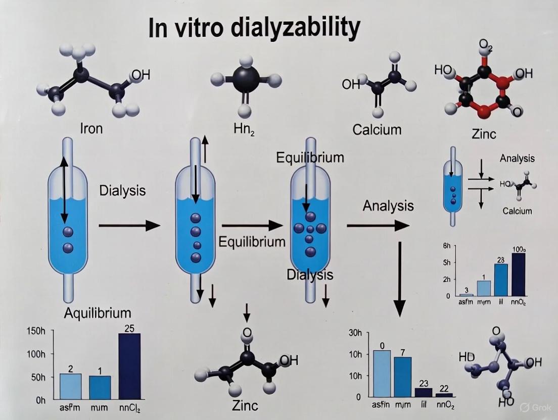
The dialyzability method simulates human gastrointestinal digestion through a two-step process (gastric and intestinal phases). It utilizes dialysis tubing with a specific molecular weight cut-off (MWCO) to separate the low molecular weight fraction of minerals that are potentially bioaccessible from the rest of the digesta [3] [1]. The fundamental premise is that dialyzable compounds represent the fraction available for absorption in the small intestine.
Equipment and Reagents
Table 2: Essential Research Reagent Solutions for Dialyzability Assays
| Reagent/Equipment | Specifications | Function in Protocol |
|---|---|---|
| Dialysis Membrane | Specific molecular weight cut-off (MWCO); typically 8,000-14,000 Da [1] | Physically separates low molecular weight, potentially bioaccessible minerals from larger complexes and undigested material |
| Pepsin | From porcine stomach; activity ~3,000 U/mg [1] | Gastric protease enzyme that initiates protein digestion in simulated gastric phase |
| Pancreatin | Porcine-derived; contains amylase, lipase, proteases [1] | Enzyme mixture that simulates pancreatic secretion for intestinal digestion phase |
| Bile Salts | Porcine bile extract [1] | Emulsifies lipids and facilitates mineral solubilization in intestinal phase |
| pH Meter | Precision of ±0.01 pH units | Critical for monitoring and adjusting pH at each digestion stage to maintain enzyme activity |
| Atomic Absorption Spectrophotometry (AAS) or ICP-AES | Element-specific detection [1] | Quantitative measurement of mineral concentration in dialyzate fraction |
Step-by-Step Procedure
Sample Preparation: Homogenize test material to ensure representative sampling. Accurately weigh approximately 5g of sample into a digestion vessel.
Gastric Phase Simulation:
- Add pepsin solution to achieve final concentration of 1-3 mg/mL in the gastric digest [1].
- Adjust pH to 2.0 using HCl to simulate adult gastric conditions (use pH 4.0 for infant simulations) [1].
- Incubate at 37°C for 1 hour with continuous agitation in a water bath to simulate body temperature and gastric mixing.
Intestinal Phase Initiation:
- Place dialysis bag containing sodium bicarbonate buffer (to simulate neutralization) into the gastric digest [1].
- Gradually adjust pH to 5.5-6.0 using NaHCO₃ before adding pancreatin-bile mixture [1].
- Add pancreatin (final concentration 0.5-2.0 mg/mL) and bile salts (final concentration 2-4 mg/mL) to simulate intestinal secretions [1].
- Readjust pH to 6.5-7.0 and incubate at 37°C for 2 hours with continuous agitation.
Dialyzate Collection:
- Carefully remove dialysis bag from the intestinal digest.
- Quantitatively transfer dialyzate content to a volumetric flask.
- Analyze mineral content in dialyzate using appropriate analytical method (AAS, ICP-AES).
Calculation:
- Calculate dialyzability as: (Mineral content in dialyzate / Total mineral content in test sample) × 100
The workflow below illustrates the sequential stages of this protocol, highlighting the critical transitions between digestive phases and the collection point for analysis.
Factors Influencing Mineral Bioaccessibility and Methodological Considerations
Dietary Factors Affecting Bioaccessibility
The bioaccessibility of minerals measured by dialyzability methods is significantly influenced by several dietary factors:
Inhibitory Compounds: Phytic acid, abundant in cereals and legumes, strongly chelates minerals like iron, zinc, and calcium, forming insoluble complexes that reduce dialyzability [4]. The critical PA-to-mineral molar ratio should be considered, with ratios >20 significantly reducing zinc absorption [4]. Oxalates, present in spinach and other vegetables, similarly bind calcium, dramatically reducing its bioaccessibility to as low as 0.1-5% in some plant foods [5].
Enhancing Factors: Short-chain fatty acids (SCFAs) produced through microbial fermentation of prebiotic fibers in the colon can acidify the luminal environment, increasing mineral solubility and potentially enhancing calcium absorption [6] [1]. Probiotic strains, including Lactobacillus plantarum and Bifidobacterium species, have demonstrated positive effects on iron, calcium, selenium, and zinc bioaccessibility through various mechanisms including microbial metabolite production and reduction of phytic acid content [6] [7].
Food Matrix Effects: Processing methods significantly impact mineral bioaccessibility. Fermentation of cereals and soaking and germination of crops can degrade phytic acid, thereby increasing mineral bioaccessibility [6]. For instance, fermented soymilk with lactic acid bacteria shows increased bioavailability of magnesium, calcium, iron, and zinc [6]. Similarly, milling reduces phytic acid in rice from 1.39-11.1 g/kg to 0.1-0.2 g/kg by removing the PA-rich surface of the kernel [4].
Methodological Standardization
Critical technical factors must be controlled to ensure reproducible dialyzability results:
pH Control: Precise pH adjustment is essential, as pepsin denatures at pH ≥5, compromising gastric digestion [1]. The final intestinal pH of 6.5-7.0 must be maintained for pancreatin activity.
Dialysis Membrane Selection: The molecular weight cut-off (MWCO) of the dialysis membrane significantly influences results, as it determines which molecular complexes can pass through [3]. Standardization of MWCO (typically 8,000-14,000 Da) is essential for inter-laboratory comparisons.
Temporal Parameters: Adherence to strict incubation time schedules for both gastric (typically 1 hour) and intestinal (typically 2 hours) phases is critical for method standardization and reproducibility [3].
Applications and Validation in Research Context
The in vitro dialyzability method serves as a valuable screening tool in multiple research contexts:
Food Fortification Strategies: Dialyzability assays effectively screen different fortification formulations. For example, calcium carbonate-fortified white bread demonstrates high calcium bioaccessibility (~30%), whereas plant-based beverages fortified with tricalcium phosphate show low bioaccessibility due to poor solubility [5]. This allows researchers to optimize mineral forms and combinations before costly human trials.
Risk Assessment: Combined with Caco-2 cell bioavailability assays, dialyzability methods provide robust toxicological assessment. In mineral clay analysis, while total arsenic and lead content exceeded guidelines, dialyzability followed by Caco-2 permeability assessment showed minimal bioavailability, indicating reduced consumer risk [2].
Nutritional Ranking: Dialyzability effectively ranks mineral availability from different meals and dietary patterns, providing a rapid assessment tool for comparing dietary strategies, though with noted limitations for certain food components like milk, proteins, and tea [3].
When applying these methods, researchers should acknowledge that dialyzability measures bioaccessibility, not full bioavailability, and results should be interpreted as the potential maximum available fraction rather than the exact amount that will be absorbed in vivo [1]. For comprehensive assessment, dialyzability can be coupled with Caco-2 cell models to evaluate actual cellular uptake and transport, providing a more complete picture of the bioavailability pathway [1] [2].
In vitro dialyzability methods serve as a crucial screening tool in mineral absorption research, providing a reliable and ethical alternative to complex human absorption studies. These methods are designed to simulate the human gastrointestinal environment to predict the bioavailability of essential minerals, such as iron and zinc, from various food matrices and pharmaceutical formulations. The core principle revolves around a two-step enzymatic digestion process followed by dialysis through a semi-permeable membrane, which collectively mimics the passage of bioavailable nutrients across the intestinal epithelium [8] [3]. For researchers and drug development professionals, this methodology offers a standardized approach to rapidly rank mineral availability from different meals or formulations, guiding further development and optimization before proceeding to costly clinical trials [8].
The Principle and Components
The Two-Step Digestion Process
The simulation begins with a sequential enzymatic digestion that replicates the gastric and intestinal phases of human digestion. During the gastric phase, the sample is incubated with pepsin at an acidic pH (typically pH 1.9), breaking down complex matrices and initiating protein hydrolysis [9]. This is followed by the intestinal phase, where the digesta is adjusted to a neutral-to-alkaline pH and subjected to pancreatin, which contains a mixture of digestive enzymes including proteases, amylases, and lipases [8] [9]. This two-step process is critical for liberating minerals from the food or formulation matrix, making them available for potential absorption.
The Dialysis Membrane
The dialyzability component utilizes a semi-permeable membrane with a defined molecular weight cut-off (MWCO), typically ranging from 1,000 to 10,000 Daltons, with 1000 MWCO being used in specific protocols [9]. This membrane acts as a selective barrier, simulating the intestinal mucosa by allowing only low molecular weight compounds, including soluble mineral complexes, to pass through. The dialyzable fraction of the mineral (the portion that crosses the membrane) is subsequently quantified and used as an estimate of the potentially bioavailable mineral [8] [3]. The membrane effectively excludes mineral bound to large molecules, which are generally considered non-bioavailable, though this represents a known limitation as some large complexes may be absorbed in vivo via alternative pathways [8].
Table 1: Core Methodological Steps and Parameters for In Vitro Dialyzability
| Phase | Key Parameters | Objective | Typical Duration |
|---|---|---|---|
| Gastric Digestion | Pepsin, pH ~1.9-2.0, 37°C [9] | Simulate stomach conditions; liberate minerals from food matrix. | 30 minutes to 2 hours |
| Intestinal Digestion | Pancreatin, pH ~5.0-8.0, 37°C [8] [9] | Simulate small intestine conditions; further digest matrix. | 2 to 24 hours [9] |
| Dialysis | Molecular Weight Cut-Off (MWCO) membrane (e.g., 1000-10,000 Da) [8] [9] | Separate bioavailable (dialyzable) mineral fraction. | Concurrent with intestinal digestion |
Detailed Experimental Protocol
Reagents and Equipment
The following "Research Reagent Solutions" and equipment are essential for executing the in vitro dialyzability protocol.
Table 2: Essential Research Reagents and Materials
| Item | Specification / Function |
|---|---|
| Pepsin | Proteolytic enzyme for gastric-phase digestion. Activity: ≥ 2500 U/mg protein. Prepared in 0.1 M HCl to achieve pH ~1.9 [9]. |
| Pancreatin | Enzyme mixture (proteases, lipases, amylases) for intestinal-phase digestion. Prepared in 0.1 M NaHCO₃ or suitable buffer to achieve pH ~8.0 [8] [9]. |
| Dialysis Membrane | Semi-permeable membrane with a defined Molecular Weight Cut-Off (MWCO), e.g., 1000-10,000 Da. Selects for low molecular weight, bioavailable mineral complexes [8] [9]. |
| Buffer Solutions | - 0.1 M HCl: For gastric simulation and pepsin activity.- 0.1 M NaHCO₃ / Phosphate Buffer: For pH adjustment and intestinal simulation [9]. |
| pH Meter | Critical for precise adjustment of pH after gastric phase and monitoring during intestinal phase [8]. |
| Water Bath or Incubator | Maintains a constant temperature of 37°C throughout the digestion process to simulate physiological conditions [9]. |
Step-by-Step Procedure
- Sample Preparation: Weigh a representative sample (e.g., 1-10 g of food homogenate or drug formulation) into a digestion vessel.
- Gastric Digestion: Add a pre-warmed (37°C) pepsin solution in 0.1 M HCl to the sample. The final pH should be approximately 1.9. Incubate the mixture in a shaking water bath at 37°C for 30 minutes to 2 hours to simulate gastric transit [9].
- pH Adjustment & Intestinal Digestion: Carefully adjust the pH of the gastric digestate to approximately 5.0 using a neutralization solution (e.g., 0.1 M NaHCO₃), and then further to pH 7.0-8.0. Add a pre-warmed pancreatin solution to initiate the intestinal phase [8] [9].
- Initiate Dialysis: Transfer the entire intestinal mixture to a dialysis tube or bag sealed at one end, which has been pre-hydrated according to manufacturer instructions. Seal the other end and place the bag into a container with a suitable volume of dialysis buffer (e.g., saline or a specific buffer at pH 7-8). The buffer may be replaced intermittently (e.g., 6-11 changes) or circulated continuously to maintain a sink condition and improve the efficiency of dialyzable product removal [9].
- Incubate: Continue incubation with shaking at 37°C for a defined period, typically 2 to 24 hours, simulating intestinal transit time [9].
- Sample Collection and Analysis: After the incubation, collect the dialysate (the fluid outside the membrane bag). Acid-digest the dialysate if necessary and analyze the mineral content (Iron, Zinc, etc.) using an appropriate analytical method, such as Atomic Absorption Spectrometry (AAS) or Inductively Coupled Plasma (ICP) techniques [8].
Critical Factors for Standardization and Limitations
Standardization and Key Parameters
The reliability and reproducibility of in vitro dialyzability methods hinge on the strict control of several critical parameters. Final pH adjustment after the gastric phase and adherence to a strict time schedule are paramount for standardization [8]. Furthermore, the selected molecular weight cut-off of the dialysis membrane and the specific analytical method used for mineral determination can significantly influence the results and must be consistently reported [8].
Benefits and Limitations
In vitro dialyzability serves as a valuable screening tool, with studies showing a good correlation with human absorption data for ranking iron and zinc availability from most meals [8]. The method is simpler and more cost-effective than sophisticated computer-controlled gastrointestinal models or human trials [8].
However, the method has notable limitations. It may not accurately predict the effects of certain dietary components, such as milk proteins, tea, and organic acids, on mineral absorption [8]. A fundamental limitation is that the method excludes mineral bound to large molecules (which are sometimes available in vivo) and includes mineral bound to small molecules (which is not always bioavailable) [8]. Therefore, results should be interpreted as an estimation of potential bioavailability.
The in vitro dialyzability method represents a cornerstone technique for predicting mineral bioavailability in nutritional research. Its development was driven by the pressing need for simple, efficient, and ethical screening tools to replace complex and expensive human absorption studies. Before its establishment, scientists relied heavily on animal models or human trials to assess mineral absorption, which were time-consuming, costly, and raised ethical concerns. The dialyzability method emerged as an elegant solution, bridging the gap between simple chemical solubility tests and complex biological systems.
The core principle of in vitro dialyzability involves simulating human gastrointestinal digestion through a controlled, two-step process that mimics the gastric and intestinal phases. This is followed by dialysis through a semi-permeable membrane with a specific molecular weight cut-off, which separates the low molecular weight fraction of minerals considered bioavailable. The dialyzable mineral fraction serves as an estimate of the amount available for absorption in the human intestine. This methodological framework has proven particularly valuable for screening large numbers of samples when human studies are impractical, enabling researchers to evaluate the effects of food processing, formulation changes, and dietary interactions on mineral availability.
The Foundational Work: Miller's Method (1981)
The seminal paper by Miller et al. in 1981 marked a turning point in mineral bioavailability research by establishing a standardized, reproducible in vitro method for estimating iron availability from meals. This foundational protocol provided the scientific community with a crucial tool that balanced physiological relevance with practical feasibility.
Original Experimental Protocol
The original methodology involved a sequential simulation of the human digestive process. The experimental workflow can be summarized as follows:
- Sample Preparation: Homogenize mixtures of foods (entire meals) to create a uniform matrix for digestion.
- Gastric Phase: Expose the homogenized sample to pepsin at pH 2.0 to simulate stomach digestion. Incubate with continuous shaking for 2 hours at 37°C to maintain physiological temperature.
- Intestinal Phase Transition: Use dialysis to gradually adjust the pH to intestinal levels (pH 5.5-6.0). Dialysis tubing with a molecular weight cutoff of 6,000-8,000 is employed, containing sodium bicarbonate solution that diffuses out to neutralize the gastric digest.
- Intestinal Digestion: Add pancreatin and bile salts to the neutralized mixture to simulate intestinal digestion. Adjust the final pH to 7.0 and incubate for an additional 2 hours at 37°C with continuous shaking.
- Measurement: Quantify the mineral content that diffused across the semi-permeable membrane into the dialysate using atomic absorption spectrophotometry or other analytical methods. This dialyzable fraction represents the bioaccessible mineral [10] [11].
Key Innovations and Validation
Miller's method introduced several critical innovations that established its reliability and widespread adoption. The protocol accurately reflected actual food iron availability by measuring both intrinsic food iron and added extrinsic radioiron, with results showing remarkable consistency between these measurement approaches. The method successfully distinguished between meals containing known iron availability enhancers (ascorbic acid, orange juice) and inhibitors (eggs, tea, coffee, cola, whole wheat bread), demonstrating its predictive capability. Furthermore, the incorporation of dialysis to adjust pH created a more physiologically relevant transition from gastric to intestinal conditions compared to simple acid-base titration [10].
Critical Technical Evolution: Refinements and Standardization
Following the establishment of Miller's foundational protocol, researchers identified several critical factors that required standardization to improve reproducibility and accuracy across laboratories. These refinements transformed the method from a novel approach into a robust scientific tool.
Key Technical Refinements
- pH Control and Timing: Research by Sandberg (2005) emphasized that final pH adjustment and adherence to a strict time schedule were critical factors for standardization. Even minor deviations in pH or incubation times significantly influenced dialyzability results [3].
- Membrane Selection: The molecular weight cut-off of the dialysis membrane was identified as a crucial parameter affecting results. Membranes with cut-offs between 6,000-8,000 Daltons became standard, as they appropriately separated low molecular weight mineral complexes that could potentially be absorbed [3] [10].
- Mineral Determination Methods: The selection of analytical methods for iron and zinc quantification (e.g., AAS, ICP-AES) influenced result accuracy and sensitivity, requiring careful method validation [3].
- Physiological Relevance: Later research revealed limitations in predicting effects of certain food components like milk proteins, tea, and organic acids, indicating where the method required complementary approaches [3].
Addressing Methodological Limitations
As the dialyzability method was applied to diverse food matrices, researchers encountered specific limitations that necessitated methodological adaptations. A significant challenge emerged with heme-iron containing samples, particularly meat products. Studies revealed that the dialyzability method consistently underestimated iron bioavailability from meat samples because it couldn't adequately simulate the unique absorption pathway of heme-iron, which bypasses the common mineral solubility constraints [12]. This limitation highlighted the importance of understanding specific mineral absorption mechanisms when interpreting dialyzability results.
The method also faced challenges in complex food matrices where mineral-binding components like phytates, fibers, and polyphenols interacted differently during digestion than in simple solutions. Researchers addressed this by incorporating additional digestion phases or modifying enzyme concentrations to better simulate physiological conditions [12] [11].
Modern Adaptations: The Contemporary Toolkit
The evolution of in vitro dialyzability methods has progressed toward miniaturization, standardization, and high-throughput capabilities while maintaining physiological relevance.
The Multi-well Plate Setup
A significant advancement came with the introduction of a modified setup using multi-well plates, which addressed practical challenges associated with the original method's larger sample volumes. Developed by Argyri et al. (2009) and further validated in subsequent studies, this adaptation preserved the fundamental principles of Miller's method while offering practical advantages [13].
Experimental Protocol - Multi-well Plate Setup:
- Apparatus: Six-well plates with specialized ring inserts to hold dialysis membranes.
- Membrane Preparation: Cut Spectrapore I dialysis tubing (MWCO 6000-8000) into 4 cm² pieces. Soak in water for at least 1 hour prior to use and store in 0.15 M PIPES buffer (pH 6.3) until needed.
- Gastric Phase: Weigh samples into wells. Add pepsin solution in HCl (pH 2.0). Seal plates and incubate for 2 hours at 37°C with continuous shaking.
- Intestinal Phase: Place prepared membrane rings into each well. Add pancreatin-bile extract mixture. Reseal plates and incubate for an additional 2 hours at 37°C with shaking.
- Sample Collection: Carefully remove dialysate from inside the membrane ring for mineral analysis.
- Analysis: Determine mineral content in dialysate using appropriate analytical methods (AAS, ICP-AES) [13].
This modified setup demonstrated excellent correlation with human absorption data for both iron (r = 0.90, p < 0.001) and zinc (r = 0.85, p < 0.001), confirming its predictive validity while offering practical advantages [13].
The INFOGEST Standardized Protocol
The INFOGEST static in vitro digestion method represents the most recent effort to standardize digestion protocols across laboratories. This comprehensive protocol specifies enzyme activities, pH values, incubation times, and salt concentrations based on physiological data [12].
Key Advantages of INFOGEST:
- Standardized enzyme activities and sources ensure inter-laboratory reproducibility.
- Detailed specifications for each digestion parameter (gastric pH, intestinal pH, transit times).
- Validation across multiple food matrices and nutrient types.
- Higher bioaccessible percentages compared to traditional dialysis methods for certain minerals [12].
Limitations: The requirement for specific enzymes (e.g., gastric lipase from rabbit stomach extracts) and kits for enzyme activity determination presents cost and availability challenges for some laboratories, particularly in resource-limited settings [12].
Comparative Analysis: Methodological Evolution
The table below summarizes the key methodological parameters across the evolutionary stages of in vitro dialyzability methods:
Table 1: Evolution of Key Parameters in In Vitro Dialyzability Methods
| Parameter | Miller's Original Method (1981) | Traditional Dialyzability (2005) | Multi-well Plate Setup (2011) | INFOGEST Protocol (2022) |
|---|---|---|---|---|
| Sample Volume | Large volume (unspecified) | Large volume | Miniaturized (well plates) | Standardized based on food type |
| Dialysis Membrane | 6,000-8,000 MWCO | 6,000-8,000 MWCO | 6,000-8,000 MWCO | Varies based on research question |
| Gastric Phase | Pepsin, pH 2.0, 2h | Pepsin, pH 2.0, 1-2h | Pepsin, pH 2.0, 2h | Pepsin + gastric lipase, pH 3.0, 2h |
| Intestinal Phase | Pancreatin + bile, pH 7.0, 2h | Pancreatin + bile, pH 7.0, 2h | Pancreatin + bile, pH 7.0, 2h | Pancreatin + bile, pH 7.0, 2h |
| pH Adjustment | Dialysis with NaHCO₃ | Dialysis with NaHCO₃ | Integrated in system | Chemical adjustment |
| Throughput | Low | Low | Medium-High (6 samples/plate) | Medium |
| Correlation with Absorption | Iron: Good [10] | Iron/Zinc: Variable [3] | Iron: r=0.90; Zinc: r=0.85 [13] | Under investigation [12] |
Quantitative Performance Comparison
The evolution of dialyzability methods has significantly impacted their predictive capability and application range:
Table 2: Performance Comparison Across Food Matrices
| Method | Iron Dialyzability Range | Zinc Dialyzability Range | Key Applications | Limitations |
|---|---|---|---|---|
| Miller's Original | 2-15% (varying meals) [10] | Not originally reported | Meal iron availability | Large sample volume, low throughput |
| Traditional Dialyzability | 1-50% (broad range) [3] | 5-30% (estimated) | Fortified foods, plant-based diets | Excludes available iron bound to large molecules [3] |
| Multi-well Plate | 3-18% (validated meals) [13] | 5-25% (validated meals) [13] | High-throughput screening, biofortified crops | Lower sample representation |
| INFOGEST | 5-40% (meat products) [12] | 10-50% (meat products) [12] | Processed foods, comparative studies | Cost, complexity for routine use |
The Scientist's Toolkit: Essential Research Reagents
Successful implementation of in vitro dialyzability methods requires carefully selected reagents and materials that maintain physiological relevance while ensuring reproducibility.
Table 3: Essential Research Reagents for In Vitro Dialyzability Studies
| Reagent/Material | Specifications | Function | Physiological Basis |
|---|---|---|---|
| Pepsin | Porcine gastric mucosa, ≥250 U/mg | Gastric protease | Simulates protein digestion in stomach |
| Pancreatin | Porcine pancreas extract | Pancreatic enzyme cocktail | Simulates intestinal digestion (proteases, amylase, lipase) |
| Bile Salts | Porcine bile extract | Emulsification | Enhances lipid solubility and micelle formation |
| Dialysis Membrane | MWCO 6,000-8,000 (e.g., Spectrapore) | Molecular size exclusion | Simulates intestinal pore size for absorption |
| PIPES Buffer | 0.15 M, pH 6.3 | Membrane storage | Maintains membrane integrity without mineral contamination |
| Sodium Bicarbonate | Analytical grade | pH adjustment | Simulates natural bicarbonate secretion in duodenum |
Visualization of Methodological Evolution
The historical trajectory from Miller's original method to contemporary adaptations demonstrates how scientific techniques evolve through critical assessment, refinement, and innovation. The core principle of simulating gastrointestinal digestion followed by dialysis remains fundamentally sound, as evidenced by the method's continued relevance over four decades. Modern adaptations have enhanced throughput, reproducibility, and practical implementation while maintaining strong correlation with human absorption data for key minerals like iron and zinc.
Future developments will likely focus on increasing physiological relevance through multi-compartmental systems, incorporation of cellular uptake models (Caco-2 cells), and personalization factors reflecting individual digestive variations. The integration of dialyzability methods with other in vitro approaches will provide comprehensive nutrient bioavailability assessment platforms, further reducing the need for animal and human studies in preliminary screening. As food fortification and biofortification programs expand globally, these refined dialyzability methods will play an increasingly crucial role in developing effective nutritional interventions to combat mineral deficiencies worldwide.
In vitro dialyzability is a screening method that estimates mineral bioaccessibility—the fraction of a mineral that is released from the food matrix during digestion and is available for intestinal absorption [1]. The method is based on simulating human gastrointestinal digestion followed by dialysis through a semi-permeable membrane with a defined molecular weight cut-off (MWCO), which separates low molecular weight, potentially absorbable minerals from larger, non-absorbable complexes [8]. This method provides a practical, cost-effective, and ethically favorable alternative to human and animal studies for the initial ranking of mineral availability from different food matrices and meal compositions [8] [1]. While results generally correlate well with human absorption studies for ranking iron and zinc availability, the method has limitations, as it may not fully predict the effects of certain dietary components like milk proteins, tea, and organic acids [8].
Key Principles and Physiological Basis
The fundamental principle of the in vitro dialyzability method is that only minerals of low molecular weight, capable of passing through the pores of a dialysis membrane, are considered bioaccessible and thus potentially available for absorption in the small intestine [1]. The method operates on the physiological basis that mineral absorption is preconditioned by digestion, which liberates minerals from the food matrix, and subsequent solubility in the gastrointestinal lumen [1].
The dialysis membrane acts as an analog to the intestinal mucosal barrier, with a typical MWCO of 3.5-10 kDa, simulating the selective permeability of the gut wall [8] [1]. Minerals that pass through this membrane (the dialyzable fraction) are considered analogous to the pool that would be available for uptake by enterocytes in vivo. It is critical to note that dialyzability measures bioaccessibility (the fraction released from the matrix), not full bioavailability (the fraction absorbed and utilized physiologically), which also depends on host factors, cellular uptake, and metabolism [1].
Experimental Protocols
Standard Two-Step In Vitro Digestion and Dialyzability Protocol
This protocol, adapted from the method initially described by Miller et al. (1981), is widely used for estimating iron, zinc, calcium, copper, and magnesium bioaccessibility [1] [14].
Reagents and Equipment
- Simulated Gastric Fluid: Prepare a pepsin solution (e.g., 0.5-1 g/L pepsin from porcine stomach) in a saline solution. Adjust pH to 2.0 using HCl to simulate adult gastric conditions. For infant models, adjust pH to 4.0 [1].
- Simulated Intestinal Fluid: Prepare a pancreatin-bile salt mixture (e.g., 0.5-1 g/L pancreatin and 3-5 g/L bile salts) in a saline solution. A sodium bicarbonate buffer is often included to facilitate pH neutralization [1].
- Dialysis Tubing: Use dialysis tubing with a MWCO of 3.5-10 kDa, pre-treated according to manufacturer's instructions.
- Water Bath or Incubator: Maintained at 37°C with continuous shaking (e.g., 60-120 oscillations per minute) to simulate body temperature and peristalsis.
- pH Meter: For precise pH adjustments.
- Analytical Equipment: Atomic Absorption Spectrophotometry (AAS) or Inductively Coupled Plasma Atomic Emission Spectroscopy (ICP-AES) for precise mineral quantification [8] [15].
Procedure
- Sample Preparation: Homogenize the test food sample. Accurately weigh a portion (typically 5-20 g) into a digestion vessel. For solid foods, a preliminary step using lingual alpha-amylase may be included [1].
- Gastric Digestion:
- Add simulated gastric fluid containing pepsin to the sample.
- Adjust pH to 2.0 with HCl for adult models.
- Incubate at 37°C for a defined period (e.g., 1-2 hours) with continuous shaking.
- Intestinal Digestion and Dialysis:
- Place the dialysis tubing, filled with a bicarbonate buffer, into the gastric digest.
- Add the simulated intestinal fluid containing pancreatin and bile salts.
- Carefully adjust the pH to 6.5-7.0 using NaOH or via the slow diffusion of bicarbonate from the dialysis bag [1].
- Continue incubation at 37°C for a further 2 hours with shaking.
- Sample Collection:
- After incubation, carefully remove the dialysis bag.
- Quantitatively collect the fluid from inside the dialysis bag (the dialyzate).
- Digest the dialyzate with concentrated nitric acid and hydrogen peroxide if required for analysis.
- Mineral Analysis:
- Determine the concentrations of iron, zinc, calcium, copper, and magnesium in the dialyzate using AAS or ICP-AES [15].
- Calculate the dialyzability as a percentage of the total mineral content in the original sample:
- Dialyzability (%) = (Amount of mineral in dialyzate / Total amount of mineral in original sample) × 100
Critical Factors for Standardization
Standardization is critical for obtaining reproducible and comparable results. Key factors include:
- pH Control: Precise adjustment during the gastric (pH 2.0) and intestinal (pH 6.5-7.0) phases is essential, as enzyme activity and mineral solubility are highly pH-dependent [8].
- Time Schedule: Adherence to a strict incubation schedule for both digestion phases is necessary [8].
- Membrane Characteristics: The MWCO of the dialysis membrane must be consistent, as it directly influences the amount of mineral that dialyzes [8].
- Enzyme Quality and Concentration: Use consistent sources and activities of digestive enzymes (pepsin, pancreatin) [1].
Applications and Data Presentation
In vitro dialyzability is extensively applied to screen and rank mineral availability from various food types, study the impact of food processing, and investigate the effects of promoters (e.g., vitamin C, organic acids) and inhibitors (e.g., phytic acid, polyphenols, certain proteins) of mineral absorption.
Mineral Dialyzability from Different Food Matrices
The table below summarizes exemplary data on the dialyzability of key minerals from selected food products, as reported in the literature.
Table 1: Dialyzability of Essential Minerals from Different Food Matrices
| Food Matrix | Iron (%) | Zinc (%) | Copper (%) | Magnesium (%) | Calcium (%) | Key Findings & Reference |
|---|---|---|---|---|---|---|
| Whole-Grain Pasta | Varies by genotype | Varies by genotype | ~15-25% | ~25-35% | Not Reported | Lower dialyzability % than white pasta, but higher total dialyzable amount for Zn and Fe due to greater total mineral content [15]. |
| White Pasta | Varies by genotype | Varies by genotype | ~25-35% | ~35-45% | Not Reported | Higher dialyzability percentage for Cu, Fe, Mg, and Zn compared to whole-grain pasta [15]. |
| Infant Formulas (Casein/Whey) | ~1.5-5.5% | ~1-3% | ~3-7% | Not Reported | Not Reported | Dialyzability varies significantly between brands. Correlations found between mineral content and dialyzability [14]. |
| Infant Formulas (Protein Hydrolysate) | ~5-10% | ~2-4% | ~4-9% | Not Reported | Not Reported | Significantly higher iron dialyzability compared to other formula types [14] [16]. |
| Infant Formulas (Soy Protein) | ~0.5-2% | ~1-2% | ~2-5% | Not Reported | Not Reported | Lower iron dialyzability, potentially due to phytate content [16]. |
Factors Influencing Mineral Dialyzability
Table 2: Key Factors Affecting Mineral Dialyzability and Underlying Mechanisms
| Factor | Effect on Dialyzability | Proposed Mechanism |
|---|---|---|
| Phytic Acid | Decreases (strong inhibitor) | Forms insoluble complexes with di- and trivalent minerals (Fe, Zn, Ca) in the intestinal lumen [15]. |
| Ascorbic Acid (Vitamin C) | Increases (for Iron) | Reduces ferric iron (Fe³⁺) to the more soluble and absorbable ferrous (Fe²⁺) form; can chelate iron [16]. |
| Certain Proteins & Peptides | Varies (can increase or decrease) | Casein and whey can inhibit; protein hydrolysates (small peptides) can enhance solubility and dialyzability [14] [16]. |
| Dietary Fiber | Decreases | Can bind minerals, physically trapping them and preventing their dialyzability [15]. |
| Other Minerals | Varies (can be antagonistic) | High levels of calcium can inhibit iron dialyzability due to competition for absorption sites or joint precipitation [16]. |
| Organic Acids (e.g., Citric, Lactic) | Increases | Act as chelating agents, forming soluble complexes with minerals and preventing precipitation [16]. |
Research Reagent Solutions
A successful in vitro dialyzability experiment requires specific reagents and equipment. The following table details the essential materials and their functions.
Table 3: Essential Research Reagents and Materials for In Vitro Dialyzability Studies
| Item | Function/Description | Key Consideration |
|---|---|---|
| Pepsin (from porcine stomach) | Enzyme for gastric-phase digestion; hydrolyzes proteins. | Activity and concentration must be standardized. pH must be maintained at ~2 for optimal activity [1]. |
| Pancreatin (from porcine pancreas) | Enzyme mixture for intestinal-phase digestion; contains proteases, amylase, lipase. | Represents the complex enzyme secretion of the pancreas [1]. |
| Bile Salts (e.g., porcine bile extract) | Biological detergent that emulsifies lipids, facilitating fat digestion. | Critical for the bioaccessibility of fat-soluble vitamins and for simulating intestinal conditions [1]. |
| Dialysis Tubing (MWCO 3.5-10 kDa) | Semi-permeable membrane that separates dialyzable minerals. | The MWCO defines the size of molecules that can pass through and must be consistent [8] [1]. |
| Atomic Absorption Spectrophotometer (AAS) | Instrument for accurate quantification of mineral elements. | The method of choice for mineral determination in many studies; offers high sensitivity [8] [15]. |
| pH Meter with Electrode | For precise monitoring and adjustment of pH during simulated digestion. | Critical for standardizing enzyme activity and mineral solubility [8]. |
| Temperature-Controlled Shaking Incubator | Maintains physiological temperature (37°C) and simulates peristalsis via shaking. | Ensures continuous mixing of the digest and temperature homogeneity [1]. |
Workflow and Process Visualization
Experimental Workflow for Mineral Dialyzability
The following diagram illustrates the sequential steps of the standard in vitro dialyzability protocol.
Principle of Membrane Dialyzability
This diagram conceptualizes the separation process occurring during the intestinal dialysis phase, where low molecular weight minerals diffuse into the dialysis bag.
Executing the Dialyzability Protocol: A Step-by-Step Guide for Standardization
In the field of mineral absorption research, in vitro dialyzability methods serve as a critical screening tool to estimate the bioaccessibility of minerals—the fraction that is released from the food matrix and becomes available for intestinal absorption [8] [11]. This Application Note details a standardized static in vitro digestion protocol, harmonized with the international INFOGEST consensus, specifically adapted for the study of mineral dialyzability [17] [18]. The method provides a physiologically relevant, reproducible, and high-throughput system for predicting mineral bioavailability, thereby reducing the need for more costly and complex animal or human trials in preliminary studies [13] [11].
Principle of the Method
The core principle involves simulating the sequential gastric and intestinal phases of human digestion under controlled in vitro conditions. During this process, the food matrix is broken down by digestive enzymes at physiologically relevant pH, temperature, and time parameters [18]. The released minerals are then separated via dialysis through a semi-permeable membrane with a specific molecular weight cut-off (typically 6-8 kDa), which mimics the passage of low molecular weight compounds across the intestinal mucosa [8] [3] [13]. The dialyzable fraction of the mineral is quantified and expressed as a percentage of the total mineral content, providing an estimate of its bioaccessibility [11].
Experimental Protocols
Reagent Preparation
Simulated digestive fluids should be prepared fresh daily or aliquoted and stored at appropriate temperatures to maintain enzyme activity.
Table 1: Simulated Digestive Fluids and Reagents
| Simulated Fluid / Reagent | Composition | Function in Digestion |
|---|---|---|
| Simulated Gastric Fluid (SGF) | Pepsin (e.g., 2000 U/mL in final mixture), NaCl, pH adjusted to 3.0 with HCl [18]. | Proteolysis in the stomach; denatures proteins and initiates peptide breakdown [19]. |
| Simulated Intestinal Fluid (SIF) | Pancreatin (e.g., 100 U/mL of trypsin activity), Bile salts (e.g., 10 mM), pH adjusted to 7.0 with NaHCO₃ [18] [11]. | Final digestion of peptides, lipids, and carbohydrates; bile salts emulsify lipids [11]. |
| PIPES Buffer | 0.15 M, pH 6.3 [13]. | Used to pre-soak and store dialysis membranes to maintain integrity and pH. |
Step-by-Step Digestion and Dialysis Protocol
Step 1: Gastric Phase
- Sample Preparation: Weigh a representative sample (typically 1-5 g) of the test food into a digestion vessel.
- Oral Phase (Optional but recommended for solid foods): Add simulated saliva (e.g., containing α-amylase) and mix for a short period (e.g., 30 s) [20].
- Gastric Digestion: Add SGF to the sample. The ratio of food bolus to digestive fluids should be kept constant (e.g., 1:1 v/v) as per the INFOGEST protocol [18].
- Incubation: Incubate the gastric mixture at 37°C for 120 minutes with continuous agitation in a shaking water bath or on an orbital shaker [17].
Step 2: Intestinal Phase with Dialysis
- Setup: Place a pre-soaked dialysis membrane (MWCO 6000-8000 Da) tightly held by a well ring and immersed in the gastric digest [13]. The dialysis bag may contain a small volume of sodium bicarbonate buffer to initiate neutralization [11].
- Initiate Intestinal Digestion: Add the pre-warmed SIF to the gastric digest. The pH of the mixture must be carefully adjusted to 7.0 using NaHCO₃ (e.g., 1M) [8] [11].
- Incubation: Continue incubation at 37°C for another 120 minutes with constant agitation [17].
- Termination: After incubation, carefully retract the dialysis bag. The content inside the bag is the dialyzable fraction, representing the bioaccessible mineral.
Step 3: Sample Analysis
- Collect the dialyzate and digestate samples.
- Analyze the mineral content (e.g., Iron, Zinc) in the dialyzate and the original food sample using appropriate analytical techniques such as Atomic Absorption Spectrophotometry (AAS) or Inductively Coupled Plasma Atomic Emission Spectroscopy (ICP-AES) [11].
- Calculate the percentage dialyzability as: Dialyzability (%) = (Amount of mineral in dialyzate / Total amount of mineral in sample) × 100 [13].
Key Experimental Considerations
- pH Control: Precise pH adjustment during the gastric (pH ~3) and intestinal (pH ~7) phases is critical for optimal enzyme activity and physiologically relevant results [8] [18].
- Standardized Timing: Adherence to a strict time schedule for each digestion phase is essential for reproducibility [8].
- Enzyme Quality: Use well-characterized enzymes from reliable sources, with activities confirmed and standardized across experiments [18].
Key Parameters and Data Presentation
The following table summarizes the core parameters of the standardized static digestion protocol based on the INFOGEST model, which is adopted by a majority of recent studies for simulating human gastrointestinal processes [17] [18].
Table 2: Standardized Parameters for Static In Vitro Digestion
| Phase | Duration (min) | pH | Key Enzymes/Chemicals | Temperature |
|---|---|---|---|---|
| Oral | 2 | 7 | α-Amylase (optional) | 37°C |
| Gastric | 120 | 3.0 | Pepsin | 37°C |
| Intestinal | 120 | 7.0 | Pancreatin, Bile Salts | 37°C |
Workflow and Logical Diagram
The entire experimental procedure, from sample preparation to data analysis, is visualized in the following workflow for clarity and easy replication.
Diagram 1: Experimental workflow for mineral dialyzability analysis.
The relationship between the in vitro dialyzability method and the broader context of mineral bioavailability is complex. The following diagram outlines the conceptual pathway and the position of the dialyzability assay within the research framework.
Diagram 2: Role of dialyzability in predicting mineral absorption.
The Scientist's Toolkit
Table 3: Essential Research Reagent Solutions
| Item | Function / Role in Experiment |
|---|---|
| Pepsin (from porcine gastric mucosa) | Primary protease in the gastric phase; breaks down proteins into smaller peptides, facilitating mineral release [18] [11]. |
| Pancreatin (porcine) | A mixture of pancreatic enzymes (including trypsin, amylase, lipase) for the intestinal phase; completes macronutrient digestion [11]. |
| Bile Salts (e.g., porcine bile extract) | Biological emulsifiers; critical for lipid solubilization and the formation of mixed micelles, which can affect mineral accessibility [11]. |
| Dialysis Membrane (MWCO 6-8 kDa) | Semi-permeable barrier that separates low molecular weight, bioaccessible minerals from larger, undigested molecules and complexes [8] [13]. |
| PIPES Buffer | An inert buffer used to condition the dialysis membrane, preventing pH shocks and maintaining membrane integrity [13]. |
In vitro dialyzability methods are indispensable tools for predicting mineral bioavailability, serving as a critical screening step before costly and complex human trials. These methods simulate the human gastrointestinal digestion process to estimate the fraction of a mineral that is available for absorption. The reliability of these simulations, however, hinges on the stringent control of several operational parameters. This application note delineates three critical procedural parameters—pH adjustment, timing, and membrane selection—within the context of mineral absorption research. Standardization of these factors is essential for obtaining reproducible and physiologically relevant data on mineral dialyzability, particularly for iron and zinc [8] [3]. The following sections provide detailed protocols and data to guide researchers in optimizing these key aspects of their experimental design.
Critical Parameter 1: pH Adjustment
The pH profile during in vitro digestion is a primary determinant of mineral solubility and dissociation from the food matrix, thereby directly influencing dialyzability.
Rationale and Impact
The simulated gastric phase, conducted under highly acidic conditions (typically pH ~2), facilitates the release of minerals from the food matrix. The subsequent transition to a neutral pH (~7) during the intestinal phase is critical for simulating the duodenal environment. This adjustment can precipitate certain mineral complexes, altering the fraction available for dialysis. Inconsistencies in the final intestinal pH have been identified as a major source of inter-laboratory variability [8] [21]. The use of buffering agents, such as Piperazine-NN-bis(2-ethane-sulfonic acid) disodium salt (PIPES) at pH 7.0, has been recommended to improve the reproducibility of the intestinal conditions [21] [22].
Recommended Reagents and Protocols
- Gastric Phase Acidification: Use HCl at a concentration of 0.08 M to achieve a consistent pH of ~2.0 for the gastric digestion step [22].
- Intestinal Phase Buffering: Adjust the pH to 7.0 ± 0.2 for the intestinal phase. This can be achieved using a 0.1 M or 1.0 M NaHCO₃ solution or, for enhanced reproducibility, a PIPES buffer [21] [22].
- Simulated Gastric Juice (SGJ): 0.32% (m/v) pepsin in 0.08 M HCl [22].
- Simulated Intestinal Juice (SIJ): 0.40% (m/v) pancreatin and 2.5% (m/v) bile salts in 0.10 M NaHCO₃ [22].
Table 1: Reagents for pH Control in a Standard Two-Step In Vitro Digestion
| Reagent | Concentration / Molarity | Function | Typical Volume Ratio (Reagent:Sample) |
|---|---|---|---|
| HCl | 0.08 M | Acidification for gastric digestion | As needed to achieve pH ~2.0 |
| NaHCO₃ | 0.1 M or 1.0 M | pH adjustment for intestinal phase | As needed to achieve pH 7.0 |
| PIPES Buffer | 0.15 M, pH 6.3-7.0 | Stabilizes intestinal pH | Pre-soak membrane; may be included in dialysate [13] [21] |
Critical Parameter 2: Timing
Adherence to a strict time schedule is vital for standardizing the dialyzability method, as it ensures consistent interaction times between the mineral, digestive enzymes, and the dialysis membrane.
Standard Digestion Phases
The consensus method involves a two-step digestion, with each phase maintained at 37°C under gentle agitation to simulate peristalsis [22]:
- Gastric Digestion: 2 hours.
- Intestinal Digestion: 2 hours.
Considering Time Delays in Dialysis
Research on microdialysis systems reveals that the measured concentration in the dialysate does not instantaneously reflect the concentration in the digest. A phenomenon known as Recovery Time (RT)—the time gap between a change in the target molecule's concentration in the digest and the formation of a stable concentration in the dialysate—must be considered. One study using a 10-minute sampling interval reported an RT of 20 minutes for calcium, due to factors beyond simple dead volume in the system, including transmembrane diffusion kinetics [23]. This implies that shorter sampling intervals may be necessary to accurately capture the kinetics of mineral dialyzability.
Diagram 1: In vitro digestion workflow with timing.
Critical Parameter 3: Membrane Selection
The dialysis membrane acts as the primary surrogate for the intestinal barrier, making its physicochemical properties a cornerstone of the method.
Molecular Weight Cut-Off (MWCO)
The MWCO determines the size of molecules that can pass through the membrane. A narrower pore-size distribution yields greater selectivity.
- Common MWCOs: 6-8 kDa and 10 kDa are widely used [13] [21] [22].
- Physiological Rationale: These MWCOs are selected to exclude large molecules (e.g., proteins, mineral complexes bound to large ligands) while allowing smaller, potentially bioavailable mineral complexes to pass through [8].
Membrane Material and Preparation
Table 2: Dialysis Membrane Specifications and Their Impact on Results
| Parameter | Typical Specifications | Impact on Dialyzability |
|---|---|---|
| Molecular Weight Cut-Off (MWCO) | 6-8 kDa, 10 kDa, 12.4 kDa [13] [21] [22] | Lower MWCO may underestimate availability of Fe/Zn bound to larger ligands; higher MWCO may overestimate by including unavailable small complexes [8]. |
| Membrane Material | Regenerated cellulose (Spectra/Por) [13] [22] | Affects hydrophilicity, protein adsorption, and non-specific binding. |
| Membrane Surface Area | Standardized piece (e.g., 4 cm²) [13] | Influences the total area available for diffusion, affecting the absolute amount of mineral dialyzed. |
Integrated Experimental Protocol for Mineral Dialyzability
This protocol provides a detailed method for assessing the dialyzability of minerals such as iron and zinc from food samples, integrating the critical parameters discussed above.
Sample Preparation
- Prepare a homogeneous sample.
- Accurately weigh a portion (e.g., 5-10 g) into the digestion vessel.
- All glassware must be meticulously cleaned, including soaking overnight in 1 N HCl and rinsing with deionized water to prevent mineral contamination [13] [22].
Gastric Digestion
- Add SGJ to the sample at a ratio of 3 g pepsin solution per 10 g of food [21].
- Adjust the pH to 2.0 ± 0.1 using 1 M HCl.
- Incubate the mixture at 37°C for 2 hours in a shaking water bath.
Intestinal Digestion & Dialysis
- Place a pre-soaked dialysis bag containing 20-50 mL of PIPES buffer (pH 6.3-7.0) into the gastric digest [13] [21].
- Adjust the pH of the mixture to 7.0 ± 0.2 using a sterile 0.1 M or 1.0 M NaHCO₃ solution.
- Add SIJ (pancreatin and bile salts).
- Incubate the system at 37°C for 2 hours with continuous shaking.
Sample Collection and Analysis
- After the intestinal digestion, carefully retrieve the dialysis bag.
- Quantitatively transfer the dialysate (the fluid inside the bag) to an acid-washed container.
- The dialyzable mineral fraction can be analyzed directly by ICP-OES or ICP-MS after acidification, or subjected to further processing [21] [22].
Diagram 2: Relationship between critical parameters and outcomes.
The Scientist's Toolkit: Essential Research Reagents and Materials
Table 3: Key Reagents and Materials for In Vitro Dialyzability Studies
| Item | Function / Role | Specification / Notes |
|---|---|---|
| PIPES Buffer | Stabilizes pH during intestinal digestion; used to pre-soak membranes and/or as dialysate [13] [21]. | 0.15 M, pH ~6.3-7.0. |
| Dialysis Membrane | Semi-permeable barrier simulating intestinal absorption; critical for size-based fractionation. | Regenerated cellulose; MWCO 6-8 kDa or 10 kDa [13] [22]. |
| Enzymes (Pepsin, Pancreatin) | Simulate enzymatic breakdown of food matrix during gastric (pepsin) and intestinal (pancreatin) phases. | Porcine origin. Use concentrations as per protocol (e.g., 0.32% pepsin, 0.4% pancreatin) [22]. |
| Bile Salts | Emulsifies fats, simulating the role of bile in the small intestine. | ~50% sodium cholate & 50% sodium deoxycholate; typical concentration 2.5% (m/v) in SIJ [21] [22]. |
| ICP-OES / ICP-MS | Highly sensitive and accurate quantification of multi-element concentrations in dialyzable fractions. | Preferred over FAAS for multi-element analysis and lower detection limits [21] [22]. |
The standardization of pH adjustment, timing, and membrane selection is non-negotiable for generating reliable and meaningful in vitro dialyzability data. Adherence to detailed protocols for these parameters significantly enhances the method's reproducibility and its correlation with human absorption studies for minerals like iron and zinc [8] [13]. While these simplified dialyzability methods are powerful screening tools, researchers must remain cognizant of their limitations, such as the inability to predict the effects of certain dietary components like milk proteins or tea, and the fact that they do not account for active transport or mucosal uptake mechanisms [8] [24]. Mastery of these critical parameters ensures that in vitro dialyzability remains a robust and valuable technique in mineral bioavailability research.
Analytical Techniques for Quantifying Dialyzable Minerals (AAS, ICP-AES)
The assessment of mineral bioavailability is a critical component of nutritional science, providing insight into the fraction of an ingested nutrient that is available for utilization in physiological functions. Within this framework, in vitro dialyzability methods have emerged as a vital screening tool, simulating human gastrointestinal digestion to predict mineral absorption [1]. These methods rely on sophisticated analytical techniques for accurate quantification, with Flame Atomic Absorption Spectrometry (F AAS) and Inductively Coupled Plasma Optical Emission Spectrometry (ICP OES), also referred to as ICP-AES, being extensively employed [25]. This document details the application of these analytical techniques within the context of in vitro dialyzability research, providing validated protocols, key methodological considerations, and illustrative data to support scientists in drug development and nutritional research.
Principles of In Vitro Dialyzability
Bioaccessibility, defined as the fraction of a compound that is released from its food matrix and becomes available for intestinal absorption, is a key predictor of bioavailability [1]. The in vitro dialyzability method estimates this by using a two-step digestion process that simulates the gastric and intestinal phases, followed by dialysis through a semi-permeable membrane with a specified molecular weight cut-off [8] [3]. The fundamental premise is that dialyzable minerals represent the fraction that is potentially absorbable by the human body [1].
This approach offers a practical compromise between simplicity and physiological relevance. While sophisticated computer-controlled gastrointestinal models exist, simple dialyzability methods are often preferable for screening purposes, as they are less expensive, faster, and allow for better control of experimental variables [8] [1]. These methods have demonstrated good correlation with human absorption studies for ranking the availability of minerals like iron and zinc from various meals, though predictions can be affected by factors such as the presence of milk, certain proteins, and tea [3].
Core Analytical Techniques
Atomic Absorption Spectrometry (AAS)
Flame Atomic Absorption Spectrometry (F AAS) is a robust and widely used technique for determining mineral concentrations in dialyzable fractions. Its principle relies on the absorption of optical radiation by free, ground-state atoms in a flame. F AAS is valued for its sensitivity, simplicity, and relatively low cost, making it a common choice for laboratories analyzing specific elements at low parts-per-billion (μg/L) levels [25] [26]. It is particularly well-suited for analyzing minerals such as calcium (Ca) and magnesium (Mg) in bioaccessible fractions [25].
Key operational settings for F AAS, as applied in dialyzability studies, include the use of specific analytical lines (e.g., 422.7 nm for Ca and 285.2 nm for Mg), spectral band-passes (e.g., 0.7 nm), and optimized flow rates for an air-acetylene flame [25]. For even lower detection limits, Graphite Furnace AAS (GFAAS) is employed, which is particularly useful for analyzing elements like aluminum in complex biological matrices such as serum and urine [26].
Inductively Coupled Plasma Optical Emission Spectrometry (ICP OES)
ICP OES is a multi-elemental technique that offers a powerful alternative to AAS. It uses an argon plasma to atomize and excite sample atoms, and the emitted light is measured for quantitative analysis. A significant advantage of ICP OES is that it is relatively free of the chemical interferences that can affect AAS, due to the high temperature of the plasma [25] [26]. This makes it an excellent tool for the simultaneous determination of a wide range of elements in dialyzates.
While ICP OES is a robust technique, potential interferences include intense emission from elements like calcium, which can elevate the background for other elements and affect detection limits [26]. Nevertheless, its ability to rapidly quantify multiple elements, such as Al, Ba, Ca, Cr, Cu, Fe, Mg, Mn, Ni, Sr, and Zn, in a single analysis makes it highly efficient for comprehensive mineral bioaccessibility studies [25].
Experimental Protocol: Quantifying Dialyzable Minerals from Food Matrices
The following section provides a detailed, step-by-step protocol for an in vitro dialyzability assay, from sample preparation to elemental analysis, adaptable for various food and beverage samples.
The diagram below illustrates the complete experimental workflow for the in vitro dialyzability assay and subsequent mineral analysis.
Materials and Reagents
Table 1: Essential Research Reagents and Materials for In Vitro Dialyzability Assay
| Item | Function / Specification | Example / Notes |
|---|---|---|
| Pepsin | Gastric phase enzyme. | From porcine gastric mucosa [1]. |
| Pancreatin | Intestinal phase enzymes. | A cocktail of amylase, lipase, and proteases [1]. |
| Bile Salts | Emulsifier for fat digestion. | Porcine bile extract [25]. |
| Dialysis Membrane | Simulates intestinal barrier. | Semi-permeable tube with specific Molecular Weight Cut-Off (MWCO) [8]. |
| HCl / NaOH | pH adjustment. | For simulating gastric (pH ~2) and intestinal (pH ~6.5-7) conditions [25] [1]. |
| Standard Solutions | Instrument calibration. | High-purity single/multi-element standards for AAS/ICP OES [25]. |
| F AAS / ICP OES | Elemental quantification. | Perkin-Elmer F AAS; Sequential or simultaneous ICP OES [25] [27]. |
Step-by-Step Procedure
- Sample Preparation: Prepare the test material (e.g., brew coffee from ground or instant powders using hot water) and allow it to cool to room temperature [25].
- Gastric Digestion:
- Mix a known volume or weight of the sample with a pepsin solution. The concentration of pepsin can vary (e.g., 0.001–16%), but must be specified for reproducibility [25].
- Adjust the pH to ~2.0 using dilute HCl (e.g., 0.1 M) to simulate the acidic environment of the stomach.
- Incubate the mixture for 2 hours at 37°C with constant, gentle shaking or agitation to mimic gastric peristalsis [25] [1].
- Intestinal Digestion with Dialysis:
- Introduce a dialysis tube or bag, filled with a sodium bicarbonate (NaHCO₃) buffer solution, into the gastric digest [1].
- Slowly add a mixture of pancreatin (e.g., 0.015–3.04%) and bile salts (e.g., 0.15–2.8%) to the beaker outside the dialysis bag.
- Carefully adjust the pH of the external solution to ~6.5–7.0 using a Na₂CO₃ solution (e.g., 0.1 or 1.0 mol L⁻¹) or NaOH. The NaHCO₃ inside the bag will diffuse out and aid in neutralization.
- Incubate the entire system for another 2 hours at 37°C with gentle shaking [25] [1].
- Collection of Dialyzable Fraction:
- Sample Analysis:
- Analyze the dialyzate directly by F AAS or ICP OES without any further digestion. This direct analysis has been validated as a precise and accurate preparation procedure [25].
- Prepare appropriate calibration standards and blanks in a matrix that matches the dialyzate (e.g., similar pH and salt content) to ensure accurate quantification.
Data Analysis and Validation
Calculation of Bioaccessible Fraction
The bioaccessible fraction of each element is calculated as the percentage of the total mineral content in the original sample that is found in the dialyzable fraction.
Formula:
% Bioaccessible Fraction = (Concentration in Dialyzate × Volume of Dialyzate) / (Total Concentration in Sample × Mass/Volume of Sample) × 100
Exemplary Data from Coffee Brew Analysis
The following table summarizes validation data and bioaccessibility results for minerals in coffee brews, obtained using the direct analysis (P2) method with F AAS and ICP OES [25].
Table 2: Method Validation Parameters and Bioaccessible Fractions of Elements in Coffee Brews
| Element | Precision (% RSD) | Recovery (%) | LOD (μg L⁻¹) | LOQ (μg L⁻¹) | Bioaccessible Fraction (%) | |
|---|---|---|---|---|---|---|
| Ground Coffee (GCs) | Instant Coffee (ICs) | |||||
| Al | N.R. | 104 | N.R. | N.R. | 19.0 | 23.0 |
| Ba | N.R. | 98.0 | 0.095 | 0.32 | 42.8 | 48.4 |
| Ca | N.R. | N.R. | N.R. | N.R. | 35.0 | 38.9 |
| Cu | 5.9 | N.R. | N.R. | N.R. | 15.0 | 14.3 |
| Fe | 0.54 | N.R. | N.R. | N.R. | 5.08 | 2.81 |
| Mg | N.R. | N.R. | N.R. | N.R. | 32.2 | 37.9 |
| Mn | N.R. | N.R. | N.R. | N.R. | 28.1 | 29.1 |
| Ni | N.R. | N.R. | 1.8 | 6.0 | 40.9 | 60.0 |
| Sr | N.R. | 98.0 | N.R. | N.R. | 43.2 | 45.6 |
| Zn | N.R. | N.R. | N.R. | N.R. | 11.5 | 9.57 |
N.R. = Not explicitly reported in the summary of the cited source [25].
Method Validation
The chosen analytical procedure must be rigorously validated. Key performance characteristics include:
- Precision: Expressed as relative standard deviation (RSD). For the direct analysis method, precision can be excellent, ranging from 0.54% for Fe to 5.9% for Cu [25].
- Accuracy: Assessed through recovery experiments, where known amounts of a standard are added to the sample. Recovery rates for the direct analysis method have been reported between 98.0% (Sr) and 104% (Al) [25].
- Sensitivity: Determined by the Limit of Detection (LOD) and Limit of Quantification (LOQ). For example, LODs for Ba and Ni were reported as 0.095 μg L⁻¹ and 1.8 μg L⁻¹, respectively [25].
Critical Considerations and Limitations
While in vitro dialyzability is a valuable screening tool, researchers must be aware of its limitations and critical control points.
- Standardization is Key: The final pH adjustment in the intestinal phase and adherence to a strict time schedule are critical for obtaining reproducible and comparable results [8] [3].
- Membrane Selection: The molecular weight cut-off (MWCO) of the dialysis membrane directly influences which complexes can pass through and must be carefully selected and reported [8].
- Predictive Limitations: The method excludes minerals bound to large molecules that might be available in vivo (e.g., through brush border membrane hydrolysis) and includes minerals bound to small molecules that may not always be absorbed [3]. Effects of certain dietary components like milk proteins and tea may not be fully predicted [3].
- Contamination Control: Due to the low concentrations of minerals in dialyzates, stringent precautions against contamination from reagents, glassware, and the laboratory environment are essential for accurate results, particularly for ubiquitous elements like aluminum [26].
In vitro dialyzability is a critical screening tool in nutritional science, providing a rapid, cost-effective method for estimating mineral absorption potential from complex food matrices. This method simulates human digestion through a two-step process (gastric and intestinal phases), using dialysis tubing with a specific molecular weight cut-off to separate minerals released from the food matrix that would be available for absorption in the small intestine [1] [3]. For researchers and drug development professionals, these methods offer valuable preliminary data on bioaccessibility—the amount of an ingested nutrient released from the food matrix and potentially available for absorption—before proceeding to more complex and expensive human trials [1] [28]. While true bioavailability (the amount absorbed and available for physiological functions) requires human studies, dialyzability methods provide crucial screening, ranking, and categorization capabilities for evaluating nutritional interventions [1] [11].
The following application notes present specific case studies and protocols for evaluating mineral absorption from infant formula, fortified foods, and plant-based meals, contextualized within mineral absorption research.
Case Study 1: Zinc Bioaccessibility in Infant Formulas
Research Context and Objectives
Infant formula must provide adequate bioavailable zinc to support critical growth and developmental processes. This case study applied in vitro solubility and dialyzability methods to assess zinc bioaccessibility from various infant formula types, with parallel analysis using size-exclusion chromatography coupled to inductively coupled plasma-mass spectrometry (SEC-ICP-MS) to characterize zinc-binding biomolecules [29].
Experimental Protocol & Methodology
Sample Preparation:
- Selected commercial infant formulas included milk-based, soy-based, and lactose-free varieties
- Prepared samples according to manufacturer instructions simulating reconstitution for feeding
In Vitro Gastrointestinal Digestion:
- Gastric Phase: Samples acidified to pH 2 with pepsin addition, incubated at 37°C for 1 hour with continuous agitation
- Intestinal Phase: pH adjusted to 5.5-6 followed by pancreatin/bile addition, final pH adjustment to 6.5-7, further incubation at 37°C for 2 hours
Solubility Assay:
- Centrifuged intestinal digests at high speed to separate soluble and insoluble fractions
- Analyzed zinc in supernatant using atomic absorption spectrophotometry (AAS)
- Calculated percent solubility as: (Soluble Zn / Total Zn in sample) × 100
Dialyzability Assay:
- Following gastric digestion, dialysis tubing containing sodium bicarbonate buffer added to digest
- After intestinal digestion, collected dialysate and measured zinc content via AAS
- Calculated percent dialyzability as: (Dialyzable Zn / Total Zn in sample) × 100
SEC-ICP-MS Analysis:
- Extracted soluble proteins with 100 mM Tris-HCl buffer (pH 6.8)
- Separated zinc-containing biomolecules using size-exclusion chromatography
- Detected and quantified zinc using ICP-MS
Key Findings and Data Analysis
Table 1: Zinc Bioaccessibility in Infant Formulas Using Different Assessment Methods
| Formula Type | % Zinc in Soluble Protein Fraction | % Zinc Solubility | % Zinc Dialyzability | Molecular Weight of Zn-Binding Compounds |
|---|---|---|---|---|
| Milk-Based | ~90% | 70% | 10% | 1-7 kDa |
| Soy-Based | ~7% | 30% | 1% | 1-7 kDa |
| Lactose-Free | ~24% | 30% | 1% | 1-7 kDa |
The data revealed significant differences in zinc distribution between formula types. Despite high solubility (30-70%) across all formulas, dialyzability was remarkably low (1-10%) [29]. SEC-ICP-MS analysis demonstrated that zinc was bound to low-molecular-weight compounds (1-7 kDa) in all formulas, suggesting the dialysis method may underestimate bioaccessible zinc. This discrepancy highlights the importance of complementary speciation studies for validating dialyzability data [29].
Figure 1: Experimental workflow for assessing zinc bioaccessibility in infant formulas
Case Study 2: Iron Bioaccessibility in Plant-Based Foods
Research Context and Objectives
Plant-based foods contain exclusively non-heme iron, which has lower bioavailability than heme iron from animal sources due to inhibitors like phytic acid, tannins, and dietary fiber [28]. This case study applied in vitro dialyzability methods to assess iron bioaccessibility from various plant-based matrices, examining the effects of processing and compositional factors on iron availability.
Experimental Protocol & Methodology
Sample Selection and Preparation:
- Selected plant-based foods: legumes (beans, lentils), cereals (millet, sorghum), and vegetables
- Prepared samples representing raw, cooked, and processed forms
INFOGEST Standardized In Vitro Digestion:
- Oral Phase: Mixed samples with simulated salivary fluid containing amylase, incubated 2 minutes at 37°C
- Gastric Phase: Mixed with simulated gastric fluid containing pepsin, acidified to pH 3, incubated 2 hours at 37°C with continuous agitation
- Intestinal Phase: Mixed with simulated intestinal fluid containing pancreatin and bile salts, adjusted to pH 7, incubated 2 hours at 37°C
Dialyzability Assay:
- Used dialysis membrane with molecular weight cut-off of 3.5 kDa
- Following intestinal digestion, collected dialysate and measured iron content via AAS or ICP-MS
- Calculated percent dialyzable iron as: (Dialyzable Fe / Total Fe in sample) × 100
Inhibitor Analysis:
- Quantified phytic acid and tannin content in original samples and digests
- Conducted correlation analysis between inhibitor levels and iron dialyzability
Key Findings and Data Analysis
Table 2: Iron Dialyzability from Plant-Based Food Matrices
| Food Matrix | Processing Method | Phytic Acid Content (mg/g) | Tannin Content (mg/g) | % Iron Dialyzability |
|---|---|---|---|---|
| Pearl Millet | Raw | 8.5 | 2.1 | 3.2% |
| Pearl Millet | Soaked & Germinated | 4.2 | 1.8 | 7.8% |
| Common Beans | Raw | 10.2 | 1.5 | 2.1% |
| Common Beans | Cooked | 6.8 | 1.4 | 5.4% |
| Lentils | Raw | 6.3 | 0.9 | 4.5% |
| Lentils | Cooked | 3.5 | 0.8 | 8.9% |
| Spinach | Raw | 1.2 | 0.3 | 12.3% |
| Spinach | Steamed | 0.8 | 0.3 | 15.6% |
The data demonstrated that processing methods significantly impact iron bioaccessibility. Soaking, germination, and cooking reduced phytic acid content, correlating with increased iron dialyzability [28]. Pearl millet showed particularly low iron dialyzability due to combined effects of phytic acid and tannins. Physical barriers like cell walls in beans' cotyledons also limited iron release, which processing methods only partially disrupted [28].
Case Study 3: Mineral Fortification in Complementary Foods
Research Context and Objectives
Complementary foods for infants must provide adequate bioavailable minerals to support the nutritional needs during the transition from exclusive breastfeeding. This case study evaluated mineral bioaccessibility in three novel complementary food formulations designed with different ingredient matrices to assess how composition affects iron and zinc availability [30].
Experimental Protocol & Methodology
Food Formulations:
- BF1: Chickpeas, rice, artichoke, carrots, orange peel
- BF2: Corn, egg white, spinach, carrots, orange peel
- BF3: Potato, mushroom, beet, carrots, orange peel
Sample Preparation:
- Dried plant raw materials at 50°C for 48 hours
- Powdered using mechanical grinder
- Mixed formulations according to predetermined weights
- Cooked samples (10g formula + 100ml water) at 70°C for 15 minutes
In Vitro Dialyzability Assessment:
- Conducted two-step in vitro digestion simulating gastric and intestinal phases
- Used dialysis tubing with molecular weight cut-off of 8-10 kDa
- Measured iron and zinc in dialysate using atomic absorption spectrophotometry
- Analyzed correlation between formulation composition and mineral dialyzability
Nutritional Composition Analysis:
- Determined protein content via Kjeldahl method
- Analyzed vitamin content using HPLC
- Assessed mineral content with atomic absorption spectrophotometry
Key Findings and Data Analysis
Table 3: Composition and Mineral Dialyzability in Complementary Food Formulations
| Parameter | BF1 | BF2 | BF3 |
|---|---|---|---|
| Primary Ingredients | Chickpeas, Rice, Artichoke | Corn, Egg White, Spinach | Potato, Mushroom, Beet |
| Protein Content (%) | 15.2 | 12.8 | 8.9 |
| Iron Content (mg/100g) | 4.2 | 3.8 | 3.5 |
| Zinc Content (mg/100g) | 2.8 | 2.4 | 1.9 |
| % Iron Dialyzability | 8.9 | 7.2 | 10.3 |
| % Zinc Dialyzability | 12.4 | 9.8 | 14.1 |
| Phytic Acid Content (mg/g) | 3.2 | 2.8 | 1.9 |
| Sensory Acceptance Score | 6.8/10 | 5.4/10 | 7.5/10 |
BF1 demonstrated higher protein content but moderate mineral dialyzability, potentially due to higher phytic acid content from chickpeas and rice. BF3 showed the highest mineral dialyzability despite lower overall mineral content, likely due to lower levels of mineral-chelating compounds [30]. Sensory evaluation revealed BF3 as most acceptable, followed by BF1 and BF2, indicating the importance of balancing nutritional quality with sensory properties in complementary food development [30].
Figure 2: Research framework for evaluating mineral bioaccessibility in complementary foods
Standardized Experimental Protocols
In Vitro Dialyzability Protocol for Mineral Absorption Assessment
Principle: This protocol simulates gastrointestinal digestion to estimate mineral bioaccessibility by measuring the fraction that passes through a dialysis membrane after digestion, representing the fraction available for intestinal absorption [1] [3].
Reagents and Equipment:
- Pepsin (porcine stomach origin)
- Pancreatin (porcine pancreas origin)
- Bile salts (porcine origin)
- Dialysis tubing (molecular weight cut-off 3.5-10 kDa)
- Atomic Absorption Spectrophotometer or ICP-MS
- Incubation shaker with temperature control (37°C)
- pH meter
Procedure:
Sample Preparation:
- Homogenize test material to particle size <0.5 mm
- Precisely weigh test sample (typically 1-10g) into digestion vessel
Gastric Phase:
- Add pepsin solution (typically 0.5-1.0 mg/mL in final mixture)
- Adjust pH to 2.0 with HCl for adult digestion or pH 4.0 for infant digestion
- Incubate at 37°C for 1-2 hours with continuous agitation
Intestinal Phase:
- Transfer gastric digest to dialysis tube suspended in buffer
- Add pancreatin solution (typically 0.5-1.0 mg/mL in final mixture)
- Add bile salts (typically 1-2 mg/mL in final mixture)
- Adjust pH to 6.5-7.0 with NaHCO₃
- Incubate at 37°C for 2 hours with continuous agitation
Sample Collection and Analysis:
- Collect dialysate and measure mineral content via AAS or ICP-MS
- Analyze undialyzed sample for total mineral content
- Calculate percent dialyzability: (Mineral in dialysate / Total mineral in sample) × 100
Critical Factors for Standardization:
- Strict adherence to pH adjustments and time schedules
- Consistent molecular weight cut-off of dialysis membrane
- Enzyme activity and concentration
- Liquid-to-solid ratio in digestion mixture [3]
The Scientist's Toolkit: Essential Research Reagents and Equipment
Table 4: Essential Research Reagents and Equipment for In Vitro Dialyzability Studies
| Category | Item | Specification/Function | Application Notes |
|---|---|---|---|
| Enzymes | Pepsin | Porcine origin, activity ≥250 U/mg | Simulates gastric proteolysis; activity pH-dependent |
| Pancreatin | Porcine pancreas extract | Provides mix of amylase, lipase, proteases for intestinal digestion | |
| Digestion Components | Bile Salts | Porcine bile extract | Emulsifies lipids, facilitates micelle formation |
| Dialysis Membrane | MWCO 3.5-10 kDa | Selects for low molecular weight, potentially absorbable fractions | |
| Equipment | Atomic Absorption Spectrophotometer | Elemental detection at ppm-ppb levels | Quantifies minerals in dialysate and original samples |
| ICP-MS | High-sensitivity elemental analysis | Preferred for speciation studies and low-concentration minerals | |
| Temperature-Controlled Incubator | Maintains 37°C with agitation | Simulates body temperature and digestive motility | |
| Reference Materials | Certified Reference Materials | NIST, BCR food matrices | Validates analytical accuracy and method performance |
Limitations and Methodological Considerations
While in vitro dialyzability methods provide valuable screening data, researchers must acknowledge several limitations. Dialyzability methods exclude iron bound to large molecules that might be available in vivo and include iron bound to small molecules that may not always be available [3]. Effects of certain food components like milk proteins, tea compounds, and organic acids may not be accurately predicted [3]. The methods cannot assess uptake rates, transport kinetics, or nutrient competition at absorption sites [1].
Recent advancements in the INFOGEST standardized protocol have improved inter-laboratory comparability, but validation against human studies remains essential [28]. Sophisticated models like TIM gastrointestinal systems and Caco-2 cell cultures offer more comprehensive assessment but with increased cost and complexity [1]. For most screening applications, dialyzability methods provide the optimal balance of practicality and predictive value when interpreted with understanding of their limitations.
In vitro dialyzability methods provide researchers with valuable tools for preliminary screening of mineral bioaccessibility from infant formulas, fortified foods, and plant-based meals. The case studies presented demonstrate the method's application across diverse food matrices and its utility in evaluating the impact of formulation and processing on mineral accessibility. While these methods cannot fully replicate human absorption, they offer cost-effective, reproducible approaches for ranking mineral bioavailability and screening intervention strategies before proceeding to human trials. As fortification and plant-based food innovation continue to advance, dialyzability methods will remain essential tools in developing nutritionally optimized products that effectively address global micronutrient deficiencies.
Overcoming Limitations and Enhancing Dialyzability Assay Accuracy
In vitro dialyzability methods serve as vital screening tools in mineral absorption research, providing a practical means to estimate the bioaccessibility of essential minerals—the fraction that is released from the food matrix and available for intestinal absorption [11]. These methods simulate human gastrointestinal digestion through a controlled two-step process (gastric and intestinal phases), using dialysis through a semi-permeable membrane to separate the absorbable fraction of minerals [8] [3]. The reliability of these models in predicting mineral bioavailability depends heavily on the precise standardization of critical parameters, primarily enzyme sources, pH conditions, and digestion time [12] [8]. This protocol details the optimized application of the in vitro dialyzability method, contextualized within mineral absorption research, to ensure reproducible and physiologically relevant results for researchers and drug development professionals.
Critical Factors and Their Optimization
The following table summarizes the three critical methodological factors and their standardized specifications for reproducible mineral dialyzability assessment.
Table 1: Critical Factors in In Vitro Dialyzability Methods
| Factor | Specification | Physiological Rationale | Impact on Results |
|---|---|---|---|
| Enzyme Sources | Gastric: Pepsin (porcine stomach origin)Intestinal: Pancreatin (porcine pancreatic extract containing amylase, lipase, proteases) and bile salts [11] | Mimics the natural enzymatic cocktail encountered in the human GI tract [31] [32] | Source and activity of enzymes directly influence the efficiency of food matrix breakdown and mineral release [12]. |
| pH Conditions | Gastric Phase: pH 2.0 (simulates adult fasting state)Intestinal Phase: pH 6.5-7.0 after neutralization [8] [11] | Optimizes activity of pepsin (low pH) and pancreatic enzymes/ bile (neutral pH) [31] [33] | Final pH adjustment is a critical factor for standardization; deviations alter enzyme efficacy and mineral solubility [8] [3]. |
| Digestion Time | Gastric Phase: 1-2 hoursIntestinal Phase: 30 minutes to 2 hoursA strict time schedule is mandatory [8] [3] | Reflects typical gastric emptying and small intestinal transit times [31] | Digestion time directly affects the extent of mineral release and dialyzability [12]. |
Detailed Experimental Protocol for Mineral Dialyzability
Principle
This method estimates mineral bioaccessibility by simulating the gastric and intestinal digestion of a food sample. The dialyzable mineral fraction, which passes through a membrane with a specific molecular weight cut-off (typically 6-8 kDa), is considered bioaccessible and is quantified analytically [13] [8].
Materials and Equipment
- Digestion Reagents:
- Pepsin (from porcine gastric mucosa, e.g., Sigma P7000)
- Pancreatin (from porcine pancreas, e.g., Sigma P1750)
- Bile salts (e.g., Sigma B8756)
- Hydrochloric Acid (HCl)
- Sodium Bicarbonate (NaHCO₃)
- PIPES or other suitable buffer
- Equipment:
- Water bath or metabolic shaker, maintained at 37°C
- pH Meter
- Dialysis tubing or membranes (MWCO 6000-8000, e.g., Spectra/Por 1)
- Six-well plates with ring inserts (for modern setup) [13]
- Centrifuge
- Analytical Instrumentation: Atomic Absorption Spectrophotometry (AAS) or Inductively Coupled Plasma Optical Emission Spectroscopy (ICP-OES) [12] [11].
Procedure
- Sample Preparation: Homogenize the test food material. Accurately weigh a sample (typically 1-10 g) into a digestion vessel.
- Gastric Digestion:
- Add a pre-warmed (37°C) solution of pepsin in HCl to the sample. The final concentration of pepsin should be relevant to physiological conditions.
- Adjust the pH of the mixture to 2.0 using 1M HCl.
- Incubate the mixture for 1 hour in a shaking water bath at 37°C to simulate gastric motility and temperature.
- Intestinal Digestion & Dialysis:
- Place a prepared dialysis bag, containing a buffer solution (e.g., NaHCO₃), into the gastric digest. In the modern setup, a dialysis membrane is held tightly with a well ring and immersed in the sample within a six-well plate [13].
- Adjust the pH of the mixture to 5.5-6.0 using a saturated NaHCO₃ solution.
- Add a solution of pancreatin and bile salts to the mixture.
- Finally, adjust the pH to 6.5-7.0 to simulate the intestinal environment.
- Incubate the system for 2 hours at 37°C with constant agitation. The buffer inside the dialysis bag will diffuse out and help maintain the pH, while low molecular weight compounds (including bioaccessible minerals) diffuse into the bag.
- Sample Collection:
- After incubation, carefully retrieve the dialysis bag (or the dialysate from the well insert).
- The liquid inside the dialysis bag is the dialyzate. Note the final volume accurately.
- Centrifuge the dialyzate if necessary to remove any particulate matter.
- Mineral Analysis:
- Analyze the dialyzate for the mineral(s) of interest (e.g., Fe, Zn, Ca, Mg) using AAS or ICP-OES [12].
- Calculate the Dialyzability (%) as follows: Dialyzability (%) = (Amount of mineral in dialyzate / Total amount of mineral in original sample) × 100
The Scientist's Toolkit: Key Research Reagent Solutions
Table 2: Essential Materials for In Vitro Dialyzability Experiments
| Item | Function/Application in the Protocol |
|---|---|
| Pepsin (Porcine) | Simulates gastric proteolysis, breaking down proteins that may bind minerals, thereby facilitating mineral release [31] [11]. |
| Pancreatin & Bile Salts | Simulates intestinal digestion; pancreatin enzymes break down fats and carbs, while bile salts emulsify lipids, all crucial for liberating minerals [31] [32]. |
| Dialysis Membrane (MWCO 6-8 kDa) | Acts as an intestinal barrier analogue, selectively allowing only low molecular weight, potentially absorbable mineral complexes to pass through [12] [8]. |
| PIPES Buffer | Maintains a stable pH environment during the intestinal dialysis phase, which is critical for consistent enzyme activity and mineral solubility [13]. |
| Atomic Absorption Spectrophotometry (AAS) | Provides highly sensitive and specific quantification of individual mineral concentrations in the complex dialyzate matrix [12] [11]. |
Workflow and Data Interpretation
The following diagram illustrates the logical workflow of the in vitro dialyzability protocol, from sample preparation to data interpretation.
Diagram 1: In vitro dialyzability workflow.
Validation and Correlation with Absorption
The in vitro dialyzability method has been validated for certain minerals. For instance, studies have shown that iron and zinc dialyzability from meals, when measured using a properly standardized setup, correlates well with absorption data obtained from human studies [13]. This correlation confirms its utility as a predictive screening tool. However, researchers must be aware of limitations. The method may not accurately predict the effects of certain food components like milk, specific proteins, or tea on absorption [8] [3]. Furthermore, it is generally inadequate for assessing heme-iron bioaccessibility from meat products, as the dialysis mechanism does not effectively simulate its unique absorption pathway [12].
Methodological Comparisons
Different in vitro digestion protocols can yield varying results. Research on processed meat products demonstrated that the INFOGEST protocol (a standardized international method) generally yielded higher bioaccessibility percentages for Fe, Zn, Ca, and Mg compared to traditional dialysis assays [12]. This highlights the importance of clearly specifying the protocol used and exercising caution when comparing data across studies that employ different methodological setups.
The in vitro dialyzability method is a critical tool for screening iron and zinc bioavailability from meals, functioning as a precursor to more complex and expensive human trials [13]. This method simulates human gastrointestinal digestion and uses dialysis membranes to estimate the proportion of mineral available for absorption [8]. However, while these methods generally correlate well with human absorption data for many food types, specific dietary components—namely tea, milk, and organic acids—can induce predictive inconsistencies that are not fully accounted for by standard models [8] [13]. This application note details the specific effects of these compounds and provides refined experimental protocols to enhance the predictive accuracy of in vitro dialyzability assays, thereby narrowing the gap between in vitro predictions and in vivo outcomes.
Quantitative Data on Inhibitory and Enhancing Compounds
The following tables summarize the quantitative effects of key dietary components on iron and zinc dialyzability, as established in the literature.
Table 1: Effects of Dietary Components on Iron Dialyzability
| Dietary Component | Experimental Context | Effect on Iron Dialyzability/Absorption | Proposed Mechanism |
|---|---|---|---|
| Tea (Polyphenols) | Consumption with an iron-containing porridge meal [34] | Notable decrease in nonheme iron absorption [34] | Polyphenols (catechins/tannins) form insoluble complexes with iron [34] |
| Tea (Polyphenols) | Consumption with NaFeEDTA [34] | Reduction in iron absorption by >85% [34] | Polyphenols (catechins/tannins) form insoluble complexes with iron [34] |
| Tea (Polyphenols) | Consumption with rice meal (1-2 cups) [34] | Reduction in iron absorption by 49%-66% [34] | Polyphenols (catechins/tannins) form insoluble complexes with iron [34] |
| Milk Proteins | In vitro dialyzability assessment [8] | Effect on iron availability not reliably predicted [8] | Potential mineral-binding by proteins (e.g., casein) or interference with dialyzability method [8] |
| Organic Acids | In vitro dialyzability assessment [8] | Effect on iron availability not reliably predicted [8] | Mechanism not specified in assessed literature [8] |
Table 2: Association Between Tea Consumption Patterns and Iron Deficiency (ID)
| Tea Consumption Variable | Study Findings | Correlation with Iron Deficiency |
|---|---|---|
| Number of Teacups Consumed | Increased cups associated with higher ID odds [34] | Adjusted Odds Ratio = 7.282 (95% CI: 3.580–14.812) [34] |
| Infusion Time | Majority (60.0%) infused for >5 minutes; 64.1% had moderate strength [34] | Associated with iron deficiency; shorter infusion recommended [34] |
| Timing Relative to Meals | Majority consumed within 1 hour before or after meals [34] | Consuming tea outside mealtimes (1hr before/after) recommended [34] |
Detailed Experimental Protocols
Standard In Vitro Dialyzability Protocol for Minerals
This protocol is adapted from established methods [13] and serves as the baseline for assessing mineral bioavailability.
3.1.1 Primary Objective: To estimate the bioaccessible fraction (dialyzability) of iron and zinc from food samples. 3.1.2 Principle: The method involves a two-step simulated gastrointestinal digestion (gastric and intestinal phases), followed by dialysis through a semi-permeable membrane with a defined molecular weight cut-off (e.g., 6,000-8,000 Da). The dialyzable mineral is used as an estimation of the available fraction for absorption [8] [13].
3.1.3 Materials and Reagents:
- Six-well plates and custom ring inserts [13].
- Dialysis membrane (e.g., Spectra/Por 1, MWCO 6,000-8,000) [13].
- PIPES buffer (0.15 M, pH 6.3) for membrane storage.
- Enzymes: Pepsin (porcine, for gastric phase); Pancreatin and Porcine bile extract (for intestinal phase).
- Chemicals for digestive simulants: HCl, NaCl, NaHCO₃, KCl, KH₂PO₄, MgCl₂, (NH₄)₂CO₃.
- All glassware must be acid-washed (soaked in 1 N HCl overnight) and rinsed with deionized water to minimize mineral contamination [13].
3.1.4 Procedure:
- Sample Preparation: Homogenize test meals/solutions. Accurately weigh samples for analysis.
- Gastric Digestion:
- Mix the sample with a simulated gastric fluid (e.g., containing pepsin, pH adjusted to 2.0 with HCl).
- Incubate for a strict duration (e.g., 1-2 hours) at 37°C with constant agitation.
- Intestinal Digestion and Dialysis:
- Raise the pH of the gastric digest to ~5-6 using NaHCO₃ solution.
- Add a simulated intestinal fluid (e.g., containing pancreatin and bile salts).
- Immediately attach a dialysis membrane, held tightly with a well ring and containing a small volume of PIPES buffer, to the well plate.
- Readjust the final pH to 7.0 and incubate the system for a further 2 hours at 37°C.
- The final pH adjustment and adherence to a strict time schedule are critical for standardization [8].
- Sample Collection:
- After incubation, carefully retract the dialysis membrane.
- The content inside the membrane is the dialyzable fraction.
- Analyze this fraction for iron and zinc content using appropriate methods (e.g., Atomic Absorption Spectrometry).
Modified Protocol for Problematic Compounds (Tea, Milk, Organic Acids)
To address predictive gaps, the standard protocol requires modifications for meals containing tea, milk, or high levels of organic acids.
3.2.1 Objective: To improve the predictive accuracy of iron and zinc dialyzability for meals containing compounds known to cause discrepancies between in vitro and in vivo results.
3.2.2 Modifications:
- For Tea-Rich Meals:
- Standardized Tea Preparation: Prepare tea infusions at a fixed strength (e.g., 1-2 g leaves/100 mL water) with a controlled infusion time (e.g., 5 minutes) to mimic human consumption patterns [34].
- Dose-Response Analysis: Include a range of tea concentrations in the test meals to model the non-linear inhibitory relationship observed in vivo [34].
- For Milk-Containing Meals:
- Protein Characterization: Document the specific type and concentration of milk proteins (e.g., casein, whey) in the test system, as their effects may not be adequately captured by standard dialyzability [8].
- Consider Alternative Assays: If dialyzability results are inconsistent with literature-based in vivo data, supplement with a Caco-2 cell model, which includes cellular uptake mechanisms [13].
- For Meals High in Organic Acids:
- pH Monitoring and Control: Implement more frequent pH monitoring and micro-adjustments during the intestinal phase, as organic acids may buffer the system and alter digestion kinetics.
- Include a Positive Control: Use a meal with known enhancing effects (e.g., ascorbic acid-fortified meal) to validate the system's response to promoters.
3.2.3 Data Interpretation:
- Compare the dialyzability results from modified protocols with a robust database of published human absorption values for similar meals [13].
- Acknowledge that for these specific compounds, in vitro dialyzability may provide a reliable ranking of meals rather than an absolute prediction of bioavailability [8].
The Scientist's Toolkit: Research Reagent Solutions
Table 3: Essential Materials for In Vitro Dialyzability Studies
| Reagent/Equipment | Function/Application | Key Considerations |
|---|---|---|
| Dialysis Membrane (MWCO 6,000-8,000 Da) | Separates dialyzable (bioaccessible) mineral from the food matrix during intestinal digestion [13] | Molecular weight cut-off is critical; must be pre-soaked and stored in buffer [13] |
| Pepsin | Simulates gastric proteolysis in the gastric phase of digestion [13] | Activity and concentration must be standardized for reproducible results [8] |
| Pancreatin & Bile Salts | Simulates intestinal digestion in the intestinal phase; bile salts aid in lipid emulsification [13] | Batch-to-batch variability can affect outcomes; use from a consistent supplier [8] |
| PIPES Buffer | Maintains stable pH in the dialyzate compartment during the intestinal phase [13] | pH 6.3 is typical for storage; final intestinal pH should be adjusted to 7.0 [13] |
| Six-Well Plates with Ring Inserts | Holds sample and allows for efficient, parallel dialysis in a modern setup [13] | Enables smaller sample volumes and higher throughput compared to older systems [13] |
| Simulated Gastric & Intestinal Fluids | Chemically defined media that mimic the ionic composition and enzymes of human gut secretions [8] | Final pH adjustment and strict adherence to time schedules are critical for standardization [8] |
Experimental Workflow and Compound Effects Diagram
The following diagram illustrates the core experimental workflow and the points where key dietary compounds influence the process.
Diagram 1: In Vitro Dialyzability Workflow and Compound Interference Points. The diagram outlines the key stages of the dialyzability protocol. Red arrows indicate points where specific dietary compounds (tea, milk, organic acids) are known to interfere, potentially creating a gap between in vitro results and in vivo absorption.
The predictive gaps associated with tea polyphenols, milk proteins, and organic acids in in vitro dialyzability methods represent a significant challenge in mineral bioavailability research. By understanding the specific effects of these compounds—documented through both epidemiological data and controlled in vitro experiments—and by implementing the refined protocols detailed in this document, researchers can significantly improve the correlation between their experimental results and in vivo outcomes. Adherence to standardized, modified methodologies is essential for generating reliable data that can effectively inform the development of fortified foods and nutritional policies.
The bioavailability of essential minerals, particularly iron, is significantly influenced by its dietary source and the presence of other food matrix components. Heme iron, found in animal-based foods like red meat, and non-heme iron, found in plant-based foods, exhibit fundamentally different absorption mechanisms and bioavailability [35]. A primary challenge for plant-based iron sources is the presence of phytate (myo-inositol hexaphosphate), an antinutrient concentrated in cereal bran and legumes which strongly chelates minerals and inhibits their absorption [36] [37]. The in vitro dialyzability method serves as a critical screening tool in this context, simulating human digestion to estimate the fraction of mineral available for absorption and allowing researchers to efficiently investigate these matrix-specific effects and potential mitigation strategies [8] [3] [13].
Theoretical Background & Key Concepts
Iron Forms and Absorption Pathways
Iron absorption occurs primarily in the duodenum and upper jejunum. The body's ability to absorb iron is influenced by both physiological needs and the chemical form of dietary iron [35].
- Heme Iron: This form, found in meat, poultry, and seafood, is bound within a porphyrin ring structure (hemoglobin, myoglobin). It is absorbed via a specific, highly efficient pathway on enterocytes that is less influenced by other dietary components. Approximately 25% of consumed heme iron is absorbed, and its absorption is relatively unaffected by an individual's iron status [35].
- Non-Heme Iron: This inorganic form, found in plant foods (e.g., legumes, spinach, fortified grains) and animal foods to a lesser degree, is absorbed via a different pathway. Its bioavailability is significantly lower, with about 17% or less being absorbed, and is highly susceptible to the presence of both dietary inhibitors and enhancers in the meal [35].
The following diagram illustrates the divergent absorption pathways and key influencers for heme and non-heme iron.
Phytate as an Antinutrient
Phytate is the primary storage form of phosphorus in plants, abundant in whole grains, legumes, nuts, and seeds, where it can constitute 1–2% of the seed weight [37]. Its strong negative charge allows it to chelate positively charged mineral cations like iron, zinc, and calcium in the digestive tract, forming insoluble complexes that are poorly absorbed [36]. The content of phytate in foods is variable, influenced by plant genetics, agricultural practices, and environmental conditions during growth, such as precipitation and temperature [37].
Impact of Red Meat Intake on Iron Status
Table 1: Meta-analysis of intervention studies on red meat intake and iron status biomarkers in adults (2025) [38].
| Intervention Factor | Biomarker | Raw Mean Change Difference (RMCD) | 95% Confidence Interval | P-value | Clinical Interpretation |
|---|---|---|---|---|---|
| Overall Effect | Serum Ferritin | 1.87 µg L⁻¹ | -0.73 to 4.48 | 0.139 | No significant effect |
| Overall Effect | Hemoglobin (Hb) | 2.36 g L⁻¹ | 0.71 to 4.02 | 0.011 | Significant positive effect |
| Duration ≥8 weeks | Serum Ferritin | 2.27 µg L⁻¹ | 0.87 to 3.67 | Not specified | Positive effect emerges |
| Duration >16 weeks | Serum Ferritin | 5.62 µg L⁻¹ | 0.67 to 10.6 | Not specified | Stronger positive effect |
Iron Absorption and Phytate Content
Table 2: Bioavailability of iron from different sources and the impact of dietary factors [35] [39] [36].
| Dietary Component | Typical Absorption Rate | Key Influencing Factors | Estimated Impact on Non-Heme Iron Absorption |
|---|---|---|---|
| Heme Iron (from meat) | ~25% | Less affected by dietary factors; absorbed via specific transporter. | N/A |
| Non-Heme Iron (mixed diet) | 14% - 18% | Highly dependent on meal composition. | N/A |
| Non-Heme Iron (plant-based diet) | 5% - 12% | High phytate content reduces bioavailability. | N/A |
| Phytate | N/A | Forms insoluble complexes with iron. | High levels can reduce absorption by >50% [39]. |
| Vitamin C | N/A | Reduces ferric iron (Fe³⁺) to more soluble ferrous (Fe²⁺); counters phytate. | Can increase absorption several-fold [35]. |
| MFP Factor (Meat, Fish, Poultry) | N/A | Promotes non-heme iron absorption via luminal carriers. | Can increase absorption 2 to 3 fold [35]. |
Detailed Experimental Protocols
Protocol 1: In Vitro Dialyzability for Iron Bioavailability
This protocol is adapted from established methods [8] [3] [13] and is used to estimate available iron from food samples.
1. Principle: A two-step enzymatic digestion simulates the human gastric and intestinal phases. Low-molecular-weight iron, considered available for absorption, is separated from the digesta by dialysis through a semi-permeable membrane.
2. Equipment & Reagents:
- Six-well cell culture plates and custom ring inserts to hold dialysis membranes.
- Spectra/Por 1 dialysis tubing (MWCO: 6,000-8,000 Da).
- Pepsin (porcine, ≥2500 U/mg).
- Pancreatin (porcine) and bile salts.
- pH meter, water bath, or incubator shaker set to 37°C.
- Atomic Absorption Spectrophotometry (AAS) or ICP-MS for iron analysis.
3. Procedure:
- Sample Preparation: Homogenize test meal (e.g., 10 g of cooked grain or meat mixture). Record exact weight.
- Gastric Phase: Adjust sample pH to 2.0 with HCl. Add pepsin solution (e.g., 1 mL of 16 g/L solution per g of sample). Incubate for 1 hour at 37°C with constant agitation.
- Intestinal Phase & Dialysis: Raise pH to 6.3-6.5 using NaHCO₃ solution. Add pancreatin-bile extract mixture (e.g., 2.5 mL of 4 g/L pancreatin and 25 g/L bile salts per g of sample). Immediately attach a dialysis bag filled with 15-20 mL of PIPES buffer (pH 6.3) to the ring insert and immerse it in the digest. Incubate for 30 minutes at 37°C with agitation.
- Analysis: Carefully retrieve the dialysis bag. The fluid inside (dialyzate) contains the bioavailable iron fraction. Digest the dialyzate with nitric acid if necessary, and determine iron concentration using AAS/ICP-MS.
- Calculation: Calculate dialyzable iron as a percentage of the total iron in the original sample.
The workflow for this protocol is visualized below.
Protocol 2: Assessing the Inhibitory Effect of Phytate
This protocol modifies Protocol 1 to quantitatively evaluate phytate's impact and potential countermeasures.
1. Principle: The dialyzability of iron from a high-phytate food (e.g., whole-grain cereal) is tested alone and in the presence of a known absorption enhancer (e.g., Vitamin C or meat), quantifying the change in bioavailable iron.
2. Experimental Groups:
- Control: High-phytate test meal (e.g., 50 g of cooked whole-wheat porridge).
- Test Group 1: Control meal + Vitamin C source (e.g., 100 mg ascorbic acid in water).
- Test Group 2: Control meal + MFP factor (e.g., 50 g of cooked lean ground beef).
3. Procedure:
- Follow the in vitro dialyzability procedure (Protocol 1) for each experimental group.
- Ensure all meals are standardized for total iron content at the beginning of the experiment for accurate comparison.
- Analyze the dialyzate for iron content as described.
4. Data Interpretation: Compare the percentage of dialyzable iron across the control and test groups. A significant increase in dialyzable iron in Test Groups 1 and 2 demonstrates the potential of Vitamin C and the MFP factor to overcome phytate inhibition.
The Scientist's Toolkit: Essential Reagents & Materials
Table 3: Key research reagents and materials for in vitro iron dialyzability studies.
| Item | Specification / Example | Primary Function in Protocol |
|---|---|---|
| Dialysis Membrane | Spectra/Por 1, MWCO 6,000-8,000 Da | Physically separates dialyzable (bioavailable) mineral fraction from digest [8] [13]. |
| Enzymes | Pepsin (from porcine gastric mucosa), Pancreatin (from porcine pancreas) | Simulate proteolytic and digestive activities of the gastric and intestinal phases [13]. |
| Bile Salts | Porcine bile extract | Emulsifies lipids, simulating the role of bile in the intestine [13]. |
| Buffers | HCl, NaHCO₃, PIPES buffer (pH 6.3) | Precisely control pH during the simulated gastric and intestinal digestion steps [8] [3]. |
| Analytical Instrument | Atomic Absorption Spectrophotometry (AAS) or Inductively Coupled Plasma Mass Spectrometry (ICP-MS) | Precisely quantifies iron concentration in the dialyzate and original samples [13]. |
| Phytate Assay Kit | Megazyme Phytic Acid/Total Phosphorus Assay Kit | Quantifies phytate content in plant-based food samples for correlation with dialyzability data [37]. |
Discussion & Research Implications
The data confirms that the food matrix is a decisive factor for iron bioavailability. While heme iron from red meat is a highly effective source for improving hemoglobin levels, its effect on iron stores (ferritin) requires longer-term consumption [38]. The strong inhibitory effect of phytate can be a major nutritional concern, particularly for populations relying on plant-based diets [39] [36]. However, the in vitro dialyzability method provides a valuable tool for researching practical solutions.
Notably, research suggests potential for dietary adaptation to high phytate intake over 8 weeks, possibly increasing iron absorption by 41% [36]. Furthermore, the MFP factor and Vitamin C have been consistently shown to counteract phytate, significantly enhancing non-heme iron absorption [35]. These findings highlight that the overall meal composition is more important than the iron content of a single ingredient. Future research should leverage in vitro methods to screen a wider variety of traditional meals, fermented foods, and novel processing techniques aimed at reducing phytate content, thereby improving mineral bioavailability in diverse global diets.
The in vitro dialyzability method serves as a crucial predictive tool for estimating mineral bioavailability in food and nutritional research. This method simulates the human gastrointestinal digestion process to determine the proportion of minerals that are released from the food matrix and become available for absorption. Continuous-flow systems and novel setup designs represent significant advancements in this field, addressing limitations of traditional batch methods by improving simulation accuracy, throughput, and correlation with human absorption data. These protocol modifications enhance the method's applicability in screening fortified foods, biofortified crops, and pharmaceutical formulations, providing researchers with more reliable tools for predicting iron and zinc bioavailability without resorting to expensive and time-consuming human trials [8] [13].
The evolution of these methodologies reflects a broader trend in life sciences toward innovative in vitro models that bridge the gap between conventional cell culture and in vivo conditions. While this application note focuses specifically on mineral dialyzability, the underlying principles of improving physiological relevance through system design share common ground with advancements in complex in vitro models (CIVMs), including microfluidic platforms and three-dimensional culture systems that are transforming drug discovery and safety assessment [40] [41] [42].
Continuous-Flow Dialysis Systems
Fundamental Principles and Design
Continuous-flow dialysis systems maintain a constant flow of fresh dialysate along the peritoneal membrane, effectively eliminating stagnant fluid layers that accumulate metabolic waste and deplete nutrient gradients. This dynamic flow environment enhances the diffusive transport of solutes by sustaining favorable concentration gradients between the plasma and dialysate throughout the treatment duration. The mass transfer area coefficient (MTAC) of solutes increases under continuous flow conditions due to reduced diffusion resistance and an expansion of the effective membrane surface area available for exchange [43].
These systems typically employ either a single-pass configuration, where dialysate passes through the abdominal cavity once before disposal, or a recirculating approach with integrated regeneration mechanisms. Technological innovations have enabled dialysate regeneration using sorbent cartridge technology, particularly the Recirculation DialYsis (REDY) system, which purifies spent dialysate for reuse. This regeneration capability significantly reduces the total dialysate volume required per treatment, enhancing the practicality and convenience of continuous-flow methodologies [43].
System Configuration and Implementation
The physical implementation of continuous-flow dialysis typically utilizes dual-catheter arrangements, with configurations including two single-lumen catheters or one double-lumen catheter. Research indicates optimal solute clearance with dialysate flow rates ≥100 mL/min, substantially higher than conventional methods. Catheter placement follows several established patterns: the "one up, one down" configuration (inflow near the liver, outflow in the true pelvis), opposed positioning (left vs. right abdominal regions), or inflow between the umbilicus and upper iliac crest with outflow in the standard pelvic location [43].
Table 1: Continuous-Flow System Configurations and Performance Characteristics
| System Component | Configuration Options | Performance Characteristics | Research Findings |
|---|---|---|---|
| Catheter Type | Two single-lumen catheters; Double-lumen catheter; Single-lumen with rapid cycling | Dual-catheter approaches generally enable higher flow rates | Small solute clearance appears significantly higher with two catheters at flows ≥100 mL/min [43] |
| Catheter Placement | "One up, one down" configuration; Opposite placement (left vs. right); Standard position with secondary inflow | Placement affects flow dynamics and clearance efficiency | Optimal configuration may depend on individual patient anatomy [43] |
| Dialysate Flow | Single-pass mode; Recirculating with regeneration; Sorbent-based regeneration | Higher flows generally improve clearance up to a physiological maximum | Flow rates ≥100 mL/min show enhanced small solute clearance compared to conventional PD [43] |
| Dialysate Volume | Large volume (conventional); Reduced volume with regeneration | Regeneration systems minimize storage requirements | Sorbent-based systems can considerably reduce required dialysate volume [43] |
A specialized continuous-flow dialysis system with inductively coupled plasma optical emission spectrometry (ICP-OES) has been developed specifically for in vitro estimation of mineral bioavailability. This automated system enables real-time monitoring of mineral dialyzability, providing enhanced analytical precision and reduced processing time compared to manual methods. The integration of continuous-flow dialysis with advanced detection techniques represents a significant methodological improvement for nutritional bioavailability studies [44].
New Setup Designs for In Vitro Dialyzability
Modified Multi-Well Dialyzability System
A significant innovation in dialyzability setup design utilizes a modified six-well plate approach that enhances practicality while maintaining methodological principles. This system performs simulated gastrointestinal digestion directly in six-well plates, with dialysis membranes tightly secured by well rings immersed in the incubating samples. This configuration solves practical problems associated with traditional setups, particularly the requirement for large sample volumes that limited application in research settings with material constraints [13].
The modified setup preserves the essential two-step digestion process simulating gastric and intestinal phases, followed by dialysis through a semi-permeable membrane with a molecular weight cut-off typically between 6,000-8,000 Da. This membrane selectively separates dialyzable minerals (those potentially available for absorption) from larger molecules and complexes. Standardization remains critical, with strict adherence to time schedules and precise pH adjustments throughout the simulated digestion process to ensure reproducible results [8] [13].
Validation and Correlation with Absorption Data
The modified six-well plate setup has demonstrated strong correlation with human absorption data for both iron and zinc. Research incorporating a series of test meals showed that iron dialyzability measured with this system correlated significantly with previously published iron absorption values from human studies (P < 0.001). Similarly, zinc dialyzability results showed a positive correlation with zinc absorption data, validating the method's predictive capability for multiple minerals [13].
Table 2: Validation of Modified Dialyzability Setup Against Human Absorption Data
| Mineral | Test Meals/Solutions | Correlation with Human Absorption | Statistical Significance | Key Findings |
|---|---|---|---|---|
| Iron | Series of meals from published absorption studies | Positive correlation | P < 0.001 | The modified setup provided predictions comparable to more complex methods [13] |
| Zinc | Series of meals from published absorption studies | Positive correlation | P < 0.001 | Setup applicable for zinc bioavailability prediction [13] |
| General Performance | Various composite meals | Good agreement with literature values | Spearman's rho = 0.74-0.89 for mineral content | Method suitable for screening applications [13] |
This validation confirms that simplified dialyzability methods can provide reliable bioavailability predictions for screening purposes, particularly when sophisticated cell culture models or human trials are impractical. The methodological simplicity and lower resource requirements make these modified setups particularly valuable for research groups with infrastructure limitations [8] [13].
Experimental Protocols
Protocol for Modified Six-Well Plate Dialyzability Method
Principle: This method estimates bioavailable iron and zinc through in vitro simulation of gastrointestinal digestion, followed by dialysis to separate the bioaccessible fraction [13].
Reagents and Materials:
- Spectrapore dialysis tubing (molecular weight cutoff: 6,000-8,000 Da)
- Six-well plates and compatible ring inserts
- Pepsin (porcine, EC 3.4.23.1)
- Pancreatin (porcine pancreas)
- Bile salts (porcine)
- PIPES buffer (0.15 M, pH 6.3)
- All chemicals and acids of analytical grade
Equipment:
- Laboratory water purification system
- pH meter with combination electrode
- Metabolic shaking water bath (37°C)
- Atomic absorption spectrophotometer or ICP-OES
- Laminar flow cabinet (for sterile operations)
Procedure:
- Sample Preparation: Homogenize test meals/samples. Accurately weigh duplicate or triplicate portions equivalent to 2-5 g of dry weight.
Gastric Phase:
- Add samples to six-well plates.
- Adjust pH to 2.0 with 1 M HCl.
- Add pepsin solution (0.5-1.0 mL of 16% w/v in 0.1 M HCl).
- Incubate at 37°C for 1-2 hours with continuous shaking.
Intestinal Phase:
- Elevate pH to 6.3-6.5 using 1 M NaHCO₃.
- Add pancreatin-bile extract mixture (2.5 mL of 4% w/v pancreatin and 2.5% w/v bile extract in 0.1 M NaHCO₃).
- Insert dialysis membranes secured with rings.
Dialysis:
- Continue incubation at 37°C for 30-120 minutes.
- Maintain pH at 6.3-6.5 throughout with periodic adjustment.
Sample Collection and Analysis:
- Carefully retract dialysis membranes.
- Quantitatively transfer dialysates to volumetric flasks.
- Digest samples with concentrated nitric acid if necessary.
- Analyze iron and zinc content by AAS or ICP-OES.
Calculations:
Quality Control: Include reagent blanks, standard reference materials, and control samples with known dialyzability in each batch [8] [13].
Protocol for Continuous-Flow Dialysis System with ICP-OES
Principle: This automated system enables continuous digestion, dialysis, and real-time monitoring of mineral dialyzability using flow injection and inductively coupled plasma optical emission spectrometry [44].
System Configuration:
- Continuous-flow dialysis unit with proportional pumps
- Dialysis membrane (molecular weight cutoff: 6,000-8,000 Da)
- Temperature-controlled digestion chamber
- Flow injection analysis system
- Inductively coupled plasma optical emission spectrometer
Procedure:
- System Setup:
- Install appropriate dialysis membrane in flow unit.
- Connect digestive solution reservoirs to proportional pumps.
- Calibrate ICP-OES with mineral standard solutions.
Sample Introduction:
- Homogenize samples and load into autosampler.
- Program flow injection sequence.
Continuous Digestion and Dialysis:
- Mix sample stream with simulated gastric juice (pepsin in HCl, pH 2.0).
- Pass through digestion coil (37°C, 1-2 hours residence time).
- Merge with intestinal juice (pancreatin and bile in NaHCO₃, pH 6.3-6.5).
- Direct through dialysis unit with counter-current buffer flow.
Real-Time Monitoring:
- Introduce dialysate stream to ICP-OES via flow injection.
- Monitor selected elemental emission lines continuously.
- Acquire time-resolved data for kinetic analysis.
Data Analysis:
- Integrate emission signals versus time.
- Calculate dialyzable mineral fractions from calibration curves.
- Express results as percentage dialyzability or absolute amounts.
Advantages: This approach provides improved precision, minimal manual operation, reduced contamination risk, and the capability for kinetic studies of mineral release [44].
Visualization of Experimental Workflows
Workflow for Modified Dialyzability Methods
Continuous-Flow Dialysis System Architecture
The Scientist's Toolkit: Essential Research Reagents and Materials
Table 3: Key Research Reagent Solutions for In Vitro Dialyzability Studies
| Reagent/Material | Specifications | Function in Protocol | Considerations |
|---|---|---|---|
| Dialysis Membrane | Molecular weight cut-off: 6,000-8,000 Da; Material: Spectrapore or equivalent | Separates dialyzable minerals from macromolecules | Cut-off selection critical; pre-soaking required [13] |
| Pepsin | Porcine origin; Activity: ≥250 units/mg | Simulates gastric protein digestion | Concentration and incubation time affect mineral release [8] [13] |
| Pancreatin | Porcine pancreas extract; Contains proteases, amylase, lipase | Simulates intestinal digestion | Must include sufficient enzyme activities [13] |
| Bile Salts | Porcine bile extract | Emulsifies lipids and facilitates mineral solubilization | Concentration affects micelle formation and mineral availability [13] |
| PIPES Buffer | 0.15 M, pH 6.3 | Maintains intestinal pH during dialysis | pH critical for simulating physiological conditions [13] |
| Six-Well Plates | Standard cell culture grade | Platform for digestion and dialysis | Compatibility with ring inserts essential [13] |
| Atomic Absorption Spectrophotometer | Or ICP-OES | Quantification of mineral content | Sensitivity and detection limits must accommodate expected concentrations [13] [44] |
Continuous-flow systems and new setup designs for in vitro dialyzability represent significant methodological advancements that enhance the predictive capability and practical application of mineral bioavailability assessment. The modified six-well plate approach offers a validated, accessible method that correlates well with human absorption data for both iron and zinc, making it particularly valuable for screening applications in nutritional research and food development. Meanwhile, continuous-flow systems with integrated detection technologies provide automated, precise analytical options for more sophisticated laboratory settings.
These protocol modifications maintain the fundamental principles of in vitro dialyzability while addressing practical limitations of traditional methods. As research in this field continues to evolve, further standardization and validation across diverse food matrices will strengthen the method's utility. The integration of these improved dialyzability approaches with emerging in vitro models, including gut-on-a-chip technologies and advanced cellular systems, presents promising avenues for future development that may further bridge the gap between in vitro prediction and in vivo absorption [8] [40] [41].
Benchmarking Dialyzability: Correlation with In Vivo Data and Alternative Models
Within mineral nutrition research, the bioavailability of an ingested mineral is defined as the proportion that is absorbed and becomes available for the body's physiological functions or storage [13]. Accurately predicting this bioavailability is crucial for evaluating the nutritional quality of foods and the efficacy of fortified products. While human clinical trials are the reference standard, they are complex, expensive, and time-consuming. Consequently, in vitro dialyzability methods have been developed as efficient screening tools to estimate mineral bioavailability [3] [11].
This application note details the validation of in vitro dialyzability against human absorption studies. We summarize the quantitative evidence supporting its predictive power, provide a standardized protocol for its implementation, and contextualize its strengths and limitations within a broader research framework. The method's core principle is that the dialyzable fraction of a mineral—the portion that passes through a semi-permeable membrane after simulated gastrointestinal digestion—correlates with the amount available for absorption in the human small intestine [11].
Quantitative Correlations with Human Data
The utility of the in vitro dialyzability method is demonstrated by its significant correlations with data from human absorption studies for key minerals. The table below summarizes validation data for iron and zinc from a series of test meals, demonstrating the method's strong predictive value.
Table 1: Correlation between In Vitro Dialyzability and Human Absorption for Iron and Zinc
| Mineral | Number of Meals Tested | Correlation with Human Absorption | Reference for Validation |
|---|---|---|---|
| Iron | 14 | Spearman's rho = 0.91, P < 0.001 [13] | Argyri et al., 2011 [13] |
| Zinc | 9 | Spearman's rho = 0.90, P < 0.001 [13] | Argyri et al., 2011 [13] |
These robust correlations confirm that dialyzability is a valid tool for the rapid ranking and categorization of the potential bioavailability of iron and zinc from various meals [13] [11]. However, it is critical to note that the results can be influenced by the specific experimental setup. For instance, one study found that the INFOGEST in vitro digestion protocol yielded higher bioaccessibility percentages for iron, zinc, calcium, and magnesium in meat products compared to traditional dialysis assays [12].
Detailed Experimental Protocol
The following section provides a standardized protocol for assessing mineral dialyzability, based on established methods with modifications for modern laboratory equipment [13] [11].
Materials and Reagents
Table 2: Essential Research Reagent Solutions and Materials
| Item | Specification/Function |
|---|---|
| Dialysis Membrane | Molecular weight cut-off of 6,000-8,000 Da [13]. Simulates the intestinal pore barrier. |
| Pepsin | From porcine gastric mucosa, for protein digestion in the gastric phase [11]. |
| Pancreatin | A cocktail of pancreatic enzymes (e.g., amylase, lipase, trypsin) for intestinal digestion [11]. |
| Bile Salts | Emulsifiers that simulate the role of bile in fat digestion [11]. |
| PIPES Buffer | 0.15 M, pH 6.3. Used to pre-soak the dialysis membrane and maintain pH during dialysis [13]. |
| Six-Well Plates & Ring Inserts | A modern setup that allows for smaller sample volumes and higher throughput [13]. |
Step-by-Step Workflow
The diagram below outlines the key stages of the in vitro digestion and dialysis procedure.
- Sample Preparation: Homogenize the food sample. Accurately weigh a portion into the vessel (e.g., a well of a six-well plate).
- Gastric Phase: Add a pepsin solution to the sample. Adjust the pH to 2.0 using HCl to simulate stomach acidity. Seal the vessel and incubate at 37°C for a defined period (e.g., 1-2 hours) with continuous agitation [11].
- Intestinal Phase & Dialysis Setup: After gastric digestion, neutralize the mixture to a pH between 5.5 and 6.0. Add a solution of pancreatin and bile salts. A dialysis membrane or bag, pre-soaked in PIPES buffer and tightly secured with a ring insert, is now immersed in the digest. Adjust the final pH to 6.5–7.0 [13] [11].
- Dialysis: Incubate the entire system at 37°C for a predetermined time (e.g., 30 minutes to 2 hours) with agitation. The buffer inside the dialysis bag slowly diffuses out to maintain pH, while low molecular weight minerals diffuse into the bag [13] [11].
- Sample Analysis & Calculation: After incubation, carefully retrieve the dialysate (the fluid inside the dialysis membrane). Analyze the mineral content (e.g., Iron, Zinc) in the dialysate using appropriate analytical techniques such as Atomic Absorption Spectrophotometry (AAS) or Inductively Coupled Plasma (ICP) spectroscopy. Calculate the percentage dialyzability as follows:
- % Dialyzability = (Amount of mineral in dialysate / Total amount of mineral in original sample) × 100 [11].
Strengths, Limitations, and Research Context
The in vitro dialyzability method is best understood not as a replacement for human studies, but as a powerful screening tool within a larger research strategy.
Strengths and Advantages
- High-Throughput Screening: The method is significantly faster, less expensive, and offers better control of experimental variables than human or animal studies, making it ideal for screening a large number of samples, such as novel foods or biofortified crops [13] [11].
- Strong Predictive Correlation: As shown in Section 2, the method provides a reliable ranking or categorizing of the relative bioavailability of minerals from different meals [13].
- Practical and Accessible: Compared to more sophisticated models like Caco-2 cell cultures or computer-controlled gastrointestinal models (TIM), the dialyzability method requires less specialized infrastructure and training, enhancing its accessibility for many research groups [13].
Limitations and Considerations
- Not a Direct Measure of Bioavailability: It is critical to recognize that dialyzability measures bioaccessibility—the fraction released from the food matrix that is potentially available for absorption. It does not account for subsequent uptake by intestinal cells or host-related factors (e.g., nutritional status, genotype) that influence true bioavailability [11].
- Methodological Variability: Results can be sensitive to specific protocol parameters, including final pH, digestion time, type and source of enzymes, and the molecular weight cut-off of the dialysis membrane. This necessitates careful standardization for comparable results [3] [12].
- Compound-Specific Shortcomings: The method may be inadequate for certain mineral forms. For example, it has been shown to underestimate the bioaccessibility of heme-iron from meat products [12]. It also excludes iron bound to large molecules that might be available in vivo and includes iron bound to small molecules that may not always be available [3].
Integration with Other Models
For a more comprehensive absorption assessment, dialyzability can be combined with other models. The diagram below illustrates this integrated research framework.
For instance, the bioaccessible fraction obtained from dialyzability can be applied to Caco-2 human intestinal cell models to study cellular uptake and transport, providing a deeper layer of bioavailability assessment [11]. This tiered approach allows researchers to efficiently narrow down promising candidates before committing to costly human trials.
Within the realm of mineral absorption research, in vitro methods serve as critical tools for the initial, rapid screening of bioavailability. Two foundational techniques employed for this purpose are dialyzability and solubility assays. This document provides a detailed comparative analysis of these methods, framing them within the context of researching mineral absorption. The core distinction lies in their operational principles: solubility assays measure the concentration of a mineral in solution after simulated digestion, representing the maximum potentially available pool. Dialyzability assays extend this by incorporating a dialysis membrane to simulate the passive absorption of minerals across the intestinal mucosa, providing a more refined estimate of bioaccessible fraction [8] [3]. The following sections will delineate the principles, protocols, and applications of each method, supported by structured data and visual workflows to aid researchers in selection and implementation.
Conceptual Foundations
Solubility Assay: The solubility of a substance is defined as the saturation mass concentration in water at a given temperature [45]. In mineral nutrition, this translates to the concentration of a mineral that remains in solution following a simulated gastrointestinal digestion. It is a thermodynamic or kinetic measurement that indicates the maximum dissolved fraction of the mineral, which is a primary prerequisite for absorption. However, a soluble mineral is not necessarily bioavailable, as other factors can inhibit or enhance its uptake into the body [46] [47].
Dialyzability Assay: This method involves a two-step in vitro digestion process simulating the gastric and intestinal phases, followed by dialysis through a semi-permeable membrane with a defined molecular weight cut-off (MWCO) [8] [3]. The dialyzable fraction (the mineral that passes through the membrane) is used as an estimation of the potentially absorbable fraction. This method more closely mimics the physiological barrier presented by the intestinal mucosa, where only low-molecular-weight compounds can typically pass [48].
The table below summarizes the core attributes, applications, and limitations of each method, providing a clear, side-by-side comparison for researchers.
Table 1: Comparative Overview of Solubility and Dialyzability Assays
| Feature | Solubility Assay | Dialyzability Assay |
|---|---|---|
| Primary Objective | Determine the saturation concentration of a mineral in a solution after simulated digestion [45] [29]. | Estimate the fraction of a mineral that is potentially absorbable by simulating passive diffusion [8] [3]. |
| Core Principle | Dissolution and chemical equilibrium. | Simulated digestion followed by passive dialysis through a semi-permeable membrane [8]. |
| Key Outcome Measure | Percentage or concentration of soluble mineral. | Percentage or concentration of dialyzable mineral [29]. |
| Physiological Relevance | Low; identifies the potentially available pool but does not account for absorption barriers. | Moderate; incorporates a physical barrier (membrane) that mimics intestinal uptake of low-molecular-weight complexes [3]. |
| Typical Applications | Early-stage screening of mineral availability from different food matrices [8]. | Ranking relative bioavailability of minerals from different meals or fortificants [46] [8]. |
| Main Limitations | Poor predictor of actual iron absorption in humans [46]. May overestimate bioavailability. | Does not predict effects from milk, certain proteins, tea, and organic acids. May include small, unavailable molecules and exclude some available large molecules [8] [3]. |
| Method Complexity | Relatively simple. | More complex, requiring a two-step digestion and dialysis process [8]. |
Experimental Protocols
Detailed Protocol: In Vitro Dialyzability Assay for Minerals
This protocol is adapted from established methods for assessing iron and zinc dialyzability [8] [3] [29].
3.1.1 Research Reagent Solutions and Essential Materials
Table 2: Essential Materials for Dialyzability and Solubility Assays
| Item | Function / Specification | Example / Note |
|---|---|---|
| Semi-permeable Membrane | Separation based on molecular size; critical for dialyzability. | Regenerated cellulose dialysis tubing, typically with a MWCO of 8-14 kDa [8] [48]. |
| Gastric & Intestinal Enzymes | Simulate human digestion. | Pepsin (for gastric phase); Pancreatin and bile salts (for intestinal phase) [8]. |
| pH Meter | Precisely adjust and monitor pH during digestion. | Final pH adjustment to 7.0-7.5 is critical for standardization [8]. |
| Incubation Shaker | Maintain constant temperature and agitation. | Typically 37°C, to simulate body temperature [8]. |
| Analytical Instrument | Quantify mineral concentration. | Inductively Coupled Plasma Mass Spectrometry (ICP-MS) or Atomic Absorption Spectrometry [29]. |
| Chemicals for Buffer | Maintain physiological pH during intestinal phase. | 0.5 M NaOH and 0.5 M HCl for pH adjustments [8]. |
3.1.2 Step-by-Step Workflow
- Sample Preparation: Weigh a representative sample of the test food or meal (e.g., 10 g) into a digestion flask.
- Gastric Phase:
- Add a volume of simulated gastric juice (typically containing pepsin in a low-pH buffer) to the sample.
- Adjust the pH to 2.0 using 6 M HCl.
- Flush the headspace with nitrogen gas to prevent oxidation.
- Incubate in a shaking water bath at 37°C for a set time (e.g., 1-2 hours), simulating stomach digestion.
- Intestinal Phase:
- After gastric digestion, slowly raise the pH to 5-6 using 0.5 M NaOH.
- Add a simulated intestinal juice solution (containing pancreatin and bile salts).
- Adjust the final pH to 7.0-7.5. This step is identified as a critical factor for standardization [8].
- Dialysis:
- Transfer the entire intestinal digest into a dialysis bag or tube sealed at one end, which has been pre-treated according to manufacturer instructions.
- Seal the other end and place the bag into a container with a known volume of dialysis buffer (e.g., pH 7.5). The buffer volume is typically 200-500 times the sample volume [48].
- Incubate at 37°C with continuous stirring for a predetermined time (e.g., 30-180 minutes), adhering to a strict time schedule for reproducibility [8].
- Sample Collection and Analysis:
- After dialysis, carefully retract the dialysate (the solution outside the bag).
- Analyze the mineral content in the dialysate using an appropriate quantitative method (e.g., ICP-MS).
The percentage of dialyzable mineral is calculated as: (Amount of mineral in dialysate / Total amount of mineral in original sample) × 100.
Detailed Protocol: Solubility Assay for Minerals
3.2.1 Thermodynamic Solubility (Shake-Flask Method)
This method is considered to measure the "true" or thermodynamic solubility of a compound and is a critical parameter in formulation development [47] [49].
- Sample Preparation: An excess amount of the pure mineral compound or a digested food sample is placed in a vial or flask.
- Saturation: A buffered solution (e.g., Tris-HCl, 100 mM, pH 6.8) is added to the sample. The buffer composition may be adjusted to reflect the physiological conditions of interest [29].
- Equilibration: The mixture is shaken or stirred at a constant temperature (e.g., 37°C) for a prolonged period (e.g., 12-24 hours) to reach equilibrium between the solid and dissolved phases [49].
- Separation: After equilibration, the solution is centrifuged at high speed or filtered using a 0.45 μm filter to separate the undissolved solid from the saturated solution.
- Analysis: The concentration of the mineral in the clear supernatant or filtrate is determined quantitatively (e.g., by LC/MS or ICP-MS) [49]. The solubility is expressed as the saturation mass concentration (e.g., μg/mL or mM).
Workflow Visualization
The following diagram illustrates the logical relationship and key differences between the two assay workflows.
Assay Workflow Comparison
Critical Methodological Considerations and Validation
Standardization and Limitations
Both methods require careful standardization to yield reproducible and meaningful data. For dialyzability, the final pH adjustment, strict time schedule, molecular weight cut-off (MWCO) of the membrane, and the method of mineral determination have been identified as critical factors influencing results [8] [3]. A key limitation of the dialyzability method is that it may exclude iron bound to large molecules that could, in some cases, be available in vivo. Conversely, it may include iron bound to small molecules that is not always available for absorption [3]. Solubility assays, while simpler, are noted to be poor predictors of actual iron absorption in humans [46].
Data Interpretation and Correlation with Bioavailability
While in vitro solubility and dialyzability methods often correlate with human absorption studies for ranking iron and zinc availability from various meals, exceptions exist. The effects of dietary components like milk, certain proteins, tea, and organic acids may not be accurately predicted by dialyzability tests [8]. Furthermore, speciation studies using techniques like size-exclusion chromatography coupled to ICP-MS (SEC-ICP-MS) are valuable for validating dialyzability data. For instance, such studies have revealed that zinc in infant formula gastrointestinal extracts is bound to biomolecules of 1-7 kDa, suggesting that low dialyzability scores should be interpreted with caution as the metal may still be associated with potentially bioavailable low-molecular-weight compounds [29].
Solubility and dialyzability assays are foundational tools in mineral absorption research. The solubility assay provides a basic, rapid indication of the potentially available mineral pool, while the dialyzability assay offers a more physiologically relevant estimate of the bioaccessible fraction by incorporating a membrane barrier. The choice between methods depends on the research question: solubility is suitable for initial screening, whereas dialyzability is preferable for ranking the relative bioavailability from different food matrices. Researchers must be aware of the limitations of each method and adhere to standardized protocols to ensure data reliability. The integration of these in vitro methods with advanced speciation analysis represents a powerful approach for deepening our understanding of mineral bioavailability.
Within the broader research on in vitro dialyzability methods for mineral absorption, scientists increasingly rely on more sophisticated tools to enhance predictive accuracy. While simple dialyzability methods provide valuable screening data, they are limited to modeling luminal interactions and cannot predict absorption at the level of the whole organism [50]. This application note details two advanced methodologies—Caco-2 cell models and computer-controlled TNO Gastro-Intestinal Models (TIM)—that address these limitations. These systems provide a bridge between basic dialyzability screens and human trials, offering deeper mechanistic insights and improved predictive capability for the bioavailability of minerals, nutrients, and pharmaceutical compounds [51] [50] [52].
Caco-2 Intestinal Epithelial Models
The Caco-2 cell line, derived from human colon adenocarcinoma, spontaneously differentiates into enterocyte-like cells that form a polarized monolayer. This system models the intestinal epithelial barrier, accounting for both passive diffusion and active transport processes involving uptake and efflux transporters [52]. Standard Caco-2 assays are conducted in physiological buffers, but recent advancements incorporate mucin protection and biorelevant media to enhance physiological relevance and protect cell integrity from solubilizing components like bile salts [52].
Computer-Controlled TIM Systems
TIM systems are dynamic, computer-controlled models that simulate the physiological conditions of the human gastrointestinal tract. The systems mimic essential parameters including temperature, pH, gastric and pancreatic secretions, peristaltic mixing, and transit times [51] [53]. A key feature is the incorporation of dialysis membranes or filtration units to separate the bioaccessible fraction of compounds—the portion available for intestinal absorption [51]. TIM settings can be adapted to simulate various conditions such as fasted/fed states, age, and co-medication [51].
Table 1: Key Characteristics of Caco-2 and TIM Systems
| Feature | Caco-2 Model | TIM System |
|---|---|---|
| Physiological Basis | Human intestinal cell line | Computer-controlled mechanical simulator |
| Primary Output | Apparent permeability (Papp) | Bioaccessible fraction |
| Complexity | Moderate | High |
| Throughput | Medium to High | Low to Medium |
| Key Measurement | Cellular transport and uptake | Luminal availability during digestion |
| Simulated Processes | Transcellular/paracellular transport, active transport | Digestion, transit, dissolution, solubilisation |
Application in Mineral Absorption Research
Insights from Dialyzability Studies
In vitro dialyzability methods involve a two-step digestion process simulating gastric and intestinal phases, with dialyzable iron or zinc serving as an estimate of available mineral [8] [3]. Research demonstrates that iron and zinc dialyzability measured with properly standardized setups correlates well with absorption data from human studies [13]. These methods are particularly valuable for screening the effects of dietary factors on mineral bioavailability from fortified foods, biofortified crops, and complete meals [13].
However, dialyzability methods have inherent limitations. They exclude mineral bound to large molecules (which may sometimes be available) and include mineral bound to small molecules (which may not always be available) [3]. More significantly, they model only luminal effects and lack the cellular component required to fully predict absorption [50].
Advantages of Integrated Approaches
Combining the strengths of TIM and Caco-2 models creates a powerful tool for bioavailability prediction. The TIM system can generate digested samples that more accurately reflect the complex luminal environment, which are then applied to Caco-2 cells to study absorption mechanisms [51] [52]. This combination allows researchers to deconstruct the sequential processes of digestion and absorption, identifying where dietary factors exert their greatest influence on mineral bioavailability.
Experimental Protocols
Protocol: Mucin-Protected Caco-2 Assay for Mineral Absorption
Principle: This protocol enhances the standard Caco-2 assay by adding a protective mucin layer, enabling compatibility with complex biorelevant media and intestinal fluids generated during digestion [52].
Materials:
- Caco-2 cells (passage 30-50)
- Dulbecco's Modified Eagle Medium (DMEM) with 10% FBS, 1% non-essential amino acids, 1% L-glutamine
- Millicell 96-cell culture insert plates (0.4 µm pore size)
- Porcine mucin (Type III)
- Hank's Balanced Salt Solution (HBSS) with HEPES
- Biorelevant media (e.g., FaSSIF, FeSSIF) or TIM-derived intestinal fluids
- Test minerals or mineral-containing meals
Procedure:
- Cell Culture: Seed Caco-2 cells at a density of 400,000 cells/mL (100 µL per insert) and culture for 7-8 days until transepithelial electrical resistance (TEER) values indicate confluent monolayer formation [52].
- Mucin Application: On the day of experiment, add porcine mucin (50 mg/mL in HBSS) to the apical compartment and incubate for 30 minutes at 37°C to form a protective layer [52].
- Sample Application: Replace mucin solution with test samples dissolved in biorelevant media or TIM-derived intestinal fluids.
- Permeability Assay: Incubate for 2-4 hours at 37°C with gentle shaking. Sample from basolateral compartment at regular intervals.
- Analysis: Quantify mineral transport using appropriate analytical methods (e.g., atomic absorption spectroscopy, ICP-MS).
- Data Calculation: Calculate apparent permeability (Papp) using standard formulas and correlate with fraction absorbed in humans using established sigmoidal relationships [52].
Protocol: TIM System for Mineral Bioaccessibility Assessment
Principle: The TIM system dynamically simulates gastrointestinal digestion to determine the bioaccessible fraction of minerals released from food matrices and available for absorption [51] [53].
Materials:
- TIM apparatus (TIM-1 for stomach/small intestine; TIM-2 for colon)
- Simulated gastric and intestinal fluids
- Dialysis membranes with appropriate molecular weight cut-off
- pH electrodes and control units
- Peristaltic pump systems
- Test meals or mineral formulations
Procedure:
- System Setup: Calibrate all system parameters (temperature at 37°C, pH sensors, secretion rates) according to the desired physiological condition (fasted/fed state, specific age group) [51].
- Sample Introduction: Introduce the test meal or mineral formulation into the gastric compartment.
- Dynamic Digestion: Initiate the computer-controlled program that simulates:
- Gastric emptying with decreasing pH
- Addition of simulated gastric and pancreatic secretions
- Peristaltic mixing throughout the system
- Transit through intestinal compartments with appropriate residence times [51]
- Dialysate Collection: Collect the dialysate containing bioaccessible minerals from the dialysis units at regular intervals.
- Analysis: Quantify mineral content in dialysate fractions using appropriate analytical methods.
- Data Interpretation: Express results as the percentage of total mineral that becomes bioaccessible during digestion. Validate against human absorption data when available [51].
Data Presentation and Analysis
Quantitative Comparison of Model Predictions
Research validation studies demonstrate the predictive performance of these advanced models compared to human data. The table below summarizes key performance metrics for mineral bioavailability prediction.
Table 2: Predictive Performance of Advanced In Vitro Models
| Model System | Correlation with Human Absorption | Key Strengths | Recognized Limitations |
|---|---|---|---|
| In Vitro Dialyzability | Good correlation for iron and zinc from meals (Spearman's rho >0.89) [13] | Simple, cost-effective screening tool | Cannot predict magnitude of absorption in humans [50] |
| Caco-2 Cell Models | Consistency in identifying enhancers/inhibitors of bioavailability [50] | Models cellular uptake mechanisms; medium throughput | Requires specialized facilities and technical expertise [13] |
| TIM Systems | High predictive quality for bioaccessibility [51] | Models full digestive process; high physiological relevance | Sophisticated equipment; lower throughput [51] |
| TIM + Caco-2 Combination | Improved mechanistic understanding of absorption [52] | Integrates digestion and absorption processes | Complex experimental setup; resource-intensive |
Visualizing Experimental Workflows
The following diagrams illustrate the standard workflows for the dialyzability method and the integrated TIM-Caco-2 approach, highlighting the sequential processes in mineral bioavailability assessment.
Diagram 1: Dialyzability method workflow for mineral bioavailability.
Diagram 2: Integrated TIM-Caco-2 approach for mineral absorption studies.
The Scientist's Toolkit: Essential Research Reagents
Table 3: Key Reagents for Advanced Bioavailability Studies
| Reagent/Material | Function/Application | Examples/Specifications |
|---|---|---|
| Porcine Mucin (Type III) | Protects Caco-2 monolayer from bile salts in biorelevant media; enhances physiological relevance [52] | 50 mg/mL in HBSS; 30-minute pre-incubation |
| Simulated Intestinal Fluids (SIF) | Provides biorelevant solubilization conditions for dissolution and permeability studies [52] | FaSSIF (fasted state), FeSSIF (fed state) |
| Dialysis Membranes | Separates low-molecular-weight bioaccessible fraction from digestive bolus [8] | 6,000-8,000 MWCO; Spectra/Por 1 |
| TIM Effluent Media | Complex biorelevant medium representing digested chyme; used for absorption studies [52] | Collected from TIM system digestion |
| Stable Isotope Tracers | Allows tracking of mineral absorption and metabolism in complex systems [53] | ⁵⁷Fe, ⁶⁷Zn for mineral studies |
Advanced in vitro models including Caco-2 cells and TIM systems represent significant improvements over simple dialyzability methods for predicting mineral bioavailability. While dialyzability remains a valuable screening tool, these sophisticated approaches offer enhanced physiological relevance and mechanistic insight. The integration of TIM-generated bioaccessible fractions with mucin-protected Caco-2 assays represents a particularly powerful approach for investigating the complex interplay between digestion and absorption. When properly validated against human data, these models can significantly reduce the need for animal studies and increase the success rate of subsequent human trials, ultimately advancing the development of mineral-fortified foods and pharmaceutical formulations [51] [52].
The accurate prediction of mineral absorption in humans is a critical challenge in nutritional science and drug development. Traditional in vitro dialyzability methods, which simulate human digestion to estimate mineral bioavailability, provide valuable data but lack the biological context of intestinal absorption. By integrating these methods with advanced cell culture models, researchers can create a more physiologically relevant system that bridges the gap between simple solubility assays and complex human trials. This integrated approach offers enhanced predictive capability for mineral bioavailability from foods, supplements, and pharmaceutical formulations, potentially reducing development timelines and improving reliability of pre-clinical data.
This Application Note provides detailed protocols and data for establishing these combined systems, focusing specifically on mineral absorption research. The frameworks described herein are particularly valuable for screening novel mineral formulations, evaluating food-fortification strategies, and investigating factors that influence mineral uptake.
Theoretical Foundation and Benefits of Model Integration
The Principle of In Vitro Dialyzability
In vitro dialyzability methods simulate human gastrointestinal digestion through a two-step process that replicates the gastric and intestinal phases [8] [3]. The method involves placing a digested sample within a compartment separated by a semi-permeable membrane with a defined molecular weight cut-off (MWCO) [48]. During the intestinal phase, low-molecular-weight compounds, including potentially bioaccessible minerals, diffuse through the membrane into the dialysate, while larger molecules and complexes are retained [8]. The dialyzable fraction of minerals—particularly iron and zinc—is subsequently quantified and serves as an estimation of the potentially available amount for absorption [8] [3].
Critical factors for standardization include strict adherence to time schedules, precise pH adjustment during the intestinal phase, and careful selection of membrane cut-off specifications [8] [3]. The MWCO of the membrane determines the size of molecules that can permeate, typically retaining molecules larger than 10-14 kDa, which approximates the size exclusion properties of the intestinal mucosa [48].
Rationale for Integration with Cell Cultures
While dialyzability methods effectively estimate bioaccessibility (release from the food matrix), they cannot predict subsequent biological uptake and transport across the intestinal epithelium [8]. Integrated models address this limitation by positioning relevant cell cultures (e.g., Caco-2 human intestinal cells) as the next step after in vitro digestion, enabling investigation of both dissolution and cellular uptake mechanisms [54].
This combination provides several significant advantages:
- Enhanced Physiological Relevance: Incorporates cellular transport mechanisms and metabolic processes absent in chemical assays alone [54].
- Mechanistic Insights: Allows researchers to distinguish between factors affecting mineral release from the food matrix and those influencing cellular uptake and transport.
- Improved Prediction Accuracy: Correlates more closely with human absorption studies for ranking iron and zinc availability from different meals, though exceptions exist for specific dietary components like milk, certain proteins, tea, and organic acids [8] [3].
- Cost Efficiency: Reduces reliance on more expensive and ethically challenging animal studies and human trials for preliminary screening [54].
Quantitative Data on Mineral Dialyzability from Food Matrices
The dialyzability of essential minerals varies significantly across different food matrices and is influenced by processing methods and the presence of inhibitors or enhancers. The following tables summarize key findings from recent in vitro studies.
Table 1: Dialyzability of Essential Minerals from Pasta Products (Data adapted from [15])
| Pasta Type | Mineral | Total Content (mg/100g) | Dialyzability (%) | Dialyzable Amount (mg/100g) |
|---|---|---|---|---|
| White Flour Pasta | Copper (Cu) | 0.17 | 31.4 | 0.053 |
| Iron (Fe) | 1.02 | 15.9 | 0.162 | |
| Magnesium (Mg) | 29.50 | 38.8 | 11.45 | |
| Zinc (Zn) | 1.22 | 24.1 | 0.294 | |
| Whole-Grain Pasta (Cyclonic Mill) | Copper (Cu) | 0.41 | 13.9 | 0.057 |
| Iron (Fe) | 2.61 | 5.4 | 0.141 | |
| Magnesium (Mg) | 82.10 | 19.3 | 15.85 | |
| Zinc (Zn) | 3.52 | 6.8 | 0.239 |
Table 2: Bioaccessibility of Minerals from Processed Meat Products Using Different In Vitro Methods (Data adapted from [12])
| Mineral | Pork Products Content (mg/100g) | Beef Products Content (mg/100g) | Dialysis Method Bioaccessibility (%) | INFOGEST Method Bioaccessibility (%) |
|---|---|---|---|---|
| Iron (Fe) | 0.78 ± 0.24 | 1.51 ± 0.51 | 0.6 - 13.5 | 9.5 - 39.7 |
| Zinc (Zn) | 1.79 ± 0.55 | 3.57 ± 1.42 | 5.8 - 36.5 | 20.2 - 56.6 |
| Calcium (Ca) | 13.24 ± 9.99 | 20.29 ± 10.45 | 1.6 - 18.5 | 15.5 - 51.6 |
| Magnesium (Mg) | 19.43 ± 2.78 | 26.15 ± 7.33 | 15.2 - 42.8 | 30.1 - 72.4 |
Table 3: Magnesium Bioavailability from Various Sources (Data adapted from [55])
| Mg Source | Total Mg Content (mg/g or as noted) | Bioavailability (%) | Key Influencing Factors |
|---|---|---|---|
| Daily Food Rations | Variable | 48.74 - 52.51 | Nutritional composition of diet |
| Dietary Supplements | Variable | Higher than food sources | Chemical form of Mg, pharmaceutical formulation |
| Medicinal Products | Variable | Highest among sources | Pharmaceutical form, purity |
Key observations from these data sets:
- Food Matrix Effects: Whole-grain pasta contains significantly higher total mineral content than refined pasta, but demonstrates lower dialyzability percentages, likely due to higher phytate content which binds minerals [15].
- Methodological Influence: The INFOGEST protocol consistently yields higher bioaccessibility estimates compared to traditional dialysis methods, particularly for heme-iron in meat products, highlighting the importance of standardized methodology [12].
- Mineral-Specific Patterns: Magnesium shows generally higher dialyzability compared to iron and zinc across different food matrices, suggesting different binding affinities and complex formation [55] [15].
Experimental Protocols
Protocol 1: Standard In Vitro Dialyzability for Minerals
This protocol adapts established methods for assessing mineral dialyzability from food samples [8] [15] [12].
Materials and Equipment
- Dialysis membranes: Regenerated cellulose, MWCO 10-14 kDa [48]
- Digestive enzymes: Pepsin (for gastric phase), pancreatin-bile extract (for intestinal phase)
- pH meter and stat
- Metabolic water bath with agitation capability
- Inductively Coupled Plasma Optical Emission Spectrometry (ICP-OES) for mineral quantification [55]
- Gastric solution: 0.1 M HCl, pH ≈ 2.0
- Intestinal solution: 0.1 M NaHCO₃, pH ≈ 7.0
Step-by-Step Procedure
- Sample Preparation: Homogenize test sample (typically 0.5-1.0 g dry weight) with gastric solution (10-20 mL) in dialysis tube.
- Gastric Digestion: Add pepsin solution (final concentration 0.5-1.0 g/L), incubate at 37°C for 1 hour with continuous agitation.
- Intestinal Phase Transition: Adjust pH to 7.0 using NaHCO₃ solution.
- Intestinal Digestion: Add pancreatin-bile extract mixture (final concentration 0.5-1.0 g/L pancreatin, 3-6 g/L bile extract), incubate at 37°C for 2 hours with continuous agitation.
- Dialysis Termination: Carefully remove dialysate from the outer compartment.
- Mineral Quantification: Digest dialysate with nitric acid and analyze mineral content via ICP-OES [55].
Critical Parameters
- Maintain strict pH control throughout the process, especially during the gastric to intestinal transition [8] [3].
- Standardize membrane surface area to sample volume ratio to ensure reproducible kinetics [48].
- Use consistent agitation speed (typically 60-100 rpm) to maintain hydrodynamics without damaging the membrane.
Protocol 2: Integration with Intestinal Cell Cultures
This protocol describes the coupling of the dialyzability assay with intestinal cell models to create a combined system for assessing both bioaccessibility and bioavailability.
Materials and Equipment
- Cell culture model: Caco-2 cell line (human colorectal adenocarcinoma)
- Transwell filter inserts (porous membranes, 0.4-3.0 μm pore size)
- Cell culture reagents: DMEM medium, fetal bovine serum, non-essential amino acids, penicillin-streptomycin
- Differentiation media for inducing intestinal epithelial phenotype (21 days)
- Incubator maintaining 37°C, 5% CO₂
Step-by-Step Procedure
- Cell Culture Establishment: Seed Caco-2 cells on Transwell filters at high density (typically 50,000 cells/cm²) and culture for 21 days to allow differentiation into enterocyte-like cells [54].
- Transepithelial Electrical Resistance (TEER) Monitoring: Regularly measure TEER values to confirm formation of tight junctions (acceptable TEER > 300 Ω·cm²).
- Dialysate Preparation: Collect dialyzable fraction from Protocol 1 and adjust pH to 7.0-7.4 if necessary.
- Apical Application: Replace apical compartment medium with dialysate sample.
- Incubation and Sampling: Incubate cells for 2-4 hours at 37°C, then collect basolateral medium for mineral analysis.
- Cell Harvest and Analysis: Lyse cells to determine intracellular mineral accumulation.
- Transport Calculation: Quantify mineral transport as percentage of applied dose appearing in basolateral compartment.
Critical Parameters
- Confirm cell monolayer integrity via TEER measurement both before and after experiments.
- Include appropriate controls (e.g., blank dialysate, reference minerals) to normalize for batch-to-batch variability.
- Consider using serum-free media during transport assays to prevent mineral binding to serum proteins.
Visualization of Experimental Workflows
Integrated Dialyzability-Cell Culture System
Dialysis Membrane Function Principle
The Scientist's Toolkit: Essential Research Reagents
Table 4: Essential Research Reagents for Integrated Dialyzability-Cell Culture Studies
| Category | Specific Item | Specifications | Function & Application Notes |
|---|---|---|---|
| Dialysis Membranes | Regenerated Cellulose | MWCO: 10-14 kDaThickness: 12-30µm | Selective barrier simulating intestinal pore size; minimal protein adsorption [48] |
| Digestive Enzymes | Pepsin | Activity: ≥250 U/mgSource: Porcine gastric mucosa | Gastric phase digestion; optimal activity at pH 2.0 |
| Pancreatin-Bile Extract | Pancreatin: 0.5-1.0 g/LBile: 3-6 g/L | Intestinal phase digestion; emulsifies lipids and provides digestive enzymes | |
| Cell Culture | Caco-2 Cell Line | HTB-37Human colorectal adenocarcinoma | Differentiates into enterocyte-like cells; forms tight junctions in 21 days |
| Transwell Inserts | Pore size: 0.4-3.0 µmMaterial: Polycarbonate | Permeability support for cell growth and transport studies | |
| Analytical Instruments | ICP-OES | Detection limits: ppb rangeMulti-element capability | Quantification of mineral content in dialysate and cellular fractions [55] |
| TEER Meter | Electrodes: Chopstick or cup-style | Monitors integrity of cell monolayers by measuring electrical resistance | |
| Buffer Components | Simulated Gastric Fluid | 0.1 M HClpH ≈ 2.0 | Provides optimal environment for pepsin activity |
| Simulated Intestinal Fluid | 0.1 M NaHCO₃pH ≈ 7.0 | Neutralizes gastric digest and provides optimal pancreatic enzyme environment |
The integration of in vitro dialyzability methods with intestinal cell cultures represents a significant advancement in mineral absorption research. This combined approach provides a more comprehensive prediction of mineral bioavailability by simulating both the digestive process and subsequent intestinal absorption. The protocols and data presented in this Application Note provide researchers with a robust framework for implementing these integrated models, potentially enhancing the predictive accuracy of pre-clinical studies while reducing reliance on more complex and costly in vivo methods.
As these technologies evolve, further standardization and validation against human clinical data will strengthen their utility in nutritional science, food development, and pharmaceutical research. The continued refinement of these integrated systems promises to accelerate the development of more bioavailable mineral formulations and fortified food products with demonstrated efficacy.
Conclusion
In vitro dialyzability remains a valuable, cost-effective screening tool for rapidly ranking the bioaccessibility of minerals from various food matrices and formulations. Its well-established correlation with human absorption data for iron and zinc makes it particularly useful for initial assessments. However, researchers must be cognizant of its limitations, including its inability to predict the effects of certain food components and its potential inadequacy for specific mineral forms like heme-iron. Standardization of critical parameters is essential for reproducibility. The future of mineral bioavailability assessment lies in the strategic integration of methods, where dialyzability serves as a primary screen before employing more complex, physiologically relevant models like co-cultured Caco-2/HT29-MTX cells, organoids, or gut-on-a-chip microfluidic systems. This multi-method approach will provide a more holistic and accurate prediction of in vivo outcomes, ultimately accelerating development in functional foods, biofortified crops, and pharmaceutical formulations.
References
- 1. Application of in vitro bioaccessibility and bioavailability ... [https://pmc.ncbi.nlm.nih.gov/articles/PMC3429087/]
- 2. In-vitro bioaccessibility and bioavailability of heavy metals ... [https://www.nature.com/articles/s41598-020-65449-4]
- 3. Methods and options in vitro dialyzability; benefits ... [https://pubmed.ncbi.nlm.nih.gov/16711473/]
- 4. Enhancing micronutrient absorption through simultaneous ... [https://pmc.ncbi.nlm.nih.gov/articles/PMC10290024/]
- 5. A comparison of the bioaccessible calcium supplies ... [https://www.sciencedirect.com/science/article/pii/S0963996923013431]
- 6. Bioaccessibility and Bioavailability of Minerals in Relation to a ... [https://pmc.ncbi.nlm.nih.gov/articles/PMC8268569/]
- 7. Exploring the intricacies of mineral absorption in ... [https://www.sciencedirect.com/science/article/pii/S2590157523005102]
- 8. Methods and Options for in vitro Dialyzability [https://www.imrpress.com/journal/IJVNR/75/6/10.1024/0300-9831.75.6.395]
- 9. Assessment of Protein Digestibility by In Vitro Enzymatic ... [https://www.sciencedirect.com/science/article/pii/S002231662319907X]
- 10. An in vitro method for estimation of iron availability from ... [https://pubmed.ncbi.nlm.nih.gov/6794346/]
- 11. Application of in vitro bioaccessibility and bioavailability ... [https://www.frontiersin.org/journals/physiology/articles/10.3389/fphys.2012.00317/full]
- 12. Static in vitro digestion methods for assessing essential ... [https://www.sciencedirect.com/science/article/abs/pii/S0963996922001788]
- 13. Iron or zinc dialyzability obtained from a modified in vitro ... [https://www.sciencedirect.com/science/article/abs/pii/S0308814611000343]
- 14. Dialyzability of iron, zinc, and copper of different types ... [https://link.springer.com/article/10.1007/BF02784110]
- 15. In vitro dialyzability of essential minerals from white and ... [https://www.sciencedirect.com/science/article/abs/pii/S0308814618308033]
- 16. Infant formula iron dialysability related to other nutrients [https://www.sciencedirect.com/science/article/abs/pii/S0308814604004169]
- 17. Experimental Protocols Used to Mimic Gastrointestinal ... [https://pmc.ncbi.nlm.nih.gov/articles/PMC11314321/]
- 18. A standardised static in vitro digestion method suitable for food [https://pubs.rsc.org/en/content/articlehtml/2014/fo/c3fo60702j]
- 19. Phases of digestion [https://en.wikipedia.org/wiki/Phases_of_digestion]
- 20. Contribution of the proximal and distal gastric phases to the ... [https://www.sciencedirect.com/science/article/pii/S0963996922003271]
- 21. Evaluation of an in vitro method to estimate trace elements ... [https://www.sciencedirect.com/science/article/abs/pii/S0039914010005734]
- 22. Simplified Method of Multi-Elemental Analysis ... [https://link.springer.com/article/10.1007/s12011-019-01828-x]
- 23. Time Delay of Microdialysis in vitro - PMC [https://pmc.ncbi.nlm.nih.gov/articles/PMC3624718/]
- 24. Upgrading In Vitro Digestion Protocols with Absorption ... [https://www.mdpi.com/2076-3417/14/18/8320]
- 25. a Bioaccessibility Study | Food Analytical Methods [https://link.springer.com/article/10.1007/s12161-018-1352-5]
- 26. ANALYTICAL METHODS - Toxicological Profile for Aluminum [https://www.ncbi.nlm.nih.gov/books/NBK597316/]
- 27. Elemental / Mineral Analyses [https://aescl.missouri.edu/mineral.html]
- 28. A Narrative Review: In-vitro Methods for Assessing Bio- ... [https://www.frontiersin.org/journals/sustainable-food-systems/articles/10.3389/fsufs.2021.727533/full]
- 29. Availability of zinc from infant formula by in vitro methods ... [https://www.sciencedirect.com/science/article/pii/S0022030216307160]
- 30. Comparative nutritional analysis and sensory evaluation of ... [https://pmc.ncbi.nlm.nih.gov/articles/PMC12136798/]
- 31. Physiology, Digestion - StatPearls - NCBI Bookshelf - NIH [https://www.ncbi.nlm.nih.gov/books/NBK544242/]
- 32. Chemical Digestion and Absorption: A Closer Look [https://courses.lumenlearning.com/suny-ap2/chapter/chemical-digestion-and-absorption-a-closer-look/]
- 33. What Are Enzymes, Pancreas, Digestion & Liver Function [https://my.clevelandclinic.org/health/articles/21532-enzymes]
- 34. Consumption Pattern of Tea Is Associated with Serum ... [https://pmc.ncbi.nlm.nih.gov/articles/PMC10997237/]
- 35. Dietary Iron - StatPearls - NCBI Bookshelf [https://www.ncbi.nlm.nih.gov/books/NBK540969/]
- 36. Phytate and Mineral Absorption: An Updated Perspective [https://sniglobal.org/phytate-and-mineral-absorption-an-updated-perspective/]
- 37. Phytate Content in Cereals Impacted by Cropping System ... [https://www.mdpi.com/2304-8158/14/3/446]
- 38. Effect of Increasing Red Meat Intake on Iron Status ... [https://pubmed.ncbi.nlm.nih.gov/40063701/]
- 39. Bioavailability of iron, zinc, and other trace minerals from ... [https://www.sciencedirect.com/science/article/pii/S0002916522033652]
- 40. In Vitro Human Models Grow Up [https://www.genengnews.com/topics/drug-discovery/in-vitro-human-models-grow-up/]
- 41. Complex in vitro model: A transformative model in drug ... [https://pmc.ncbi.nlm.nih.gov/articles/PMC10828975/]
- 42. Bridging the gap: Innovative human-based in vitro ... [https://www.sciencedirect.com/science/article/pii/S2452074824000430]
- 43. Evidence on continuous flow peritoneal dialysis: A review [https://pmc.ncbi.nlm.nih.gov/articles/PMC9796292/]
- 44. A continuous-flow dialysis system with inductively coupled ... [https://pubs.rsc.org/en/content/articlelanding/2005/ja/b508639f]
- 45. Test No. 105: Water Solubility [https://www.oecd.org/en/publications/1995/07/test-no-105-water-solubility_g1gh28cf.html]
- 46. The use of solubility, dialyzability, and Caco-2 cell methods ... [https://pubmed.ncbi.nlm.nih.gov/18214016/]
- 47. Solubility Assay [https://www.creative-biolabs.com/solubility-assay.html]
- 48. Dialysis Methods for Protein Research [https://www.thermofisher.com/us/en/home/life-science/protein-biology/protein-biology-learning-center/protein-biology-resource-library/pierce-protein-methods/dialysis-methods-protein-research.html]
- 49. Aqueous Solubility Assay [https://enamine.net/biology/services/admet-and-pharmacokinetics/aqueous-solubility-assay]
- 50. Evidence for the usefulness of in vitro dialyzability, Caco-2 ... [https://pubmed.ncbi.nlm.nih.gov/16711476/]
- 51. Results from a Validated in vitro Gastrointestinal Model ... [https://esmed.org/MRA/mra/article/view/3136]
- 52. Mucin-Protected Caco-2 Assay to Study Drug Permeation ... [https://pmc.ncbi.nlm.nih.gov/articles/PMC9032137/]
- 53. The TNO In Vitro Model of the Colon (TIM-2) - NCBI - NIH [https://www.ncbi.nlm.nih.gov/books/NBK500161/]
- 54. A Dialysis Membrane-Integrated Microfluidic Device for ... [https://www.mdpi.com/2072-666X/16/7/745]
- 55. In Vitro Evaluation of Bioavailability of Mg from Daily Food ... [https://pubmed.ncbi.nlm.nih.gov/40077618/]