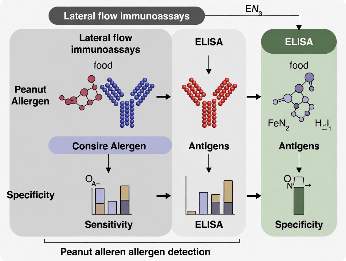
ELISA vs. Lateral Flow: A Critical Sensitivity Comparison for Peanut Allergen Detection
This article provides a comprehensive analysis of the sensitivity and operational characteristics of Enzyme-Linked Immunosorbent Assay (ELISA) and Lateral Flow Immunoassay (LFIA) for detecting major peanut allergens, including Ara h...
ELISA vs. Lateral Flow: A Critical Sensitivity Comparison for Peanut Allergen Detection
Abstract
This article provides a comprehensive analysis of the sensitivity and operational characteristics of Enzyme-Linked Immunosorbent Assay (ELISA) and Lateral Flow Immunoassay (LFIA) for detecting major peanut allergens, including Ara h 1, Ara h 2, and Ara h 3. Tailored for researchers, scientists, and drug development professionals, it explores the foundational principles of these immunoassays, details methodological protocols and real-world applications, discusses troubleshooting and optimization strategies to overcome technical limitations, and presents a rigorous validation and comparative assessment of performance metrics. The synthesis of current research aims to inform method selection for food safety monitoring, clinical diagnostics, and the development of next-generation allergen detection technologies.
Understanding Peanut Allergens and Immunoassay Fundamentals
Peanut allergy is a significant global health concern, potentially triggering severe and life-threatening anaphylactic reactions. The major peanut allergens—Ara h 1, Ara h 2, Ara h 3, and Ara h 6—are seed storage proteins with distinct molecular characteristics and clinical impacts. This review compares the clinical significance of these allergens, focusing on their prevalence, stability, and role in diagnosis. Furthermore, it critically evaluates two primary immunochemical detection methodologies, Enzyme-Linked Immunosorbent Assay (ELISA) and Lateral Flow Immunoassay (LFIA), within the context of peanut allergen research. By synthesizing data on protein stability, IgE reactivity, and assay performance, this guide provides researchers and drug development professionals with a foundation for selecting appropriate analytical tools for allergen detection, risk assessment, and therapeutic development.
Peanut allergy affects an estimated 1-2% of the population in Western nations and typically persists into adulthood, creating a lifelong risk of anaphylaxis for sensitized individuals [1]. The allergic response is an IgE-mediated process where initial exposure to peanut allergens activates T helper 2 (Th2) cells, leading to IgE antibody production. These IgE antibodies bind to Fc epsilon receptors on mast cells and basophils, sensitizing the immune system. Upon re-exposure, the allergen cross-links surface IgE molecules, triggering degranulation and release of inflammatory mediators like histamine and leukotrienes, which cause the clinical symptoms of allergy [2]. The severity of these reactions is influenced by which specific peanut proteins an individual is sensitized to, with certain allergens carrying a higher risk for systemic responses.
Clinical Significance of Major Peanut Allergens
The clinical importance of a peanut allergen is determined by its prevalence of IgE recognition among allergic populations and its inherent biophysical properties, which influence how it interacts with the immune system. The major allergens Ara h 1, Ara h 2, Ara h 3, and Ara h 6 are recognized by the serum IgE of over 50% of peanut-allergic patients and are responsible for the majority of severe reactions [3] [1].
Table 1: Molecular Characteristics and Clinical Prevalence of Major Peanut Allergens
| Allergen | Protein Family | Molecular Function | IgE Recognition Prevalence | Resistance to Digestion | Risk Association |
|---|---|---|---|---|---|
| Ara h 1 | Cupin (7S vicilin) | Seed storage protein, forms trimers and higher oligomers [3] | Major allergen (>50% of patients) [3] | Rapidly hydrolyzed by pepsin [4] | Increased risk of severe symptoms when sensitized [1] |
| Ara h 2 | Prolamin (2S albumin) | Seed storage protein, trypsin inhibitor, 5-helix bundle with 4 disulfide bonds [3] | Major allergen, considered one of the most potent [3] | Highly resistant to pepsin and trypsin [4] | Strongly associated with severe symptoms and anaphylaxis [1] |
| Ara h 3 | Cupin (11S legumin) | Seed storage protein, trypsin inhibitor, forms hexamers [3] | Major allergen (>50% of patients) [3] | Rapidly hydrolyzed by pepsin [4] | Increased risk of severe symptoms when sensitized [1] |
| Ara h 6 | Prolamin (2S albumin) | Seed storage protein, shares 59% sequence identity with Ara h 2 [3] | Major allergen, similar allergenic activity to Ara h 2 [3] | Highly resistant to pepsin [4] | Strongly associated with severe symptoms and anaphylaxis [1] |
Key Differentiators of Clinical Severity
The data in Table 1 reveals critical factors that differentiate the clinical impact of these allergens:
- Protein Stability: Ara h 2 and Ara h 6 demonstrate remarkable stability against digestion due to their dense, disulfide-bonded structures [4] [3]. This resistance allows them to reach the intestinal immune system in a largely intact, immunologically active form, which is a key factor in their potency.
- Abundance vs. Potency: While Ara h 1 and Ara h 3 are abundant storage proteins in peanut, Ara h 2 and Ara h 6 are recognized more frequently and strongly by IgE from peanut-allergic individuals. It is estimated that 97% of peanut allergy patients are sensitized to at least one of Ara h 1, Ara h 2, or Ara h 3 [1]. Despite lower abundance, Ara h 2 and Ara h 6 are considered the most clinically relevant due to their high IgE-binding capacity [5].
- Cross-Reactivity: Ara h 2 and Ara h 6, with their 59% sequence identity and nearly identical 3D structures, show high IgE cross-reactivity. Sensitization to one often implies reactivity to the other, and they are collectively considered the most potent peanut allergens [3].
Detection Methodologies: ELISA vs. Lateral Flow Immunoassays
The accurate detection and quantification of peanut allergens are crucial for food safety, regulatory compliance, and clinical research. ELISA and LFIA are two cornerstone techniques in this field, each with distinct advantages and limitations.
Enzyme-Linked Immunosorbent Assay (ELISA)
ELISA is a widely used laboratory technique known for its high sensitivity, quantitative results, and reliability. It is often considered the reference method for allergen quantification.
- Experimental Protocol for Peanut Allergen Detection: A sensitive and specific competitive ELISA protocol for detecting Ara h 2 has been developed. The method involves coating a microtiter plate with purified Ara h 2. Sample extracts or standards are then mixed with rabbit anti-Ara h 2 polyclonal antibodies and added to the wells. The free antibodies bind to the immobilized Ara h 2. After washing, an enzyme-conjugated secondary antibody (e.g., Goat anti-Rabbit IgG) is added, followed by a colorimetric substrate. The resulting signal is inversely proportional to the allergen concentration in the sample [6] [5].
- Performance Data: The developed competitive Ara h 2 ELISA demonstrates a limit of detection (LOD) of 0.5 ppm (mg/kg) of peanut protein and a quantitative range of 0.8–6.8 ppm. This assay showed superior performance in recovering peanut from processed matrices like fried and high-pressure processed (HPP) pastries compared to some commercial kits [6]. Another ELISA protocol designed for peanut seeds achieved precise quantification of five allergens (Ara h 1, Ara h 2, Ara h 3, Ara h 6, and Ara h 8) across a wide range of concentrations in different peanut genotypes [5].
Lateral Flow Immunoassay (LFIA)
LFIA, or lateral flow test, is a rapid, user-friendly, and portable technology ideal for on-site screening. Its design is typically less complex than ELISA, relying on capillary flow to move the sample across a nitrocellulose membrane containing test and control lines.
- Experimental Protocol: In a typical sandwich format LFIA for allergen detection, the sample is applied to a sample pad. It migrates to a conjugate pad containing colored or fluorescent nanoparticles (e.g., carbon, gold) coated with capture antibodies. If the target allergen is present, it binds to these antibodies. The complex continues to flow along the strip until it reaches the test line, which is immobilized with a second allergen-specific antibody. This results in the capture of the complex and a visible line. A control line confirms the test has functioned correctly [7].
- Performance Data and Innovations: Research into improving LFIAs has yielded very fast assays, with some achieving results in as little as 1 minute, though most are completed within 10 minutes [7]. The limits of detection for multiplexed LFIAs (detecting both hazelnut and peanut) have been reported as low as 0.1 ppm for total hazelnut protein and 0.5 ppm for total peanut protein in buffer, and 0.5-1 ppm for both when spiked into a food matrix [7]. A key innovation is the use of smartphone analysis for result interpretation, which improves objectivity and allows for data transmission [7].
Table 2: Comparative Analysis of ELISA and LFIA for Peanut Allergen Detection
| Feature | ELISA | Lateral Flow Immunoassay (LFIA) |
|---|---|---|
| Format | Microtiter plate, laboratory-based | Compact, single-use strip, portable |
| Assay Time | Several hours | 1 - 20 minutes [7] |
| Data Output | Quantitative | Typically qualitative or semi-quantitative |
| Sensitivity | High (LOD for Ara h 2: 0.5 ppm) [6] | Variable; can be very high (LOD for peanut: 0.5 ppm in matrix) [7] |
| Throughput | High (can run many samples simultaneously) | Low (single sample per strip) |
| Ease of Use | Requires technical expertise and equipment | Simple, minimal training required |
| Multiplexing Capability | Low (typically single analyte per well) | Good, but design is challenging [7] |
| Key Limitation | Time-consuming, not for on-site use | Susceptible to hook effect at high allergen concentrations [7] |
| Ideal Application | Regulatory testing, quantitation, research | Rapid screening, field tests, point-of-care |
IgE-Mediated Allergic Response Pathway
The Scientist's Toolkit: Essential Research Reagents and Materials
The development and execution of reliable immunoassays for peanut allergen detection depend on a suite of critical reagents. The following table details key components used in state-of-the-art protocols.
Table 3: Essential Research Reagents for Peanut Allergen Immunoassays
| Reagent / Material | Function in the Assay | Specific Examples from Literature |
|---|---|---|
| Purified Allergen Standards | Serve as a reference for calibration and quantification; essential for generating standard curves. | Natural Ara h 1, Ara h 2, Ara h 3, Ara h 6, Ara h 8 purified from peanut flour [5]. |
| Monoclonal & Polyclonal Antibodies | Provide the core specificity of the immunoassay; used as both capture and detection antibodies. | Anti-Ara h 2 polyclonal antibodies from rabbits [6]; MAbs 2C12 (Ara h 1), 1E8 (Ara h 3), 3B8 (Ara h 6) [5]. |
| Enzyme Conjugates & Substrates | Enable signal generation in ELISA; an enzyme linked to an antibody catalyzes a colorimetric reaction. | Goat anti-Rabbit IgG conjugated to Horseradish Peroxidase (HRP); ABTS as the chromogenic substrate [5]. |
| Solid-Phase Supports | Provide the surface for immobilizing capture antibodies or antigens. | Microtiter plates for ELISA; Nitrocellulose membranes with specific flow rates (e.g., 140 CN) for LFIA [7] [5]. |
| Labeling Nanoparticles | Act as the signal tag in LFIA, creating a visible line for detection. | Carbon nanoparticles ("Spezial Schwartz 4") used in flow-through and lateral flow assays [7]. |
Lateral Flow Immunoassay Workflow
The major peanut allergens Ara h 1, Ara h 2, Ara h 3, and Ara h 6 are of paramount clinical significance, with Ara h 2 and Ara h 6 standing out due to their exceptional stability and potent IgE reactivity. The choice between ELISA and LFIA for their detection is not a matter of superiority but of application. ELISA remains the gold standard for laboratory-based quantification requiring high sensitivity and precision, as evidenced by assays with LODs down to 0.5 ppm. Conversely, LFIA offers an unparalleled advantage in speed and portability for rapid screening, with modern designs achieving results in minutes and approaching the sensitivity of ELISA. For comprehensive risk assessment and product safety, the synergistic use of both methods—using LFIA for initial screening and ELISA for confirmation and quantification—represents the most robust strategy. Future advancements in multiplexing, smartphone-based quantification, and the development of antibodies with even higher affinity and specificity will further empower researchers and clinicians in the ongoing effort to manage and mitigate peanut allergy.
Enzyme-Linked Immunosorbent Assay (ELISA) is a foundational technique in biomedical and food safety research, renowned for its high sensitivity and quantitative capabilities. In the context of peanut allergen detection, ELISA serves as a benchmark against which rapid, point-of-need tests like lateral flow immunoassays (LFIAs) are measured. This guide objectively compares the performance of these two methodologies, providing researchers with critical data to inform their analytical choices.
Core Principles of the ELISA Technique
The fundamental principle of ELISA is the specific binding between an antigen and an antibody, with the signal generated by an enzyme-linked reporter molecule that catalyzes a colorimetric, fluorescent, or chemiluminescent reaction. This signal, measured with a plate reader, is proportional to the amount of target analyte in the sample, enabling precise quantification.
Two primary formats are widely used for allergen detection:
- Sandwich ELISA: Ideal for detecting complex antigens, this format uses a capture antibody immobilized on a plate and an enzyme-conjugated detection antibody. The analyte is "sandwiched" between them, enhancing specificity. It is typically used for quantification in non-processed or mildly processed foods [8] [9].
- Competitive ELISA: Often employed for detecting small molecules or thermally processed allergens, this format involves sample antigens competing with a labeled antigen for a limited number of antibody binding sites. The signal is inversely proportional to the analyte concentration, making it highly effective for detecting degraded allergens in complex matrices [6].
Performance Comparison: ELISA vs. Lateral Flow Immunoassays
Direct comparisons of ELISA and LFIA reveal a consistent performance trade-off between sensitivity and speed. The following tables summarize experimental data from recent peer-reviewed studies.
Sensitivity and Detection Limits for Peanut Allergens
| Target Allergen | Assay Format | Reported Sensitivity (LOD) | Specificity | Source/Kit (if specified) |
|---|---|---|---|---|
| Ara h 1 | Sandwich ELISA | 10 ng/mL | Highly selective; no cross-reactivity with tree nuts | [9] |
| Ara h 1 | Lateral Flow (LFIA) | 500 ng/mL | Highly selective; no cross-reactivity with tree nuts | [9] |
| Ara h 3 | DAS-ELISA | 39.06 ng/mL | Low cross-reactivity; recovery rates 78.48-90.93% | [8] |
| Ara h 3 | Lateral Flow (LFIA) | 1.6 µg/mL (1600 ng/mL) | No cross-reactivity with other proteins | [8] |
| Peanut (via Ara h 2) | Competitive ELISA | 0.5 ppm (mg/kg) peanut protein | Cross-reactivity only with cloves from 45 ingredients tested | In-house developed assay [6] |
| Almond & Peanut | Lateral Flow Microimmunoassay (LFµIA) | Almond: 185 µg/kg; Peanut: 229 µg/kg | 77% selectivity | Multiplexing microarray format [10] |
Comparative Analysis of Operational Characteristics
| Characteristic | ELISA | Lateral Flow Immunoassay (LFIA) |
|---|---|---|
| Quantitative Capability | Fully quantitative, with standard curves and high precision (CVs < 10%) [8] | Primarily qualitative or semi-quantitative; advanced microarray formats enable improved quantification [10] |
| Throughput | High; can process 96+ samples simultaneously in a plate | Low to medium; single tests or small batches |
| Assay Time | Several hours (2-4 hours including incubation and washing steps) [10] | Minutes (typically 10-20 minutes) [10] [9] |
| Ease of Use | Requires trained personnel, multiple liquid handling steps, and plate washing | Simple; minimal training required, often a single-step procedure |
| Infrastructure Needs | Requires plate washers, readers, and laboratory environment | Portable; can be used at the point-of-need with visual or smartphone readout [10] |
| Multiplexing Potential | Low per plate; typically measures one analyte per well | Moderate; can detect multiple allergens on a single strip with dedicated test lines [10] |
| Cost per Test | Lower reagent cost, but higher overall cost when labor and equipment are factored in | Generally higher per-test cost, but lower operational overhead |
Detailed Experimental Protocols
To illustrate the experimental foundation of the data presented, here are detailed methodologies for key studies comparing both techniques.
This protocol outlines the development of a Double Antibody Sandwich (DAS) ELISA to evaluate the reduction of Ara h 3 antigenicity after heat-moisture treatment.
1. Antibody Production:
- Immunogen: Ara h 3 was purified from peanuts. Rabbits were immunized with the antigen to generate polyclonal antibodies.
- Antibody Purification: Antisera from immunized rabbits were purified using a Protein G column to isolate IgG antibodies.
2. Assay Procedure:
- Coating: Microtiter plates were coated with the purified capture antibody and incubated overnight at 4°C.
- Blocking: Plates were blocked with a protein-based blocking buffer (e.g., 4% skimmed milk) for 2 hours at room temperature to prevent non-specific binding.
- Sample Incubation: Samples and standards (heat-treated and native Ara h 3) were added to the wells and incubated.
- Detection Antibody Incubation: An enzyme-conjugated detection antibody (HRP-labeled) was added to form the "sandwich" complex.
- Washing: Plates were washed multiple times with a PBS-Tween buffer between each step to remove unbound material.
- Signal Development: A substrate solution (e.g., TMB) was added. The enzymatic reaction was stopped after a fixed time with a stop solution (e.g., sulfuric acid).
- Quantification: The absorbance was measured at 450 nm. A standard curve was generated from known concentrations of Ara h 3, and sample concentrations were interpolated from this curve.
3. Validation:
- Sensitivity: The limit of detection (LOD) was calculated to be 39.06 ng/mL.
- Precision: Intra-plate and inter-plate coefficients of variation were below 6.79% and 3.05%, respectively.
- Accuracy: Recovery rates from spiked samples ranged from 78.48% to 90.93%.
This protocol describes the development of a monoclonal antibody-based LFIA for rapid peanut detection.
1. Antibody and Conjugate Preparation:
- Antibody Generation: Monoclonal antibodies (MAbs) were generated against the native Ara h 1 protein.
- Conjugate Pad Preparation: Gold nanoparticles (AuNPs) or other labels were conjugated to the purified anti-Ara h 1 monoclonal antibodies.
2. Strip Assembly and Test Principle:
- Sample Pad: The sample is applied here, and the extracted food matrix migrates via capillary action.
- Conjugate Pad: Contains the dried antibody-conjugated labels. The analyte (Ara h 1) binds to the conjugates upon contact.
- Nitrocellulose Membrane: The core reaction site containing two key lines:
- Test Line: Immobilized with a second anti-Ara h 1 antibody (capture antibody) that binds the analyte-conjugate complex.
- Control Line: Immobilized with an anti-species antibody (e.g., anti-mouse) to capture the remaining conjugated antibodies, validating strip function.
- Absorbent Pad: Acts as a sink to wick the fluid through the strip.
3. Assay Execution and Reading:
- The extracted food sample is applied to the sample pad.
- The result is read visually within 10-20 minutes. The appearance of both control and test lines indicates a positive result. The absence of a test line indicates a negative result. The test is invalid if the control line does not appear.
Visualizing the Workflows
The following diagrams illustrate the fundamental workflow and key difference between the two assay formats.
The Scientist's Toolkit: Key Research Reagent Solutions
Successful implementation of these immunoassays relies on specific, high-quality reagents. The table below details essential components.
| Reagent / Material | Critical Function in Assay | Key Considerations |
|---|---|---|
| Capture & Detection Antibodies | Provide the core specificity for the target allergen (e.g., Ara h 1, Ara h 2, Ara h 3). | Specificity, affinity, and isotype are critical. Monoclonal antibodies offer high consistency; polyclonal can increase signal [9]. |
| Enzyme Conjugates | Enzymes like Horseradish Peroxidase (HRP) are linked to detection antibodies to generate a measurable signal. | Conjugation efficiency and enzyme activity stability directly impact assay sensitivity and background noise. |
| Protein Extraction Buffer | Extracts allergenic proteins from complex food matrices for analysis. | Must efficiently solubilize the target protein without degrading its antigenic epitopes, especially from processed foods [8]. |
| Nitrocellulose Membrane | The porous matrix in LFIA where capture antibodies are immobilized at test and control lines. | pore size affects flow rate and sensitivity. Requires precise dispensing of antibodies [10]. |
| Nanoparticle Reporters | Gold nanoparticles or colored latex beads used as labels in LFIA for visual detection. | Size, shape, and surface chemistry affect conjugation efficiency, stability, and visual intensity [10]. |
| Microtiter Plates | The solid phase for ELISA, typically 96-well plates, where the capture antibody is immobilized. | Binding capacity and well-to-well consistency are vital for reproducible quantitative results. |
| Blocking Buffers | Solutions of irrelevant proteins (e.g., BSA, casein, skim milk) used to cover non-specific binding sites. | Effectiveness determines the signal-to-noise ratio by minimizing background. |
The choice between ELISA and lateral flow immunoassay is not a matter of which is superior, but which is most fit-for-purpose. ELISA remains the undisputed gold standard for laboratory-based quantification, offering unparalleled sensitivity, precision, and robustness for compliance monitoring and in-depth research, particularly when analyzing complex or processed food samples [8] [6]. Lateral flow immunoassays provide an indispensable tool for rapid, on-site screening, enabling decision-making at the processing line or in supply chain audits, albeit with higher detection limits [10] [9].
The ongoing development of epitope-specific antibodies [8] and advanced multiplexing lateral flow platforms [10] continues to narrow the performance gap. However, for the core task of sensitive and quantitative allergen detection in a controlled laboratory setting, ELISA maintains its foundational status.
Lateral Flow Immunoassays (LFIAs) represent a cornerstone technology in point-of-care diagnostics, offering an unparalleled combination of speed, portability, and user-friendliness. These paper-based platforms enable the detection and quantification of analytes in complex mixtures within 5-30 minutes, requiring no external equipment or specialized technical expertise [11]. The fundamental principle behind LFIA is simple: a liquid sample moves via capillary action through various zones of polymeric strips containing molecules that interact with the target analyte [11]. First described in the late 1970s, LFIAs gained widespread recognition through home pregnancy tests and have since expanded into clinical diagnostics, food safety, environmental monitoring, and veterinary medicine [11] [12]. Their low development costs, ease of production, long shelf life, and minimal storage requirements make them particularly valuable for resource-limited settings, remote regions, and battlefield medicine [11].
This guide examines the core principles of LFIA technology, with a specific focus on performance comparisons with Enzyme-Linked Immunosorbent Assays (ELISAs) in the critical context of peanut allergen detection. Peanut allergy represents one of the most severe and persistent food allergies, affecting 1-3% of the population in developed countries, where sub-milligram levels of peanut protein can trigger reactions in sensitive individuals [13]. Accurate detection of peanut allergens like Ara h 1, Ara h 2, and Ara h 6 is therefore essential for food safety and public health. We present experimental data, methodological protocols, and technical specifications to provide researchers and drug development professionals with a comprehensive understanding of how LFIAs compare to traditional ELISA methods in sensitivity, specificity, and practical application.
Core Principles and Components of Lateral Flow Immunoassays
Fundamental Architecture and Mechanism
The operational principle of LFIAs centers on capillary action moving a liquid sample through a series of overlapping membranes mounted on a backing card for stability [11]. A typical LFIA device consists of four essential components: sample pad, conjugate pad, nitrocellulose membrane, and absorbent pad [11] [14]. The sample application pad serves as the entry point, where the liquid sample is applied. This pad is often impregnated with buffer salts, proteins, and surfactants that prepare the sample for optimal interaction with detection components [11]. The treated sample then migrates to the conjugate release pad, which contains detection antibodies specific to the target analyte. These antibodies are conjugated to colored or fluorescent particles, most commonly colloidal gold (40-80 nm) or latex microspheres (100-300 μm) [11] [14].
The sample-conjugate mixture continues flowing onto the nitrocellulose membrane, which contains pre-immobilized capture antibodies in defined test and control lines. As the complex passes through these lines, specific binding events occur, generating visible or detectable signals [11]. The control line validates proper assay function, while the test line indicates analyte detection. Finally, the absorbent pad at the terminal end wicks excess fluid, maintaining consistent flow and preventing backflow [11]. The entire process typically completes within 5-30 minutes, with results interpretable by eye or dedicated readers [11].
Assay Formats and Detection Modalities
LFIAs employ primarily three formats tailored to different analytical targets. Sandwich assays are ideal for larger analytes with multiple antigenic sites, such as proteins or whole pathogens. In this format, the target analyte is immobilized between two complementary antibodies - one conjugated to a detector particle and the other immobilized on the test line [11] [15]. The presence of the analyte generates a visible test line, indicating a positive result [11]. Competitive assays are suited for small molecules with single antigenic determinants that cannot bind two antibodies simultaneously. In this format, the analyte in the sample blocks binding sites on test line antibodies, preventing interaction with the labeled conjugate. Thus, a positive result is indicated by the absence or diminished intensity of the test line [11] [15]. Direct detection assays utilize antigens immobilized on the test line to capture specific antibodies from the sample, with signal generation achieved through labeled anti-species antibodies [14].
Recent advancements have introduced sophisticated detection labels beyond traditional colloidal gold. Fluorescent nanoparticles, quantum dots, paramagnetic particles, and gap-enhanced Raman tags (GERTs) offer enhanced sensitivity and quantification capabilities [14] [16]. GERTs, with Raman reporter molecules embedded in nanogaps between metallic core and shell structures, demonstrate exceptional photostability and signal reproducibility, enabling detection limits as low as 0.65 pg/mL for cardiac troponin I - an improvement of 2-3 orders of magnitude over conventional colloidal gold [16]. These innovations address traditional LFIAs' limitations in sensitivity and quantification while maintaining the platform's fundamental advantages.
Table 1: Key Components of a Lateral Flow Immunoassay Strip
| Component | Material Options | Primary Function | Key Considerations |
|---|---|---|---|
| Sample Pad | Cotton, glass fiber, cellulose | Receives sample; distributes evenly; may filter particulates | Wicking rate, volume capacity, pre-treatment requirements |
| Conjugate Pad | Glass fiber, polyester, sintered polyethylene | Stores and releases labeled bioreagents | Stability during drying, conjugate release efficiency |
| Nitrocellulose Membrane | Nitrocellulose with proprietary surfactants | Matrix for immobilizing capture reagents; site for detection | Capillary flow rate, protein binding capacity, uniformity |
| Absorbent Pad | High-density cellulose | Drives fluid flow; wicks excess reagents | Capacity, wicking rate, compatibility with membrane |
Diagram 1: Lateral Flow Immunoassay Workflow and Components. This diagram illustrates the sequential flow of a liquid sample through the key components of a lateral flow test strip, with each component performing specific functions in the assay process.
Sensitivity Comparison: LFIA vs. ELISA for Peanut Allergen Detection
Quantitative Performance Metrics
The comparative analysis between LFIAs and ELISAs for peanut allergen detection reveals a consistent pattern: while ELISAs generally offer superior sensitivity, modern LFIAs have closed this gap significantly through technological advancements. In direct comparisons for walnut trace detection, ELISA demonstrated a sensitivity of 0.25 µg/g of walnut protein in spiked food matrices, whereas LFIA achieved 0.5 µg/g [17]. Both methods exhibited high specificity, with cross-reactivity observed only with pecan nut among 88 food ingredients tested [17]. For peanut allergen Ara h 1 detection, ELISA reached a remarkable sensitivity of 10 ng/mL, while LFIA achieved 500 ng/mL using the same monoclonal antibodies [9].
Recent innovations have substantially enhanced LFIA sensitivity. Surface-enhanced Raman scattering (SERS)-based LFIAs utilizing gap-enhanced Raman tags (GERTs) have demonstrated detection limits approaching 0.65 pg/mL for cardiac troponin I, surpassing conventional fluorescence-based methods and rivaling ELISA sensitivities [16]. This 2-3 order of magnitude improvement over traditional colloidal gold LFIAs highlights the potential of advanced detection methodologies. Furthermore, a novel IgE epitope-specific antibodies-based sandwich ELISA for Ara h 2 detection achieved a limit of detection of 0.98 ng/mL with high accuracy (mean bias of 0.88%) and precision (relative standard deviation <16.50%) [13], showcasing the continued evolution of both technologies.
Table 2: Sensitivity Comparison of ELISA and LFIA for Allergen Detection
| Allergen Target | Assay Type | Detection Limit | Specificity Notes | Reference |
|---|---|---|---|---|
| Walnut Protein (Jug r 1) | Sandwich ELISA | 0.25 µg/g (food matrix) | Cross-reactivity only with pecan nut (88 ingredients tested) | [17] |
| Walnut Protein (Jug r 1) | Lateral Flow Immunoassay | 0.5 µg/g (food matrix) | Identical specificity profile to ELISA | [17] |
| Peanut Ara h 1 | Sandwich ELISA | 10 ng/mL (peanut meal) | No cross-reactivity with tree nut meals (100-0.4 µg/mL) | [9] |
| Peanut Ara h 1 | Lateral Flow Immunoassay | 500 ng/mL (peanut meal) | Identical specificity profile to ELISA | [9] |
| Peanut Ara h 2 | IgE Epitope-specific ELISA | 0.98 ng/mL | Specific to IgE epitopes of Ara h 2 | [13] |
Experimental Protocols and Methodologies
ELISA Protocol for Peanut Allergen Detection
The development of a sensitive ELISA for peanut allergen detection typically follows a sandwich immunoassay format. For Ara h 1 detection, researchers generated monoclonal antibodies against the target protein through hybridoma technology [9]. The optimized protocol involves: (1) coating microplate wells with capture antibody (1-2 µg/mL in carbonate-bicarbonate buffer, pH 9.6) overnight at 4°C; (2) blocking with protein-based blockers (BSA or casein) for 1-2 hours at room temperature; (3) adding samples and standards diluted in PBS-Tween buffer, incubating 1-2 hours; (4) adding detection antibody conjugated to horseradish peroxidase (HRP) or biotin, incubating 1-2 hours; (5) adding substrate solution (TMB or other chromogenic substrates) and incubating 15-30 minutes; and (6) stopping the reaction with acid and measuring absorbance at 450 nm [9] [13]. The entire process requires 4-6 hours with multiple washing steps between each stage.
For enhanced specificity towards allergenic potential, researchers have developed IgE epitope-specific ELISAs. In the case of Ara h 2 detection, this involved generating a monoclonal antibody against a major IgE epitope of Ara h 2 as the capture antibody, and a polyclonal antibody against twelve IgE epitopes of Ara h 2 as the detection antibody [13]. This approach specifically detects IgE-reactive epitopes rather than merely the protein presence, potentially correlating better with clinical allergenicity.
LFIA Protocol for Peanut Allergen Detection
The development of an LFIA for peanut allergen detection shares similarities with ELISA in antibody selection but differs substantially in execution. For Ara h 1 detection, the same monoclonal antibodies used in ELISA were adapted to the lateral flow format [9]. The standard protocol involves: (1) conjugating detection antibodies to colloidal gold nanoparticles (40 nm) through passive adsorption in low ionic strength buffer, followed by blocking with proteins like BSA or casein; (2) applying the conjugate to a glass fiber pad; (3) striping capture antibodies and control antibodies on nitrocellulose membranes at concentrations of 1-3 µg/cm using precision dispensing systems; (4) assembling the component layers (sample pad, conjugate pad, membrane, absorbent pad) on a backing card with 1-2 mm overlaps; and (5) cutting the assembled card into individual test strips [11] [9]. During operation, the sample is applied to the sample pad, migrates through the conjugate pad to solubilize and bind the detection conjugates, and continues flowing across the membrane where test and control lines capture the complexes. Results are typically available within 5-20 minutes.
Critical to LFIA performance is the meticulous selection of membrane properties, particularly the capillary flow time of nitrocellulose membranes, which balances assay speed and sensitivity [15] [18]. Fast-flowing membranes (e.g., Hi-Flow Plus 65) suit speed-critical tests, while slow-flowing membranes (e.g., Hi-Flow Plus 180) benefit high-sensitivity applications requiring longer interaction times [18].
The Scientist's Toolkit: Essential Research Reagents and Materials
Successful development of either ELISA or LFIA for peanut allergen detection requires careful selection of core reagents and materials. Antibodies stand as the most critical component, with monoclonal antibodies generally preferred for their minimal batch-to-batch variability, especially as capture antibodies [15]. For LFIAs intended for commercial production, the substantial antibody quantities required (approximately 1 gram per million tests) make consistent supply and quality paramount [14].
Table 3: Essential Research Reagents for Allergen Immunoassay Development
| Reagent/Material | Function | Key Selection Criteria | Examples/Alternatives |
|---|---|---|---|
| Monoclonal Antibodies | Primary detection and capture reagents | Affinity (fast kon, slow koff), specificity, lot-to-lot consistency | Anti-Ara h 1 MAbs, Anti-Jug r 1 MAbs |
| Polyclonal Antibodies | Detection or control line reagents | Broad epitope recognition, sensitivity, species specificity | Anti-species IgG, epitope-specific pAbs |
| Colloidal Gold Nanoparticles | Visual detection label in LFIA | Particle size (40-80 nm), stability, conjugation efficiency | 40 nm colloidal gold |
| Latex Microspheres | Visual/fluorescent detection label | Size (100-300 μm), dye incorporation, surface chemistry | Estapor Microspheres |
| Nitrocellulose Membranes | Porous matrix for test/control lines | Capillary flow time, protein binding capacity, uniformity | Hi-Flow Plus Membranes |
| Sample/Conjugate Pads | Sample application and conjugate storage | Material composition, wicking rate, compatibility | SureWick Pad Materials |
| HRP Enzymes & Substrates | Signal generation in ELISA | Catalytic efficiency, substrate sensitivity, background | TMB, HRP-Conjugated Antibodies |
Membranes represent another crucial component, with nitrocellulose remaining the gold standard for its optimal balance of capillary flow and protein binding characteristics [11] [18]. The capillary flow time - the time required for liquid to travel through and completely fill a membrane segment - serves as a more accurate selection parameter than pore size alone [11]. For high-sensitivity applications, slower flow rates (e.g., 180-200 seconds for 4 cm) increase interaction times between analytes and capture antibodies, enhancing detection limits [18].
Detection labels significantly influence sensitivity and readout options. While colloidal gold offers intense color and ease of use, fluorescent labels like Estapor Microspheres enable quantitative measurements and multiplexing [18]. For ultimate sensitivity, emerging technologies like gap-enhanced Raman tags (GERTs) provide photostable, reproducible signals with detection limits in the pg/mL range, though they require specialized readers [16]. The choice between these options depends on the specific application requirements regarding sensitivity, quantification needs, equipment availability, and cost constraints.
Lateral Flow Immunoassays and ELISAs represent complementary technologies in peanut allergen detection, each with distinct advantages suited to different applications. ELISAs maintain superiority in laboratory settings where maximum sensitivity, precision, and throughput are paramount, with detection limits reaching 0.98 ng/mL for Ara h 2 using epitope-specific approaches [13]. LFIAs, while generally less sensitive, offer rapid results (5-30 minutes), simplicity of use, and portability for point-of-need testing, detecting walnut traces at 0.5 µg/g in complex food matrices [17]. The core principles of LFIA - capillary flow through porous membranes, specific antibody-antigen interactions, and visual or instrument-based signal detection - make them indispensable tools for field testing, manufacturing facilities, and clinical point-of-care applications.
Technological advancements continue to narrow the sensitivity gap between these platforms. Innovations in label technology, particularly SERS-based tags like GERTs, have enhanced LFIA sensitivity by 2-3 orders of magnitude, approaching ELISA-level performance while maintaining the rapid, portable format [16]. Similarly, the development of epitope-specific antibodies in ELISA formats enables more biologically relevant detection of allergenic potential rather than mere protein presence [13]. For researchers and drug development professionals, the selection between ELISA and LFIA ultimately depends on the specific application requirements, balancing sensitivity needs with operational constraints. As both technologies continue to evolve, their complementary roles in food safety and clinical diagnostics will expand, offering increasingly sophisticated tools for protecting allergic individuals from accidental exposure to peanut allergens.
The accurate detection of food allergens, such as peanut, is paramount for protecting public health. Immunoassays, primarily Lateral Flow Immunoassays (LFA) and Enzyme-Linked Immunosorbent Assays (ELISA), are cornerstone techniques for this purpose. The reliability of the data generated by these methods hinges on a clear understanding and rigorous application of key performance metrics: the Limit of Detection (LOD), Limit of Quantitation (LOQ), Specificity, and Cross-Reactivity. These parameters form the foundation of any robust analytical method, ensuring that results are not only detectable but also accurate, precise, and specific to the target analyte. Within the context of peanut allergen research, comparing these metrics between the rapid, on-site LFA and the laboratory-based, quantitative ELISA is critical for method selection and data interpretation. This guide provides an objective comparison of these technologies, underpinned by experimental data and detailed protocols.
Defining the Core Analytical Metrics
Limit of Blank (LoB), Limit of Detection (LOD), and Limit of Quantitation (LOQ)
The lowest levels of analyte detection and quantification are formally defined by a hierarchy of metrics. The Limit of Blank (LoB) is the highest apparent analyte concentration expected to be found when replicates of a blank sample (containing no analyte) are tested. It is calculated as LoB = mean_blank + 1.645(SD_blank), identifying the threshold above which a signal is unlikely to be merely background noise [19].
The Limit of Detection (LOD) is the lowest analyte concentration that can be reliably distinguished from the LoB. It is determined using both the LoB and test replicates of a sample containing a low concentration of analyte. The formula LOD = LoB + 1.645(SD_low concentration sample) ensures that 95% of measurements at the LOD will correctly be identified as positive [19]. Simpler approaches, accepted by guidelines like ICH Q2(R1), calculate LOD directly based on the standard deviation of the response and the slope of the calibration curve (LOD = 3.3 * σ / S) or by using a signal-to-noise ratio of 3:1 [20].
The Limit of Quantitation (LOQ) is the lowest concentration at which the analyte can not only be detected but also quantified with acceptable precision and trueness (bias). It represents a higher threshold of confidence than the LOD and is calculated as LOQ = 10 * σ / S or defined by a signal-to-noise ratio of 10:1 [19] [20]. The LOQ may be equivalent to the LOD, but is often at a much higher concentration, and it is at this level that predefined goals for bias and imprecision must be met [19].
Specificity and Cross-Reactivity
Specificity refers to an assay's ability to exclusively measure the intended biomarker or analyte without interference from other substances present in the sample matrix [21]. In immunoassays, this characteristic is primarily determined by the antibodies used. A highly specific assay will generate a true positive signal only for the target analyte, minimizing false positives.
Cross-Reactivity is the practical challenge to specificity. It occurs when assay reagents, particularly antibodies, bind to molecules structurally similar to the intended target, leading to a false positive signal [21]. For example, an assay for walnut might show cross-reactivity with pecan nut due to protein similarities [17]. Cross-reactivity is quantitatively expressed as a percentage, calculated by comparing the assay's response to the interfering substance versus its response to the target analyte [22].
Experimental Comparison: LFA vs. ELISA for Peanut Allergens
The following table summarizes experimental data for peanut allergen detection from recent studies, allowing for a direct comparison of LFA and ELISA performance.
Table 1: Comparative Performance Metrics for Peanut Allergen Detection using LFA and ELISA
| Immunoassay Format | Specific Target | Reported LOD | Reported LOQ | Matrix | Assay Time | Key Cross-Reactivity Findings |
|---|---|---|---|---|---|---|
| Lateral Flow (LFµIA) [10] | Total Peanut Protein | 229 µg/kg | Not Specified | Food | 10 min | High selectivity (77%) reported; specific cross-reactants not listed. |
| Lateral Flow (Optimized) [7] | Total Peanut Protein (TPP) | 0.5 ppm (µg/g) | Not Specified | Buffer & Food Matrix | 10 min | No significant cross-reactivity with hazelnut (co-detected). |
| Sandwich ELISA [22] | Synthetic Cannabinoid (JWH-200) | 0.04 ± 0.02 ng mL⁻¹ | Not Specified | Oral Fluids | > several hours | Excellent specificity; cross-reactivity with AM-2233 (5.6%) and AM-1220 (4.7%). |
| Lateral Flow (This Context) [22] | Synthetic Cannabinoid (JWH-200) | 0.08 ± 0.04 ng mL⁻¹ | Not Specified | Oral Fluids | 15 min | Excellent specificity; cross-reactivity with AM-2233 (5.6%) and AM-1220 (4.7%). |
Table 2: Methods for Determining LOD and LOQ [19] [20]
| Evaluation Method | Description | Typical Application | Calculation (Simplified) |
|---|---|---|---|
| Signal-to-Noise (S/N) | Compares analyte signal against baseline noise. | Instrumental methods (e.g., HPLC). | LOD: S/N = 3; LOQ: S/N = 10 |
| Standard Deviation & Slope | Uses the SD of the response and the slope of the calibration curve. | Quantitative methods (e.g., ELISA, photometry). | LOD = 3.3 * (σ/S); LOQ = 10 * (σ/S) |
| Visual Examination | Determination by analyzing samples with known concentrations. | Non-instrumental methods (e.g., microbial inhibition). | Established by observed minimum level. |
Detailed Experimental Protocols
To ensure reproducibility, the core experimental workflows for developing and validating both LFA and ELISA are outlined below.
Protocol 1: Development of a Lateral Flow Immunoassay for Peanut Allergen Detection [10] [7]
- Reagent Preparation: Polyclonal antibodies against peanut protein extracts are raised in rabbits. Antibodies are purified using affinity columns (e.g., HiTrap Protein G HP). Gold nanoparticles (AuNPs) are synthesized and characterized.
- Conjugate Pad Preparation: The purified anti-peanut antibodies are bioconjugated to the AuNPs. This conjugate is dispensed onto a glass fiber pad and dried.
- Membrane Biofunctionalization: A nitrocellulose membrane is printed in a microarray format using a precision dispenser. The following are immobilized:
- Test Zones: Capture antibodies (anti-peanut) at a defined concentration (e.g., 2.0 mg/mL).
- Control Zone: An anti-species antibody (e.g., goat anti-rabbit IgG) to confirm proper flow.
- Hook Effect Spots: Pure peanut protein (e.g., 2.0 mg/mL) to identify high-dose hook effects.
- Assembly: The biofunctionalized membrane, conjugate pad, sample pad, and absorbent pad are assembled onto a laminated backing card and cut into individual strips.
- Assay Procedure: A sample extract (100 µL) is applied to the sample pad. The result is read after 10 minutes, either visually or quantitatively using a smartphone and image analysis software (e.g., Image J).
- Validation: The LOD is calculated as the mean optical density (OD) of the blank + 3 standard deviations (SD). The dynamic range is determined from the calibration curve.
Protocol 2: Enzyme-Linked Immunosorbent Assay (ELISA) [23] [24]
- Coating: A 96-well polystyrene microtiter plate is coated with a capture antibody specific to the target allergen (e.g., 100 µL in carbonate buffer, pH 9.6). The plate is incubated and then washed to remove unbound antibody.
- Blocking: Remaining binding sites on the plate are blocked with a protein solution (e.g., 1% BSA or casein) to reduce nonspecific binding.
- Sample Incubation: Standards (calibrators) and unknown samples are added to the wells. Allergenic proteins in the sample bind to the immobilized capture antibodies. The plate is incubated and washed.
- Detection Antibody Incubation: A second, enzyme-linked detection antibody (e.g., HRP-conjugated) specific to the allergen is added. This forms an antibody-analyte-antibody "sandwich." The plate is incubated and washed again.
- Signal Generation: A substrate solution is added (e.g., TMB for HRP). The enzyme catalyzes a reaction, producing a color change.
- Signal Measurement & Quantification: The reaction is stopped (e.g., with sulfuric acid), and the absorbance is measured with a spectrophotometer. Color intensity is proportional to the allergen concentration, which is determined by interpolation from the standard curve.
Visualizing Metrics and Workflows
Figure 1: Relationship between LoB, LOD, and LOQ. The LOD is statistically derived from both blank and low-concentration sample data, while the LOQ represents a higher threshold for reliable quantification [19].
Figure 2: The impact of specificity and cross-reactivity on assay results. A highly specific assay generates a signal only from the target, while cross-reactivity with similar molecules leads to false positives [21].
Essential Research Reagent Solutions
The performance of any immunoassay is critically dependent on the quality of its reagents. The table below details key materials required for developing and running LFA and ELISA for allergen detection.
Table 3: Essential Research Reagents for Immunoassay Development [23] [22] [10]
| Reagent Category | Specific Examples | Critical Function in the Assay |
|---|---|---|
| Antibodies | Anti-peanut protein polyclonal or monoclonal antibodies; Purified IgG. | Form the core of assay specificity; used for both capture and detection of the target analyte. |
| Solid Supports | Nitrocellulose membrane (LFA); Polystyrene microtiter plates (ELISA). | Provides the surface for immobilizing capture bioreagents and the platform for the assay workflow. |
| Labels & Conjugates | Gold nanoparticles (AuNPs); Horseradish Peroxidase (HRP); Alkaline Phosphatase (AP). | Generates a detectable signal (colorimetric, chemiluminescent) proportional to the analyte concentration. |
| Blocking Buffers | Bovine Serum Albumin (BSA); Casein; Host serum; Commercial protein-free blockers. | Reduces nonspecific binding by blocking unused sites on the solid support, lowering background noise. |
| Buffer Systems | PBS; TBS; Carbonate-Bicarbonate (Coating); with additives like Tween-20. | Maintains optimal pH and ionic strength for immunoreactions; washing removes unbound material. |
| Signal Generation | TMB (HRP substrate); pNPP (AP substrate); Chemiluminescent substrates. | The enzyme acts on the substrate to produce a measurable signal for detection and quantification. |
The choice between Lateral Flow Immunoassay and ELISA for peanut allergen research is not a matter of one being superior to the other, but rather which is fit-for-purpose. The experimental data confirms that ELISA consistently offers superior sensitivity (lower LOD) and provides robust quantification, making it the gold standard for confirmatory laboratory analysis. In contrast, LFA provides a powerful tool for rapid, on-site screening, with moderately higher but still clinically relevant LODs, delivering results in minutes rather than hours. Both methods can achieve high specificity, which is ultimately governed by the quality and selectivity of the antibody reagents used. Researchers must weigh the need for sensitivity and precise quantification against the requirements for speed, cost, and portability when selecting the appropriate analytical platform for their specific application in safeguarding food safety.
Methodologies, Protocols, and Real-World Deployment
Standardized ELISA Protocols for Precise Quantification of Allergens in Food Matrices
The Enzyme-Linked Immunosorbent Assay (ELISA) is a cornerstone analytical technique for the specific detection and quantification of biomolecules, playing an indispensable role in food safety by reliably identifying allergens and ensuring product integrity and regulatory compliance [25]. For researchers and drug development professionals, the selection of an appropriate, standardized ELISA protocol is critical for generating accurate, reproducible data on allergen concentrations in complex food matrices. This is particularly vital within the broader context of analytical method comparison, especially when evaluating the sensitivity of lateral flow immunoassays (LFIA) against the established benchmark of ELISA for challenging targets like peanut allergens. ELISA's high sensitivity and specificity, which accurately mirror the chemical reaction in the body during an allergic response, make it the gold standard for routine allergen screening [25] [26]. The technique's versatility allows for adaptation into various formats—including indirect, direct, sandwich, and competitive ELISA—each with distinct advantages tailored to different analytical needs, such as detecting whole proteins, small molecules, or achieving ultra-low detection limits [25].
ELISA Formats: Principles and Applications
Understanding the fundamental principles and procedural workflows of different ELISA formats is the first step in selecting the right tool for precise allergen quantification.
Comparative Workflow of Common ELISA Formats
The following diagram illustrates the key steps and components in two common ELISA formats: the Sandwich ELISA (often used for large proteins) and the Competitive ELISA (often used for small molecules).
Table 1: Comparison of Major ELISA Formats for Allergen Analysis.
| Format | Principle | Best Suited For | Key Advantage in Allergen Detection |
|---|---|---|---|
| Sandwich ELISA [25] | Two antibodies bind the antigen at different epitopes, "sandwiching" it. | Detecting large, multivalent antigens like food allergens (peanut, egg, milk). | High specificity and sensitivity; ideal for detecting allergens at low concentrations in complex food matrices [25]. |
| Competitive ELISA [25] | Sample antigen and a labeled antigen compete for binding to a limited amount of antibody. | Detecting small molecules (e.g., mycotoxins, pesticides) or monovalent antigens. | Useful for detecting small contaminants where two antibodies cannot bind simultaneously [25]. |
| Indirect ELISA [25] [27] | A primary antibody binds the antigen, and a labeled secondary antibody binds to the primary. | Detecting contaminants and pathogens; immune response monitoring (antibody quantification). | Increased sensitivity and flexibility; allows for signal amplification [25]. |
| Direct ELISA [25] | An enzyme-linked antibody directly detects the immobilized antigen. | Rapid screening of specific foodborne pathogens or allergens in simple samples. | Quick and straightforward, reducing steps and potential errors [25]. |
Experimental Data: Performance Comparison of Commercial Kits
Theoretical principles must be validated with empirical performance data. Studies comparing commercial ELISA kits reveal critical variations in performance based on the target allergen and food matrix.
Kit Performance for Egg Allergen Detection
A 2023 study directly compared seven commercial egg ELISA kits across nine different food matrices, highlighting the profound impact of the matrix on kit performance [28].
Table 2: Recovery Performance of Seven Commercial Egg ELISA Kits Across Various Food Matrices. [28]
| Food Matrix | Number of Kits Performing Within Set Recovery Criteria | Notes on Matrix Interference |
|---|---|---|
| Cookie, Chocolate, Stock Cube, Wine | 7 out of 7 | All kits performed satisfactorily in these matrices. |
| Pasta, Vegetable Drink & Milk, Ice Cream, Salad Dressing | 4 out of 7 | Recovery was acceptable for a majority, but not all, kits. |
| Meat/Meat Replacers | 1 out of 7 | Showed high matrix effects; performance was challenging for most kits. |
The study concluded that while all seven kits could qualitatively detect egg at the clinically relevant VITAL3 ED01 level of 0.2 mg total egg protein, their quantitative performance varied significantly, underscoring the necessity for kit validation in a specific matrix before use [28].
Broader Comparability of Commercial ELISA Kits
The challenge of variability is not limited to allergen testing. A 2017 study comparing four commercial ELISA kits for quantifying corticosterone in rat serum found statistically significant differences in the absolute values obtained from identical samples [29]. While the kits showed high correlation (allowing for relative comparisons within a study), their precision in determining the true concentration was low [29]. This finding reinforces the critical need for researchers to use the same kit and protocol throughout a study and to be cautious when comparing absolute values from different kits.
Standardized and Optimized ELISA Protocols
To ensure reliability and reproducibility, especially for in-house assay development, rigorous optimization and standardization are mandatory. The process involves systematically refining each step of the ELISA procedure [30].
Key Optimization Steps
- Antigen Coating: The concentration of the capture antibody or antigen immobilized on the plate is foundational. The optimal concentration is one that covers the entire well surface in a monolayer, providing the highest specific signal without wasting reagent [30].
- Blocking: After coating, remaining binding sites on the plate must be blocked to prevent nonspecific binding of detection antibodies. Common blocking agents include Bovine Serum Albumin (BSA), non-fat dried milk, or casein. The optimal blocking buffer must be determined empirically to minimize background noise [30].
- Antibody Concentrations: The concentrations of both the capture and detection antibodies are critical. A checkerboard titration, where these concentrations are varied against each other, is the most effective way to identify the optimal dilution that yields a strong signal with low background [31].
- Sample Diluent and Matrix Effects: The sample diluent should match the sample matrix as closely as possible. Components of the sample matrix can interfere with antigen-antibody binding. Spike-and-recovery experiments are essential to assess this interference and ensure accurate quantification [31] [30].
Example Optimized Protocol: Indirect ELISA
An optimized protocol for an indirect ELISA, designed for quantifying virus-specific antibodies but adaptable to other targets, outlines the following key specifications [27]:
- Coating Buffer: 1x DPBS (pH 7.4).
- Blocking & Sample Diluent: 1x DPBS (pH 7.4) with 1% BSA and 0.05% Tween-20, filtered through a 0.22 µm filter.
- Wash Buffer: 1x DPBS (pH 7.4) with 0.05% Tween-20.
- Critical Reagents: Recombinant target protein, specific primary antibody, and HRP-conjugated secondary antibody.
- Quality Controls: Inclusion of pre-characterized positive and negative control samples is required to establish assay cutoff values, sensitivity, and specificity [27].
The Scientist's Toolkit: Essential Reagents and Materials
Table 3: Key Research Reagent Solutions for ELISA Development and Execution.
| Reagent / Material | Function | Examples & Notes |
|---|---|---|
| Coated Microplate | Solid phase for immobilizing the capture molecule (antigen or antibody). | Medium-binding, flat-well polystyrene plates are standard [27]. |
| Capture Molecule | Binds and immobilizes the target analyte. | Specific antibody (for Sandwich ELISA) or antigen (for indirect ELISA). Affinity-purified antibodies are recommended for optimal signal-to-noise [31]. |
| Blocking Buffer | Saturates unused binding sites to reduce nonspecific background. | BSA (1-5%), non-fat dried milk, or casein. Must be optimized for each assay [30]. |
| Detection Antibodies | Binds to the target analyte and carries a label for signal generation. | Primary or secondary antibodies conjugated to enzymes like HRP or AP. Recommended concentration: 0.5-5 µg/mL for affinity-purified antibodies [31]. |
| Enzyme Substrate | Reacts with the enzyme to produce a measurable signal (color, light). | TMB (colorimetric-HRP), PNPP (colorimetric-AP), or chemiluminescent substrates for higher sensitivity [31]. |
| Buffers | Provide a stable chemical environment for interactions and washing. | Coating: PBS or carbonate buffer. Wash/Sample Diluent: PBS with 0.05% Tween-20 (PBST) [27]. |
The precise quantification of allergens in food matrices via ELISA remains a sophisticated process that balances robust standardized protocols with the need for matrix-specific optimization. The sandwich ELISA format stands out for its high sensitivity and specificity in detecting proteinaceous allergens like those from peanuts, making it a relevant benchmark for comparing emerging technologies such as lateral flow immunoassays. However, as comparative studies show, researchers must be acutely aware of the significant performance variations that can exist between commercial kits and the profound influence of the food matrix on quantitative results. Therefore, a deep understanding of ELISA principles, coupled with rigorous in-house optimization and validation using standardized protocols and controls, is non-negotiable for generating reliable data that can inform drug development, food safety risk assessments, and regulatory compliance.
LFIA Workflows for Rapid Environmental Monitoring and Surface Swab Testing
Lateral Flow Immunoassays (LFIAs) have emerged as indispensable tools for rapid environmental monitoring and surface swab testing in food safety and allergen control programs. These immunochromatographic tests provide a simple, cost-effective, and rapid means to detect specific analytes in complex samples, making them particularly valuable for on-site verification of sanitation protocols and allergen control measures [32]. Within the broader context of analytical sensitivity, LFIAs are often compared to the more traditional Enzyme-Linked Immunosorbent Assay (ELISA), with each method offering distinct advantages tailored to different application requirements. For peanut allergen research specifically, understanding the performance characteristics of both methodologies is crucial for selecting the appropriate detection strategy based on the needed sensitivity, throughput, and operational constraints [8] [33].
The fundamental principle underlying LFIA technology is the capillary action-driven migration of a liquid sample along a series of porous membranes, facilitating specific antigen-antibody interactions that yield a visually detectable signal, typically within 5-15 minutes [34] [35]. This simple operational workflow, combined with no requirement for specialized equipment or extensive technical training, has positioned LFIAs as the premier platform for point-of-care testing and rapid screening in industrial settings [32] [35]. For allergen management, this translates to the ability to perform immediate swab tests on food contact surfaces and processing equipment to verify cleaning efficacy and prevent cross-contamination events.
Comparative Sensitivity: LFIA Versus ELISA for Peanut Allergen Detection
Quantitative Performance Comparison
The analytical sensitivity of LFIAs and ELISA methods varies significantly based on the target allergen, antibody specificity, and sample matrix. For peanut allergen detection, particularly for the major allergen Ara h 3, recent research provides direct comparative data on method performance.
Table 1: Sensitivity Comparison for Peanut Allergen Detection
| Method Format | Target Allergen | Limit of Detection | Assay Time | Key Applications |
|---|---|---|---|---|
| DAS-ELISA [8] | Ara h 3 | 39.06 ng/mL | Several hours (incl. incubation steps) | Laboratory quantification of processed peanut allergens |
| LFIA [8] | Ara h 3 | 1.6 μg/mL | < 15 minutes | Rapid screening of processed peanut products |
| Commercial ELISA Kits [33] | Various peanut allergens (Ara h 1-6) | Variable; highest recovery with Romer and R-Biopharm kits | Several hours | Laboratory quantification of specific peanut proteins |
| Commercial Allergen LFIAs [32] | Peanut and other food allergens | Matrix-dependent; qualitative/semi-quantitative | 5-15 minutes | Surface swab testing, environmental monitoring |
The data reveals a clear sensitivity gap between the two methodologies, with ELISA demonstrating approximately 40-fold greater sensitivity for Ara h 3 detection compared to LFIA [8]. This differential is consistent across multiple studies comparing immunoassay formats, with ELISA typically achieving detection limits in the low nanogram per milliliter range, while LFIAs generally operate in the microgram per milliliter range [8]. This distinction fundamentally dictates their appropriate applications: ELISA remains the gold standard for quantitative analysis requiring high sensitivity, while LFIAs excel in rapid screening scenarios where immediate results are prioritized over ultimate sensitivity.
Implications for Peanut Allergen Research
The sensitivity differential between these methods significantly impacts their application in peanut allergen research. ELISA's superior detection limits make it essential for characterizing low-level contamination and quantifying allergen reduction during processing techniques like heat-moisture treatment [8]. Research demonstrates that heat-moisture treatment at temperatures exceeding 90°C for more than 20 minutes significantly reduces Ara h 3 antigenicity, with the most substantial decline observed following treatment at 90°C for 30 minutes [8]. Such precise quantification of antigenicity changes requires the sensitivity provided by ELISA methodologies.
Conversely, LFIAs offer distinct advantages for monitoring the effectiveness of sanitation protocols and environmental monitoring programs. Their rapid format enables food production facilities to perform immediate verification of cleaning efficacy on food contact surfaces and processing equipment [32]. Multiple commercial LFIA products are specifically validated for surface swab testing, including the RIDAQUICK series and bioavid Lateral Flow tests for peanut and other allergens [32] [36]. This rapid feedback loop allows for immediate corrective actions when allergen contamination is detected, preventing cross-contamination events before production resumes.
LFIA Workflow for Environmental Monitoring and Surface Testing
Fundamental LFIA Components and Mechanism
The lateral flow immunoassay operates on a elegantly simple principle, incorporating several core components mounted on an inert backing material and typically housed in a plastic cassette for handling convenience [34]. The fundamental architecture consists of:
- Sample Pad: An absorbent pad treated with salts and surfactants to optimize sample composition for subsequent reactions [34] [37].
- Conjugate Pad: Contains detection antibodies specific to the target analyte, labeled with visible particles (typically colloidal gold or colored latex beads) [34] [35].
- Nitrocellulose Membrane: The critical reaction matrix where capture antibodies are immobilized in distinct lines (test and control zones) [34] [37].
- Absorbent Pad: Serves as a fluid sink to maintain consistent capillary flow and prevent backflow [34] [35].
The analytical process follows a sequential flow path driven by capillary action. When a liquid sample is applied to the sample pad, it migrates to the conjugate pad, solubilizing the labeled antibodies. If the target analyte is present, it forms complexes with these detection antibodies. The solution continues to migrate along the nitrocellulose membrane until it reaches the test line, where antibody-antigen complexes are captured, generating a visible signal. The fluid continues to the control line, which captures excess labeled antibodies to confirm proper assay function [34] [35]. For peanut allergen detection, this typically generates results within 5-15 minutes, enabling rapid decision-making [32].
Diagram: Lateral Flow Immunoassay Workflow
Surface Sampling and Testing Protocol
The application of LFIA technology to environmental monitoring and surface swab testing follows a standardized protocol designed to ensure representative sampling and accurate results:
Surface Sampling: Using specialized swabs included with commercial test kits, systematically sample a defined surface area (typically 10x10 cm). The swab is moistened with appropriate extraction buffer to enhance allergen recovery from the surface [32].
Sample Extraction: Transfer the swab to a extraction tube containing specified buffer volume. Vigorously mix the swab in the buffer to elute any captured allergens from the swab matrix [32] [36].
Assay Activation: Apply the extracted sample to the sample well of the LFIA device using the provided transfer pipette. The volume is precisely controlled by the device design [32].
Result Interpretation: After the specified development time (typically 5-15 minutes, depending on the specific test), visually inspect the test and control lines. A visible test line alongside the control line indicates a positive result for allergen contamination. The control line must always appear for the test to be considered valid [32] [35].
For quantitative or semi-quantitative analysis, dedicated readers such as the RIDAQUICK SCAN or smartphone-based applications like the RIDASMART APP can be employed to measure test line intensity, providing objective results and documentation capabilities [32] [38].
Experimental Protocols for LFIA Development and Validation
Antibody Production and Characterization
The foundation of any sensitive LFIA is a highly specific antibody pair capable of recognizing distinct epitopes on the target analyte. For peanut allergen detection, particularly for thermally processed products, this requires specialized antibody development:
- Immunogen Preparation: Purified native or recombinant allergen proteins (e.g., Ara h 3) serve as immunogens. For detecting processed allergens, antibodies targeting epitopes disrupted by heat-moisture treatment may be specifically developed [8].
- Animal Immunization: Immunize host animals (typically rabbits or larger species for polyclonal production) following established protocols. For peanut allergen research described in the search results, rabbit experiments were approved by the Animal Experimentation Ethics Committee, with all animals receiving humane care according to Institutional Animal Care and Use Committee guidelines [8].
- Antibody Characterization: Assess antiserum specificity and titer using immunoassays. Select antibodies recognizing different epitopes for the conjugate and capture lines to avoid competition [8] [34].
LFIA Assembly and Optimization
The assembly process requires precise integration of all components with careful attention to reagent concentrations and membrane characteristics:
- Conjugate Pad Preparation: Label specific antibodies with colloidal gold or other detection moieties. Optimize the conjugate application to the pad to ensure consistent release and stability [34] [37].
- Membrane Coating: Immobilize capture antibodies at the test line position and species-specific anti-immunoglobulin antibodies at the control line using precision dispensing systems [34] [35].
- Assembly and Packaging: Mount all components sequentially on backing cards, ensuring slight overlaps to facilitate continuous capillary flow. House assembled strips in plastic cassettes with sample port and result window [34] [37].
Assay Validation Protocols
Comprehensive validation is essential to establish assay performance characteristics:
- Sensitivity and Limit of Detection: Determine the lowest analyte concentration consistently producing a positive signal. For the Ara h 3 LFIA, this was established at 1.6 μg/mL [8].
- Specificity and Cross-Reactivity: Evaluate assay performance against related allergens and common food matrices to ensure minimal cross-reactivity. The Ara h 3 LFIA demonstrated no cross-reactivity with other proteins [8].
- Stability Studies: Assess shelf life under various storage conditions, with quality LFIAs remaining stable for up to 3 years under controlled humidity [34] [37].
The Scientist's Toolkit: Essential Reagents and Materials
Table 2: Key Research Reagent Solutions for Allergen LFIA Development
| Reagent/Material | Function | Application Notes |
|---|---|---|
| Epitope-Specific Antibodies [8] | Recognition of target allergens | Critical for detecting processed allergens; should target epitopes stable after heat treatment |
| Colloidal Gold Nanoparticles [34] [35] | Visual detection label | Most common label; provides strong color with consistent size; ~40nm particles often used |
| Nitrocellulose Membranes [34] [37] | Matrix for capillary flow and antibody immobilization | Pore size affects flow rate and sensitivity; requires specialized dispensing equipment |
| Sample Pads & Conjugate Release Pads [34] [37] | Sample application and conjugate storage | Typically glass fiber; often pre-treated with buffers, surfactants, and stabilizers |
| LFIA Readers [32] [38] | Quantitative result measurement | Include dedicated devices (RIDAQUICK SCAN) or smartphone-based systems (RIDASMART APP) |
| Surface Swab Kits [32] [36] | Environmental sample collection | Specialized swabs with optimized extraction buffers for maximum allergen recovery |
Lateral Flow Immunoassays represent a strategically important technology within the hierarchy of analytical methods for peanut allergen detection and environmental monitoring. While ELISA maintains superiority in sensitivity and quantitative precision, LFIAs offer unparalleled advantages in speed, simplicity, and suitability for on-site testing [8] [33]. The 40-fold sensitivity differential between the methods clearly delineates their respective application spaces: ELISA for precise laboratory quantification of allergen reduction during processing, and LFIA for rapid verification of sanitation efficacy and environmental monitoring [8] [32].
The development of epitope-specific antibodies targeting heat-modified allergens, as demonstrated in recent peanut allergen research, significantly enhances the utility of both methodologies for evaluating processed food products [8]. Furthermore, advancements in reader technologies, particularly smartphone-based quantification systems, are bridging the historical gap between the qualitative nature of LFIAs and the quantitative requirements of comprehensive allergen control programs [32] [38].
For researchers and food safety professionals, the strategic integration of both technologies provides a comprehensive approach to allergen management: LFIA for rapid, high-frequency monitoring of critical control points in processing environments, and ELISA for definitive quantification and method validation. This dual-method approach leverages the respective strengths of each technology to create a robust allergen control system capable of both preventing cross-contamination through immediate feedback and generating definitive data for process validation and regulatory compliance.
Sample preparation is a critical step in food allergen analysis, directly influencing the sensitivity and accuracy of subsequent detection methods like Lateral Flow Immunoassays (LFIAs) and Enzyme-Linked Immunosorbent Assays (ELISAs). The complexity of food matrices, especially following processing, poses significant challenges for efficient allergen extraction. This guide objectively compares key methodologies, focusing on their application in peanut allergen research.
The Impact of Matrix and Processing on Allergen Extraction
The efficiency of allergen extraction is highly dependent on the food matrix and the processing it has undergone. Complex matrices such as chocolate and thermally processed foods (e.g., baked biscuits) are particularly challenging. These matrices can entrap allergenic proteins or introduce interfering compounds that reduce protein solubility and availability for immunoassay detection [39].
For instance, studies optimizing the recovery of 14 specific allergens from incurred food matrices found that chocolate-containing matrices and those subjected to thermal processing consistently yielded lower recovery rates, often below the ideal 50-150% range achieved for less complex foods [39]. Furthermore, thermal processing can induce conformational changes in proteins. For peanut allergen Ara h 3, heat-moisture treatment was shown to significantly reduce its antigenicity by destroying key epitopes, a factor that must be considered when developing detection methods for processed foods [8].
Extraction Buffer Composition and Optimization
The choice of extraction buffer is paramount to disrupt matrix interactions, solubilize allergens, and ensure they are available for immunoassay quantification. Research indicates that a single "universal" extraction buffer is elusive, but optimized formulations can significantly improve recovery across multiple allergens and matrices [39].
The table below summarizes key extraction buffers used for challenging food matrices, as identified in recent research:
Table 1: Composition and Application of Optimized Allergen Extraction Buffers
| Buffer Identifier | Formulation | Key Additives | Reported Performance |
|---|---|---|---|
| Buffer D [39] | 0.05 M sodium carbonate/bicarbonate, pH 9.6 | 10% Fish Gelatine | One of two buffers providing optimized recovery (50-150%) for 14 allergens from complex incurred matrices. |
| Buffer J [39] | PBS, 2% Tween-20, 1 M NaCl, pH 7.4 | 10% Fish Gelatine, 1% PVP (Polyvinylpyrrolidone) | Second optimized buffer for multiplex analysis. Additives combat polyphenol interference in chocolate. |
| Buffer B [39] | PBS, 2% Tween-20, 1 M NaCl, pH 7.4 | 10% Fish Gelatine | Used for improved recovery from challenging matrices. |
| Buffer C [39] | 0.1 M Tris, 1% SDS, 0.1 M sodium sulphite, pH 8.5 | SDS, Sodium Sulphite | A denaturing buffer cited for notable improvements in allergen recovery. |
Additive Functions: Common buffer additives serve specific purposes [39]:
- Detergents (e.g., Tween-20, SDS): Disrupt hydrophobic and ionic matrix interactions to release allergens.
- Blocking Agents (e.g., Fish Gelatine, BSA, NFDM): Prevent surface binding and protein aggregation.
- Polymers (e.g., PVP): Particularly effective for binding and neutralizing polyphenolic compounds in cocoa and other matrices.
- Salts (e.g., 1 M NaCl): Increase ionic strength to solubilize proteins.
Detailed Experimental Protocols
Protocol for Optimized Multiplex Allergen Extraction
This protocol, adapted from research on multiplex allergen detection, is designed for challenging processed matrices [39].
Table 2: Key Research Reagent Solutions
| Reagent/Material | Function in the Protocol |
|---|---|
| Incurred Food Matrix | Sample for analysis; provides real-world context of allergen within a food. |
| Extraction Buffers (e.g., D or J) | To solubilize and release allergenic proteins from the matrix. |
| Orbital Incubator | Provides controlled temperature and agitation for efficient extraction. |
| Refrigerated Centrifuge | Separates clarified extract from insoluble pellet post-extraction. |
Workflow Steps:
- Sample Preparation: Weigh 1 g of homogenized incurred food matrix (e.g., baked biscuit, chocolate dessert) into a suitable extraction tube.
- Buffer Addition: Add a 1:10 ratio of extraction buffer (e.g., 10 mL of Buffer J) to the sample.
- Extraction: Vortex the mixture for 30 seconds to ensure thorough mixing. Incubate for 15 minutes in an orbital incubator set to 60°C and 175 rpm.
- Clarification: Centrifuge at 1250 rcf (relative centrifugal force) for 20 minutes at 4°C.
- Supernatant Collection: Carefully collect the clarified supernatant from the middle layer, avoiding the insoluble pellet and any separated fat.
Protocol for Detecting Processed Peanut Allergen Ara h 3
This protocol specifically addresses the detection of the peanut allergen Ara h 3 after heat-moisture treatment, which alters its antigenicity [8].
Workflow Steps:
- Heat-Moisture Treatment: Subject peanut samples to controlled heat-moisture processing (e.g., >90°C for >20 minutes) to simulate industrial processing.
- Allergen Extraction: Extract proteins from the processed sample using a suitable buffer (e.g., PBS with detergents).
- DAS-ELISA Analysis:
- Coat a microtiter plate with a capture antibody specific for Ara h 3.
- Block the plate to prevent non-specific binding.
- Add the extracted sample and standards. The target allergen binds to the capture antibody.
- Add a detector antibody specific for a different epitope on Ara h 3, conjugated to Horseradish Peroxidase (HRP).
- Add enzyme substrate (e.g., TMB) and measure the colorimetric change.
- LFIA Analysis:
- Apply the extracted sample to the sample pad of the LFIA strip.
- The allergen binds to labeled detector antibodies, and the complex is captured at the test line by immobilized antibodies.
- A visual or smartphone-read signal indicates presence and quantity.
Sensitivity Comparison: LFIA vs. ELISA
The following table compares the performance of modern LFIA and ELISA methods for peanut allergen detection, based on recent experimental data.
Table 3: Sensitivity Comparison of LFIA and ELISA for Peanut Allergen Detection
| Method | Target Allergen | Limit of Detection (LOD) | Recovery Rate | Key Context |
|---|---|---|---|---|
| DAS-ELISA [8] | Ara h 3 | 39.06 ng/mL | 78.48% - 90.93% | Specific for heat-moisture treated Ara h 3 |
| LFIA (Qualitative) [8] | Ara h 3 | 1.6 μg/mL | Not Specified | First LFIA reported for detecting processed Ara h 3 |
| LFµIA (Microarray) [40] | Peanut Proteins | 229 μg/kg | RSD 5-25% (Precision) | Multiplex format for almond and peanut |
| Sandwich LFIA [41] | Total Peanut Protein | 0.5 ppm (in buffer) | Validated in 20 matrices | Used with smartphone readout |
While ELISA traditionally offers lower detection limits, advancements in LFIA technology are closing this gap. The development of allergen-specific immunoassays that utilize purified component allergens for standards and antibodies has improved the relevance, specificity, and standardization of measurements for both techniques [39]. Furthermore, the integration of smartphone-based readout systems for LFIA introduces a quantitative, user-friendly interface capable of low-ppm sensitivity, enhancing its utility for point-of-need testing [41].
In food manufacturing, preventing allergen cross-contact is a paramount safety requirement. Cross-contact occurs when an allergenic food is inadvertently transferred to a non-allergenic food, potentially leading to life-threatening allergic reactions in consumers [42]. Unlike microbial pathogens, allergens are chemical hazards that are not eliminated by heat or sanitation alone, making their physical management through processes like Cleaning-in-Place (CIP) systems essential [43]. Effective validation of cleaning efficacy is therefore critical, requiring highly sensitive detection methods to verify that allergenic residues have been reduced to safe levels.
Among food allergens, peanut is particularly significant due to the prevalence and severity of peanut allergies [7]. This creates an urgent need for reliable, sensitive, and rapid detection methods to validate cleaning effectiveness in CIP systems. Lateral Flow Immunoassays (LFIAs) and Enzyme-Linked Immunosorbent Assays (ELISAs) represent two established immunological approaches for allergen detection, each with distinct performance characteristics, advantages, and limitations. This guide provides an objective comparison of these technologies within the context of peanut allergen detection for CIP validation, presenting experimental data to inform researchers, scientists, and drug development professionals.
Lateral Flow Immunoassays (LFIAs)
LFIAs are paper-based biosensors that operate via capillary action to move samples across a nitrocellulose membrane. In a typical sandwich format assay for protein detection, the sample containing the allergen is applied to a sample pad. As it migrates, it rehydrates conjugated pads containing labeled antibodies (often gold nanoparticles or carbon nanoparticles). If the target allergen is present, it forms a complex with these labeled antibodies. This complex continues to flow until it reaches the test line, where immobilized capture antibodies bind the complex, generating a visible signal. A control line confirms proper assay function [7]. LFIAs are celebrated for their speed, simplicity, and portability, providing a binary (yes/no) or semi-quantitative result, typically within 1-20 minutes [7] [44].
Enzyme-Linked Immunosorbent Assays (ELISAs)
ELISAs are plate-based quantitative assays performed in a laboratory setting. The competitive ELISA format, often used for small molecules or complex matrices, involves the target allergen in a sample competing with a labeled allergen (enzyme-conjugated) for a limited number of antibody binding sites on a plate. In the context of peanut detection, a recently developed competitive ELISA for Ara h 2—a major peanut allergen—demonstrates this principle [6]. After the competitive binding step, a substrate is added, which the enzyme converts into a colored product. The intensity of this color, measured spectrophotometrically, is inversely proportional to the amount of allergen in the sample, allowing for precise quantification against a standard curve [6] [44].
Experimental Comparison: LFIA vs. ELISA for Peanut Allergen Detection
Key Performance Metrics
Direct comparison of peer-reviewed studies reveals significant differences in the performance of optimized LFIAs and ELISAs for peanut allergen detection. The quantitative data below summarizes the capabilities of each method for detecting total peanut protein (TPP).
Table 1: Performance Comparison of Peanut Allergen Detection Methods
| Performance Metric | Lateral Flow Immunoassay (LFIA) | Competitive ELISA (Ara h 2 specific) |
|---|---|---|
| Limit of Detection (LOD) | 0.5 ppm (TPP in matrix) [7] | 0.5 ppm (peanut protein) [6] |
| Quantification Range | Semi-quantitative | 0.8 - 6.8 ppm (peanut protein) [6] |
| Assay Time | 1 - 10 minutes [7] | Several hours (typical for multi-step ELISA) |
| Throughput | Low (single samples) | High (96-well plate format) |
| Result Interpretation | Visual or smartphone readout | Instrument-based (spectrophotometer) |
| Multiplexing Capability | Possible with careful design [7] | Typically single-analyte |
Detailed Experimental Protocols
To ensure reproducibility and provide a clear basis for comparison, the following subsections detail the specific experimental protocols from the cited research.
Protocol for Optimized LFIA
The following methodology was used to develop a high-performance, multiplex LFIA for hazelnut and peanut allergens [7]:
- Antibody Labeling: Carbon nanoparticles ("Spezial Schwartz 4") were used as the label. Antibodies (50-6B12 for hazelnut and 51-2A12/51-12D2 for peanut) were buffer-exchanged into 5 mM borate buffer (pH 8.8) before conjugation.
- Membrane and Assembly: Assays were developed on 140 CN nitrocellulose membranes (Unisart, Sartorius) secured on a plastic backing. Goat anti-mouse IgG was used for the control line.
- Test Procedure: The sample was applied to the sample pad. The assay relied on passive, capillary flow across the membrane. The total assay time was optimized to be within 10 minutes.
- Data Acquisition: Results were analyzed using smartphone imaging in a device-independent color space to minimize inter-device variability, allowing for more objective interpretation of the test line signal.
Protocol for Competitive Ara h 2 ELISA
The developed competitive ELISA for peanut quantification followed this procedure [6]:
- Antibody Production: Ara h 2 was purified from light roast peanut flour to raise polyclonal IgG antibodies in rabbits. High-titer sera were then purified to obtain the specific IgG used in the assay.
- Plate Coating and Assay: The specific protocol for plate coating was not detailed in the abstract. However, as a competitive ELISA, it involved incubating the sample (containing the target peanut allergen) with a limited amount of the anti-Ara h 2 antibody and a defined amount of enzyme-labeled Ara h 2. These components competed for binding sites during the incubation step.
- Detection and Quantification: After a washing step to remove unbound material, a substrate was added. The enzymatic reaction produced a color change measured spectrophotometrically. The absorbance was used to calculate the peanut protein concentration based on a standard curve generated with known standards.
Analysis of Strengths and Limitations in CIP Validation Context
Operational Considerations
The choice between LFIA and ELISA for validating CIP efficacy often depends on the operational needs of the manufacturing environment.
- Speed and On-Site Utility: LFIAs provide results in minutes, enabling rapid decision-making on the production floor. This allows for almost immediate verification of cleaning efficacy before a new production run begins, significantly reducing downtime [7] [44]. Their simplicity allows them to be used by production staff with minimal training.
- Quantitative Precision and Data Robustness: ELISAs provide precise, numerical data essential for establishing baseline cleanliness, tracking trends over time, and meeting rigorous regulatory documentation standards. The high throughput of the 96-well plate format makes ELISA ideal for testing multiple samples simultaneously, such as various sampling points within a single CIP circuit or comparing different cleaning protocols [6] [44].
Technical Challenges and Mitigations
- Hook Effect: A phenomenon known as the "hook effect" can occur in one-step, sandwich format LFIAs when extremely high allergen concentrations saturate both the labeled and capture antibodies, leading to a false negative or a falsely weak signal [7]. This risk can be mitigated by determining the correct working range of the assay and, for some products, performing a sample dilution if high contamination is suspected.
- Matrix Interference: Complex food matrices can interfere with both assay types. The competitive Ara h 2 ELISA demonstrated minimal cross-reactivity, with interference observed only with cloves from a test of 45 food ingredients [6]. For both methods, appropriate sample extraction and dilution are critical to minimize matrix effects and ensure accurate results.
The Scientist's Toolkit: Essential Research Reagents and Materials
Successful development and implementation of allergen detection assays require specific, high-quality reagents and materials.
Table 2: Key Research Reagent Solutions for Allergen Immunoassays
| Reagent/Material | Function in Assay | Specific Examples & Notes |
|---|---|---|
| Capture & Detection Antibodies | Specific binding to the target allergen protein; determine assay specificity and sensitivity. | Monoclonal (e.g., 51-2A12 for peanut) or polyclonal (e.g., rabbit anti-Ara h 2 IgG). Antibody kinetics screening via SPR is recommended for LFIA [7] [6]. |
| Labeling System | Provides a detectable signal for the antigen-antibody complex. | Carbon nanoparticles (LFIA) [7]; Enzyme conjugates like Horseradish Peroxidase (ELISA) [6]. |
| Solid Support | Matrix for immobilizing capture antibodies and facilitating fluidics. | Nitrocellulose membranes, characterized by capillary flow rate (e.g., 120-150 s/4 cm for LFIA); 96-well polystyrene plates (ELISA) [7]. |
| Sample Buffer | Medium for sample preparation and dilution; helps maintain pH and protein stability while reducing non-specific binding. | Borate Buffer (e.g., 5 mM, pH 8.8) [7]; PBS with additives like BSA and Tween-20 [7]. |
| Reference Standards | Calibrate the assay and enable quantification. | Purified allergen proteins (e.g., Ara h 2 from peanut flour); defined total protein extracts (e.g., Total Peanut Protein - TPP) [7] [6]. |
Both LFIAs and ELISAs are powerful tools for the sensitive detection of peanut allergens, playing a critical role in validating the efficacy of CIP processes to prevent life-threatening cross-contact. The choice between them is not a matter of which is superior, but which is most appropriate for a specific application. LFIAs excel as rapid, on-site tools for routine monitoring and immediate line-clearance decisions after cleaning. In contrast, ELISAs serve as the gold standard for quantitative, high-precision analysis in a central laboratory, ideal for validation studies, troubleshooting, and comprehensive regulatory documentation. For a robust food safety program, many facilities find that the strategies are complementary, leveraging the strengths of both to ensure that their cleaning validation is both efficient and scientifically defensible.
This guide provides an objective comparison of the performance characteristics between traditional Enzyme-Linked Immunosorbent Assays (ELISAs), multiplexed arrays, and flow-through immunoassays, with a specific focus on applications in peanut allergen detection. The data presented below, synthesized from recent scientific literature and commercial technical resources, reveals a nuanced landscape where each platform offers distinct advantages depending on the specific research requirements, sample availability, and desired throughput.
Table 1: Core Characteristics of Immunoassay Platforms
| Feature | Traditional ELISA | Multiplex Bead Arrays | Flow-Through Immunoassays |
|---|---|---|---|
| Detection Capacity | Single analyte per assay [45] [46] | Simultaneous detection of multiple analytes (up to 80+ proteins) [45] [47] | Can be configured for single or multiple allergens [7] |
| Sample Volume | Larger volume required for multiple analytes [46] | Significantly lower (25-50 µL for multiple analytes) [47] | Variable; can be very low [7] |
| Sensitivity | High (e.g., 0.5 ppm for peanut Ara h 2) [6] | High to ultra-high (fg/mL to pg/mL range) [45] [48] | Moderate to High (e.g., 0.05-0.5 ppm for hazelnut/peanut) [7] |
| Assay Time | ~4 hours [47] | ~1.5 - 3.5 hours [47] [48] | Very fast (1 - 10 minutes) [7] |
| Throughput | Moderate (96-well format) | High (96-well or 384-well format) [47] | Rapid, single or few samples |
| Cost Profile | Lower cost for single-analyte tests [46] | Higher initial cost, cost-effective for multi-analyte studies [46] [47] | Presumed low cost, minimal equipment |
Detailed Performance Data in Allergen Detection
Sensitivity and Specificity in Peanut Allergen Detection
Table 2: Experimental Performance in Detecting Food Allergens
| Assay Format | Target Allergen | Limit of Detection (LOD) | Key Performance Findings | Source |
|---|---|---|---|---|
| Competitive ELISA | Peanut (Ara h 2) | 0.5 ppm (mg/kg) peanut protein | Quantification range of 0.8–6.8 ppm; outperformed some commercial kits in recovery from processed matrices [6]. | [6] |
| Sandwich ELISA | Walnut (Jug r 1) | 0.25 µg/g (ppm) walnut protein | High specificity with cross-reactivity only to Pecan nut; high recovery (71–97%) in incurred foods [17]. | [17] |
| Lateral Flow (LFIA) | Walnut (Jug r 1) | 0.5 µg/g (ppm) in food; 0.1 µg on surfaces | Reliable detection in complex foods and on environmental surfaces; results within minutes [17]. | [17] |
| Active Flow-Through | Peanut & Hazelnut | 0.05 ppm (buffer); 0.5-1 ppm (matrix) | Achieved lower LOD than passive flow-through and LFIA formats in a controlled comparison [7]. | [7] |
| Multiplex Array (Simoa) | Cytokines (General) | fg/mL range | Demonstrated 1-2 orders of magnitude higher sensitivity than some other multiplex platforms [45]. | [45] |
Platform-Specific Advantages and Limitations
ELISA: Remains the "gold standard" for single-analyte quantification due to its high sensitivity, specificity, and well-established, reproducible protocols [46]. Its simplicity, cost-effectiveness for focused studies, and extensive validation history make it ideal for labs requiring precise measurement of a known allergen like Ara h 2 [6]. Its main limitation is low throughput for multi-analyte profiles.
Multiplex Bead Arrays (e.g., Luminex): The primary advantage is high-content data from minimal sample volume, creating a comprehensive biomarker profile from a single run [49] [47]. This is invaluable for studying complex biological networks or when sample volume is scarce (e.g., pediatric studies). Challenges include potentially higher initial equipment costs, more complex data analysis, and the need for rigorous optimization to minimize cross-reactivity between antibodies [50] [46].
Flow-Through & Lateral Flow Immunoassays: The standout feature is speed and portability, providing results in minutes suitable for point-of-care or rapid screening in industrial settings [7] [17]. Newer flow-through designs can overcome mass transport limitations of lateral flow, improving sensitivity and speed, and can be adapted for smartphone readout [7]. They may, however, have a more limited dynamic range and higher detection limits compared to plate-based assays.
Experimental Protocols for Key Platforms
Protocol: Competitive ELISA for Peanut Ara h 2
This protocol is adapted from the development of a sensitive Ara h 2-specific assay [6].
- 1. Antibody Preparation: Purify polyclonal IgG antibodies from rabbit sera immunized with Ara h 2 purified from light roast peanut flour.
- 2. Plate Coating: Coat microtiter plate wells with the purified anti-Ara h 2 antibodies and block remaining sites to prevent non-specific binding.
- 3. Competitive Incubation: Simultaneously incubate the food sample extract (containing the target peanut proteins) and a defined amount of enzyme-labeled Ara h 2 antigen in the coated wells. The native and labeled antigens compete for binding to the limited number of capture sites on the immobilized antibody.
- 4. Wash and Develop: Wash the plate to remove unbound material. Add an enzyme substrate to produce a colorimetric signal.
- 5. Quantification: Measure the signal intensity. The signal is inversely proportional to the concentration of peanut allergen in the sample, with higher allergen levels resulting in a weaker signal. Quantify against a standard curve of known Ara h 2 concentrations.
Protocol: Bead-Based Multiplex Immunoassay
This generalized protocol is based on Luminex xMAP technology [47].
- 1. Bead Incubation: Mix color-coded magnetic beads, each set coated with a unique capture antibody for a specific allergen or biomarker, with the sample in a well of a microtiter plate.
- 2. Sandwich Formation: After a wash step, add a mixture of biotinylated detection antibodies, which bind to the captured analytes, forming a sandwich complex.
- 3. Signal Development: Introduce streptavidin-conjugated R-phycoerythrin (S-RPE), which binds to the biotin on the detection antibodies.
- 4. Flow Cytometric Reading: The bead suspension is hydrodynamically focused and passed through a dual-laser system in a multiplex reader. One laser identifies the bead color (and thus the specific analyte), while the second laser quantifies the fluorescent signal from the R-PE, which is proportional to the amount of captured analyte.
Technology Workflow Diagrams
Diagram 1: Core Experimental Workflows
This diagram visualizes the fundamental procedural differences between the three immunoassay formats, highlighting the sequential steps in ELISA versus the mixed-bead approach of multiplex arrays and the rapid flow mechanism of LFIAs.
The Scientist's Toolkit: Essential Research Reagents
Table 3: Key Reagents for Immunoassay Development
| Reagent / Material | Function in the Assay | Example from Literature |
|---|---|---|
| Capture Antibody | Immobilized on plate/bead/membrane to specifically bind the target analyte. | Anti-Ara h 2 IgG for peanut ELISA [6]; Anti-Jug r 1 for walnut LFIA [17]. |
| Detection Antibody | Binds to a different epitope on the captured analyte; often conjugated for signal generation. | Biotin-conjugated antibody in multiplex assays [47]; Carbon nanoparticle-labeled antibody in LFIA [7]. |
| Signal Reporter | Generates a measurable signal (e.g., color, fluorescence, luminescence). | Streptavidin-R-Phycoerythrin (S-RPE) [47]; Enzyme-Horseradish Peroxidase (HRP) with substrate [6]. |
| Solid Phase | The substrate to which capture antibodies are immobilized. | 96-well microtiter plates [6]; Color-coded magnetic beads [47]; Nitrocellulose membrane [7]. |
| Blocking Buffer | Prevents non-specific binding of proteins to the solid phase, reducing background noise. | Bovine Serum Albumin (BSA) in borate buffer [7]. |
The choice between ELISA, multiplex arrays, and flow-through immunoassays is not a matter of identifying a superior technology, but of selecting the right tool for the specific research question. ELISA remains the benchmark for highly accurate and sensitive quantification of individual allergens like peanut Ara h 2. Multiplex arrays are unparalleled for comprehensive biomarker profiling when sample volume is limited, offering high-throughput capabilities. Flow-through and lateral flow assays provide rapid, on-site results crucial for quick screening and control checks.
Emerging technologies like the "Lab-in-a-Tip" (LIT) system promise to further reshape this landscape by integrating the advantages of different platforms—offering high sensitivity, extreme speed, and minimal sample use simultaneously [48]. For the foreseeable future, a multi-modal approach, leveraging the strengths of each format, will be the most effective strategy for advancing research and ensuring safety in peanut allergen detection and beyond.
Overcoming Technical Challenges and Enhancing Assay Performance
The lateral flow immunoassay (LFIA) is a mainstay in point-of-care diagnostics, prized for its rapid results, cost-effectiveness, and ease of use [51] [52]. However, its quantitative accuracy, especially for high-abundance analytes, is critically limited by the "hook effect," a phenomenon leading to false-negative results or significant underestimation of analyte concentration [53]. This effect is exclusively associated with sandwich-type immunoassays, where the signal intensity increases with analyte concentration until a threshold is crossed, after which the signal paradoxically decreases [53] [54]. For researchers and scientists, particularly in fields like food allergen detection where target concentrations can vary widely, understanding and mitigating the hook effect is paramount for developing reliable assays.
Within the specific context of peanut allergen research, the hook effect presents a substantial risk. Assays designed to detect major allergens like Ara h 3 could fail to accurately quantify heavily contaminated samples, leading to a dangerous underestimation of allergenicity [8]. This review systematically compares the performance of LFIA with the more traditional enzyme-linked immunosorbent assay (ELISA), explores the mechanistic basis of the hook effect, and summarizes advanced design solutions to overcome this limitation, thereby enhancing the reliability of allergen detection platforms.
Performance Comparison: LFIA vs. ELISA in Peanut Allergen Research
While both LFIA and ELISA are immunological methods based on specific antigen-antibody binding, their operational formats, ease of use, and susceptibility to interference differ significantly. The table below provides a direct comparison of these two techniques, with a focus on applications in food allergen research.
Table 1: Performance Comparison between ELISA and LFIA for Allergen Detection
| Feature | ELISA | LFIA |
|---|---|---|
| Format & Throughput | Microtiter plate-based; suitable for medium-to-high throughput [8] | Single-use test strip; designed for single tests or low throughput [8] [52] |
| Assay Time | Several hours, including incubation and washing steps [8] | Typically several minutes, with no washing steps required [8] [52] |
| Data Output | Quantitative, via plate reader [8] | Qualitative or semi-quantitative, via visual assessment or reader systems [52] |
| Ease of Use | Requires trained personnel and laboratory infrastructure [8] | User-friendly, suitable for point-of-care or field use [52] [55] |
| Hook Effect | Susceptible, though sample dilution is more readily incorporated [53] | Highly susceptible in sandwich formats; a major cause of false negatives [53] [54] |
Quantitative data from peanut allergen research underscores this comparison. A study developing methods to detect the peanut allergen Ara h 3 after heat treatment established a DAS-ELISA with a detection limit of 39.06 ng/mL and recovery rates of 78.48% to 90.93% [8]. The corresponding LFIA developed in the same study showed a lower sensitivity, with a detection limit of 1.6 μg/mL for Ara h 3 [8]. This difference in sensitivity highlights a common trade-off, though both methods were effective for detecting the allergen in processed peanut products [8].
Experimental Insights: Protocols for Evaluating Allergen Detection
To ensure the reliability of allergen detection methods, rigorous experimental protocols are employed. The following methodologies are critical for evaluating assay performance, particularly in the context of processed foods where allergen structure may be altered.
Protocol for Assessing Heat-Treatment Impact on Allergenicity
- Objective: To evaluate how thermal processing reduces the antigenicity of an allergen, such as Ara h 3.
- Procedure:
- Heat-Moisture Treatment: Subject purified allergen or peanut material to controlled temperatures (e.g., 90°C) and durations (e.g., 20-30 minutes) [8].
- Indirect ELISA:
- Coat microtiter plates with untreated (native) allergen.
- Add heat-treated allergen samples to the wells.
- Use allergen-specific antibodies (e.g., from immunized rabbits) to probe the remaining antigenic sites.
- Quantify the binding using an enzyme-conjugated secondary antibody and a colorimetric substrate.
- Data Analysis: A significant reduction in signal compared to untreated controls indicates a loss of antigenicity due to epitope destruction [8].
Protocol for Establishing a Sandwich DAS-ELISA
- Objective: To develop a quantitative assay for a specific allergen.
- Procedure:
- Antibody Preparation: Generate and purify epitope-specific antibodies. For processed allergens, this may involve developing antibodies specific to epitopes destroyed by heat treatment [8].
- Plate Coating: Immobilize the capture antibody on a microtiter plate.
- Sample Incubation: Add the allergen standard or sample to the wells for binding.
- Detection: Add an enzyme-conjugated detection antibody to form a "sandwich" complex.
- Signal Development: Introduce an enzyme substrate and measure the color intensity. The limit of detection (LOD), inter-/intra-assay precision, and recovery rates should be calculated for validation [8].
Design Solutions to Overcome the Hook Effect in LFIA
The hook effect arises in sandwich LFIAs because an excessively high concentration of analyte saturates both the capture antibodies on the test line and the detection antibodies on the reporter particles. This prevents the formation of the critical "sandwich" complex at the test line, as each antibody tends to bind a separate analyte molecule, causing the signal to drop [53]. The following design strategies have been developed to mitigate this issue.
Assay Format Selection: Competitive LFAs
For small molecules or targets where high concentrations are expected, a competitive format is inherently robust against the hook effect [54].
- Mechanism: The signal generation is inverse. A high analyte concentration prevents the binding of labeled reporters to the test line, resulting in a weak or absent signal. There is no signal to "hook" and decrease [54].
- Application: While less intuitive for users, this format is widely used for small molecules like toxins, hormones, and drugs [54].
(Diagram 1: Competitive LFIA Signal Logic)
Kinetic Analysis via Real-Time Imaging
A novel method to overcome the hook effect in sandwich LFIAs involves using real-time imaging of signal development kinetics rather than relying on an end-point measurement [53].
- Mechanism: A smartphone app records time-lapse images of the test (T) and control (C) lines as the strip develops. The ratio of their intensities (T/C) over time is tracked.
- Data Modeling: The kinetic profile of the T/C ratio is fitted to empirical equations. The fitting parameters correlate with the analyte concentration over a much wider range, effectively seeing "past" the hook effect [53].
- Performance: This approach has been shown to expand the dynamic range of a commercial pregnancy test from 2 to 3 orders of magnitude without any physical modification to the strip [53].
(Diagram 2: Kinetic Workflow for Hook Effect Mitigation)
Computational Modeling for Design Optimization
Computational models are powerful tools for predicting and mitigating the hook effect during the LFIA design phase, reducing extensive experimental trial-and-error.
- Model Basis: Finite element simulations in software like COMSOL can model the complete 3D domain of an LFIA strip, incorporating capillary fluid flow, analyte transport, and reaction kinetics at the test and control lines [51].
- Design Insights: These models allow developers to simulate how parameters like reporter particle concentration, capture antibody density on the test line, and reaction rate constants affect the onset and severity of the hook effect. For instance, optimizing these parameters can effectively enhance detection sensitivity and broaden the working range in sandwich LFIA [51].
The Scientist's Toolkit: Key Reagents and Materials
The development of robust LFIAs, particularly those resistant to the hook effect, relies on a specific set of reagents and materials. The table below details essential components for research in this field.
Table 2: Key Research Reagent Solutions for LFIA Development
| Item | Function in LFIA | Research Context |
|---|---|---|
| Epitope-Specific Antibodies | Serve as the primary capture and detection bioreceptors; specificity is critical for accurate detection. | Antibodies targeting epitopes destroyed by processing (e.g., heat) can monitor effective desensitization in food production [8]. |
| Reporter Particles | Generate the detectable signal (e.g., color, fluorescence). | Composite nanoparticles (e.g., Fe3O4@Au, SiO2@QDs) can enhance sensitivity and provide diverse readout options, potentially mitigating matrix effects [52]. |
| Nitrocellulose Membrane | The porous matrix on which capture antibodies are striped to form the test and control lines. | The membrane's pore size, flow rate, and protein-binding capacity are key optimization parameters that influence assay sensitivity and hook effect threshold [51]. |
| Recombinant Allergens | Used as standardized antigens for assay development, calibration, and antibody production. | Pure recombinant allergens (e.g., Ara h 3) are essential for coating plates in ELISA development and for controlled spiking experiments [8]. |
| Computational Modeling Software | Simulates fluid dynamics and reaction kinetics within the LFIA strip. | Used to predict the impact of design parameters on performance, including the onset of the hook effect, before physical prototyping [53] [51]. |
Impact of Food Processing and Heat-Moisture Treatment on Allergen Detection
Food allergies represent a significant global public health concern, with accurate detection of allergenic residues in processed foods being a critical challenge for the food industry, regulatory bodies, and allergic consumers [56]. Food processing techniques, particularly thermal treatments, induce structural and conformational changes in allergenic proteins that directly impact their immunoreactivity and detectability by analytical methods [57] [58] [59]. This creates a complex analytical landscape where the effectiveness of detection methods varies considerably based on the specific processing parameters applied and the intrinsic properties of each allergen.
The fundamental challenge lies in the protein modifications induced by processing. Heat treatment can lead to protein unfolding, aggregation, and chemical modification, which may mask or destroy antibody recognition sites (epitopes) crucial for immunological detection methods [59]. While these modifications can reduce allergenicity, they also complicate accurate detection, potentially leading to false-negative results and undecalared allergen presence in food products [59]. This review comprehensively examines how processing affects allergen detection, with particular focus on performance comparisons between established and emerging detection platforms for allergen analysis in complex food matrices.
Effects of Processing on Allergen Detection
Thermal Processing and Allergen Structural Modifications
Thermal processing, including boiling, baking, roasting, and canning, induces significant changes in the structural integrity of allergenic proteins. These modifications directly impact the detectability of allergens through standard immunoassays. For the major peanut allergen Ara h 3, heat-moisture treatment at temperatures exceeding 90°C for more than 20 minutes significantly reduces antigenic activity, as measured by indirect ELISA [8]. The conformational changes and possible aggregation of proteins during thermal processing can mask or destroy linear and conformational epitopes, thereby reducing antibody binding capacity and detectability [59].
The reduction in immunoreactivity varies considerably based on the specific thermal processing parameters. Research demonstrates that the detectability of peanut allergens by ELISA diminishes by 25-50% following heat treatment, directly impacting the accuracy of allergen control programs [8]. This presents a significant challenge for food manufacturers implementing allergen control plans, as conventional immunoassays may fail to detect processed allergen residues that retain their allergenic potential despite reduced immunoreactivity [57] [59].
Table 1: Impact of Processing Methods on Allergen Detection
| Processing Method | Target Allergen | Impact on Detection | Key Findings |
|---|---|---|---|
| Heat-moisture treatment (≥90°C, >20 min) | Peanut (Ara h 3) | Significant reduction in ELISA detection | Alters protein structure, destroying antigenic epitopes; reduces antibody binding capacity [8] |
| Various thermal processes | Milk proteins | Diminished ELISA recovery | Protein structural changes and aggregation reduce solubility and antibody recognition [59] |
| Baking, boiling, roasting | Multiple allergens (egg, milk, peanut) | Variable effects on immunodetection | Effectiveness of detection method depends on processing conditions and matrix [58] |
| Fermentation/Hydrolysis | Multiple allergens | Potential for substantially reduced detection | Can extensively break down proteins, potentially eliminating antibody recognition sites [57] |
Implications for Detection Method Selection
The variability in processing effects necessitates careful selection of detection methodologies. Immunoassays utilizing antibodies developed against native proteins may demonstrate significantly reduced efficiency when analyzing thermally processed foods [59] [8]. This has prompted the development of novel antibodies specifically targeting epitopes that remain accessible or are newly formed following processing. For instance, researchers have successfully developed epitope-specific antibodies for heat-moisture treated Ara h 3, enabling more accurate detection of processed peanut allergens [8].
The food matrix itself further influences detection efficiency. Complex matrices containing chocolate, ice cream, or baked goods present additional challenges through matrix components that may interfere with protein extraction or antibody binding [17]. Consequently, effective allergen detection strategies must account for both the processing history and the composition of the food matrix to ensure accurate results.
Comparison of Major Allergen Detection Platforms
Enzyme-Linked Immunosorbent Assay (ELISA)
ELISA remains one of the most widely utilized methods for allergen detection due to its high sensitivity, specificity, and potential for quantification. The technique leverages the specific binding between antibodies and allergenic proteins, with enzymatic amplification providing sensitive detection. For walnut detection targeting Jug r 1, sandwich ELISA has demonstrated detection capabilities of 0.25 µg/g walnut protein in spiked complex matrices and 0.5 µg/g in incurred foods such as chocolate, ice cream, and bread [17]. Recovery rates for well-optimized ELISA methods typically range between 71-97%, with acceptable precision across analyses [17].
Recent advancements have focused on improving ELISA for processed allergen detection. The double antibody sandwich ELISA (DAS-ELISA) format has shown particular promise for detecting heat-processed allergens. For Ara h 3 detection following heat-moisture treatment, DAS-ELISA achieved a detection limit of 39.06 ng/mL, with inter-plate and intra-plate coefficients of variation below 3.05% and 6.79%, respectively [8]. Recovery rates ranging from 78.48% to 90.93% indicate reliable performance across various sample types.
Lateral Flow Immunoassays (LFIAs)
Lateral flow immunoassays have gained significant traction for rapid allergen screening due to their simplicity, portability, and quick results. These devices typically provide results within 1-10 minutes, making them ideal for point-of-use testing in production facilities [7]. Recent technological improvements have enhanced their sensitivity to approach that of ELISA methods in some applications.
For walnut detection (Jug r 1), LFIA demonstrated detection capabilities of 0.5 µg/g walnut protein in spiked matrices and 1.5 µg/g in incurred foods [17]. The assay showed remarkable robustness, detecting as little as 0.1 μg of walnut protein on environmental surfaces, highlighting its utility for sanitation verification [17]. For processed peanut allergen detection, LFIA specific to heat-moisture treated Ara h 3 achieved a detection limit of 1.6 μg/mL, with no observed cross-reactivity with other tested proteins [8].
A significant advantage of modern LFIAs is their compatibility with smartphone-based readout systems, which introduces user-friendly interfaces and enables wireless data transmission to relevant stakeholders [7]. This digital integration improves result interpretation consistency and facilitates data management in allergen control programs.
Table 2: Performance Comparison of ELISA and LFIA for Allergen Detection
| Parameter | ELISA | Lateral Flow Immunoassay (LFIA) |
|---|---|---|
| Sensitivity | High (e.g., 39.06 ng/mL for Ara h 3 [8]; 0.25 µg/g for walnut [17]) | Moderate to High (e.g., 1.6 μg/mL for Ara h 3 [8]; 0.5 µg/g for walnut [17]) |
| Assay Time | Several hours (including incubation steps) | Rapid (1-10 minutes) [7] |
| Quantification | Excellent (standard curves with high precision) | Semi-quantitative to quantitative (with reader systems) |
| Throughput | High (can process multiple samples simultaneously) | Low to medium (individual tests) |
| Ease of Use | Requires technical expertise and laboratory equipment | Simple, minimal training required [7] |
| Portability | Low (laboratory-based) | High (field-deployable) [7] |
| Cost per Test | Moderate to high | Low to moderate |
| Data Quality | Excellent precision and accuracy | Good, but generally lower precision than ELISA |
Complementary and Emerging Detection Technologies
While immunoassays dominate routine allergen detection, other analytical platforms provide complementary capabilities. DNA-based methods (e.g., PCR) offer advantages for detecting highly processed allergens where protein integrity is compromised but DNA fragments remain detectable [60]. Mass spectrometry (LC-MS/MS) provides unparalleled specificity and multiplexing capability, enabling detection and quantification of multiple allergens simultaneously, though with higher infrastructure requirements and technical expertise [56] [8].
Biosensor technologies represent an emerging frontier in allergen detection, with demonstrated potential for rapid, sensitive, and on-site analysis [60]. When integrated with microfluidics and smartphone interfaces, these systems promise to deliver laboratory-quality results in field-deployable formats, potentially overcoming traditional limitations of both ELISA and conventional LFIAs.
Experimental Protocols for Method Comparison
Antibody Production for Processed Allergen Detection
Objective: Generate antibodies specifically recognizing heat-modified epitopes of Ara h 3 for improved detection of processed peanut allergens [8].
Procedure:
- Antigen Preparation: Purify native Ara h 3 from peanut extracts. Subject a portion to heat-moisture treatment at 90°C for 30 minutes to induce structural modifications characteristic of processing.
- Animal Immunization: Immunize rabbits with heat-treated Ara h 3 using standard protocols with Freund's adjuvant. Administer booster injections at predetermined intervals.
- Antibody Characterization: Test antiserum reactivity against both native and heat-treated Ara h 3 using indirect ELISA. Select sera showing higher affinity for processed allergen.
- Antibody Purification: Isolate immunoglobulin fractions using ammonium sulfate precipitation or protein A/G affinity chromatography.
This protocol yields antibodies with enhanced recognition capabilities for processed allergens, addressing a key limitation of conventional immunoassays [8].
DAS-ELISA for Processed Ara h 3 Detection
Objective: Establish a sensitive and quantitative DAS-ELISA specific to heat-moisture treated Ara h 3 [8].
Procedure:
- Coating: Adsorb capture antibody (anti-Ara h 3 monoclonal) to microplate wells overnight at 4°C.
- Blocking: Incubate with blocking buffer (e.g., 1% BSA in PBS) for 1-2 hours at 37°C to prevent nonspecific binding.
- Sample Incubation: Add standards or extracted food samples to wells. Incubate 1-2 hours at 37°C.
- Detection Antibody Incubation: Add HRP-conjugated detection antibody (preferably specific to heat-modified epitopes). Incubate 1 hour at 37°C.
- Substrate Reaction: Add enzyme substrate (e.g., TMB). Incubate 15-30 minutes for color development.
- Signal Measurement: Stop reaction with stop solution. Measure absorbance at 450 nm.
Optimization Notes: Antibody pairing, incubation times, and buffer compositions require systematic optimization. For processed allergen detection, incorporating antibodies specific to heat-stable epitopes significantly improves assay performance [8].
LFIA Development for Processed Allergen Detection
Objective: Create a rapid, user-friendly LFIA for detection of processed walnut or peanut allergens in food products and on environmental surfaces [8] [17].
Procedure:
- Conjugate Preparation: Immobilize detection antibodies (e.g., anti-Jug r 1 for walnut) to nanoparticle labels (gold, carbon, or latex).
- Membrane Assembly: Dispense capture antibodies at test line(s) and control antibodies at control line on nitrocellulose membrane. Assemble with sample pad, conjugate pad, and absorbent pad on backing card.
- Assay Execution: Apply extracted food sample or surface wash to sample pad. Allow capillary flow to transport sample across membrane.
- Result Interpretation: Visually inspect or use dedicated readers after 5-15 minutes for line development.
Critical Parameters: Membrane selection (pore size, flow rate), antibody-label conjugation efficiency, and test line configuration significantly impact assay sensitivity, specificity, and reproducibility [7] [8].
Experimental Workflow and Signaling Pathways
The following diagram illustrates the conceptual workflow and key recognition elements in immunological allergen detection:
Figure 1: Allergen Detection Workflow and Method Comparison
This diagram illustrates the fundamental principles shared by immunological detection methods, highlighting how processing-induced structural changes impact the key molecular recognition event between antibody and allergen. The parallel workflows for ELISA and LFIA demonstrate how this recognition principle is implemented in different technical formats with varying complexity and time requirements.
Research Reagent Solutions
Table 3: Essential Research Reagents for Allergen Detection Studies
| Reagent Category | Specific Examples | Function and Application Notes |
|---|---|---|
| Capture/Detection Antibodies | Anti-Ara h 3 mAb [8]; Anti-Jug r 1 pAb [17] | Critical for specificity; monoclonal antibodies offer consistency; polyclonal may detect multiple epitopes. Antibodies against processed allergens improve detection [8]. |
| Labeling Systems | Horseradish Peroxidase (HRP) [8]; Gold nanoparticles [17]; Carbon nanoparticles [7] | Signal generation for detection. HRP for ELISA with colorimetric/chemiluminescent substrates; nanoparticles for LFIA visual detection. |
| Blocking Agents | Bovine Serum Albumin (BSA) [8]; Casein; Non-fat dry milk | Reduce non-specific binding in immunoassays, improving signal-to-noise ratio. |
| Extraction Buffers | PBS; Borate buffer [7]; Commercial extraction kits | Efficiently recover proteins from complex matrices while maintaining immunoreactivity. |
| Membrane Materials | Nitrocellulose (various pore sizes) [7]; Nylon [7] | Solid support for LFIA; pore size affects flow rate and sensitivity. |
| Reference Materials | Purified native and processed allergens (Ara h 3, Jug r 1) [8] [17] | Essential for assay development, standardization, and validation. |
The impact of food processing, particularly heat-moisture treatments, on allergen detection presents both challenges and opportunities for method development. Thermal processing induces structural modifications in allergenic proteins that can significantly reduce their detectability by conventional immunoassays, creating a critical need for detection strategies that account for processing history.
ELISA remains the gold standard for sensitive, quantitative allergen detection, with advanced formats like DAS-ELISA achieving detection limits in the ng/mL range even for processed allergens [8]. Lateral flow immunoassays offer compelling advantages in speed, simplicity, and portability, with modern implementations demonstrating progressively improved sensitivity approaching that of ELISA methods [7] [17]. The choice between these platforms depends fundamentally on the specific application requirements, balancing needs for sensitivity, throughput, speed, and operational complexity.
Future methodological advances will likely focus on developing antibodies specifically targeting processed epitopes, integrating digital readout systems for improved quantification, and creating multiplexed platforms capable of simultaneously detecting multiple allergens in complex processed food matrices. Such innovations will enhance the accuracy of allergen risk assessments and contribute significantly to improved public health protection for allergic consumers.
Strategies to Minimize Matrix Interference in Complex Food Samples
Matrix interference presents a significant challenge in the immunoanalytical detection of food allergens, particularly when comparing the performance of enzyme-linked immunosorbent assays (ELISA) and lateral flow immunoassays (LFIAs). Complex food matrices contain various components—including proteins, lipids, carbohydrates, and pigments—that can interfere with antibody-antigen binding, leading to either suppression or enhancement of signals and ultimately compromising analytical accuracy [61] [62]. For researchers focusing on peanut allergen detection, understanding and mitigating these matrix effects is crucial for developing reliable detection methods that can ensure food safety for allergic consumers.
This guide provides a comprehensive comparison of strategies to minimize matrix interference when employing ELISA and LFIA for peanut allergen research, supported by experimental data and detailed methodologies. The approaches discussed are particularly relevant within the broader context of sensitivity comparison between these two immunoassay platforms, as matrix components can disproportionately affect their performance characteristics, including limits of detection, dynamic range, and reproducibility.
Understanding Matrix Interference in Food Samples
Matrix interference in immunoassays refers to the combined effect of all sample components that disrupt the specific binding interaction between the target analyte and the detection antibodies [62]. In food samples, this interference can manifest through multiple mechanisms:
- Direct interaction with antibodies or target proteins, potentially blocking epitope recognition sites [61]
- Alteration of pH or viscosity that affects antibody binding kinetics [62]
- Non-specific binding to solid surfaces or detection components [63]
- Interference with enzymatic reactions in signal generation systems [61]
The complexity of food matrices varies significantly depending on the sample type. For peanut allergen detection, challenging matrices might include chocolate, baked goods, sauces, and other processed foods that contain high levels of fats, sugars, or phenolic compounds [17] [64]. These components can reduce the extractability of allergenic proteins or mask their recognition by antibodies, leading to false negatives, or alternatively, cause false positives through cross-reactivity [64].
Table: Common Matrix Interferents in Food Allergen Detection
| Interferent Category | Specific Components | Primary Effects on Immunoassays |
|---|---|---|
| Proteins | Non-target proteins, enzymes | Non-specific binding, competition for binding sites |
| Pigments | Chlorophyll, carotenoids | Absorption interference, antibody binding disruption |
| Carbohydrates | Sucrose, fructose, glucose | Increased viscosity, molecular interactions with antibodies |
| Lipids | Fats, oils | Surface coating, reduced antigen accessibility |
| Phenolic Compounds | Tannins, polyphenols | Protein precipitation, antibody denaturation |
Comparative Strategies for Matrix Interference Minimization
Sample Preparation and Extraction Methods
Effective sample preparation is the first line of defense against matrix interference in both ELISA and LFIA platforms:
Dilution Approach: Simple sample dilution remains one of the most practical strategies for reducing matrix effects. By diluting the sample, the concentration of interfering substances decreases while maintaining detectable levels of the target allergen. Researchers must empirically determine the Minimum Required Dilution (MRD) for each food matrix to ensure optimal detection while minimizing interference [62]. For peanut allergen detection in complex matrices, dilution factors typically range from 1:2 to 1:50, depending on the sample complexity and assay sensitivity.
Buffer Modification: Using specialized extraction buffers can significantly improve target protein recovery while reducing interference. High-salt or high-pH buffers have demonstrated enhanced efficiency in extracting proteins bound to cell wall components [64]. For example, Tris buffer (50 mM, pH 7.4) has been successfully employed for extracting almond and peanut proteins from defatted samples, providing clean extracts with minimal interference [10].
Acetic Acid Treatment: Recent research on parathion detection in vegetables has demonstrated that acetic acid treatment effectively reduces matrix interference. One study reported that acetic acid treatment significantly decreased the matrix interference index (Im) from 16-26% to 10-13%, while yielding satisfactory average recovery rates of 80-113% in spike-and-recovery experiments [61]. While this approach was applied to pesticide detection, the principle may be adapted for allergen analysis.
Supported Liquid Extraction (SLE): Although more common in LC-MS applications, SLE principles can be adapted for immunoassay sample preparation. This technique effectively separates analytes from matrix components through liquid-liquid partitioning, providing cleaner samples for analysis [65].
Assay Modification and Optimization
Matrix-Matched Calibration: Preparing calibration standards in the same matrix as the sample (or a representative blank matrix) can compensate for matrix effects by ensuring that standards and samples experience similar interference [62]. This approach is particularly valuable for quantitative applications but requires access to appropriate blank matrices that are free of the target allergen.
Assay Procedure Adjustment: Modifying the ELISA protocol itself can mitigate matrix effects. Strategies include reducing sample volume, extending incubation times, or employing simultaneous incubation approaches where the sample is incubated together with the enzyme-conjugated antibody [62]. These modifications can enhance assay robustness against matrix interferents.
Buffer Composition Optimization: The use of specialized assay diluents containing blocking agents, detergents, or stabilizers can neutralize specific interferents. Commercial ELISA kits often include matrix-specific diluents designed to minimize interference in biological samples such as plasma, serum, and cell culture supernatants [62].
Innovative Assay Formats and Materials
Microarray-Based Lateral Flow Immunoassays: Recent advancements in LFIA technology have led to the development of lateral flow microimmunoassays (LFµIA) that incorporate microarray formats with up to 36 spots within a single strip. These platforms include internal calibration spots, positive and negative controls, and hook effect controls, providing built-in mechanisms to identify and compensate for matrix effects [10].
Broad-Specificity Antibodies: The development of antibodies with broad specificity profiles can enhance detection capabilities in complex matrices. These antibodies recognize multiple similar compounds or metabolites, improving the chances of detecting the target even when matrix components interfere with specific epitopes [63].
Bispecific Monoclonal Antibodies: BsMAbs containing two distinct antigen-binding sites can recognize two different target analyses simultaneously, offering improved efficiency and reduced matrix interference through dual recognition mechanisms [63].
Experimental Comparison: ELISA vs. LFIA for Peanut Allergen Detection
Performance Metrics and Experimental Data
Direct comparison of ELISA and LFIA performance in peanut allergen detection reveals distinct advantages and limitations for each platform, particularly regarding matrix interference management:
Table: Sensitivity Comparison of ELISA and LFIA for Nut Allergen Detection
| Assay Format | Target Allergen | Matrix | Limit of Detection | Recovery Rate | Reference |
|---|---|---|---|---|---|
| Sandwich ELISA | Walnut (Jug r 1) | Chocolate, ice cream, bread | 0.5 µg/g walnut protein | 71-97% | [17] |
| Lateral Flow Immunoassay | Walnut (Jug r 1) | Chocolate, ice cream, bread | 1 µg/g walnut protein | Not specified | [17] |
| Lateral Flow Microimmunoassay | Peanut | Commercial snacks | 229 µg/kg | RSD 5-25% | [10] |
| Lateral Flow Immunoassay | Walnut | Working surfaces | 0.1 μg walnut protein | Not specified | [17] |
The data demonstrate that while ELISA typically offers lower detection limits, modern LFIA platforms can achieve comparable sensitivity with the advantage of rapid analysis and point-of-need applicability. The recovery rates for ELISA (71-97%) indicate acceptable matrix tolerance, though matrix-specific optimization remains necessary.
Detailed Experimental Protocols
Standard ELISA Protocol for Peanut Allergen Detection:
- Coating: Add peanut-specific capture antibody to 96-well microplate and incubate at 37°C for 2 hours [61]
- Blocking: Treat plates with 2% BSA or other blocking agent for 1 hour to minimize non-specific binding [61]
- Sample Incubation: Add extracted food samples (appropriately diluted) and incubate for 2 hours
- Detection Antibody Incubation: Add enzyme-conjugated detection antibody and incubate for 1 hour
- Signal Development: Add enzyme substrate (e.g., TMB) and incubate for 15-30 minutes
- Signal Measurement: Measure absorbance at appropriate wavelength [61]
Standard LFIA Protocol for Peanut Allergen Detection:
- Sample Preparation: Extract peanut proteins from food samples using appropriate extraction buffer (e.g., Tris 50 mM, pH 7.4) [10]
- Sample Application: Apply 100 μL of extracted sample to the sample pad of the LFIA strip [10]
- Lateral Flow: Allow sample to migrate through the strip via capillary action (typically 10-15 minutes)
- Signal Detection: Visually inspect test and control lines or use smartphone-based quantification for more precise results [10]
Matrix Effect Evaluation Protocol:
- Spike-and-Recovery Experiment: Fortify blank matrix with known concentrations of peanut protein standard
- Sample Processing: Extract and analyze spiked samples using the established protocol
- Recovery Calculation: Compare measured concentration to expected concentration [62]
- Matrix Effect Assessment: Use the formula: Recovery (%) = (Measured Concentration/Expected Concentration) × 100 [62]
- Acceptance Criteria: Recovery rates of 80-120% are generally considered acceptable [62]
The Scientist's Toolkit: Essential Research Reagents and Materials
Table: Key Research Reagent Solutions for Allergen Immunoassays
| Reagent/Material | Function | Application Notes |
|---|---|---|
| Anti-Jug r 1 Antibodies | Specific recognition of walnut allergen | Cross-reactivity only with pecan nut among 88 food ingredients tested [17] |
| Anti-Peanut Protein Polyclonal Antibodies | Detection of peanut allergens in LFIA and ELISA | Raised in rabbits; purified using Protein G column [10] |
| Gold Nanoparticles (AuNPs) | Signal generation in LFIA | Synthesized via Turkevich method; functionalized with antibodies [10] |
| HRP-Conjugated IgG | Signal generation in ELISA | Critical for colorimetric detection with TMB substrate [61] |
| TMB Substrate Solution | Enzymatic signal development | Single-component solutions enhance reproducibility [61] |
| Nitrocellulose Membranes | Solid support for LFIA | Pore size affects flow rate and sensitivity [10] |
| Blocking Agents (BSA, Casein) | Reduce non-specific binding | Concentration optimization critical for sensitivity [61] |
| Extraction Buffers (Tris, PBS) | Protein solubilization and extraction | pH and ionic strength affect protein recovery [64] |
Visualization of Experimental Workflows
The following diagrams illustrate key experimental workflows and relationships discussed in this guide:
Diagram 1: Matrix Interference Minimization Workflow. This diagram illustrates the relationship between complex food samples, matrix interference sources, and minimization strategies that enhance detection accuracy in both ELISA and LFIA platforms.
Diagram 2: Matrix Effect Evaluation Protocol. This workflow outlines the decision process for assessing and addressing matrix effects in allergen detection assays, emphasizing the importance of recovery rate evaluation before proceeding with definitive analysis.
Matrix interference remains a significant challenge in food allergen detection, but strategic approaches can effectively minimize its impact on both ELISA and LFIA platforms. While ELISA generally offers superior sensitivity and quantitative precision, modern LFIA formats have closed this gap through innovations such as microarray layouts and internal calibration systems. The choice between these platforms for peanut allergen detection ultimately depends on the specific application requirements, with ELISA being preferable for laboratory-based quantification and LFIA offering advantages for rapid screening and point-of-need testing.
Successful management of matrix effects requires a multifaceted approach incorporating appropriate sample preparation, assay optimization, and validation using spike-and-recovery experiments. As immunoassay technologies continue to evolve, particularly with advancements in antibody engineering and signal detection systems, the resilience of these platforms to matrix interference will further improve, enhancing their reliability for protecting consumers with food allergies.
The accuracy of peanut allergen detection, crucial for food safety and clinical diagnostics, is fundamentally dependent on the choice of antibodies and a deep understanding of their target epitopes. Researchers developing immunoassays such as Enzyme-Linked Immunosorbent Assay (ELISA) and Lateral Flow Immunoassay (LFIA) must navigate the critical decision between monoclonal and polyclonal antibodies, a choice that directly influences assay sensitivity, specificity, and reproducibility. This decision is framed within a broader thesis comparing the inherent sensitivity of LFIA and ELISA platforms. While LFIAs offer rapid, on-site results, ELISAs typically provide superior quantitative sensitivity in a laboratory setting. However, the performance of both platforms is profoundly affected by the antibody reagents used. The emergence of epitope-specific antibodies represents a significant advancement, enabling the detection of specific linear amino acid sequences on allergens, even when the overall protein structure is denatured by food processing. This guide provides an objective, data-driven comparison of monoclonal and polyclonal antibody performance, supported by experimental data and detailed protocols from recent peanut allergen research, to empower scientists in selecting the optimal reagents for their specific diagnostic goals.
Fundamental Antibody Characteristics and Comparison
Antibodies are Y-shaped proteins produced by the immune system that recognize and bind to unique molecular structures known as antigens. The specific region on an antigen to which an antibody binds is called an epitope. Monoclonal and polyclonal antibodies represent two distinct classes defined by their production methods and biological properties [66] [67].
- Monoclonal Antibodies (mAbs): These are identical antibodies produced by a single clone of immune cells. They are derived from a single B-cell parent and bind to one specific epitope on an antigen with high uniformity [66] [67]. Their production involves immunizing an animal (e.g., a mouse), fusing its antibody-producing B-cells with immortal myeloma cells to create hybridomas, and then screening and selecting clones for the one producing the desired antibody [66].
- Polyclonal Antibodies (pAbs): These are a heterogeneous mixture of antibodies produced by different B-cell clones within an animal. Consequently, they recognize and bind to multiple different epitopes on the same antigen. They are generated by immunizing an animal, followed by serial bleeds to collect serum containing the antibody mixture, which is then purified [66] [67].
The table below summarizes the key differences between these two antibody types.
Table 1: Key Characteristics of Monoclonal vs. Polyclonal Antibodies
| Characteristic | Monoclonal Antibodies | Polyclonal Antibodies |
|---|---|---|
| Origin & Clonality | Derived from a single B-cell clone; homogeneous population [67] | Derived from multiple B-cell clones; heterogeneous mixture [67] |
| Epitope Recognition | Binds to a single, specific epitope on an antigen [66] [67] | Binds to multiple different epitopes on the same antigen [66] [67] |
| Specificity | Very high for a single epitope [67] | Moderate to high, but with a greater likelihood of some off-target binding [67] |
| Batch-to-Batch Consistency | Extremely high and consistent [68] [67] | Can vary significantly between different production batches and animals [68] [67] |
| Production Process & Time | Longer, more complex, and costly (hybridoma development) [66] [67] | Faster, less complex, and generally less expensive [66] [67] |
| Typical Use Cases | Therapeutics, in-vitro diagnostics, research requiring high specificity [66] [67] | Research applications (e.g., Western blot), immunoassays, anti-toxins [66] [67] |
Performance Data in Peanut Allergen Detection
Direct, side-by-side comparisons in rigorous scientific studies provide the most reliable evidence for antibody performance. The following data, drawn from research on key peanut allergens, illustrates how antibody choice impacts key assay metrics.
Table 2: Experimental Performance Data in Peanut Allergen Detection
| Allergen Target | Antibody Type & Specificity | Assay Platform | Key Performance Metrics | Study Findings & Conclusion |
|---|---|---|---|---|
| Multiple Histone Modifications [68] | Monoclonal vs. Polyclonal Pairs | ChIP-seq | Specificity, number of peaks identified, pattern of reads | Overall performance was highly similar for 4/5 monoclonal/polyclonal pairs. Monoclonal antibodies performed equivalently and offer superior lot-to-lot consistency [68]. |
| Serum Lipoprotein(a) [69] | Monoclonal vs. Polyclonal | ELISA | Correlation coefficient, regression line, precision | Results were highly similar (r=0.99). The monoclonal assay exhibited excellent sensitivity, specificity, and accuracy, well-suited for clinical use [69]. |
| Ara h 1 [9] | Monoclonal | Sandwich ELISA & LFIA | Sensitivity: ELISA: 10 ng/mL; LFIA: 500 ng/mL | MAbs enabled sensitive and selective detection. No cross-reactivity with tree nut meals was observed, demonstrating high specificity [9]. |
| Ara h 3 [8] | Epitope-specific (targeting heat-disrupted epitopes) | DAS-ELISA & LFIA | Sensitivity: DAS-ELISA: 39.06 ng/mL; LFIA: 1.6 μg/mL | The epitope-specific antibody effectively detected antigenicity changes due to heat-moisture treatment, demonstrating utility for processed foods [8]. |
Experimental Protocols for Epitope Characterization and Assay Development
Epitope Mapping of a Major Peanut Allergen
A foundational study cloned the cDNA for the peanut allergen Ara h 3 and mapped its linear IgE-binding epitopes, a methodology that can be adapted for characterizing antibody specificity [70].
- Objective: To identify and characterize the linear IgE-binding epitopes of the Ara h 3 allergen.
- Methodology:
- cDNA Cloning and Protein Expression: The Ara h 3 cDNA was isolated from a peanut cDNA library and the recombinant protein was expressed in a bacterial system [70].
- Patient Sera Collection: Serum IgE from patients with documented peanut hypersensitivity (positive skin prick test and a positive food challenge or history of anaphylaxis) was used [70].
- Epitope Mapping: Overlapping synthetic peptides spanning the entire Ara h 3 sequence were constructed. These peptides were probed with serum IgE from allergic patients to identify which linear sequences were recognized as epitopes [70].
- Mutational Analysis: Critical amino acids within the identified epitopes were mutated to determine their role in IgE binding [70].
- Key Findings: Four linear epitopes between 10 and 15 amino acids in length were identified. One epitope was recognized by all Ara h 3–allergic patients. Single amino acid changes within these peptides could lead to a reduction or loss of IgE binding [70].
Development of a Bead-Based Epitope Assay for Peanut Allergy Diagnosis
A high-accuracy diagnostic test was developed based on measuring IgE binding to specific sequential (linear) epitopes, highlighting the power of epitope-specific analysis [71].
- Objective: To develop and validate a blood test for diagnosing peanut allergy with superior accuracy to existing methods.
- Methodology:
- Peptide Selection: 64 clinically relevant sequential peptides from Ara h 1, Ara h 2, and Ara h 3 were synthesized [71].
- Bead Coupling: These peptides were covalently coupled to color-coded LumAvidin beads (Luminex technology) to create a multiplex assay [71].
- Plasma Incubation: Patient plasma was incubated with the bead master mix, allowing allergen-specific IgE antibodies to bind to their target peptides on the beads [71].
- Detection: A PE-conjugated mouse anti-human IgE secondary antibody was used for detection, and the median fluorescence intensity (MFI) was quantified using a Luminex instrument [71].
- Data Analysis: A diagnostic model was developed based on the levels of IgE binding to two specific Ara h 2 epitopes (Ara h 2008 and Ara h 2019) [71].
- Key Findings: In validation cohorts, this bead-based epitope assay correctly diagnosed 93% of subjects, demonstrating higher accuracy than skin prick testing or component-specific IgE testing [71].
Establishing DAS-ELISA and LFIA for Processed Ara h 3
This study developed epitope-specific antibodies to monitor the reduction in allergenicity after heat-moisture treatment, a common food processing step [8].
- Objective: To establish detection methods (DAS-ELISA and LFIA) for the peanut allergen Ara h 3 after heat-moisture treatment.
- Methodology:
- Antibody Preparation: A specific antibody targeting Ara h 3 epitopes that are destroyed by heat-moisture processing was developed [8].
- DAS-ELISA Protocol:
- Capture: A monoclonal antibody against Ara h 3 was coated onto the microplate.
- Blocking: Blocking was performed with 1% OVA.
- Antigen Incubation: Samples and standards were added.
- Detection: The biotinylated detection antibody (the newly prepared epitope-specific antibody) was added, followed by streptavidin-HRP.
- Signal Development: TMB substrate was added, and the reaction was stopped with H₂SO₄ for absorbance reading [8].
- LFIA Protocol:
- Conjugate Pad: The detection antibody was labeled with colloidal gold nanoparticles.
- Test Line: A monoclonal antibody against Ara h 3 was immobilized on the nitrocellulose membrane.
- Control Line: Goat anti-mouse IgG was immobilized.
- Sample Application: The sample was applied, and the result was read within 10 minutes [8].
- Key Findings: Heat-moisture treatment significantly reduced Ara h 3 antigenicity. The developed DAS-ELISA and LFIA effectively detected this change, providing tools for assessing desensitization in food production [8].
Diagram: Immunoassay Development Workflow
The Scientist's Toolkit: Key Research Reagent Solutions
Successful development of allergen detection immunoassays relies on a suite of critical reagents and tools. The table below details essential items for such projects.
Table 3: Essential Research Reagents and Materials for Allergen Immunoassay Development
| Reagent / Material | Function and Role in Assay Development |
|---|---|
| Hybridoma Cell Lines | Renewable source for producing consistent, monospecific monoclonal antibodies [68] [66]. |
| Peptide Libraries / Synthesized Epitopes | Defined linear peptide sequences used for epitope mapping, antibody specificity testing, and assay standardization [71] [72]. |
| Luminex Bead Arrays | Multiplexing platform enabling simultaneous measurement of IgE antibodies against dozens of different allergen epitopes from a small sample volume [71]. |
| Colloidal Gold Nanoparticles | Commonly used label for Lateral Flow Immunoassays (LFIA), providing a visual color signal upon binding at the test line [8]. |
| Horseradish Peroxidase (HRP) | Enzyme frequently conjugated to detection antibodies for use in ELISA; catalyzes a colorimetric reaction with substrates like TMB for signal generation [8]. |
| Anti-Species IgG & Anti-IgE Antibodies | Secondary antibodies used for detecting the primary antibody (e.g., human IgE in patient serum) in indirect or sandwich assay formats [70] [71]. |
The choice between monoclonal and polyclonal antibodies is not a matter of one being universally superior, but rather of selecting the right tool for the specific research or diagnostic objective. Monoclonal antibodies provide unparalleled specificity, consistency, and are the definitive choice for therapeutic development and quantitative diagnostics where reproducibility is paramount [68] [67]. Polyclonal antibodies offer advantages in sensitivity, the ability to detect multiple epitopes, and can be more tolerant of antigen denaturation, making them suitable for many research and detection applications [66] [67].
The critical advancement in the field is the move towards epitope-level characterization. Whether using monoclonal or polyclonal preparations, understanding the precise epitopes that antibodies recognize allows for the rational design of assays. This is particularly crucial for detecting allergens in processed foods, where heat and other treatments can destroy conformational epitopes while leaving linear epitopes intact [8]. The development of epitope-specific monoclonal antibodies [8] and sophisticated epitope-mapping tools like the bead-based assay [71] and nanoallergen platform [72] represents the future of highly accurate and informative allergen analysis. For researchers comparing ELISA and LFIA, it is clear that while the platform defines the broad operational characteristics (sensitivity vs. speed), the antibodies selected ultimately determine the assay's fundamental specificity and its ability to reliably detect the target allergen under real-world conditions.
Optimization of Nitrocellulose Membranes and Flow Rates in LFIA
Lateral Flow Immunoassays (LFIAs) have emerged as indispensable point-of-care diagnostic tools across clinical, food safety, and environmental monitoring fields. Their popularity stems from advantages including rapid results, user-friendliness, and cost-effectiveness. However, widespread adoption has been limited by challenges in achieving the sensitivity required for detecting low-abundance analytes. This comprehensive guide examines the critical relationship between nitrocellulose membrane properties and fluid flow dynamics in optimizing LFIA performance, with particular emphasis on applications in food allergen detection such as peanut proteins.
Within food safety, particularly for peanut allergen detection, the sensitivity demands are exceptionally high due to the severe health consequences for allergic individuals. Research demonstrates that enzyme-linked immunosorbent assays (ELISAs) currently achieve superior sensitivity for peanut allergen detection (0.5 ppm for Ara h 2) compared to conventional LFIAs (500 ng/mL for Ara h 1) [6] [9]. This sensitivity gap highlights the importance of methodical optimization of LFIA components, with nitrocellulose membrane structure and flow rate control representing two of the most impactful factors.
Fundamental Principles of LFIA Technology
LFIA Component Architecture and Function
A standard LFIA strip consists of multiple overlapping components, each serving a specific function in the assay mechanism. The typical configuration includes, from proximal to distal: a sample pad for application, a conjugate pad containing labeled antibodies, a nitrocellulose membrane with test and control lines, and an absorbent pad for waste containment and maintaining flow [73]. The proper assembly of these components is crucial, with research indicating that optimal performance is achieved when the sample pad overlaps the conjugate pad, which in turn overlaps the nitrocellulose membrane, while the absorbent pad overlaps the opposite end of the nitrocellulose membrane [73].
The nitrocellulose membrane serves as the critical substrate for both immunoreactions and fluid transport. Its three-dimensional porous structure creates capillary forces that drive sample migration while providing ample surface area for antibody-antigen interactions at the test and control lines. The membrane's properties directly influence key performance parameters including assay time, sensitivity, and reproducibility.
Comparative Detection Methodologies in LFIA
Table 1: Comparison of LFIA Detection Methodologies
| Detection Method | Limit of Detection | Advantages | Limitations | Representative Applications |
|---|---|---|---|---|
| Colorimetric (Gold nanoparticles) | ~500 ng/mL (peanut meal) [9] | Naked-eye readout, simple, low cost | Limited sensitivity, semi-quantitative | Peanut allergen Ara h 1 detection [9] |
| Fluorescence (Europium chelates) | 0.41 ng/mL (AMH) [74] | Quantitative, high sensitivity | Requires reader equipment | Anti-Müllerian Hormone detection [74] |
| Bioluminescence (Nanoluciferase) | 0.26 ng/mL (AFB1 in buffer) [75] | High signal-to-noise, no excitation needed | Requires substrate addition | Aflatoxin B1 detection [75] |
Nitrocellulose Membrane Optimization Strategies
Membrane Formation and Structural Characteristics
Nitrocellulose membranes for LFIAs are typically manufactured using the evaporation-induced phase separation (EIPS) technique, wherein NC is dissolved in a solvent/non-solvent mixture and cast as a thin film [76]. As the solvent evaporates, liquid-liquid phase separation occurs, creating a polymer-rich phase that forms the matrix and a polymer-lean phase that generates the porous structure after complete evaporation [76]. The resulting three-dimensional architecture determines the membrane's flow characteristics and binding capacity.
The thermodynamic and kinetic parameters during membrane formation critically influence pore structure. Research demonstrates that the choice of non-solvent significantly impacts membrane morphology, with longer alkyl chain alcohols (e.g., n-butanol) and branched alcohols (e.g., isobutanol) acting as stronger non-solvents that promote more rapid phase separation [76]. Additionally, the relative evaporation rates of solvent and non-solvent must be carefully controlled, as faster solvent evaporation tends to produce asymmetric structures with dense skin layers, while balanced evaporation promotes symmetric, interconnected pores ideal for LFIA applications [76].
Structural Influence on LFIA Performance
Table 2: Nitrocellulose Membrane Structural Properties and Performance Relationships
| Structural Property | Influence on LFIA Performance | Optimization Approach | Experimental Evidence |
|---|---|---|---|
| Pore Size Distribution | Determines flow rate and surface area for binding; uniform distribution promotes consistent flow fronts | Control solvent/non-solvent ratio and evaporation conditions | Membranes with bi-continuous symmetric structure showed more uniform flow and higher sensitivity [76] |
| Porosity | Higher porosity (>70%) facilitates transport of large probe particles and complexes | Adjust non-solvent content in casting solution | High porosity membranes enabled better nanoparticle migration and improved test line intensity [76] |
| Membrane Symmetry | Asymmetric structures with dense skin layers impede flow; symmetric structures promote uniform capillary flow | Precisely control evaporation process to allow complete phase separation | Symmetric membranes demonstrated 30% faster flow and more consistent test line development [76] |
| Pore Interconnectivity | Determines fluid pathway continuity; higher interconnectivity reduces flow resistance | Use appropriate alcohol non-solvents (e.g., n-butanol, isobutanol) | Ternary phase diagram analysis guided formulation for optimal interconnectivity [76] |
Experimental Protocol: Membrane Characterization
To systematically evaluate nitrocellulose membrane properties, researchers employ the following characterization methods:
Microstructural Analysis: Scanning electron microscopy (SEM) examination of both surface and cross-sectional morphology at appropriate magnifications (typically 1000-5000X) to assess pore size distribution, symmetry, and interconnectivity [76].
Wicking Rate Measurement: Cut membrane strips to standard dimensions (e.g., 5mm × 60mm), clamp vertically with the bottom edge immersed in deionized water containing a visible dye, and measure the liquid front advancement over time [76]. Calculate the wicking rate as mm/sec or determine the total time for fluid to traverse a fixed distance.
Flow Time Quantification: Assemble complete LFIA strips with optimized component overlap and apply a standard volume (typically 75-100 μL) of sample buffer. Record the time required for fluid to travel from the sample pad to the absorbent pad completely [73]. Optimal flow times generally range from 3-10 minutes depending on the specific assay requirements.
Performance Correlation: Test membranes with different structural properties using the target assay (e.g., peanut allergen detection) to correlate membrane characteristics with sensitivity (limit of detection) and signal intensity at the test line [76].
Flow Rate Optimization Methodologies
Theoretical Basis of Flow Dynamics
The flow of liquid through nitrocellulose membranes is governed by capillary action, with the flow velocity (v) described by the Washburn equation: v = (rγcosθ)/(4η), where r represents the effective pore radius, γ the liquid surface tension, θ the contact angle, and η the liquid viscosity [76]. In LFIA applications, the flow rate must be balanced to allow sufficient incubation time for immunocomplex formation while maintaining practical assay duration.
Research has demonstrated that the lateral flow rate in NC membranes is primarily determined by the porous structure in the thickness direction rather than the lateral direction [76]. Furthermore, studies establish a direct relationship between flow rate and sensitivity: lower flow rates increase the residence time of antigen-antibody complexes at the test line, resulting in higher effective concentrations and improved capture efficiency [76]. This principle explains why strategies that moderate flow velocity often enhance assay sensitivity.
Practical Flow Modulation Techniques
Component Assembly Optimization: The physical configuration of LFIA components significantly influences flow dynamics. Studies indicate that the optimal assembly features sample pad overlapping on the conjugate pad that overlaps on the nitrocellulose membrane at one end, with the absorbent pad overlapping on the NC membrane from the opposite end [73]. This configuration promotes uniform, leveled fluid flow with minimal turbulence or bubble formation.
Thermal Treatment: Controlled drying of assembled strips affects membrane morphology and flow characteristics. Research shows that drying at 55°C for 20 minutes produces optimal flow properties [73]. Excessive temperatures can collapse pore structures, while insufficient drying fails to stabilize the matrix against subsequent hydration effects.
Flow-Barrier Integration: Incorporating dissolvable polyvinyl alcohol (PVA) membranes as time-barrier films has proven effective for flow rate control [77]. These barriers temporarily delay sample migration, increasing immunoreaction time. Studies demonstrate that optimizing PVA concentration creates a balance between flow delay and complete dissolution, with specific concentrations (e.g., 1-3% w/v) providing appropriate delay without impeding eventual flow completion [77].
Comparative Sensitivity: LFIA vs. ELISA for Peanut Allergens
Performance Metrics for Food Allergen Detection
Table 3: Sensitivity Comparison for Peanut Allergen Detection Methods
| Detection Method | Target Allergen | Limit of Detection | Assay Time | Remarks | Reference |
|---|---|---|---|---|---|
| Sandwich ELISA | Ara h 1 | 10 ng/mL | 2-3 hours | High sensitivity, laboratory equipment required | [9] |
| Lateral Flow Immunoassay (Colorimetric) | Ara h 1 | 500 ng/mL | 5-10 minutes | Rapid, suitable for field testing | [9] [78] |
| Competitive ELISA | Ara h 2 | 0.5 ppm (mg/kg) | 2-3 hours | Exceptional sensitivity, detects processed allergens | [6] |
| Fluorescence LFIA | Jug r 1 (Walnut) | 0.5 µg/g (walnut protein) | <15 minutes | Higher sensitivity than colorimetric LFIA | [17] |
The data reveals that while ELISA formats generally achieve higher sensitivity for peanut allergen detection, recent advances in LFIA technology are narrowing this performance gap. The 50-fold sensitivity difference between ELISA and conventional LFIA for Ara h 1 detection highlights the ongoing challenge in LFIA optimization [9]. However, it's important to note that LFIA provides distinct advantages in speed, simplicity, and portability that make it invaluable for rapid screening applications.
Impact of Food Processing on Allergen Detection
Food processing methods significantly impact allergen detection efficiency by altering protein structures and modifying epitope accessibility. Thermal processing can denature proteins, potentially destroying conformational epitopes while creating new linear epitopes [79]. Similarly, high-pressure processing (HPP) may modify protein tertiary structures without affecting linear epitopes. These changes differentially affect ELISA and LFIA performance based on the antibody epitope recognition.
Recent research demonstrates that ELISAs targeting specific allergenic proteins like Ara h 2 can maintain detection capability even in extensively processed foods [6]. In comparative studies, a competitive Ara h 2 ELISA outperformed commercial kits in recovering peanut from fried and HPP-treated foods, though performance decreased with extremely harsh processing conditions (600 MPa for 30 minutes or baking at 190°C for 20 minutes) [6]. These findings underscore the importance of antibody selection and assay format in detecting processed allergens.
The Scientist's Toolkit: Essential Research Reagents and Materials
Table 4: Key Research Reagent Solutions for LFIA Development
| Reagent/Material | Function | Optimization Considerations | Representative Examples |
|---|---|---|---|
| Nitrocellulose Membranes | Substrate for capillary flow and immunoreaction | Pore size, porosity, symmetry, surfactant content | HF90MC100 (Millipore), CN140 (Sartorius) [75] [74] |
| Monoclonal Antibodies | Target-specific recognition | Epitope specificity, affinity, thermal stability | Anti-Ara h 1 MAbs for peanut detection [9] |
| Polyclonal Antibodies | Broad epitope recognition, often used in capture lines | Serum pooling, cross-reactivity profiling | Rabbit anti-Jug r 1 for walnut detection [17] |
| Nanobody-Nluc Fusion Proteins | Bioluminescent detection probes | Small size (∼34 kDa), fusion protein stability | AFB1-targeting nanobody-Nluc for aflatoxin detection [75] |
| Europium Chelate Microspheres | Fluorescent reporters for quantitative detection | Surface functionalization, conjugation efficiency | 0.2 μm FluoSpheres carboxylate-modified [74] |
| Stabilizing Sugars | Preserve antibody stability in conjugate pads | Sugar type, concentration, drying conditions | Lactose (5% w/v) for long-term storage stability [77] |
| Flow Modifiers | Control migration rate through membrane | Polymer concentration, dissolution kinetics | Polyvinyl alcohol (PVA) as time-barrier film [77] |
Advanced Signaling Technologies and Future Directions
Recent innovations in signaling technologies are substantially enhancing LFIA sensitivity, narrowing the performance gap with traditional ELISA. Bioluminescence detection using nanobody-Nluc fusion proteins represents a particularly promising approach, leveraging the small size (∼34 kDa combined molecular weight) of these fusion proteins to improve membrane mobility while providing exceptional signal-to-noise ratios through the Nluc/furimazine reaction system [75]. This technology has achieved detection limits of 0.26 ng/mL for aflatoxin B1 in buffer solutions, demonstrating sensitivity approaching that of some ELISA formats [75].
Similarly, fluorescence-based LFIAs utilizing europium chelate reporters offer significant sensitivity improvements over conventional colorimetric detection. The substantial Stokes shift (excitation 365 nm, emission 610 nm) and long decay time (>500 μs) of europium (III) chelates enable time-resolved fluorescence detection that effectively eliminates background interference [74]. This approach has demonstrated limits of detection of 0.41 ng/mL for Anti-Müllerian Hormone, approximately 300-fold more sensitive than typical colloidal gold-based LFIAs [74].
The integration of these advanced detection technologies with optimized nitrocellulose membranes and controlled flow rates represents the future of high-sensitivity lateral flow testing. As these methodologies mature, the performance gap between laboratory-based ELISA and point-of-care LFIA continues to diminish, potentially enabling reliable, ultrasensitive detection of challenging analytes like peanut allergens in rapid format.
Visualizing Optimization Relationships
This diagram illustrates the multidimensional optimization pathway connecting membrane fabrication parameters to structural properties and ultimately to LFIA performance metrics. The relationships demonstrate how deliberate manipulation of manufacturing conditions creates specific structural characteristics that directly influence critical performance outcomes, with particular emphasis on the connection between flow rate and sensitivity.
Performance Validation, Comparative Metrics, and Future Directions
The precise and sensitive detection of food allergens is a critical component of public health strategies to prevent severe allergic reactions. Peanut allergies are among the most serious food allergies, often causing life-threatening anaphylaxis. For researchers and food safety professionals, selecting the appropriate analytical method is paramount for accurate allergen detection, monitoring cross-contamination in food production, and validating cleaning processes. Two predominant immunological techniques used for this purpose are the Enzyme-Linked Immunosorbent Assay (ELISA) and the Lateral Flow Immunoassay (LFIA). ELISA is widely regarded as a gold standard for quantitative laboratory analysis, while LFIA provides rapid, on-site screening capabilities. This guide provides a direct, data-driven comparison of the analytical sensitivity of these two methods for detecting key peanut allergens, supported by experimental data and methodological details to inform method selection in research and quality control environments.
Quantitative Sensitivity Comparison of ELISA and LFIA
The analytical sensitivity of a method is typically defined by its Limit of Detection (LOD), which is the lowest concentration of an analyte that can be reliably distinguished from a blank sample. The following table summarizes the LODs reported in recent studies for the detection of major peanut allergens using ELISA and LFIA formats.
Table 1: Direct Comparison of Analytical Sensitivity (LOD) for Peanut Allergen Detection
| Allergen | Method Format | Limit of Detection (LOD) | Key Experimental Context |
|---|---|---|---|
| Ara h 3 | Sandwich ELISA [80] | 0.023 μg/mL | Detection in food matrices; antibodies from purified Ara h 3. |
| Ara h 3 | DAS-ELISA [8] | 39.06 ng/mL | Detection post heat-moisture treatment; epitope-specific antibody. |
| Ara h 3 | LFIA (Gold nanoparticle-based) [8] | 1.6 μg/mL | Detection post heat-moisture treatment; same antibody as DAS-ELISA. |
| Ara h 1 | LFIA (Immunomagnetic nanoparticle-based) [81] | 0.01 μg/mL (in PBS) | Detection in processed foods; visual LOD in chocolate: 1 μg/g. |
| Ara h 1 | LFIA (Gold nanoparticle-based) [8] | 10 ng/mL [8] | Literature-cited value for Ara h 1. |
Analysis of Comparative Data
The data demonstrates a clear and consistent trend: ELISA methods generally achieve lower (better) LODs compared to standard LFIA formats. The sandwich ELISA for Ara h 3, with an LOD of 0.023 μg/mL, is approximately 70 times more sensitive than the corresponding LFIA developed in the same study [8] [80]. This superior sensitivity makes ELISA the preferred method for applications requiring the quantification of trace-level allergens or for analyzing complex matrices where allergens may be present at very low concentrations.
However, advanced LFIA formats can bridge this sensitivity gap. The immunomagnetic nanoparticle (IMNP)-based LFIA for Ara h 1 achieved an LOD of 0.01 μg/mL in buffer, a sensitivity that rivals some ELISA tests [81]. This highlights that the choice of labels and assay design significantly impacts LFIA performance. While conventional gold nanoparticle-based LFIAs are rapid and cost-effective, IMNP-based assays leverage magnetic nanoparticles for improved capture efficiency and signal generation, enhancing sensitivity.
Detailed Experimental Protocols
Understanding the underlying methodologies is crucial for interpreting sensitivity data and selecting appropriate protocols for research and development.
ELISA Protocol for Precise Allergen Quantification
The sandwich ELISA is a robust technique for high-sensitivity quantification. The following protocol is adapted from studies detecting Ara h 3 [80] and other major allergens [82].
Workflow Overview:
Step-by-Step Methodology:
- Plate Coating: Microtiter plates are coated with a capture antibody specific to the target allergen (e.g., Ara h 3). Typically, 100 μL/well of the antibody, diluted in phosphate-buffered saline (PBS) to a concentration of 1-10 μg/mL, is incubated overnight at 4°C [80] [83].
- Blocking: After washing the plate to remove unbound antibody, the remaining protein-binding sites are blocked to prevent non-specific adsorption. This is commonly done using 300 μL/well of a 4-5% solution of skimmed milk or bovine serum albumin (BSA) in a PBS-Tween buffer for 1-2 hours at room temperature [80] [83].
- Sample Incubation: Standards (of known concentration) and test samples are added to the wells (typically 100 μL) and incubated. This allows the target allergen to bind to the immobilized capture antibody. Incubation times vary but are often 1-2 hours at room temperature.
- Detection Antibody Incubation: After washing, a detection antibody (also specific to the target allergen, but binding a different epitope) is added. This antibody is conjugated to an enzyme, such as Horseradish Peroxidase (HRP). The plate is incubated again to form the "antibody-allergen-antibody" sandwich complex [80].
- Enzyme Substrate Incubation: Following a final wash step to remove unbound detection antibody, a substrate solution is added. For HRP, substrates like ABTS (2,2'-Azinobis [3-ethylbenzothiazoline-6-sulfonic acid]) are used, which produce a measurable color change upon reaction [82].
- Signal Measurement: The enzymatic reaction is stopped after a fixed time, and the absorbance of the solution in each well is measured using a microplate reader. The absorbance signal is directly proportional to the concentration of the allergen in the sample [80].
LFIA Protocol for Rapid Screening
LFIA is designed for speed and simplicity. The protocol below is based on the IMNP-based assay for Ara h 1 [81] and general LFIA principles [54].
Workflow Overview:
Step-by-Step Methodology:
- Sample Preparation & Extraction: Solid food samples are homogenized. Allergens are extracted from the sample matrix using an appropriate extraction buffer. For complex matrices like chocolate or cookies, a PBS buffer containing 1 M NaCl and 2% Tween 20, with agitation for 2.5 hours at room temperature, has been shown to be effective for extracting Ara h 1 [81].
- Conjugate Release & Binding: The extracted sample is applied to the sample pad of the LFIA strip. The sample migrates to the conjugate pad, which contains the labeled detection reagent. In the IMNP-based assay, this reagent is polyclonal anti-Ara h 1 antibody conjugated to magnetic nanoparticles. The target allergen in the sample binds to these antibodies, forming "allergen-IMNP" complexes [81].
- Lateral Flow & Capture: The complex is carried by capillary action along the nitrocellulose membrane. It first encounters the test line (T), where a second anti-allergen antibody (e.g., monoclonal anti-Ara h 1) is immobilized. If the target allergen is present, it binds to the antibody on the test line, immobilizing the IMNP complex and producing a visible band.
- Result Interpretation: The strip is interpreted after a fixed development time (e.g., 5-20 minutes). A visible test line indicates a positive result. The control line should always appear, confirming the assay functioned correctly. In competitive LFIA formats (used for small molecules), the presence of the target causes the test line to disappear or weaken [54].
The Scientist's Toolkit: Key Research Reagent Solutions
The performance of both ELISA and LFIA is fundamentally dependent on the quality and specificity of core reagents.
Table 2: Essential Reagents for Allergen Immunoassay Development
| Research Reagent | Critical Function in Assay | Application & Consideration |
|---|---|---|
| Capture & Detection Antibodies | Primary agents for specific allergen recognition. | Monoclonal antibodies offer high specificity; polyclonal antibodies can increase signal sensitivity. Critical for defining assay specificity and cross-reactivity [80] [81]. |
| Recombinant Allergen Proteins | Serve as standardized reference materials and calibrators. | Essential for assay calibration, optimization, and determining sensitivity (LOD/LOQ). Purified natural Ara h 1, Ara h 2, etc., are used to generate standard curves [82]. |
| Enzyme Conjugates & Substrates | Generate measurable signal in ELISA. | HRP is common; choice of substrate (e.g., ABTS, TMB) affects signal intensity and sensitivity [82]. |
| Nanoparticle Labels | Visual signal generators in LFIA. | Gold nanoparticles (colorimetric) and magnetic nanoparticles (for enhanced sensitivity) are widely used. Surface chemistry is key for stable antibody conjugation [81] [54]. |
| Specialized Buffer Systems | Maintain optimal pH, ionic strength, and block non-specific binding. | Skim milk, BSA, or casein are used in blocking buffers. Extraction buffer composition (salts, detergents) is critical for efficient allergen recovery from food [81]. |
The direct comparison of analytical sensitivity reveals a clear trade-off. ELISA is the unequivocal choice for maximum sensitivity and precise quantification, with LODs capable of reaching the sub-nanogram per milliliter range. This makes it indispensable for compliance testing, threshold validation, and research requiring the highest level of accuracy. Conversely, LFIA offers a rapid, user-friendly, and portable screening solution. While generally less sensitive than ELISA, advancements like immunomagnetic nanoparticles are significantly closing this gap, making LFIAs highly valuable for on-site hygiene monitoring and rapid checks in food manufacturing environments. The choice between them should be guided by the specific application's requirements for sensitivity, speed, and operational context.
The accurate detection of peanut allergens is a critical component of food safety research and public health protection. For individuals with peanut allergies, even trace amounts of allergen can trigger severe reactions, making precise and reliable testing methodologies essential for food labeling and safety compliance. Among the various analytical techniques available, lateral flow immunoassays (LFAs) and enzyme-linked immunosorbent assays (ELISAs) represent two widely used immunological methods for allergen detection. While LFAs offer advantages in speed and portability for on-site screening, ELISAs are traditionally regarded as more quantitative and sensitive for laboratory-based analysis. This guide provides an objective comparison of these two platforms, focusing on the core validation parameters of accuracy, precision, recovery, and inter/intra-assay coefficient of variation (CV), specifically within the context of peanut allergen detection. The performance data and experimental protocols outlined herein are designed to assist researchers, scientists, and drug development professionals in selecting and optimizing the most appropriate method for their specific application needs.
Performance Comparison: LFA vs. ELISA for Peanut Allergens
The following table summarizes key performance characteristics of LFA and ELISA methods for detecting peanut allergens, based on recent experimental studies.
Table 1: Comparative Performance of LFA and ELISA for Peanut Allergen Detection
| Parameter | Lateral Flow Immunoassay (LFA) | Enzyme-Linked Immunosorbent Assay (ELISA) |
|---|---|---|
| Target Allergen | Whole peanut protein; Gluten [84] | Ara h 1, Ara h 2, Ara h 3, Ara h 6, Ara h 8 [80] [82] |
| Limit of Detection (LOD) | 3-7 ppm (for peanut) [84] | Ara h 3: 0.023 μg/mL [80] |
| Limit of Quantification (LOQ) | Not specified in results | Ara h 3: 0.033 μg/mL [80] |
| Accuracy (Recovery) | Prediction error of 13 ± 11% for best-performing smartphone-LFA [84] | Recovery of 0.63% - 4.08% (for Ara h 3) [80] |
| Precision (Intra-Assay CV) | CV <15% [84] | Intra-assay precision: 0.63 - 4.08% (for Ara h 3) [80] |
| Precision (Inter-Assay CV) | Not specified in results | Inter-assay precision: 3.56% - 5.78% (for Ara h 3) [80] |
| Key Strengths | Rapid, on-site capability; smartphone quantification possible [84] | High sensitivity and specificity; validated for precise quantification in complex matrices [80] [82] |
Experimental Protocols for Method Validation
Protocol for Smartphone-Driven LFA Quantification
The protocol for a secure, quantitative LFA pipeline using a smartphone reader involves the following key steps [84]:
- Sample Preparation: Food samples are processed and extracted using a suitable buffer (e.g., phosphate-buffered saline) to solubilize the target peanut allergens.
- LFA Test Execution: The extracted sample is applied to a commercial peanut protein LFA test strip (e.g., 3M Peanut Protein Rapid Kit or Romer Peanut AgraStrip) and allowed to develop.
- Image Acquisition: The developed LFA strip is photographed using a smartphone (e.g., Samsung Galaxy S21 Ultra). To maximize data quality, the use of 16-bit raw (DNG) image format is critical, as it provides a broader dynamic range and superior sensitivity compared to standard 8-bit JPEG images.
- Image and Data Analysis: The raw image is processed using a dedicated algorithm. Ratiometric ambient light correction is applied to ensure robustness against varying lighting conditions. The intensity of the test line is quantified, and the allergen concentration is determined against a calibration curve. For peanut, these curves have demonstrated excellent analytical parameters (R² = 0.97–0.99) [84].
- LC-MS Validation (Optional): To verify the LFA result and its reliability, proteins can be extracted directly from the used LFA strip and analyzed via liquid chromatography–mass spectrometry (LC-MS). This allows for direct quantification of allergen-specific peptides, providing a confirmatory control measure [84].
Protocol for a Validated Sandwich ELISA
The following detailed protocol is adapted from studies developing and validating ELISAs for the major peanut allergen Ara h 3 [80] [82]:
- Antibody Production: Purify the target allergen (e.g., Ara h 3 from defatted peanut powder using ammonium sulfate fractionation and gel filtration chromatography). Immunize animals to generate specific capture and detection antibodies [80].
- Plate Coating: Coat the wells of a 96-well microtiter plate with the capture antibody diluted in a coating buffer (e.g., 50 mM carbonate/bicarbonate buffer, pH 9.6). Incubate at 4°C for 24 hours [80].
- Blocking: Wash the plates three times with a washing buffer (e.g., PBS with 0.05% Tween-20). Block the remaining protein-binding sites by adding a blocking buffer (e.g., PBS containing 1% BSA) and incubating at 37°C for 1 hour [80].
- Sample and Standard Incubation: Wash the plate. Add known concentrations of the allergen standard (for the calibration curve) or prepared food sample extracts to the wells. Simultaneously, add the detection antibody. Incubate at 37°C for 1 hour to form the antibody-antigen sandwich complex [80].
- Enzyme Conjugate Incubation: Wash the plate to remove unbound material. Add an enzyme-conjugated secondary antibody (e.g., Horseradish Peroxidase, HRP, linked to an antibody that binds the detection antibody) and incubate at 37°C for 1 hour [80].
- Signal Development and Measurement: Wash the plate thoroughly. Add an enzyme substrate (e.g., 3,3',5,5'-Tetramethylbenzidine, TMB) and incubate in the dark for a fixed time (e.g., 15 minutes at 37°C). The enzyme catalyzes a color change in the substrate.
- Reaction Stopping and Reading: Stop the reaction by adding a stop solution (e.g., 2M sulfuric acid). Measure the absorbance of the solution in each well at a specific wavelength (e.g., 450 nm) using a microplate reader [80].
- Data Analysis: Generate a standard curve by plotting the absorbance against the known standard concentrations. Use this curve to interpolate the allergen concentration in the unknown samples. The precision (intra- and inter-assay CV) and accuracy (recovery) are calculated from repeated measurements of quality control samples [80].
The Scientist's Toolkit: Essential Research Reagents
Successful execution and validation of LFA and ELISA methods require specific, high-quality reagents. The following table details key solutions and their functions in these assays.
Table 2: Essential Research Reagents for Peanut Allergen Immunoassays
| Research Reagent | Function in Assay | Application Context |
|---|---|---|
| Capture & Detection Antibodies | Bind specifically to the target allergen (e.g., Ara h 2, Ara h 3). The core of assay specificity [80] [82]. | Critical for both LFA and ELISA. Monoclonal antibodies offer high specificity; polyclonal may increase detection likelihood in processed foods [85]. |
| Enzyme Conjugates (e.g., HRP-linked) | Catalyzes a colorimetric or chemiluminescent reaction for signal generation in ELISA [80]. | Used in ELISA for detection and quantification. |
| Gold Nanoparticles (AuNPs) | Serve as the visual or spectroscopic label in LFA strips [84] [86]. | Conjugated to a detector reagent in LFA. The foundation of the test line signal. |
| Nitrocellulose Membrane | The matrix on which capture antibodies are immobilized and the sample migrates in LFA [86]. | The central component of an LFA strip. |
| Protein Standards (Allergens) | Used to generate the calibration curve for quantifying the allergen in unknown samples [82]. | Essential for quantitative ELISA and for calibrating quantitative LFA readers. |
| Blocking Buffers (e.g., BSA) | Prevents non-specific binding of proteins to surfaces like microplates or LFA membranes [80] [86]. | Used in both ELISA (plate blocking) and LFA (membrane treatment) to reduce background noise. |
| Chromogenic Substrates (e.g., TMB) | A colorless solution that produces a colored product when catalyzed by an enzyme like HRP [80]. | Used in ELISA for signal development. The intensity of color is proportional to the allergen concentration. |
The choice between LFA and ELISA for peanut allergen detection hinges on the specific requirements of the testing scenario. ELISA remains the gold standard for laboratory-based quantification, offering superior sensitivity, precision, and accuracy, as evidenced by the low LOD, LOQ, and CV values reported for specific allergens like Ara h 3 [80]. It is the preferred method for obtaining highly reliable quantitative data, especially in regulated environments. In contrast, LFA provides a powerful tool for rapid, on-site screening. Advances such as smartphone-based quantification using 16-bit raw images and ratiometric correction are significantly enhancing the quantitative reliability and data security of LFA systems, bridging the performance gap with laboratory methods [84]. Ultimately, the two methods can be viewed as complementary. LFA enables efficient, decentralized initial screening, while ELISA and confirmatory techniques like LC-MS provide the rigorous validation needed for compliance and precise threshold management, together creating a robust framework for food safety [84].
Peanut allergy is a significant global health concern, capable of inducing severe and sometimes life-threatening anaphylactic reactions in sensitized individuals. Among the numerous allergenic proteins in peanuts, Ara h 1 and Ara h 3 are classified as major allergens due to their abundance and their recognition by IgE antibodies in a majority of peanut-allergic patients. The detection and quantification of these specific allergens are paramount for food safety, enabling preventive public health strategies and accurate food labeling. This case study provides a systematic comparison of the detection capabilities for Ara h 1 and Ara h 3, focusing on two predominant immunoassay technologies: the enzyme-linked immunosorbent assay (ELISA) and the lateral flow immunoassay (LFIA). The core of this analysis lies in objectively comparing the limits of detection (LOD) achieved for each allergen across these platforms, data critical for researchers and food safety professionals in selecting appropriate analytical methods for allergen monitoring and risk assessment.
Quantitative Comparison of Detection Limits
The sensitivity of an immunoassay, defined by its Limit of Detection (LOD), is a fundamental parameter that determines its utility in ensuring food safety. The following tables summarize the reported LODs for Ara h 1 and Ara h 3 across different immunoassay platforms and research studies.
Table 1: Comparative Limits of Detection (LOD) for Ara h 1 Across Assay Platforms
| Assay Platform | Reported LOD | Linear Range | Key Characteristics | Source |
|---|---|---|---|---|
| Sandwich ELISA | 0.01 μg/mL (10 ng/mL) | 0.05 - 20 μg/mL | High sensitivity, selective for peanut varieties | [9] [78] |
| Sandwich ELISA | 0.03 μg/mL (30 ng/mL) | Not Specified | Monoclonal antibody-based, specific for Ara h 1 | [87] [88] |
| Lateral Flow Immunoassay (LFIA) | 0.5 μg/mL (500 ng/mL) | 0.1 - 5 μg/mL | Rapid result (~10 min), suitable for visual or digital readout | [9] [78] |
| Lateral Flow Device | 0.5 ppm (μg/mL) | Not Specified | Qualitative swab test for environmental surfaces | [89] |
Table 2: Comparative Limits of Detection (LOD) for Ara h 3 in ELISA
| Assay Parameter | Reported Value | Key Characteristics | Source |
|---|---|---|---|
| LOD (Limit of Detection) | 0.023 μg/mL | Validated sandwich ELISA | [80] |
| LOQ (Limit of Quantification) | 0.033 μg/mL | Validated sandwich ELISA | [80] |
| Intra-day Precision | 0.63% - 4.08% CV | High repeatability | [80] |
| Specificity | No cross-reactivity | No cross-reaction with soybeans, cashew nuts, sesame | [80] |
Key Findings from Comparative Data
- Sensitivity Gap between Platforms: For Ara h 1, ELISA demonstrates significantly higher sensitivity than LFIA, with LODs as low as 10-30 ng/mL compared to 500 ng/mL for LFIA [9] [87] [78]. This makes ELISA the preferred method for quantifying trace-level contamination, whereas LFIA is suited for rapid, qualitative or semi-quantitative screening.
- Comparative Allergen Detectability: Direct comparison of the most sensitive assays for each allergen shows that the documented LOD for Ara h 3 (23 ng/mL) [80] is in a similar range to the most sensitive assays for Ara h 1 (10-30 ng/mL) [9] [87], indicating that both major allergens can be detected with high and comparable precision using well-optimized ELISA methods.
- Influence of Antibody and Assay Design: The high sensitivity and specificity achieved in these assays are directly attributable to the use of monoclonal antibodies developed against the purified allergens [90] [80] [87]. The sandwich ELISA format, which uses a matched antibody pair, contributes to high specificity by requiring the presence of two distinct epitopes on the target protein.
Detailed Experimental Protocols
The accuracy and sensitivity of allergen detection are contingent upon robust and well-characterized experimental protocols. The following sections detail the standard methodologies used for developing and performing the key assays discussed in this comparison.
Monoclonal Antibody Production
The foundation of sensitive and specific immunoassays is high-affinity monoclonal antibodies (mAbs). A 2023 protocol describes the development of mouse anti-peanut IgE mAbs as follows:
- Immunization: BALB/c mice are sensitized and challenged with crude peanut extract (CPE) to initiate a robust immune response [90].
- Cell Fusion & Hybridoma Selection: Spleen cells from immunized mice are fused with SP2/0-Ag4 myeloma cells using polyethylene glycol. The resulting hybridomas are selected in HAT medium and screened for the production of anti-CPE IgE antibodies using ELISA [90].
- Antibody Purification: Positive, clonal hybridomas are cultured, and the secreted IgE mAbs are purified from the culture supernatant using DEAE ion-exchange chromatography [90].
Sandwich ELISA for Ara h 1 and Ara h 3
The sandwich ELISA is a workhorse for quantitative allergen analysis. While the specifics of antibody pairs vary, the general workflow is consistent, as detailed in recent studies [80] [87] [78].
- Coating: A capture monoclonal antibody specific to the target allergen (e.g., Ara h 1 or Ara h 3) is adsorbed onto the wells of a high-binding ELISA plate.
- Blocking: The plate is blocked with a protein solution like Bovine Serum Albumin (BSA) or casein to prevent non-specific binding of other proteins.
- Sample Incubation: The test sample or allergen standard is added to the wells. The target allergen binds to the immobilized capture antibody during incubation.
- Detection Antibody Incubation: A second, biotin-labeled detection antibody specific to a different epitope on the target allergen is added, forming an antibody-allergen-antibody "sandwich."
- Enzyme Conjugate Incubation: Streptavidin conjugated to an enzyme (e.g., Horseradish Peroxidase, HRP) is added, which binds to the biotin on the detection antibody.
- Signal Development & Measurement: A colorless enzyme substrate (e.g., ABTS) is added. The enzyme converts the substrate into a colored product. The intensity of the color, measured spectrophotometrically, is proportional to the amount of allergen present in the sample.
The workflow is depicted in the following diagram.
Lateral Flow Immunoassay (LFIA) for Ara h 1
Lateral flow assays provide a rapid, user-friendly alternative to ELISA, ideal for on-site screening.
- Principle: The sample is applied to a strip containing conjugated detection antibodies labeled with colored particles (e.g., gold nanoparticles). As the sample migrates, the allergen binds to these antibodies. The complex is captured by immobilized capture antibodies at the test line, producing a visible signal. A control line confirms proper flow [9] [78].
- Procedure: For a commercial test like "Rapid Test Easy for Peanut," the process involves sampling an environmental surface with a provided swab, immersing the swab in a buffer solution, and then inserting the lateral flow test device. Results are typically available within 10 minutes [89].
The Scientist's Toolkit: Essential Research Reagents
The development and execution of sensitive immunoassays rely on a core set of specialized reagents and materials. The following table lists key components and their critical functions in allergen detection research.
Table 3: Essential Reagents and Materials for Peanut Allergen Immunoassays
| Reagent/Material | Function in Assay | Specific Examples |
|---|---|---|
| Monoclonal Antibodies (mAbs) | Serve as highly specific capture and detection reagents; the cornerstone of assay sensitivity and specificity. | Anti-Ara h 1 mAbs 2F7, 2F8 [87]; Anti-Ara h 3 mAbs [80]. |
| Allergen Standards | Purified native proteins used to generate a standard curve for accurate quantification of the target allergen in unknown samples. | Purified natural Ara h 1, Ara h 2, Ara h 3 [5]. |
| Detection Enzyme Conjugates | Enzymes linked to detection antibodies that catalyze a colorimetric reaction, generating a measurable signal. | Streptavidin-Horseradish Peroxidase (HRP) [5]. |
| Chromogenic Substrates | Colorless molecules converted by the detection enzyme into a colored product, enabling signal measurement. | ABTS (2,2'-azino-bis(3-ethylbenzothiazoline-6-sulfonic acid)) [5]. |
| Solid-Phase Supports | Surfaces to which capture antibodies are immobilized, forming the solid phase of the assay. | High-binding 96-well ELISA plates [90]; Lateral flow nitrocellulose membranes [9] [89]. |
| Blocking Buffers | Protein solutions used to coat unused binding sites on the solid phase, minimizing non-specific background signal. | Bovine Serum Albumin (BSA), casein [90] [5]. |
This comparative analysis clearly demonstrates that while ELISA platforms offer superior sensitivity for the quantification of both Ara h 1 and Ara h 3, reaching detection limits in the low nanogram per milliliter range, lateral flow immunoassays provide a critical capability for rapid, on-site screening. The choice between these technologies should be guided by the specific application requirements: ELISA for precise quantification in a laboratory setting and LFIA for speed and portability in field or production environments. The continued development and validation of high-affinity monoclonal antibodies against defined allergenic epitopes remain the primary driver of sensitivity and specificity across all platforms. For researchers and food safety professionals, this data underscores the importance of a method-selection strategy that aligns analytical capabilities with public health objectives to effectively mitigate the risks associated with peanut allergens.
Comparative Analysis of Assay Time, Cost, Ease-of-Use, and Throughput
For researchers and scientists working on peanut allergens, selecting the appropriate analytical method is crucial for accurate detection and quantification. The two predominant techniques employed are the Enzyme-Linked Immunosorbent Assay (ELISA) and the Lateral Flow Immunoassay (LFA). Each method offers a distinct balance of sensitivity, speed, cost, and operational complexity. This guide provides an objective, data-driven comparison of these platforms, focusing on their application in the detection of major peanut allergens like Ara h 3. The thesis central to this analysis is that while ELISA provides superior sensitivity and quantification for foundational research, LFA offers unparalleled speed and ease-of-use for rapid screening and point-of-need applications, with emerging technologies like machine learning (ML) poised to bridge the performance gap [91] [8] [80].
Performance Comparison at a Glance
The following tables summarize the core operational and performance characteristics of ELISA and LFA based on recent experimental data for peanut allergen detection.
Table 1: Direct Comparison of Key Performance Metrics for Peanut Allergen Detection
| Performance Parameter | ELISA (Sandwich Format) | Lateral Flow Assay (LFA) |
|---|---|---|
| Assay Time | Several hours (∼3-4 hours) [80] | 5-15 minutes [92] [93] |
| Detection Limit (Ara h 3) | 0.023 - 0.039 μg/mL [8] [80] | 1.6 μg/mL (Visual Readout) [8] |
| Quantitative Capability | High (Standard curve-based) [80] | Low (Visual); Emerging with readers [93] |
| Sample Throughput | High (96-well plate format) | Low to Moderate (Single test per device) |
| Ease-of-Use / Steps | Complex (Multiple incubation and washing steps) [83] | Simple (User-friendly, minimal steps) [92] [94] |
| Equipment Requirements | Plate washer, plate reader, incubator [83] | Minimal (Optional reader for quantification) [93] |
| Personnel Skill Level | Requires trained technical staff [91] | Suitable for minimally trained users [91] [94] |
Table 2: Experimental Precision and Recovery Data for ELISA vs. LFA (Ara h 3 Detection)
| Validation Metric | ELISA Performance | LFA Performance |
|---|---|---|
| Intra-assay Precision (CV%) | 0.63% - 4.08% [80] | High stability reported [8] |
| Inter-assay Precision (CV%) | 3.56% - 5.78% [80] | High stability reported [8] |
| Recovery Rate in Food | 90.4% - 108.7% [80] | 78.48% - 90.93% [8] |
| Cross-Reactivity | No cross-reactivity with soy, cashew, sesame [80] | No cross-reactivity with other proteins [8] |
Detailed Experimental Protocols
Understanding the methodological underpinnings is essential for interpreting performance data. Below are the standard protocols for the two assay formats as cited in recent literature.
Protocol for Sandwich ELISA Detection of Ara h 3
The following protocol is adapted from established methods for detecting peanut allergen Ara h 3 in food matrices [80].
- Plate Coating: Coat microtiter plates with 100 μL/well of purified capture antibody (specific to Ara h 3) diluted in phosphate-buffered saline (PBS). Incubate overnight at 4°C.
- Washing: Remove the coating solution and wash the plate three times with a wash buffer (e.g., PBS containing 0.1% Tween-20).
- Blocking: Add 300 μL/well of a blocking buffer (e.g., 4% skimmed milk in PBS) to prevent non-specific binding. Incubate for 2 hours at room temperature (RT).
- Washing: Repeat the washing step as above.
- Sample & Standard Incubation: Add 100 μL/well of the peanut protein extract samples or Ara h 3 standard solutions to the plate. Incubate for 1 hour at RT.
- Washing: Wash the plate three times to remove unbound antigens.
- Detection Antibody Incubation: Add 100 μL/well of the detection antibody (conjugated to an enzyme such as Horseradish Peroxidase, HRP). Incubate for 1 hour at RT.
- Washing: Wash the plate thoroughly three times to remove unbound detection antibody.
- Substrate Addition: Add 100 μL/well of a colorimetric enzyme substrate (e.g., TMB). Incubate in the dark for 15-30 minutes at RT.
- Reaction Stopping: Add 50 μL/well of a stop solution (e.g., 1M sulfuric acid).
- Absorbance Measurement: Measure the absorbance immediately at 450 nm using a microplate reader. The absorbance intensity is proportional to the concentration of Ara h 3 in the sample.
Protocol for Lateral Flow Immunoassay Detection of Ara h 3
This protocol outlines the steps for a sandwich-style LFA used to detect processed Ara h 3 [8].
- Sample Preparation: Prepare the food sample or surface swab extract in an appropriate running buffer.
- Test Strip Assembly: The LFA strip consists of a sample pad, conjugate pad (containing labeled antibodies specific to Ara h 3), nitrocellulose membrane (with immobilized test and control line antibodies), and an absorbent pad.
- Sample Application: Apply 75-100 μL of the prepared sample directly onto the sample pad.
- Capillary Flow: The sample migrates via capillary action. If Ara h 3 is present, it binds to the labeled antibodies in the conjugate pad, forming a complex.
- Complex Capture: This complex continues to flow across the membrane until it is captured by antibodies at the test line (T line), generating a visible signal. The intensity of this line is inversely proportional to the analyte concentration in competitive formats, but directly proportional in sandwich formats like the one described for Ara h 3 [8] [54].
- Control Line Activation: The sample continues to flow, activating the control line (C line) to confirm the test has functioned correctly.
- Result Interpretation: Visually interpret the results after 5-10 minutes. A visible T line indicates a positive result. For quantitative results, a lateral flow reader can be used to measure the signal intensity [93].
Workflow and Signaling Pathways
The fundamental principles of ELISA and LFA can be visualized through their respective workflows. The diagrams below illustrate the key steps and signaling mechanisms for each assay.
ELISA Experimental Workflow
LFA Signaling Principle
The Scientist's Toolkit: Key Research Reagents and Materials
The performance of both ELISA and LFA is heavily dependent on the quality and specificity of core reagents. The following table details essential components for developing and running these assays for peanut allergen research.
Table 3: Essential Research Reagents for Peanut Allergen Immunoassays
| Reagent/Material | Function in Assay | Key Considerations |
|---|---|---|
| Capture & Detection Antibodies | Specific binding to target allergen (e.g., Ara h 3 epitopes). Critical for both ELISA and LFA. | Specificity and affinity are paramount. Monoclonal antibodies offer consistency; polyclonal can increase signal but risk cross-reactivity [8] [80]. |
| Recombinant Allergen Proteins | Used as standards for calibration and quality control (e.g., Ara h 3). | Essential for creating a standard curve in quantitative ELISA and for optimizing LFA sensitivity [83] [80]. |
| Enzyme-Labeled Conjugates (HRP) | Conjugated to detection antibody in ELISA; catalyzes colorimetric reaction. | HRP is common. Stability of the enzyme-antibody conjugate directly impacts assay precision and shelf-life [80]. |
| Nitrocellulose Membrane | The matrix in LFA where test and control lines are immobilized and capillary flow occurs. | Pore size and flow rate significantly impact LFA sensitivity and development time [91] [94]. |
| Labeled Nanoparticles | Signal generators in LFA (e.g., colloidal gold, latex, fluorescent tags). | The type of nanoparticle influences the limit of detection and readout method (visual vs. digital reader) [91] [94]. |
| Blocking Buffer (e.g., BSA, Skim Milk) | Prevents non-specific binding of proteins to solid surfaces in both ELISA and LFA. | Optimized formulation is crucial to reduce background noise and improve the signal-to-noise ratio [8] [80]. |
The choice between ELISA and LFA for peanut allergen detection is not a matter of one being universally superior, but rather which is optimal for a specific research objective. ELISA remains the gold standard for laboratory-based research requiring high sensitivity, precise quantification, and high-throughput analysis of multiple samples, as evidenced by its lower detection limits for Ara h 3 and robust validation metrics [80]. Conversely, LFA is the definitive technology for rapid, on-site screening, offering results in minutes with minimal equipment and technical expertise [92] [93]. The future of these technologies points toward convergence: the integration of machine learning for intelligent signal interpretation and the use of advanced nanomaterials are progressively enhancing LFA's sensitivity and quantitative capability, potentially narrowing the performance gap with ELISA for future applications in food safety and clinical diagnostics [91] [94].
Lateral flow immunoassays (LFIAs) have evolved significantly beyond simple rapid tests, incorporating advanced technologies to meet growing demands for sensitivity, multiplexing, and quantitative analysis. This guide objectively compares two transformative technological advancements—smartphone-based readout systems and microarray lateral flow—within the context of peanut allergen detection research. For scientists evaluating these platforms, the critical comparison revolves around their ability to bridge the sensitivity gap with gold-standard ELISA methods while introducing field-deployable capabilities. The data demonstrate that while microarray LFIAs achieve superior multiplexing and sensitivity approaching ELISA, smartphone-based systems offer unprecedented accessibility and data integration for point-of-need testing.
Table 1: Core Characteristics of Emerging LFIA Technologies
| Technology | Key Innovation | Optimal Application Context | Multiplexing Capacity |
|---|---|---|---|
| Smartphone-Based Readout | AI-powered image analysis & device-independent color space interpretation | Consumer-focused testing, remote monitoring, field deployment | Moderate (typically 1-4 targets) |
| Microarray LFIA (μLFIA) | High-density spotting of capture molecules with luminescent reporters | Laboratory-quality on-site screening, regulatory testing | High (5+ targets demonstrated) |
Smartphone-Based Readout Systems
Smartphone-based LFIAs integrate the device's camera and processing capabilities with advanced algorithms to interpret results. The core innovation lies in using device-independent color space analysis and artificial intelligence (AI) to overcome challenges with strip positioning, background lighting, and subjective visual interpretation [95]. These systems typically operate through a mobile application that captures strip images and uploads them to a server for analysis, with results communicated back to the user via visual and audio cues [95].
A particularly advanced system demonstrated multiplex detection of four pathogens in a single test using colored beads as reporters, with AI tools achieving robust operation independent of strip orientation and background environment [95]. This technology has reduced interpretation errors by 40% in low-contrast conditions according to recent assessments [94]. The integration of smartphone interfaces addresses critical user experience factors by providing a familiar platform that can wirelessly transmit results to relevant stakeholders [41].
Microarray Lateral Flow Immunoassays
Microarray LFIAs (μLFIAs) incorporate high-density arrays of capture molecules immobilized in distinct spots on the nitrocellulose membrane, dramatically increasing multiplexing capacity without requiring larger test strips. This format utilizes a novel luminescent organic dye for detection and often incorporates a portable electronic reader specifically designed for microarray spot quantification [96].
In a groundbreaking development for mycotoxin detection, researchers created a μLFIA that simultaneously detects five regulated mycotoxins in rice, with detection limits well below regulatory requirements [96]. The system achieved accurate quantification with recoveries of 77-127% and relative standard deviations below 25% [96]. The technology combines an eco-friendly extraction procedure using a bio-based solvent with a portable, inexpensive reader equipped with its own power supply for field use [96].
Performance Comparison with Traditional ELISA
When evaluating these emerging technologies against traditional ELISA for peanut allergen detection, sensitivity remains a crucial differentiator. Research indicates that ELISA maintains superior sensitivity for specific peanut allergens, with a developed sandwich ELISA for Ara h 3 achieving an LOD of 0.023 μg/mL and LOQ of 0.033 μg/mL [80]. In comparison, conventional LFIAs for the major peanut allergen Ara h 1 demonstrate visual limits of detection of approximately 500 ng/mL [9] [78].
However, emerging LFIA technologies are rapidly closing this sensitivity gap. The D-strip platform, which combines digital ELISA with lateral flow, exhibited a 488-fold higher sensitivity compared to conventional LFAs for SARS-CoV-2 antigen detection, with performance comparable to clinical laboratory tests [97]. This suggests similar enhancements could be applied to allergen testing.
Table 2: Quantitative Performance Comparison for Allergen Detection
| Assay Format | Target | Limit of Detection | Assay Time | Dynamic Range |
|---|---|---|---|---|
| Sandwich ELISA [80] | Ara h 3 | 0.023 μg/mL | ~2-3 hours | Not specified |
| Conventional LFIA [78] | Ara h 1 | ~500 ng/mL (visual) | 5 minutes | 0.1-5 μg/mL |
| Digital ELISA/LFA (D-strip) [97] | SARS-CoV-2 Antigen | 1.7 × 10³ copies/mL | 15 minutes | Not specified |
| Flow-through Immunoassay [7] [41] | Total Peanut Protein | 0.05-0.5 ppm (buffer) | 1-10 minutes | Not specified |
Experimental Protocols and Methodologies
Smartphone-Based LFIA Development Protocol
The development of a smartphone-based multianalyte/multicolor LFIA involves several critical stages, as demonstrated in a study detecting multiple pathogens [95]:
Conjugate Preparation: Carboxylated beads of different colors (red, blue, green, orange) are conjugated with specific oligonucleotide probes or antibodies via EDC chemistry. The beads are activated with EDC in MES buffer (pH 4.5), followed by addition of NH2-modified probes, incubation for 60 minutes with occasional stirring, washing with TE-Tween buffer, and resuspension in storage buffer [95].
Assay Assembly and Execution: The conjugated beads are applied to the conjugate pad. For detection, samples are hybridized with the bead mixture (10 minutes at 42°C), deposited on the conjugate pad, and the strip is dipped into running buffer. The capillary flow enables target capture at test zones [95].
Image Analysis and AI Interpretation: The system uses computer vision tools for strip localization and result interpretation. The AI algorithm is trained to recognize patterns independent of strip orientation and lighting conditions, classifying results as "Positive," "Negative," or "Invalid" [95].
Microarray LFIA Optimization Protocol
The methodology for μLFIA development requires careful optimization of multiple parameters, as detailed in the five-mycotoxin detection system [96]:
Antibody Concentration Optimization: Researchers tested up to 40 combinations of different antibody concentrations (3-100 μg/mL) to identify optimal concentrations for each target. The highest spot signals were achieved with 15 μg/mL (AFB1), 21 μg/mL (T2), 12 μg/mL (ZEA), 4.5 μg/mL (DON), and 18 μg/mL (FB1) [96].
Conjugate and Buffer Optimization: Different concentrations of detection conjugate (25-150 μg/mL) were evaluated, with 100 μg/mL selected for optimal signal-to-noise ratio. Running buffer composition was systematically optimized, with a mixture of 3.9% PEG 20K, 2% Tween 20, and 1% BSA providing the highest spot intensity and flow rate with low background [96].
Green Extraction Procedure: An eco-friendly sample preparation was developed using 0.1% PEG 20K supplemented with 20% ethanol in 10 mM acetate buffer (pH 5.0), which provided excellent recovery for multiple targets while being compatible with μLFIA strip components [96].
Diagram 1: Comparative workflows for emerging LFIA technologies.
Research Reagent Solutions and Essential Materials
Successful implementation of these advanced LFIA platforms requires specific reagent systems and materials optimized for each format.
Table 3: Essential Research Reagents for Advanced LFIA Development
| Reagent/Material | Function | Technology Application | Optimization Considerations |
|---|---|---|---|
| Carboxylated Colored Beads | Multiplexed detection reporters | Smartphone-based LFIA | Color selection for distinct channel discrimination; surface chemistry for biomolecule conjugation [95] |
| Luminescent Organic Dyes (M424) | High-sensitivity signal generation | Microarray LFIA | Excitation/emission profiles compatible with portable readers; conjugation efficiency with detector antibodies [96] |
| PEG-Based Green Extraction Solvents | Eco-friendly sample preparation | Both platforms | Molecular weight optimization (PEG 20K optimal); compatibility with membrane materials and assay biochemistry [96] |
| Monoclonal Antibody Pairs | Target-specific capture and detection | Both platforms | Epitope mapping for non-competitive binding; affinity screening for rapid kinetics [80] [78] |
| Nitrocellulose Membranes | Chromatographic substrate | Both platforms | Capillary flow rate optimization (120-150 s/4cm ideal); spot capacity for microarray formats [96] [7] |
The evolving landscape of lateral flow technologies demonstrates a clear trajectory toward higher sensitivity, greater multiplexing capacity, and enhanced quantification capabilities. For peanut allergen detection, both smartphone-based readout systems and microarray LFIAs offer distinct advantages for different application contexts. Smartphone-based platforms provide exceptional accessibility and user experience through familiar interfaces and AI-powered interpretation, making them ideal for consumer-focused applications and field testing. Microarray LFIAs deliver laboratory-quality multiplexing with sensitivity approaching ELISA standards, suitable for comprehensive allergen screening in quality control environments. As both technologies continue to mature, their integration may ultimately produce next-generation platforms that combine the multiplexing power of microarrays with the connectivity and intelligence of smartphone-based systems, potentially revolutionizing allergen detection across the food production chain.
Diagram 2: Technology evolution and relationships in LFIA development.
Conclusion
The comparative analysis unequivocally establishes that ELISA maintains superior sensitivity for the quantitative detection of peanut allergens, with demonstrated limits of detection for Ara h 1 and Ara h 3 in the low ng/mL range, making it indispensable for precise threshold determination. In contrast, Lateral Flow Immunoassays offer a compelling balance of adequate sensitivity (typically in the ng/mL to µg/mL range), remarkable speed, and portability, ideal for rapid screening and environmental monitoring. The choice between these techniques is not a matter of superiority but of application-specific suitability. Future directions in peanut allergen detection will be shaped by the development of rapid, multiplexed, and consumer-friendly platforms. The integration of smartphone-based quantification, microarray layouts for enhanced reliability, and the creation of antibodies resilient to food processing effects will further bridge the sensitivity-usability gap, ultimately enhancing food safety and protecting allergic consumers.
References
- 1. f13 Peanut | Thermo Fisher Scientific [https://www.thermofisher.com/phadia/us/en/resources/allergen-encyclopedia/f13.html]
- 2. Immunotherapeutic Approaches to Peanut Allergy ... [https://pmc.ncbi.nlm.nih.gov/articles/PMC11943093/]
- 3. The Molecular Basis of Peanut Allergy - PMC - PubMed Central [https://pmc.ncbi.nlm.nih.gov/articles/PMC4785306/]
- 4. Digestion of peanut , allergens , Ara , and h ... 1 Ara h 2 Ara h 3 Ara [https://pubmed.ncbi.nlm.nih.gov/20603832/]
- 5. Frontiers | An Improved Enzyme-Linked Immunosorbent Assay... [https://www.frontiersin.org/journals/nutrition/articles/10.3389/fnut.2019.00068/full]
- 6. Development of a sensitive and Ara h 2 specific ... [https://www.sciencedirect.com/science/article/pii/S0956713525003044]
- 7. A Critical Comparison between Flow-through and Lateral ... [https://pmc.ncbi.nlm.nih.gov/articles/PMC6956089/]
- 8. Establishment of DAS-ELISA and lateral flow ... [https://www.sciencedirect.com/science/article/abs/pii/S0308814625001608]
- 9. Sensitive and selective detection of peanut allergen Ara h ... [https://pubmed.ncbi.nlm.nih.gov/35843000/]
- 10. Lateral Flow Microimmunoassay (LFµIA) for the Reliable ... [https://pmc.ncbi.nlm.nih.gov/articles/PMC9688043/]
- 11. Lateral flow assays - PMC [https://pmc.ncbi.nlm.nih.gov/articles/PMC4986465/]
- 12. Multiplex Lateral Flow Immunoassay: An Overview of ... [https://pmc.ncbi.nlm.nih.gov/articles/PMC6468474/]
- 13. Frontiers | A novel IgE epitope-specific antibodies-based sandwich... [https://www.frontiersin.org/journals/nutrition/articles/10.3389/fnut.2024.1323553/full]
- 14. Lateral Flow Immunoassays - Jackson ImmunoResearch [https://www.jacksonimmuno.com/technical/products/applications/elisa/lateral-flow/immunoassays-introduction]
- 15. Best practices for lateral flow assay development [https://www.labroots.com/trending/clinical-and-molecular-dx/23166/practices-lateral-flow-assay-development?srsltid=AfmBOopWTcJLwLr6bdZfKxYJd6lhTsbN-ANhua173aJwvDDguLSbC4Ud]
- 16. Highly sensitive, rapid, efficient and portable point-of-care ... [https://www.sciencedirect.com/science/article/abs/pii/S0956566325002519]
- 17. Sensitive ELISA and lateral flow immunoassay for the ... [https://www.sciencedirect.com/science/article/pii/S030881462302914X]
- 18. Best practices to transform lateral flow assay development [https://www.selectscience.net/best-practices-to-transform-lateral-flow-assay-development]
- 19. Limit of Blank, Limit of Detection and Limit of Quantitation [https://pmc.ncbi.nlm.nih.gov/articles/PMC2556583/]
- 20. What is meant by the limit of detection and quantification (LOD ... [https://mpl.loesungsfabrik.de/en/english-blog/method-validation/limit-of-detection-quantifcation]
- 21. Biomarker Assay Specificity → Term [https://pollution.sustainability-directory.com/term/biomarker-assay-specificity/]
- 22. Lateral flow immunoassay and enzyme linked ... [https://pmc.ncbi.nlm.nih.gov/articles/PMC5738196/]
- 23. Immunoassay Methods - Assay Guidance Manual - NCBI - NIH [https://www.ncbi.nlm.nih.gov/books/NBK92434/]
- 24. The Science of Allergen Detection: Lateral Flow and ELISA ... [https://www.prognosis-biotech.com/news-events/the-science-of-allergen-detection-lateral-flow-and-elisa-explained/]
- 25. ELISA in Food Safety Testing: A Complete Overview [https://www.hygiena.com/learning-center/technology-guide/elisa-in-food-safety-testing-a-complete-overview]
- 26. The Growing Importance of Allergen Testing: How Certified ... [https://fsns.com/the-growing-importance-of-allergen-testing-how-certified-group-ensures-food-safety-with-elisa/]
- 27. An Optimised Indirect ELISA Protocol for Detection and ... [https://pmc.ncbi.nlm.nih.gov/articles/PMC10751243/]
- 28. Comparison of commercial allergen ELISA kits for egg ... [https://www.sciencedirect.com/science/article/pii/S2405844023068950]
- 29. Comparison of commercial ELISA assays for quantification ... [https://www.nature.com/articles/s41598-017-06006-4]
- 30. Optimization, Validation and Standardization of ELISA [https://www.intechopen.com/chapters/74297]
- 31. ELISA Development and Optimization [https://www.thermofisher.com/us/en/home/life-science/protein-biology/protein-biology-learning-center/protein-biology-resource-library/pierce-protein-methods/overview-elisa/elisa-development-optimization.html]
- 32. Lateral Flow Assays: Rapid Tests for Food Analysis [https://food.r-biopharm.com/technologies/lateral-flow-assays/]
- 33. Comparison of six commercial ELISA kits for their ... [https://pubmed.ncbi.nlm.nih.gov/25651402/]
- 34. Lateral Flow Immunoassay: Methodology, Applications, ... [https://www.jacksonimmuno.com/secondary-antibody-resource/immuno-techniques/lateral-flow-immunoassay-methodology-applications-and-considerations-for-use/]
- 35. Lateral Flow Immunoassays [https://www.rapidmicrobiology.com/test-method/lateral-flow-immunoassays]
- 36. Allergen lateral flow assays [https://www.invitek.com/en/allergen-detection/allergen-lateral-flow-assays]
- 37. Lateral flow assays for viruses diagnosis - PubMed Central - NIH [https://pmc.ncbi.nlm.nih.gov/articles/PMC9252863/]
- 38. Lateral Flow Immunoassay Reader Technologies for ... [https://pmc.ncbi.nlm.nih.gov/articles/PMC9571991/]
- 39. Optimising Extraction of Specific Food Allergens from ... [https://pmc.ncbi.nlm.nih.gov/articles/PMC12563046/]
- 40. Lateral Flow Microimmunoassay (LFµIA) for the Reliable ... [https://www.mdpi.com/2079-6374/12/11/980]
- 41. A Critical Comparison between Flow-through and Lateral ... [https://www.mdpi.com/2079-6374/9/4/143]
- 42. Avoiding Cross-Contact [https://www.foodallergy.org/resources/avoiding-cross-contact]
- 43. CIP Cleaning: How to Ensure Food Safety Standards [https://tractian.com/en/blog/cip-cleaning-how-to-ensure-food-safety-standards]
- 44. How to Avoid Allergen Cross Contact in Food Manufacturing [https://certified-laboratories.com/blog/food-allergen-testing-avoiding-allergen-cross-contact/]
- 45. Multiplex Assays vs ELISA in Complex Disease States [https://www.quanterix.com/what-you-need-to-know-about-multiplex-assays-vs-elisa-when-assessing-complex-disease-states/]
- 46. ELISA vs. Multiplex Immunoassays: Why ELISA Remains ... [https://www.cusabio.com/c-21191.html?srsltid=AfmBOop7v2QbyABZy5lk-jGFtb_6Akd91MfkCUFjLay7iXleD9m09yd3]
- 47. Guide to Multiplex Immunoassays: Multiplex ELISA and More [https://www.thermofisher.com/blog/life-in-the-lab/guide-to-multiplex-immunoassays-multiplex-elisa-and-more/]
- 48. Lab-in-a-Tip: a multiplex immunoassay platform based on ... [https://www.nature.com/articles/s41467-025-59390-1]
- 49. Multiplex Bead Array Assays: Performance Evaluation and ... [https://pmc.ncbi.nlm.nih.gov/articles/PMC1534009/]
- 50. ELISA in the multiplex era: Potentials and pitfalls - PMC [https://pmc.ncbi.nlm.nih.gov/articles/PMC6680274/]
- 51. Research on the Flow Characteristics and Reaction ... [https://www.mdpi.com/1424-8220/24/6/1989]
- 52. Performance enhancement of the lateral flow ... [https://www.sciencedirect.com/science/article/abs/pii/S0165993623005289]
- 53. Development of an experimental method to overcome the ... [https://www.sciencedirect.com/science/article/abs/pii/S0925400520311035]
- 54. A comprehensive review of competitive lateral flow assays ... [https://pubs.rsc.org/en/content/articlehtml/2025/lc/d4lc01075b]
- 55. Development and evaluation of a Lateral flow ... [https://www.sciencedirect.com/science/article/pii/S2405844024059693]
- 56. Advances in Food Processing Techniques for Allergenicity ... [https://pmc.ncbi.nlm.nih.gov/articles/PMC12651889/]
- 57. Review: Food processing and allergenicity [https://www.sciencedirect.com/science/article/pii/S0278691515000848]
- 58. Effects of Food Processing on Allergenicity [https://pubmed.ncbi.nlm.nih.gov/39804418/]
- 59. Effects of Food Processing on Food Allergens - Assessment ... [https://www.nal.usda.gov/research-tools/food-safety-research-projects/effects-food-processing-food-allergens-assessment-and]
- 60. Research progress on detection methods for food allergens [https://www.sciencedirect.com/science/article/abs/pii/S0889157524009402]
- 61. Matrix Interference of Vegetable on Enzyme-Linked ... [https://pmc.ncbi.nlm.nih.gov/articles/PMC12524109/]
- 62. Matrix Interference in Sandwich ELISA Kits [https://assaybiotechnology.com/site/matrix-interference]
- 63. Application of Antibody and Immunoassay for Food Safety [https://pmc.ncbi.nlm.nih.gov/articles/PMC8948748/]
- 64. Detection of Allergenic Proteins in Foodstuffs [https://www.mdpi.com/2304-8158/11/6/878]
- 65. How to determine recovery and matrix effects for your ... [https://www.biotage.com/blog/how-to-determine-recovery-and-matrix-effects-for-your-analytical-assay]
- 66. Monoclonal vs Polyclonal Antibody Production [https://www.abbviecontractmfg.com/news-and-insights/monoclonal-vs-polyclonal-antibody-production.html]
- 67. Monoclonal vs Polyclonal Antibodies | Key Differences ... [https://biopharmaspec.com/blog/monoclonal-vs-polyclonal-antibodies/]
- 68. Systematic comparison of monoclonal versus polyclonal ... [https://pmc.ncbi.nlm.nih.gov/articles/PMC5097419/]
- 69. Comparison of monoclonal and polyclonal enzyme-linked ... [https://pubmed.ncbi.nlm.nih.gov/7602424/]
- 70. Molecular cloning and epitope analysis of the peanut ... [https://pmc.ncbi.nlm.nih.gov/articles/PMC408104/]
- 71. Accurate and reproducible diagnosis of peanut allergy ... [https://pmc.ncbi.nlm.nih.gov/articles/PMC8607840/]
- 72. Determination of Crucial Immunogenic Epitopes in Major ... [https://www.nature.com/articles/s41598-017-04268-6]
- 73. Process parameter optimization for lateral flow ... [https://www.sciencedirect.com/science/article/pii/S2589299119300175]
- 74. Development of a quantitative fluorescence lateral flow ... [https://pmc.ncbi.nlm.nih.gov/articles/PMC10172895/]
- 75. Bioluminescence readout lateral flow immunoassay using ... [https://pubs.rsc.org/en/content/articlehtml/2025/an/d5an00030k]
- 76. Development of porous nitrocellulose membrane for lateral ... [https://www.sciencedirect.com/science/article/abs/pii/S0376738825009044]
- 77. Optimizing Effective Parameters to Enhance the Sensitivity ... [https://pmc.ncbi.nlm.nih.gov/articles/PMC8869093/]
- 78. Sensitive and selective detection of peanut allergen Ara h ... [https://www.ars.usda.gov/research/publications/publication/?seqNo115=388517]
- 79. Food Allergenicity Evaluation Methods: Classification, ... [https://pmc.ncbi.nlm.nih.gov/articles/PMC12191692/]
- 80. Development of validated sandwich ELISA for detecting ... [https://www.sciencedirect.com/science/article/abs/pii/S0308814624004060]
- 81. An immunochromatographic assay utilizing magnetic ... [https://www.sciencedirect.com/science/article/abs/pii/S0308814621028508]
- 82. An Improved Enzyme-Linked Immunosorbent Assay ... [https://pmc.ncbi.nlm.nih.gov/articles/PMC6560202/]
- 83. Comparative evaluation of in-house ELISA and two ... [https://www.nature.com/articles/s41598-025-97050-y]
- 84. Secure Food-Allergen Determination by Combining ... [https://pmc.ncbi.nlm.nih.gov/articles/PMC9753063/]
- 85. UK FSA's review of main allergen detection methods [https://allergenbureau.net/uk-fsas-review-of-main-allergen-detection-methods/]
- 86. Development of a novel quantitative lateral flow assay for ... [https://www.nature.com/articles/s41598-025-09145-1]
- 87. Monitoring peanut allergen in food products by measuring ... [https://www.sciencedirect.com/science/article/abs/pii/S0091674902913801]
- 88. Monitoring peanut allergen in food products by measuring ... [https://www.ovid.com/journals/jaci/pdf/10.1067/mai.2003.118~monitoring-peanut-allergen-in-food-products-by-measuring-ara]
- 89. Rapid Test Easy for Peanut Cat. M2245 [https://www.morinaga-biosci.com/products/rapid_test_easy/item_28]
- 90. Development and evaluation of mouse anti-Ara h 1 ... [https://pmc.ncbi.nlm.nih.gov/articles/PMC10681920/]
- 91. Machine learning-integrated lateral flow assays: Unlocking ... [https://www.sciencedirect.com/science/article/abs/pii/S0165993625003462]
- 92. Molecular dynamics – Driven innovation in lateral flow ... [https://www.sciencedirect.com/science/article/pii/S0167701225000727]
- 93. Peanut Free Test, Rapid Food Allergen Lateral Flow Test [https://hardydiagnostics.com/e1210]
- 94. Advancements and Applications of Lateral Flow Assays ... [https://www.mdpi.com/1424-8220/25/17/5414]
- 95. Smartphone-Powered Automated Image Recognition Tool for ... [https://pmc.ncbi.nlm.nih.gov/articles/PMC12224150/]
- 96. A multiplex microarray lateral flow immunoassay device for ... [https://www.nature.com/articles/s41538-024-00342-2]
- 97. Compact Digital Immunoassay Platform Integrating ELISA ... [https://pmc.ncbi.nlm.nih.gov/articles/PMC11591789/]