Decoding Dietary Fiber: A Structural and Functional Analysis of Soluble vs. Insoluble Fractions for Biomedical Applications
This article provides a comprehensive analysis of the distinct chemical compositions, structural properties, and physiological functionalities of soluble and insoluble dietary fibers.
Decoding Dietary Fiber: A Structural and Functional Analysis of Soluble vs. Insoluble Fractions for Biomedical Applications
Abstract
This article provides a comprehensive analysis of the distinct chemical compositions, structural properties, and physiological functionalities of soluble and insoluble dietary fibers. Tailored for researchers, scientists, and drug development professionals, it synthesizes foundational biochemistry with advanced analytical methodologies. The scope spans from defining core components like cellulose, hemicellulose, lignin, and pectin to exploring cutting-edge characterization techniques such as FTIR, NIR, and TGA. It further addresses current challenges in fiber analysis and optimization, including the impact of processing on structure and the evidence-based efficacy of specific fiber types for targeted health benefits, ultimately aiming to inform the development of fiber-based therapeutics and functional foods.
The Molecular Architecture of Dietary Fibers: From Monomers to Complex Polymers
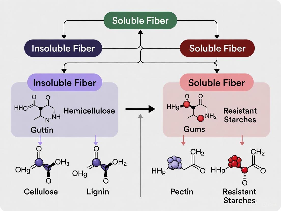
In the study of dietary fiber, the simplistic classification into soluble and insoluble types often obscures the complex chemical architecture that dictates physiological function. The core components of plant cell walls—cellulose, hemicellulose, lignin, and pectin—serve as the fundamental building blocks that determine the solubility, fermentability, and ultimate health impacts of dietary fiber [1] [2]. Understanding their distinct chemical compositions, structural roles, and interactions is crucial for researchers and drug development professionals seeking to leverage specific fiber properties for targeted health outcomes, particularly within the framework of insoluble versus soluble fiber research [3].
This whitepaper provides a technical examination of these four core polymers, detailing their quantitative characteristics, analytical methodologies for their study, and their functional roles within the broader context of fiber research.
Chemical Composition and Structural Properties
The physicochemical diversity of plant cell wall components underpins the varied physiological effects of dietary fibers, from hydration and bulking to fermentation and binding.
Table 1: Core Composition and Characteristics of Dietary Fiber Polymers
| Component | Chemical Structure | Primary Role in Plant | Fiber Solubility Classification | Key Monomeric Units | Glycosidic Linkages |
|---|---|---|---|---|---|
| Cellulose | Linear, unbranched polymer | Structural component of cell walls [1] | Insoluble [1] [4] | D-glucose [1] | β-(1→4) [4] |
| Hemicellulose | Branched, heterogeneous polymer [1] | Cell wall polysaccharide; strengthens wall [5] | Mostly Insoluble [1] | Xylose, glucose, mannose, galactose, arabinose [1] [5] | β-(1→4) and others [5] |
| Lignin | Complex, cross-linked phenylpropane polymer [1] | Non-carbohydrate cell wall component; imparts stiffness [1] [6] | Insoluble [1] [4] | Guaiacyl (G), Syringyl (S), p-Hydroxyphenyl (H) units [6] | Carbon-Carbon and ether linkages |
| Pectin | Linear polysaccharide of galacturonic acid [1] [4] | Component of primary cell wall; intercellular cement [1] | Soluble [1] [4] | D-galacturonic acid [1] [4] | α-(1→4) [4] |
Table 2: Physicochemical and Functional Properties Relevant to Physiological Effects
| Component | Fermentability by Gut Microbiota | Viscosity Forming Capacity | Water-Holding Capacity (WHC) | Primary Physiological Effects in Humans |
|---|---|---|---|---|
| Cellulose | Poorly fermented [4] | Non-viscous [7] | High WHC; provides bulking [8] [4] | Laxative effect; increases stool bulk [8] [4] |
| Hemicellulose | Varies by type and structure [1] | Generally low viscosity | Contributes to bulking | Promotes regularity; bulking effect [7] |
| Lignin | Resists bacterial degradation [1] | Non-viscous | Binds organic materials [1] | Laxative effect; may bind bile acids [1] [4] |
| Pectin | Highly fermentable [1] [4] | Highly viscous; gel-forming [1] [4] | High WHC due to gel formation [1] | Slows gastric emptying; lowers blood cholesterol & glucose [1] [4] |
Advanced Analytical Methodologies for Component Analysis
A detailed understanding of fiber composition requires sophisticated imaging and chemical analysis techniques to probe the complex architecture of plant cell walls.
Imaging and Visualization Techniques
Advanced microscopy allows for the in situ analysis of plant cell wall composition and architecture, providing insights into biomass recalcitrance and digestibility [6].
- Stimulated Raman Scattering (SRS) Microscopy: A label-free technique that utilizes the unique vibrational fingerprints of cellulose, hemicellulose, and lignin to map their distribution in native plant cell walls with high spatial resolution. It is particularly effective for real-time, non-destructive imaging of polymer distributions during conversion processes like enzymatic hydrolysis [6].
- Fluorescence Lifetime Imaging Microscopy (FLIM): Capitalizes on the autofluorescence of lignin to image its distribution. FLIM provides an additional dimension of measurement by resolving the fluorescence decay rate at each pixel, which can be correlated with lignin composition and its interaction with other cell wall polymers [6].
- Atomic Force Microscopy (AFM): Probes the surface topography of cell walls at a nanometer scale. It can be used to characterize the micro- and nanoscale structure of cellulose microfibrils and their assembly, providing information on surface roughness and mechanical properties that influence enzyme accessibility [6].
Chemical and Gravimetric Analysis
Standardized chemical methods remain fundamental for the quantitative isolation and measurement of fiber components.
- Enzymatic-Gravimetric Methods: The most common official methods for total dietary fiber analysis. These involve enzymatic digestion of protein and starch, followed by gravimetric measurement of the undigested residue. Subsequent steps can differentiate soluble and insoluble fractions [1].
- Enzymatic-Chemical Methods: These methods, such as the Uppsala method, go a step further by quantifying specific sugar monomers after hydrolysis of the fiber residue. This allows for a more detailed analysis of the polysaccharide composition of hemicellulose and pectin [1].
- Sequential Extraction and Spectroscopy: Isolating specific components often requires sequential extraction using solvents of varying pH and chelating agents (e.g., for pectin) followed by analysis using Gas Chromatography-Mass Spectrometry (GC-MS) or Fourier-Transform Infrared Spectroscopy (FTIR) for structural characterization [1].
Table 3: The Researcher's Toolkit: Key Reagents and Materials for Fiber Analysis
| Research Reagent / Material | Function in Experimental Protocol |
|---|---|
| Cellulase / Hemicellulase Enzymes | Selective hydrolysis of cellulose and hemicellulose to study polymer digestibility and sugar release profiles [6]. |
| Heat-Stable α-Amylase | Digestion of starch in samples prior to dietary fiber analysis to prevent interference [1]. |
| Protease (e.g., Pepsin) | Digestion of protein in food samples to isolate the fiber fraction for gravimetric analysis [1]. |
| Chelating Agents (e.g., CDTA, EDTA) | Sequester calcium ions to solubilize and extract homogalacturonan-rich pectins from plant cell walls [1]. |
| Dilute Acid / Alkali Solutions | Used in sequential extraction to solubilize hemicelluloses and other non-cellulosic polysaccharides [1] [5]. |
| Specific Fluorescent Dyes (e.g., Calcofluor White) | Binding to β-linked polysaccharides like cellulose for visualization under fluorescence microscopy [6]. |
The following workflow diagram illustrates a generalized protocol for the sequential analysis of core fiber components from a plant biomass sample.
Functional Roles in Soluble vs. Insoluble Fiber Context
The binary classification of fiber into soluble and insoluble types is a functional oversimplification, but it provides a valuable framework for understanding the primary physiological contributions of these core polymers.
The Insoluble Fiber Complex: Cellulose, Hemicellulose, and Lignin
The collective action of cellulose, hemicellulose, and lignin forms the insoluble, poorly fermented fiber complex that is integral to the bulking and laxative effects of dietary fiber [1] [4].
- Cellulose: As a primary contributor to this complex, its linear, crystalline structure and resistance to enzymatic digestion allow it to absorb water and increase fecal bulk throughout the gastrointestinal tract, thereby accelerating intestinal transit and preventing constipation [8] [4].
- Hemicellulose: This branched polymer complements cellulose by also holding water and contributing to stool bulk. Its fermentability varies by chemical structure, but it is generally less fermented than soluble fibers, allowing it to retain its hydrating and bulking capacity for longer periods within the colon [1] [7].
- Lignin: This non-carbohydrate polymer is highly resistant to bacterial degradation [1]. Its primary function in the gut is to provide rigidity and structure to the fecal mass. Furthermore, its hydrophobic, cross-linked nature allows it to bind to various organic materials, including bile acids, which may contribute to its observed effect of lowering serum cholesterol [1] [4].
The Soluble Gel-Forming Fiber: Pectin
Pectin is a classic example of a soluble, viscous, and readily fermented fiber whose physiological effects are directly tied to its gel-forming properties and fermentability [1] [4].
- Viscosity and Gel Formation: Upon dissolution in water, pectin forms highly viscous solutions and gels. This viscosity slows the rate of gastric emptying and nutrient absorption in the small intestine, leading to attenuated postprandial blood glucose and insulin responses [1] [4].
- Fermentability and Prebiotic Potential: Pectin is almost completely metabolized by colonic bacteria [1]. This fermentation produces short-chain fatty acids (SCFAs) like acetate, propionate, and butyrate. Propionate is particularly noted for its role in inhibiting cholesterol synthesis in the liver, contributing to the cholesterol-lowering effect of pectin [4]. Butyrate serves as the primary energy source for colonocytes and has anti-inflammatory properties [4].
The following diagram synthesizes the key signaling and metabolic pathways through which soluble, fermentable fibers like pectin exert their systemic health effects.
Cellulose, hemicellulose, lignin, and pectin are not merely structural plant polymers but are bioactive compounds whose distinct chemical properties dictate their functional roles as dietary fibers. A deep understanding of cellulose's crystallinity, hemicellulose's heterogeneity, lignin's recalcitrance, and pectin's gel-forming capacity is fundamental for moving beyond the simplistic soluble/insoluble dichotomy. For researchers and pharmaceutical developers, this knowledge enables the rational design of interventions, whether through the selection of fiber-rich botanicals with specific component profiles or the isolation and modification of individual polymers to target health outcomes such as glycemic control, cholesterol reduction, or improved gastrointestinal function. Future research will continue to elucidate the structure-function relationships of these core components, further refining our ability to harness their full therapeutic potential.
In the study of dietary fibers, the fundamental divergence in chemical composition between insoluble and soluble fractions manifests primarily in their distinct structural architectures. A core structural determinant is the highly ordered, crystalline nature of insoluble dietary fibers (IDF) compared to the disordered, amorphous character of soluble dietary fibers (SDF). This dichotomy in physical structure, arising from differences in molecular composition and bonding, dictates their physicochemical properties, physiological functions, and subsequent applications in food science and pharmaceutical development [9] [10]. Framed within a broader thesis on the chemical composition of fibers, this whitepaper delineates how crystallinity versus amorphousness serves as a primary factor influencing functionality, from gut microbiota modulation to drug delivery system design.
Structural and Chemical Basis
The classification of dietary fibers into soluble and insoluble categories is conventionally based on water solubility, but this property is intrinsically linked to their underlying chemical composition and supra-molecular organization.
- Insoluble Dietary Fibers (IDF): Primarily composed of cellulose, hemicellulose, and lignin, these components intertwine to form dense, rigid network structures [9] [10]. The extensive hydrogen bonding between linear cellulose chains facilitates their tight packing into highly crystalline domains [9]. This crystalline structure is a key reason for IDF's resistance to enzymatic hydrolysis in the human small intestine and its limited fermentability in the colon [9].
- Soluble Dietary Fibers (SDF): These include pectin, gums, inulin, and certain hemicelluloses [9]. Their chemical structures are typically branched and heterogeneous, preventing the orderly molecular packing required for crystallinity. Consequently, SDF exists in an amorphous state, which contributes to their high water solubility and rapid fermentability by gut microbiota [9] [10].
Table 1: Fundamental Composition and Properties of Insoluble vs. Soluble Fibers
| Characteristic | Insoluble Dietary Fiber (IDF) | Soluble Dietary Fiber (SDF) |
|---|---|---|
| Primary Components | Cellulose, hemicellulose, lignin [9] [10] | Pectin, gums, inulin, β-glucan [9] [10] |
| Predominant Structure | Crystalline, ordered regions [9] [11] | Amorphous, disordered matrix [10] |
| Representative Crystallinity Index (CrI) | ~25-40% (Varies by source and processing) [11] | Largely amorphous [10] |
| Water Solubility | Insoluble [9] | Soluble [9] |
| Fermentability by Gut Microbiota | Slow and limited [9] | High and rapid [9] |
Quantitative Structural Analysis
Advanced characterization techniques provide quantitative evidence of the structural differences between fiber types. The Crystallinity Index (CrI), often determined by X-ray diffraction (XRD), is a key metric for comparing the ordered structure of different fiber preparations.
Table 2: Experimental Crystallinity Data from Fiber Research
| Fiber Source & Type | Treatment/Modification | Crystallinity Index (CrI) / Key Finding | Citation |
|---|---|---|---|
| Pea Insoluble Dietary Fiber (PIDF) | Ultrafine Grinding (400 min) | CrI significantly decreased from initial state [11] | [11] |
| Date Fruit Insoluble Fiber | Fractionation | Insoluble fiber was crystalline [10] | [10] |
| Date Fruit Soluble Fiber | Fractionation | Soluble fiber was amorphous [10] | [10] |
| Quinoa Insoluble Dietary Fiber (QIDF) | Bound Phenolics Removal | CrI increased after treatment [12] | [12] |
Experimental Methodologies for Characterization
Determining the structural properties of fibers requires a suite of complementary analytical techniques. The following are detailed protocols for key experiments cited in this field.
X-Ray Diffraction (XRD) for Crystallinity Index (CrI)
Application: Quantifying the crystallinity of insoluble dietary fibers like pea IDF [11] and quinoa bran IDF [12]. Procedure:
- Sample Preparation: Compact the dried, powdered fiber sample onto an instrument sample holder.
- Data Acquisition: Use an X-ray diffractometer (e.g., D8 ADVANCE) to scan the sample across a range of diffraction angles (2θ), typically from 5° to 40°.
- Data Analysis: Identify the intensity of the diffraction peak corresponding to the crystalline region (e.g., around 22° for cellulose) and the intensity of the trough for the amorphous region (e.g., around 18°). The Crystallinity Index (CrI) is often calculated using the empirical Segal method:
- Formula:
CrI (%) = [(I_crystalline - I_amorphous) / I_crystalline] × 100[11] whereI_crystallineis the maximum intensity of the principal crystalline peak, andI_amorphousis the intensity of the amorphous background.
- Formula:
Scanning Electron Microscopy (SEM) for Morphological Analysis
Application: Visualizing the surface architecture and physical structure of fibers at the micro-scale, such as observing the dense network of PIDF or the loosened structure of QIDF after bound phenolics removal [11] [12]. Procedure:
- Sample Preparation: Adhere dried fiber powder to a metal stub using conductive tape and remove excess powder. Sputter-coat the sample with a thin layer of platinum or gold using an ion sputter (e.g., Hitachi E-1010) for approximately 2 minutes to ensure conductivity.
- Image Acquisition: Transfer the sample to a field-emission scanning electron microscope (e.g., Zeiss Gemini300). Observe the surface morphologies under a high vacuum with an accelerating voltage of 5.0 kV at various magnifications.
Gas Physisorption for Surface Area and Porosity
Application: Measuring the specific surface area (SSA) and pore volume (PV) of fibers, which are critical for understanding adsorption behaviors and are often modified by grinding [11]. Procedure:
- Sample Degassing: Place a known mass of dried fiber sample in a analysis tube and degas under a vacuum to remove contaminants.
- Gas Adsorption/Desorption: Transfer the tube to a surface area analyzer (e.g., Micromeritics Tristar II). Introduce high-purity nitrogen gas at cryogenic temperature and measure the volume of gas adsorbed and desorbed across a range of relative pressures.
- Data Analysis: Calculate the Specific Surface Area (SSA) using the Brunauer-Emmett-Teller (BET) model applied to the adsorption data in the appropriate relative pressure range. Calculate the Pore Volume (PV) and pore-size distribution using the Barrett-Joyner-Halenda (BJH) model applied to the desorption isotherm.
Structural-Functional Relationships and Pathways
The crystalline structure of IDF and the amorphous nature of SDF directly dictate their functional roles in physiological and product applications. The following diagram synthesizes the logical pathway from chemical composition to end-function.
Diagram 1: Logical pathway from chemical composition to function for insoluble and soluble fibers.
The Scientist's Toolkit: Key Research Reagents and Materials
This section details essential reagents, materials, and instruments used in the featured experiments for characterizing fiber structure and function.
Table 3: Essential Research Reagents and Materials for Fiber Analysis
| Item Name | Type/Description | Primary Function in Research |
|---|---|---|
| α-Amylase & Amyloglucosidase | Enzymes | Sequential enzymatic removal of starch and other digestible components from raw plant material to isolate pure dietary fiber [11]. |
| Alkaline Protease | Enzyme | Degradation and removal of protein contaminants during the dietary fiber extraction process [11]. |
| Nitrogen Gas (High Purity) | Analytical Gas | Adsorbate gas used in physisorption analyzers (BET/BJH method) to determine the specific surface area and pore volume of fiber samples [11]. |
| Phloroglucinol & KMnO₄ | Chemical Stains (Weisner & Mäule) | Histochemical staining for the localization and differentiation of guaiacyl and syringyl lignin units in plant tissue sections [10]. |
| Planetary Ball Mill (e.g., UBE-F2L) | Instrument | Ultrafine grinding equipment used for the physical modification of fibers to reduce particle size and disrupt crystalline structure [11]. |
| X-ray Diffractometer (XRD) | Instrument | Measures the degree of crystallinity (CrI) in fiber samples by analyzing the diffraction pattern of X-rays incident on the material [11]. |
| Surface Area & Porosimetry Analyzer | Instrument (e.g., Micromeritics Tristar II) | Characterizes the specific surface area, pore volume, and pore-size distribution of porous fiber materials via gas adsorption [11]. |
| Field-Emission SEM (e.g., Zeiss Gemini300) | Instrument | Provides high-resolution images of fiber surface morphology and microstructure [11]. |
The structural dichotomy between the crystalline architecture of insoluble fibers and the amorphous matrix of soluble fibers is a fundamental determinant of their chemical and biological behavior. Quantitative characterization through techniques like XRD and gas physisorption provides critical data linking structural parameters like CrI, SSA, and PV to functional outcomes such as glucose adsorption and fermentation kinetics. This understanding is pivotal for the rational design of functional foods and the development of advanced, fiber-based drug delivery systems, where tailoring the structural properties of fibers can lead to precise modulation of their performance and health benefits.
The functional diversity of dietary fibers (DFs), particularly the distinction between insoluble and soluble fractions, is fundamentally governed by their monosaccharide composition and glycosidic linking patterns. These structural parameters determine physicochemical properties such as solubility, viscosity, and fermentability, which in turn dictate physiological impacts including microbiota modulation, short-chain fatty acid (SCFA) production, and chronic disease intervention. This whitepaper synthesizes current research to establish a structure-function framework for DFs, providing researchers and drug development professionals with standardized characterization methodologies, quantitative structural data, and elucidated mechanisms linking chemical composition to health outcomes. Precision in DF structural analysis is critical for developing evidence-based nutritional therapies and functional food ingredients.
Dietary fibers are indigestible carbohydrates with polymerization degrees (DP) generally ≥3, resistant to human digestive enzymes [13]. The classification into soluble dietary fiber (SDF) and insoluble dietary fiber (IDF) is primarily determined by their monosaccharide building blocks and the glycosidic bonds linking them into complex polymers.
SDF, including pectins, β-glucans, and arabinogalactans, typically contains backbone and side-chain monosaccharides that create amorphous, hydratable structures. In contrast, IDF, encompassing cellulose, hemicellulose, and lignin, forms crystalline, tightly packed structures through extensive hydrogen bonding and cross-linking, resisting hydration [10] [14]. These inherent chemical differences directly impact their physiological behavior: SDF increases viscosity, modulates gastric emptying and glucose absorption, and is readily fermented by colonic microbiota to produce SCFAs. IDF primarily affects intestinal transit time, fecal bulk, and provides mechanical stimulation to the gut epithelium [15] [16].
Advancements in analytical techniques have revealed that finer structural details—specific monosaccharide ratios, glycosidic linkage positions (α- or β- configuration, 1→4, 1→3, etc.), degree of branching, and presence of functional group modifications (acetylation, methylation)—further refine these broad categories and create a spectrum of functional diversity [15] [14]. This structural complexity enables the targeted selection or design of DFs for specific research and therapeutic applications.
Structural Determinants of Fiber Function
Monosaccharide Composition and Polymerization Degree
The fundamental monomers constituting DFs directly influence their physical properties and physiological functions.
Table 1: Primary Monosaccharides in Common Dietary Fibers and Their Properties
| Monosaccharide | Common Fiber Sources | Typical Configuration | Key Functional Implications |
|---|---|---|---|
| Glucose | Cellulose, β-Glucans, Resistant Starch | β-(1→4) [Cellulose], Mix of β-(1→3) and β-(1→4) [β-Glucan] | Forms linear, rigid chains (cellulose) or viscous, soluble gels (β-glucans). |
| Galacturonic Acid | Pectin | α-(1→4) | Presence of carboxyl groups allows gel formation via cross-linking with cations; contributes to SDF viscosity. |
| Arabinose | Arabinoxylan, Pectin side chains | α-L-Arabinofuranose | Often found as side-chain substituents; disrupts crystallinity, increases solubility and microbial accessibility. |
| Xylose | Xylans, Arabinoglucuronoxylan | β-(1→4) | Forms a linear backbone; acetylation or substitution with arabinose/glucuronic acid modulates solubility. |
| Mannose | Galactomannans, Glucomannans | β-(1→4) | Forms a backbone with galactose side-chains; contributes to high viscosity and water-binding capacity. |
| Galactose | Galactomannans, Pectin side chains | β-(1→4) in backbone, α-(1→6) in side-chains | Side-chain frequency determines solubility and interaction with water; more branches increase solubility. |
| Lignin | Woody plants, Seed coats | Complex phenolic polymer | Not a carbohydrate; hydrophobic, contributes to IDF structure and fecal bulking capacity. |
The Degree of Polymerization (DP), indicating the number of monomeric units in a polysaccharide chain, is another critical parameter. Generally, lower DP fibers (e.g., oligosaccharides like FOS and XOS) ferment more rapidly in the proximal colon, while higher DP fibers may ferment more slowly and distally [14]. For instance, the molecular weight of SDF from various fruits like citrus and apples can range from 84 to 743 kDa, directly affecting its solubility, viscosity, and gelling properties [14].
Glycosidic Linkage Patterns and Functional Groups
The type, position, and stereochemistry of glycosidic bonds create specific three-dimensional architectures that define a fiber's functional role.
- Linkage Patterns: β-(1→4) linkages in cellulose create straight, rigid chains that form stable, insoluble crystalline microfibrils. In contrast, the mixed β-(1→3) and β-(1→4) linkages in β-glucans introduce kinks in the chain, preventing tight packing and resulting in solubility [14]. The α-(1→4) linked backbone of pectin, rich in galacturonic acid, provides sites for ionic gelation.
- Functional Groups: Hydroxyl groups facilitate hydrogen bonding with water, enhancing solubility. Carboxyl groups (e.g., in galacturonic acid of pectin) can ionize, increasing water binding and allowing gel formation. Acetyl esters (e.g., in acetylated galactoglucomannan from wood) protect against enzymatic degradation and can be cleaved by microbial esterases in the colon, providing a source of acetate [17]. Methyl esters on pectin affect its gelling mechanics.
- Branched vs. Linear Structures: Highly branched polysaccharides, like type II arabinogalactan found in date fruit SDF, are typically amorphous and soluble, while linear polymers tend toward crystallinity and insolubility [10].
Experimental Protocols for Structural Analysis
Accurate characterization of DF structure requires a multi-technique approach. Below are detailed protocols for key analyses.
Monosaccharide Composition Analysis
This protocol determines the qualitative and quantitative monomeric makeup of a DF sample [10].
- Sample Hydrolysis: Precisely weigh 10-20 mg of purified DF. Add 1-2 mL of 2 M trifluoroacetic acid (TFA) in a sealed tube. Heat at 121°C for 1-3 hours to hydrolyze glycosidic bonds.
- Neutralization and Drying: Cool the hydrolysate and neutralize the TFA using a stream of nitrogen gas or by adding a calculated volume of sodium hydroxide.
- Derivatization: Convert the released monosaccharides into volatile derivatives. A common method is conversion to alditol acetates by reduction with sodium borohydride followed by acetylation with acetic anhydride.
- Analysis by Gas Chromatography (GC) or GC-Mass Spectrometry (GC-MS): Inject the derivatized sample. Identify and quantify monosaccharides by comparing retention times and peak areas with authentic standards.
Determination of Glycosidic Linkage Composition
Linkage analysis reveals the bonding pattern between monosaccharides, typically performed via methylation analysis [15].
- Methylation: Suspend 1-5 mg of dry DF sample in anhydrous dimethyl sulfoxide (DMSO). Add a methylating agent (e.g., iodomethane) in the presence of a strong base (e.g., sodium hydroxide powder or dimsyl anion) to methylate all free hydroxyl groups.
- Hydrolysis and Reduction: Hydrolyze the permethylated polymer with TFA as in 3.1. Reduce the resulting partially methylated monosaccharides to partially methylated alditol acetates.
- GC-MS Analysis: The unique fragmentation pattern of each partially methylated alditol acetate in the mass spectrometer allows identification of the original linkage type (e.g., a 1,4,5-tri-O-acetyl-2,3,6-tri-O-methyl derivative indicates a 4-linked hexopyranose residue).
Fiber Fractionation and Purification
The AOAC method 991.43 is the standard for separating SDF and IDF [10].
- Enzymatic Digestion: Incubate 1 g of sample (fat-extracted if necessary) with sequential additions of heat-stable α-amylase (95°C, pH 8.2), protease (60°C, pH 7.5), and amyloglucosidase (60°C, pH 4.0-4.5) to remove digestible starch and protein.
- Precipitation of SDF: Add 4 volumes of 95% ethanol (preheated to 60°C) to the filtrate containing soluble fiber. Hold at room temperature for 1 hour to precipitate SDF.
- Filtration and Drying: Filter the mixture through a crucible. The residue on the filter is the SDF fraction (plus protein and ash), while the residue from the initial filtration is the IDF fraction. Wash both residues with 78% ethanol, 95% ethanol, and acetone. Dry and weigh.
- Correction for Protein and Ash: Analyze protein (e.g., by Kjeldahl) and ash content of the residues. Corrected SDF and IDF are calculated by subtracting protein and ash from the respective residues.
Diagram 1: Dietary Fiber Fractionation and Analysis Workflow. This outlines the key steps from raw material to purified fractions ready for structural characterization.
Functional Consequences of Structural Diversity
Impact on Gut Microbiota and SCFA Production
The gut microbiota possesses a vast arsenal of Carbohydrate-Active enZymes (CAZymes) that are highly specific to DF structure. Consequently, monosaccharide composition and linkage patterns dictate which bacterial taxa are selectively promoted.
- Structural Specificity: Bifidobacterium and Lactobacillus are known to utilize β-fructans (inulin, FOS) and α-galactooligosaccharides (α-GOS) [17] [16]. Butyrate-producing bacteria like Faecalibacterium prausnitzii and members of clostridial cluster IX are efficient at fermenting complex xylans and mannans [17]. The presence of acetyl groups on wood-derived hemicelluloses (AcGGM, AcAGX) requires specialized microbial esterases, making these fibers selective for microbes equipped with such enzymes [17].
- SCFA Profile Modulation: The baseline composition of an individual's gut microbiota influences the metabolic outcome of DF fermentation. A microbiota dominated by Bacteroidaceae and Ruminococcaceae tends to produce more butyrate upon DF supplementation, while one dominated by Prevotellaceae and Ruminococcaceae produces more propionate, regardless of the DF type [18]. Specific fibers can skew this further; for instance, acetylated galactoglucomannan (AcGGM) is strongly butyrogenic, while arabinoglucuronoxylan (AcAGX) is more propiogenic [17].
Table 2: Microbiota and SCFA Response to Specific Fiber Structures
| Dietary Fiber Type | Key Structural Features | Microbial Taxa Selectively Promoted | Primary SCFA Output |
|---|---|---|---|
| Fructooligosaccharides (FOS) | β-(2→1) fructose polymers, low DP | Bifidobacterium, Lactobacillus | Acetate |
| Xylooligosaccharides (XOS) | β-(1→4) linked xylose backbone, may be acetylated | Bifidobacterium, Bacteroides | Acetate, Butyrate |
| Galactoglucomannan (AcGGM) | β-(1→4) mannose/glucose backbone, acetylated, galactose side chains | Bifidobacterium, Bacteroides-Prevotella, F. prausnitzii, Clostridial cluster IX | Butyrate |
| Arabinoglucuronoxylan (AcAGX) | β-(1→4) xylose backbone, acetylated, glucuronic acid/arabinose side chains | Bifidobacterium, Bacteroides-Prevotella, F. prausnitzii, Clostridial cluster IX | Propionate |
| Pectin (from fruits) | α-(1→4) galacturonic acid backbone, methyl-esterified, rhammose inserts | Bacteroides, Lachnospiraceae | Acetate, Propionate |
Interactions with Other Dietary Components
DF does not exist in isolation. Its structure influences interactions with other phytochemicals, notably phenolics, forming "antioxidant dietary fiber" [13].
- Covalent and Non-covalent Bonds: Phenolic acids can be ester-linked to DF (e.g., ferulic acid cross-linking arabinoxylans) via covalent bonds. Non-covalent interactions (hydrogen bonding, hydrophobic interactions) can also occur between DF and larger polyphenols.
- Functional Property Changes: These interactions can enhance the stability of phenolics during digestion and transport them to the colon. The bound phenolics can also alter the physicochemical properties of the DF, such as its antioxidant capacity, hydration properties, and fermentability [13].
Diagram 2: Dietary Fiber-Phenolic Interactions and Outcomes. This shows how different binding types lead to complexes with enhanced functional properties.
The Scientist's Toolkit: Research Reagent Solutions
Table 3: Essential Reagents and Kits for Dietary Fiber Research
| Research Reagent / Kit | Function / Application | Example Use Case |
|---|---|---|
| Enzyme Kits (AOAC 991.43) | Sequential enzymatic digestion for SDF/IDF fractionation. Contains heat-stable α-amylase, protease, and amyloglucosidase. | Quantification of SDF and IDF content in food samples or raw materials [10]. |
| Monosaccharide Standards | High-purity sugars (e.g., L-Rhamnose, L-Arabinose, D-Galactose, etc.) for calibration in GC and HPLC. | Identification and quantification of monosaccharides in DF hydrolysates [10]. |
| Methylation Analysis Reagents | DMSO, Iodomethane, Sodium Hydride, etc., for permethylation of polysaccharides. | Determination of glycosidic linkage patterns in a DF polymer [15]. |
| Inulin/FOS, XOS, GOS Standards | Defined oligosaccharide mixtures for chromatographic calibration and as prebiotic references. | Studying fermentation kinetics of specific DF structures or quality control of prebiotic ingredients [18]. |
| Short-Chain Fatty Acid (SCFA) Standards | Pure acetate, propionate, butyrate, etc., for HPLC or GC calibration. | Quantification of SCFA production in in vitro fermentation models [17] [18]. |
| Polymer Gel Beads (e.g., Gellan Gum) | For immobilizing fecal microbiota in continuous in vitro fermentation systems (e.g., PolyFermS). | Maintaining stable, high-density, and representative gut microbiota for long-term DF intervention studies [18]. |
The functional diversity of dietary fibers is unequivocally rooted in the precise chemical details of their monosaccharide composition and linking patterns. Moving beyond the simplistic soluble vs. insoluble dichotomy to a structure-based understanding is paramount for the rational design of next-generation prebiotics and functional foods.
Future research must prioritize the development of comprehensive standardization guidelines that incorporate monosaccharide composition, DP, and linkage data to ensure product efficacy and reproducibility [15]. Furthermore, increased utilization of advanced continuous in vitro models that maintain complex microbial ecosystems will provide deeper insights into the structure-specific fermentation dynamics and cross-feeding networks. Finally, exploring the synergistic effects of DF-phenolic complexes and the impact of food processing on DF structure will unlock new possibilities for enhancing the health benefits and technological applications of dietary fibers in clinical and consumer settings.
Dietary fiber, a diverse group of carbohydrate polymers and associated compounds, resists digestion by human endogenous enzymes and undergoes varying degrees of fermentation in the large intestine. The classical binary classification system categorizes these compounds based on their solubility in water: soluble dietary fiber (SDF), which includes pectins, β-glucans, and arabinoxylans; and insoluble dietary fiber (IDF), comprising cellulose, hemicellulose, and lignin [19] [3]. This solubility dichotomy fundamentally influences their physicochemical properties and physiological effects, making their distribution in plant sources a critical area of research for understanding their health benefits and potential pharmaceutical applications.
The chemical composition of dietary fibers extends beyond solubility to include structural features such as glycosidic linkages, monomeric composition, molecular weight, and the presence of functional groups. These characteristics determine their functional properties, including water-holding capacity, viscosity, swelling ability, and fermentability [2] [3]. For researchers and drug development professionals, understanding these structural-functional relationships is essential for developing fiber-based interventions targeting specific health outcomes, such as cholesterol reduction, blood glucose regulation, and colorectal health maintenance.
Chemical Composition and Structural Characteristics
Soluble Dietary Fibers (SDF)
Soluble dietary fibers are characterized by their ability to dissolve or swell in water, forming gel-like substances with unique rheological properties. The primary SDF components include:
- Pectins: Complex polysaccharides rich in galacturonic acid with varying degrees of methyl esterification, predominantly found in fruits and vegetables [14]. Their gel-forming capability is instrumental in regulating nutrient absorption rates.
- β-Glucans: Linear polysaccharides of D-glucose monomers with mixed (1→3) and (1→4) linkages, particularly abundant in oats and barley, known for their cholesterol-lowering effects [3].
- Arabinoxylans: Hemicellulose components consisting of xylose backbones with arabinose side chains, prevalent in cereal grains, contributing to viscosity development in the gastrointestinal tract [10].
Advanced structural analysis reveals that SDFs contain various functional groups, including hydroxyl, carboxyl, and methoxyl groups, which facilitate hydrogen bonding with water molecules and cation exchange capacities [14]. Nuclear magnetic resonance (NMR) studies of SDF from fruits show chemical shifts in the range of 5.03-5.33 ppm for H1 in the →4)-α-Glcp-(1→ main chain, while →3)-β-Glcp-(1→ residues in β-glucan exhibit H1 chemical shifts between 4.42-4.57 ppm [14].
Insoluble Dietary Fibers (IDF)
Insoluble dietary fibers are characterized by their structural role in plant cell walls and resistance to dissolution in water. Key IDF components include:
- Cellulose: A linear polymer of β(1→4) linked D-glucose units that forms crystalline microfibrils providing structural integrity to plant cells [10] [3].
- Hemicellulose: A heterogeneous polymer of xylose, mannose, galactose, and other sugars with branched structures that cross-link cellulose microfibrils [10].
- Lignin: A complex polyphenolic compound that provides rigidity and resistance to microbial degradation in plants, with guaiacyl and syringyl units identified as primary subunits [10].
The structural complexity of IDF contributes to its higher thermal stability compared to SDF, as demonstrated by thermogravimetric analysis showing greater decomposition resistance in IDF fractions [10]. Crystalline regions within cellulose contribute to this thermal stability, while the amorphous regions of hemicellulose and lignin provide sites for water absorption and microbial adhesion.
Table 1: Comparative Structural and Chemical Properties of Major Dietary Fiber Components
| Fiber Component | Monomeric Units | Glycosidic Linkages | Functional Groups | Structural Features |
|---|---|---|---|---|
| Pectin (SDF) | Galacturonic acid, arabinose, galactose | α(1→4) | Carboxyl, methoxyl, hydroxyl | Branched, amorphous, gel-forming |
| β-Glucan (SDF) | Glucose | β(1→3), β(1→4) | Hydroxyl | Linear with mixed linkages, viscous |
| Arabinoxylan (SDF) | Xylose, arabinose | β(1→4) | Hydroxyl, ferulic acid | Branched, water-soluble |
| Cellulose (IDF) | Glucose | β(1→4) | Hydroxyl | Linear, crystalline, high tensile strength |
| Hemicellulose (IDF) | Xylose, mannose, glucose, others | β(1→4), various | Hydroxyl, acetyl | Branched, amorphous, heterogeneous |
| Lignin (IDF) | Guaiacyl, syringyl, p-hydroxyphenyl | C-C, C-O-C | Methoxyl, phenolic hydroxyl | Amorphous, three-dimensional network |
Analytical Methodologies for Dietary Fiber Characterization
Fiber Fractionation Protocol
The quantitative separation of soluble and insoluble fiber fractions follows standardized methodologies with specific modifications for different plant matrices. The following protocol, adapted from AOAC Method 991.43 with modifications for date fruit analysis, provides a representative experimental approach [10]:
Materials and Reagents:
- Desugared plant sample (e.g., date fruit powder, ground whole grains)
- Ethanol (78%, 95% v/v)
- Acetone (analytical grade)
- Sodium hydroxide solution
- Enzymes: Heat-stable α-amylase, protease, amyloglucosidase
- Filtration system with crucibles
Experimental Procedure:
- Sample Preparation: Finely grind plant material to 106-250 μm particle size. For fruits high in simple sugars, pre-treat with 80% ethanol (1:10 w/v) six times to remove interfering compounds, followed by drying at 50°C for 18 hours [10].
Enzymatic Digestion: Suspend 1g sample in 40mL phosphate buffer (pH 6.0). Add 50μL heat-stable α-amylase solution, incubate at 95°C for 15 minutes with continuous shaking. Cool to 60°C, add protease solution (100μL), incubate at 60°C for 30 minutes. Adjust pH to 4.5, add amyloglucosidase (200μL), incubate at 60°C for 30 minutes [10].
Soluble/Insoluble Fraction Separation: Transfer digested sample to filtration apparatus with pre-weighed crucible. Wash residue with 78% ethanol (15mL), 95% ethanol (15mL), and acetone (15mL). Retain filtrate for soluble fiber analysis.
Soluble Fiber Precipitation: Combine filtrates, evaporate to approximately 40mL total volume. Add 95% ethanol (80mL, 60°C) to precipitate soluble fiber, stand overnight at 4°C. Recover precipitate by filtration, freeze-dry, and weigh [10].
Protein and Ash Correction: Analyze both fractions for protein (Kjeldahl method, N×6.25 conversion factor) and ash content (550°C for 5 hours). Calculate corrected values:
- SDF = (Soluble fraction weight - protein - ash) / initial sample weight × 100
- IDF = (Insoluble fraction weight - protein - ash) / initial sample weight × 100 [10]
Structural Characterization Techniques
Advanced analytical methods provide detailed structural information about fiber components:
Monosaccharide Composition Analysis:
- Protocol: Hydrolyze fiber samples (1-2mg) with 2M trifluoroacetic acid at 121°C for 1 hour. Derivatize released monosaccharides to alditol acetates or conduct HPAEC-PAD analysis without derivatization. Separate using GC-MS or HPLC systems with appropriate columns (e.g., DB-225 for GC, CarboPac PA20 for HPAEC-PAD) [14].
Lignin Localization and Characterization:
- Mäule Staining: Immerse plant tissue sections in 1% (w/v) potassium permanganate solution for 5 minutes, wash with 3% HCl until color changes from dark brown to light brown, treat with 0.1M ammonia solution. Guaiacyl lignin units appear yellow-brown, syringyl units red-purple [10].
- Weisner Staining: Treat tissue sections with 1% phloroglucinol for 5 minutes, followed by 95% ethanol for 5 minutes. Add concentrated HCl to develop red-purple color specific for guaiacyl units [10].
Molecular Weight Distribution:
- Protocol: Dissolve SDF samples (2-5mg/mL) in appropriate eluent (typically 0.1-0.2M NaNO₃). Perform size-exclusion chromatography with multi-angle light scattering and refractive index detection (SEC-MALS-RI). Use pullulan or dextran standards for calibration [14].
Thermal Analysis:
- Protocol: Subject 5-10mg fiber samples to thermogravimetric analysis (TGA) with temperature ramp of 10°C/min from 25°C to 600°C under nitrogen atmosphere. Determine thermal decomposition profiles and stability parameters [10].
Whole Grains
Whole grains represent a significant source of dietary fiber, with varying ratios of soluble to insoluble components depending on the grain type and anatomical fraction. The bran fraction is particularly rich in insoluble fibers, while the endosperm contains more soluble components [3].
Table 2: Dietary Fiber Composition of Selected Whole Grains (g/100g dry weight)
| Grain Type | Total Dietary Fiber | Soluble Fiber | Insoluble Fiber | SDF:IDF Ratio | Notable Components |
|---|---|---|---|---|---|
| Oats | 60-80 | ~30 | 30-50 | 1:1 to 1:1.7 | High β-glucan (SDF) [3] |
| Barley | 20-30 | 5-10 | 15-20 | 1:2 to 1:3 | Mixed-linkage β-glucan, arabinoxylan [3] |
| Wheat | 9-38 | 1-4 | 8-34 | 1:4 to 1:9 | Arabinoxylan, cellulose, lignin [3] |
| Rye | 14-20 | 2-4 | 12-16 | 1:4 to 1:6 | Arabinoxylan, fructan [3] |
| Corn | 10-15 | 1-2 | 9-13 | 1:6 to 1:8 | Cellulose, heteroxylan [3] |
| Rice | 2-4 | 0.2-0.5 | 1.8-3.5 | 1:7 to 1:9 | Cellulose, arabinoxylan [3] |
| Sorghum | 8-12 | 0.8-1.5 | 7.2-10.5 | 1:7 to 1:9 | Cellulose, heteroxylan [3] |
The structural distribution within grains follows specific patterns. In wheat, the aleurone and starchy endosperm contain high proportions of arabinoxylans (60-70%) and β-glucans (20-30%), while the outer pericarp is rich in cellulose (30%), lignin (12%), and xylans (60%) [3]. Oat bran, comprising 30-50% of the whole grain, contributes significantly to its dietary fiber content, with β-glucan as the primary soluble component [3].
Fruits and Vegetables
Fruits and vegetables exhibit diverse dietary fiber profiles influenced by species, cultivar, maturity, and processing methods. Date fruits (Phoenix dactylifera L.) show total dietary fiber content ranging from 3.2-7.4 g/100g, with over 90% being insoluble fiber composed of crystalline lignin, cellulose, and hemicellulose [10]. The soluble fraction in dates is amorphous and primarily consists of pectin (>50%) with complex branching patterns possibly involving type II arabinogalactan [10].
Structural analyses of fruit SDF reveal diverse monosaccharide profiles. Citrus SDF primarily contains galacturonic acid and glucose, while grapefruits, lemons, pomelos, and citrus peels are rich in galacturonic acid and arabinose [14]. Dragon fruit peel SDF consists mainly of galacturonic acid, mannose, and xylose [14]. The molecular weight of SDF varies significantly by source, with citrus ranging 84-743 kDa, apple 103-485 kDa, and potato 2-1819 kDa [14].
Table 3: Dietary Fiber Content in Selected Fruits and Vegetables
| Plant Source | Total Dietary Fiber (g/100g) | Soluble Fiber (g/100g) | Insoluble Fiber (g/100g) | Notable Characteristics |
|---|---|---|---|---|
| Lima beans, cooked | 13.2 | 6.6 | 6.6 | High pectin content [20] |
| Artichoke, cooked | 9.6 | 4.8 | 4.8 | Rich in inulin-type fructans [20] |
| Navy beans, cooked | 9.6 | 4.8 | 4.8 | Balanced SDF/IDF ratio [20] |
| Green peas, cooked | 8.8 | 4.4 | 4.4 | Cellulose, pectin [20] |
| Dates | 3.0 | 0.3 | 2.7 | High lignin content [20] |
| Apple, with skin | 4.8 | ~2.4 | ~2.4 | Pectin in flesh, cellulose in skin [21] |
| Carrots, cooked | 4.8 | ~2.4 | ~2.4 | Pectic polysaccharides [21] |
| Brussels sprouts | 6.4 | ~2.5 | ~3.9 | Cellulose, hemicellulose [20] |
The Scientist's Toolkit: Research Reagent Solutions
Table 4: Essential Reagents and Materials for Dietary Fiber Research
| Reagent/Material | Specifications | Research Application | Functional Role |
|---|---|---|---|
| Heat-stable α-amylase | Bacterial source (e.g., Bacillus licheniformis), ≥50 U/mg | Starch digestion in sample preparation | Hydrolyzes α(1→4) glycosidic bonds in starch to eliminate interference [10] |
| Protease | Aspergillus oryzae or Bacillus spp., ≥300 U/mL | Protein digestion in sample preparation | Cleaves peptide bonds to remove protein content from fiber analysis [10] |
| Amyloglucosidase | Aspergillus niger, ≥100 U/mL | Starch digestion completion | Hydrolyzes α(1→6) and α(1→4) linkages in oligosaccharides [10] |
| Phloroglucinol | Analytical grade, ≥99% purity | Weisner staining for lignin detection | Specific colorimetric reaction with guaiacyl lignin units (red-purple) [10] |
| Potassium permanganate | ACS reagent, ≥99% purity | Mäule staining for lignin differentiation | Oxidizes lignin subunits to distinguish guaiacyl (yellow-brown) and syringyl (red-purple) units [10] |
| Standard reference fibers | Citrus pectin, oat β-glucan, wheat arabinoxylan, cellulose | Method validation and calibration | Quality control standards for comparative analysis and quantification [10] [3] |
| Size-exclusion columns | Sepharose, Sephacryl, or equivalent matrices | Molecular weight distribution analysis | Separation of fiber polymers by hydrodynamic volume [14] |
| Monosaccharide standards | L-Arabinose, D-Galactose, D-Glucose, etc., ≥98% purity | Compositional analysis by GC-MS/HPLC | Quantification and identification of hydrolyzed fiber components [10] [14] |
The systematic investigation of dietary fiber sources reveals complex relationships between plant origin, structural characteristics, and functional properties. The distribution of soluble and insoluble fractions across whole grains, fruits, and vegetables demonstrates significant variation, with oats and barley exhibiting higher SDF:IDF ratios (approximately 2:1 to 3:1) due to their richness in β-glucans, while wheat and rice are predominantly rich in IDF with ratios of approximately 1:4 to 1:9 [3]. Fruits such as dates show exceptionally high insoluble fiber content (>90% of TDF), with lignin composition varying significantly between cultivars [10].
The chemical composition of dietary fibers extends beyond the simplistic soluble-insoluble dichotomy to include structural features such as glycosidic linkage patterns, monosaccharide composition, molecular weight distribution, and functional groups that collectively determine their physiological effects. Advanced processing techniques including enzymatic, thermal, and mechanical treatments can modify these structural attributes, potentially enhancing their bioactive properties [14]. For pharmaceutical and nutraceutical development, understanding these structure-function relationships enables targeted selection of fiber sources for specific health applications, such as utilizing high-β-glucan oats for cholesterol management or lignin-rich preparations for prebiotic effects.
Future research directions should focus on elucidating the structure-activity relationships of less-characterized fiber components, developing standardized analytical protocols for novel fiber sources, and exploring synergistic effects between different fiber types in complex food matrices. Such investigations will advance our understanding of dietary fiber chemistry and facilitate the development of evidence-based recommendations for their application in preventive healthcare and therapeutic interventions.
Advanced Analytical Techniques for Fiber Characterization and Functional Assessment
In the field of analytical chemistry, Fourier Transform Infrared (FTIR) and Near-Infrared (NIR) spectroscopy have emerged as powerful techniques for rapid, non-destructive functional group analysis. These vibrational spectroscopic methods provide critical insights into molecular structure and composition without altering the sample, making them indispensable for a wide range of applications. Within the specific context of dietary fiber research—particularly concerning the chemical composition of insoluble versus soluble fiber—these techniques offer unprecedented capabilities for understanding structural-functional relationships that dictate physiological effects. The distinct health benefits of soluble and insoluble dietary fibers, including cholesterol reduction, glycemic control, and digestive regulation, are directly influenced by their molecular architectures and chemical functional groups [2]. This technical guide provides researchers and drug development professionals with a comprehensive framework for applying FTIR and NIR spectroscopy to elucidate these critical structural characteristics, enabling more accurate classification and prediction of fiber functionality beyond the traditional binary soluble-insoluble paradigm.
Fundamental Principles of FTIR and NIR Spectroscopy
Theoretical Foundations
FTIR and NIR spectroscopy both exploit the interactions between matter and infrared light, but they operate in distinct regions of the electromagnetic spectrum and probe different molecular phenomena.
FTIR Spectroscopy operates in the Mid-Infrared (MIR) region, typically covering wavenumbers from 4000 to 400 cm⁻¹ (wavelengths of 2.5 to 25 μm) [22] [23]. This region corresponds to the fundamental vibrational modes of chemical bonds, including stretching, bending, and rotational motions. When infrared radiation interacts with a sample, specific frequencies are absorbed that match the natural vibrational frequencies of molecular bonds within the sample. The resulting absorption spectrum represents a "molecular fingerprint" that is unique to the chemical structure and composition [24] [25]. The Fourier Transform algorithm enables simultaneous measurement of all wavelengths, significantly improving speed and signal-to-noise ratio compared to traditional dispersive infrared instruments.
NIR Spectroscopy utilizes the Near-Infrared region from approximately 750 to 2500 nanometers (13,333 to 4000 cm⁻¹) [22] [23]. This higher-energy region primarily probes overtones and combination bands of fundamental molecular vibrations, particularly those involving hydrogen atoms (O-H, N-H, C-H bonds) [22]. While NIR spectra contain broader, more overlapping bands compared to FTIR, they can be effectively interpreted using advanced multivariate statistical methods to extract quantitative and qualitative information about complex materials.
Molecular Interaction Mechanisms
The fundamental difference in the type of information provided by these techniques stems from their distinct interaction mechanisms with molecular bonds:
Table 1: Fundamental Characteristics of FTIR and NIR Spectroscopy
| Characteristic | FTIR Spectroscopy | NIR Spectroscopy |
|---|---|---|
| Spectral Range | 4000 - 400 cm⁻¹ | 13,333 - 4000 cm⁻¹ |
| Primary Transitions | Fundamental vibrations | Overtones and combination bands |
| Spectral Features | Sharp, well-defined peaks | Broad, overlapping bands |
| Penetration Depth | Shallow (surface analysis) | Deeper tissue penetration |
| Sample Preparation | Often required | Minimal to none |
The functional group sensitivity differs significantly between these techniques. FTIR provides detailed information about specific functional groups including carbonyls (C=O), hydroxyls (O-H), amines (N-H), and various carbon-carbon and carbon-oxygen bonds [24] [25]. NIR, in contrast, is particularly sensitive to molecular vibrations involving hydrogen atoms, making it exceptionally suitable for analyzing organic compounds with O-H, N-H, and C-H bonds [22] [26].
Comparative Analysis: FTIR vs. NIR for Analytical Applications
Technical Capabilities and Limitations
Each technique offers distinct advantages and limitations that must be considered when selecting an appropriate method for specific analytical challenges.
FTIR Strengths and Limitations:
- Structural Elucidation: FTIR excels at identifying unknown materials and characterizing molecular structures through detailed "fingerprint" regions [22] [23].
- Sensitivity: Higher sensitivity for specific chemical bonds and functional groups, providing superior resolution for complex mixtures [23].
- Quantitative Analysis: Suitable for both qualitative identification and quantitative determination of compound concentrations [22].
- Sample Preparation: Often requires more extensive sample preparation, including grinding, pressing, or specific thickness optimization [26] [27].
- Analysis Speed: Generally slower than NIR, making it less suitable for high-throughput applications [22].
NIR Strengths and Limitations:
- Speed and Throughput: Rapid analysis capabilities, with results typically available within seconds [22].
- Non-destructive Nature: Minimal to no sample preparation required, preserving sample integrity for subsequent analyses [22].
- Penetration Capabilities: Deeper sample penetration enables analysis of bulk properties rather than just surface characteristics [26].
- Spectral Interpretation: Broader, overlapping bands require sophisticated chemometric approaches for meaningful interpretation [26] [28].
- Concentration Sensitivity: Generally requires higher analyte concentrations compared to FTIR for reliable detection [23].
Application-Specific Performance
The selection between FTIR and NIR must be guided by the specific analytical requirements and sample characteristics:
Table 2: Application-Based Comparison of FTIR and NIR Spectroscopy
| Application Requirement | Recommended Technique | Rationale |
|---|---|---|
| Structural Identification | FTIR | Superior molecular fingerprinting capabilities |
| High-Throughput Screening | NIR | Rapid analysis (seconds per sample) |
| Quantitative Analysis | Both | FTIR for specific compounds, NIR for bulk composition |
| Intact Sample Analysis | NIR | Minimal sample preparation required |
| Surface Characterization | FTIR | Limited penetration depth ideal for surface analysis |
| Process Monitoring | NIR | Non-contact measurement capability |
Recent comparative studies demonstrate that both techniques can achieve high accuracy in classification tasks. For example, in authentication of food products like hazelnuts, both MIR (FTIR) and NIR achieved ≥93% accuracy in classifying cultivars and geographic origin, with NIR slightly outperforming FTIR for geographic discrimination [29].
Experimental Protocols for Dietary Fiber Analysis
Sample Preparation Methodologies
Proper sample preparation is critical for obtaining reliable and reproducible spectroscopic data in dietary fiber research.
FTIR Sample Preparation Protocols:
- Grinding: Reduce particle size to <500 μm using a laboratory mill to ensure homogeneity and improve spectral reproducibility [26] [28].
- ATR-FTIR Preparation: Place ground sample directly on the ATR crystal and apply consistent pressure (60-75% of maximum) to ensure optimal contact [27].
- Reflectance FTIR Preparation: Position sample on gold-coated reference plate without compression, ideal for delicate or valuable samples [27].
- Transmission FTIR Preparation: Mix finely ground sample with potassium bromide (KBr) and press into pellet form under vacuum.
NIR Sample Preparation Protocols:
- Minimal Preparation: For homogeneous samples, analysis can be performed directly on intact material with minimal processing [26] [28].
- Grinding for Heterogeneous Samples: Reduce particle size to <500 μm to improve spectral consistency and model performance [28] [30].
- Presentation: Place ground or intact samples in appropriate containers with consistent packing density and orientation.
Instrumentation and Data Collection Parameters
Standardized instrument parameters ensure comparable results across different analyses and laboratories:
FTIR Measurement Parameters:
- Spectral Range: 4000-600 cm⁻¹ for comprehensive functional group analysis [27]
- Resolution: 4 cm⁻¹ optimal for balancing detail and signal-to-noise ratio [27]
- Scans: 64-128 accumulations to improve spectral quality [27]
- Detector: Mercury Cadmium Telluride (MCT) for high sensitivity, Deuterated Triglycine Sulfate (DTGS) for routine analysis [27]
NIR Measurement Parameters:
- Spectral Range: 750-2500 nm (13,333-4000 cm⁻¹) for complete overtone coverage [28]
- Resolution: 8-16 cm⁻¹ suitable for most applications
- Scans: 32-64 accumulations typically sufficient due to stronger signal intensity
- Detector: Silicon (Si) for short-wavelength NIR, Indium Gallium Arsenide (InGaAs) for full-range analysis
Data Processing and Chemometric Analysis
Advanced data processing techniques are essential, particularly for NIR spectroscopy where bands are broad and overlapping:
Spectral Preprocessing Methods:
- Scatter Correction: Standard Normal Variate (SNV) and Multiplicative Signal Correction (MSC) to compensate for light scattering effects [27]
- Derivative Techniques: Savitzky-Golay first and second derivatives to enhance spectral resolution and remove baseline effects
- Smoothing: Moving average or Savitzky-Golay filtering to improve signal-to-noise ratio without distorting spectral features
Multivariate Analysis Techniques:
- Principal Component Analysis (PCA): For exploratory data analysis and outlier detection
- Partial Least Squares (PLS) Regression: For developing quantitative models predicting specific fiber components [26] [28]
- Discriminant Analysis: For classification of samples based on soluble/insoluble fiber characteristics [27]
The following workflow diagram illustrates the complete experimental process for spectroscopic analysis of dietary fibers:
Application to Dietary Fiber Research
Analyzing Soluble vs. Insoluble Fiber Composition
The application of FTIR and NIR spectroscopy to dietary fiber research provides unprecedented insights into the structural characteristics that determine physiological functionality. The traditional binary classification of dietary fiber as simply soluble or insoluble fails to capture the complexity of fiber structures and their diverse health effects [2]. A more comprehensive framework that accounts for backbone structure, water-holding capacity, structural charge, fiber matrix, and fermentation rate is necessary to accurately predict physiological outcomes [2].
NIR Applications in Fiber Analysis: Research has demonstrated that NIR spectroscopy can successfully predict insoluble dietary fiber content in diverse cereal products with a standard error of cross validation (SECV) of 1.54% and R² of 0.98 across a concentration range of 0-48.77% [28] [30]. Soluble dietary fiber prediction is less accurate, with SECV of 1.15% and R² of 0.82 (range 0-13.84%), potentially due to limitations in the reference method rather than the spectroscopic technique itself [28]. The expanded model incorporating samples with high fat and sugar content maintained strong performance, demonstrating the robustness of NIR for analyzing complex food matrices [30].
FTIR Applications in Fiber Analysis: FTIR provides detailed molecular-level information about functional groups present in different fiber types. Specific spectral signatures can distinguish between soluble fibers like β-glucans, pectins, and gums, and insoluble fibers such as cellulose, hemicellulose, and lignin [2]. The carbohydrate region (1200-800 cm⁻¹) shows distinct patterns for different structural organizations, while the carbonyl region (1800-1500 cm⁻¹) provides information about esterification and acetylation patterns that influence solubility and fermentability [2].
Advanced Classification Framework
The integration of spectroscopic data with the proposed comprehensive classification framework enables more accurate prediction of health outcomes:
The Researcher's Toolkit: Essential Materials and Reagents
Successful implementation of FTIR and NIR methodologies requires specific materials and analytical tools. The following table summarizes essential components for spectroscopic analysis of dietary fibers:
Table 3: Essential Research Reagents and Materials for Spectroscopic Fiber Analysis
| Item | Specification | Application | Critical Function |
|---|---|---|---|
| Laboratory Mill | Particle size <500 μm | Sample preparation | Homogenization for reproducible spectra |
| KBr Powder | FTIR grade, >99% purity | FTIR pellet preparation | Transparent matrix for transmission measurements |
| ATR Crystal | Diamond, Germanium, or ZnSe | FTIR-ATR measurements | Internal reflection element for surface analysis |
| NIR Reflectance Cup | Rotating or static | NIR measurements | Consistent presentation for powdered samples |
| Reference Materials | Certified fiber standards | Calibration validation | Quality control and method verification |
| Chemometric Software | PLS, PCA capabilities | Data analysis | Multivariate model development and prediction |
FTIR and NIR spectroscopy provide complementary approaches for rapid, non-destructive functional group analysis in dietary fiber research. FTIR excels in detailed molecular characterization and identification of unknown structures, while NIR offers superior speed and minimal sample preparation requirements ideal for high-throughput analysis. The application of these techniques to the complex challenge of dietary fiber classification moves beyond the simplistic soluble-insoluble paradigm toward a comprehensive framework that accurately predicts physiological outcomes based on structural characteristics. For researchers and drug development professionals, the integration of spectroscopic data with advanced chemometric models enables more precise formulation of fiber-enhanced products with targeted health benefits. As spectroscopic technologies continue to advance, particularly in portable instrumentation and machine learning applications, their role in nutritional science and functional food development will expand, offering new opportunities for understanding structure-function relationships in complex biological matrices.
Thermal analysis techniques, particularly Thermogravimetric Analysis (TGA) and Differential Scanning Calorimetry (DSC), serve as critical methodologies for investigating the stability and decomposition kinetics of complex biological materials. Within the context of researching the chemical composition of insoluble versus soluble fibers, these techniques provide fundamental insights into material behavior under thermal stress. TGA measures mass changes in a sample as a function of temperature or time, providing quantitative data on thermal stability and compositional analysis [31]. DSC, conversely, measures heat flows associated with phase transitions and chemical reactions as a function of temperature, enabling characterization of thermal events such as glass transitions, melting, and crystallization [32] [31]. The application of these techniques to dietary fibers—complex carbohydrates resistant to mammalian digestion—allows researchers to decipher the relationship between fiber structure (soluble vs. insoluble) and thermal properties, thereby informing their stability, processing conditions, and potential applications in pharmaceuticals and functional foods.
Theoretical Fundamentals of TGA and DSC
Principles of Thermogravimetric Analysis (TGA)
TGA operates on the fundamental principle of monitoring the mass of a sample as it undergoes a controlled temperature program. The resulting thermogram plots mass (or percentage mass) against temperature or time, revealing steps corresponding to mass loss events such as dehydration, decomposition, and oxidation [31]. In the study of dietary fibers, these mass loss events are directly attributable to the breakdown of specific polymer components. For instance, the thermal decomposition of cellulose, hemicellulose, and lignin—primary constituents of insoluble fibers—occurs at distinct temperature ranges, allowing for their identification and quantification [33] [34]. Modern TGA systems are often hyphenated with gas analyzers such as FTIR or MS, enabling the qualitative identification of volatile decomposition products and providing a deeper understanding of decomposition mechanisms [31].
Principles of Differential Scanning Calorimetry (DSC)
DSC measures the difference in heat flow between a sample and an inert reference as both are subjected to an identical temperature program. Endothermic events (e.g., melting, dehydration) require more heat to maintain the sample at the same temperature as the reference, while exothermic events (e.g., crystallization, oxidation) release heat [31]. This technique is indispensable for characterizing the glass transition temperature (Tg) of amorphous phases in fibrous materials, a critical parameter governing their physical stability, solubility, and bioavailability in pharmaceutical formulations [32] [31]. Furthermore, DSC can detect other thermal events crucial for fiber analysis, including melting points, crystallization behavior, and enthalpic relaxations associated with physical aging.
Experimental Protocols and Methodologies
Standard TGA Protocol for Fiber Analysis
A robust TGA protocol for characterizing dietary fibers involves several critical steps to ensure reproducible and meaningful data. The following workflow outlines a standard procedure adapted from research on textile fibers and biocomposites [33] [35]:
- Sample Preparation: Gently grind the fiber sample to a consistent powder to ensure uniform heat transfer. For hygroscopic fibers, pre-drying may be necessary to minimize moisture interference.
- Instrument Calibration: Calibrate the TGA balance and temperature sensor using certified reference materials (e.g., nickel or curium) as per the manufacturer's guidelines.
- Experimental Parameters:
- Sample Mass: Load 5-20 mg of sample into an alumina or platinum crucible.
- Atmosphere: Utilize an inert gas purge, such as nitrogen or argon, at a flow rate of 50-100 mL/min to simulate pyrolysis conditions and prevent oxidative degradation [33].
- Temperature Program: Employ a dynamic heating regime, typically between 5 °C/min and 20 °C/min, from ambient temperature to a final temperature of 600-800 °C to ensure complete decomposition [33].
- Data Analysis: Plot the percentage mass loss versus temperature. The derivative of the TGA curve (DTG) is calculated to pinpoint the exact temperatures of maximum decomposition rates for each component.
Standard DSC Protocol for Fiber Analysis
DSC analysis provides complementary information on the thermal transitions of fibers. The standard protocol is as follows [32] [31]:
- Sample Preparation: Precisely weigh 2-10 mg of fiber sample into a hermetically sealed aluminum crucible. An empty, sealed pan serves as the reference.
- Instrument Calibration: Calibrate the DSC cell for temperature and enthalpy using high-purity standards such as indium and zinc.
- Experimental Parameters:
- Atmosphere: Use a nitrogen purge gas (50 mL/min) to maintain an inert environment.
- Temperature Program: Typically, a heat-cool-heat cycle is used. Equilibrate at -50 °C, then heat to a temperature above the expected decomposition (e.g., 300 °C) at a scanning rate of 10 °C/min. Cool rapidly, followed by a second heating cycle to establish a stable baseline and erase thermal history.
- Data Analysis: Analyze the resulting thermogram for thermal events. The glass transition (Tg) is identified as a stepwise change in heat flow, reported as the midpoint of the transition. Melting and crystallization appear as endothermic and exothermic peaks, respectively, with the area under the peak corresponding to the transition enthalpy.
Advanced Hyphenated Techniques
Simultaneous DSC-FTIR microspectroscopy represents a powerful hyphenated technique that combines the quantitative thermal data from DSC with the chemical identification capabilities of FTIR in real-time [32]. This setup allows for one-step screening and qualitative detection of events such as intramolecular condensation, polymorphic transformation, and drug-polymer interactions as they occur, providing unparalleled insight into the stability and solid-state properties of fiber-based formulations [32].
Data Interpretation and Kinetic Analysis
Interpreting TGA and DSC Data for Fibers
The thermal profiles of insoluble and soluble fibers differ significantly due to their distinct chemical structures. Insoluble fibers like cellulose, lignin, and hemicellulose are typically more crystalline and exhibit higher thermal stability. Their TGA curves often show a major, sharp decomposition step at higher temperatures. For example, pure cotton (cellulose) decomposes at approximately 371 °C [33]. In contrast, soluble fibers such as pectins, beta-glucans, and gums often have more amorphous structures and may show broader decomposition profiles at lower temperatures, sometimes preceded by dehydration or melting events detectable by DSC [34] [36].
DSC further differentiates fiber types. Insoluble fibers may display clearer melting transitions due to their crystalline regions, while soluble fibers are more likely to exhibit prominent glass transitions. The Tg is highly sensitive to water content; moisture acts as a plasticizer, significantly lowering the Tg of hydrophilic soluble fibers, which has profound implications for their storage stability and shelf-life in pharmaceutical products [31].
Kinetic Analysis of Decomposition
Kinetic analysis of TGA data quantifies the thermal stability and predicts the material's behavior over time. Model-free isoconversional methods, such as Flynn-Wall-Ozawa (FWO) and Kissinger-Akahira-Sunose (KAS), are preferred for their robustness in analyzing complex reactions like fiber decomposition [33]. These methods calculate the activation energy (Ea) at various extents of conversion (α), revealing multi-step decomposition mechanisms.
For instance, a study on textile fibers found that the activation energy for pure cotton increased from 96.9 kJ/mol (at α = 0.1) to 195.6 kJ/mol (at α = 0.9), indicating a complex, multi-stage decomposition process where the reaction mechanism shifts as the material breaks down [33]. The Coast-Redfern (CR) method can then be applied to identify the most probable reaction model (e.g., nucleation, diffusion), offering a predictive tool for optimizing industrial processes like pyrolysis [33].
Table 1: Kinetic Parameters for Model-Free Analysis of Textile Fibers [33]
| Fiber Type | Conversion (α) | Activation Energy, Ea (kJ/mol) |
|---|---|---|
| Pure Cotton | 0.1 | 96.9 |
| 0.5 | 152.4 | |
| 0.9 | 195.6 | |
| Pure Polyester | 0.1 | 185.2 |
| 0.5 | 212.7 | |
| 0.9 | 238.1 |
The Scientist's Toolkit: Essential Research Reagents and Materials
Successful thermal analysis requires specific materials and reagents. The following table details key items used in the featured experiments and their critical functions.
Table 2: Key Research Reagent Solutions for Thermal Analysis of Fibers
| Item / Reagent | Function & Application | Experimental Context |
|---|---|---|
| Nitrogen (N₂) Gas | Creates an inert atmosphere to study pyrolysis and prevent oxidative degradation during TGA/DSC. | Used in TGA of textile fibers at multiple heating rates [33]. |
| Alumina Crucibles | Inert sample pans for TGA that withstand high temperatures without reacting with the sample. | Standard vessel for holding solid powder samples in TGA [31]. |
| Hermetic Aluminum DSC Pans | Sealed containers to prevent solvent (e.g., water) loss during DSC heating, crucial for accurate Tg measurement. | Essential for analyzing hygroscopic materials like soluble fibers [31]. |
| Model-Free Kinetic Software | Software implementing FWO, KAS, and Friedman methods to calculate activation energy from TGA data. | Used to determine complex decomposition kinetics of textile fibers [33]. |
| Phosphonium Salt Catalyst | A catalyst used in epoxy resin systems for composite materials, enhancing curing and final properties. | Used in a novel epoxy resin (EPIDIAN 11) for cryogenic composite fabrication [37]. |
| Sodium Hydroxide (NaOH) | Used for alkaline treatment of natural fibers to improve interfacial bonding in composite materials. | Treatment of sisal fibers to enhance adhesion in epoxy bio-composites [35]. |
Workflow and Data Interpretation Diagrams
Experimental Workflow for Fiber Characterization
The following diagram outlines the logical sequence of experiments from sample preparation to data interpretation for a comprehensive thermal characterization of dietary fibers.
TGA Data Interpretation Pathway
This diagram illustrates the logical process of interpreting a TGA thermogram to extract quantitative and kinetic information about a fiber sample.
Comparative Data Analysis of Fiber Composites
The application of TGA and DSC is critical in developing advanced materials, such as fiber-reinforced composites. The following table summarizes thermal properties from a study on epoxy bio-composites, demonstrating how these techniques quantify the impact of additives like carbon nanotubes (CNTs) on material performance.
Table 3: Thermal Properties of Sisal/CNT Epoxy Bio-Composites [35]
| Composite Type | CNT Content (wt.%) | Onset Degradation Temp. (°C) | Storage Modulus (GPa) | Loss Modulus (MPa) | Damping Factor (tan δ) |
|---|---|---|---|---|---|
| Baseline | 0.0 | ~250 | Baseline | Baseline | Baseline |
| CNT-Reinforced | 1.0 | ~282 (~13% increase) | +79% | +197% | -56% |
TGA and DSC are indispensable tools for probing the stability and decomposition kinetics of insoluble and soluble fibers. Through carefully designed experimental protocols, these techniques provide quantitative data on composition, thermal resilience, and reaction kinetics. The integration of advanced methods like simultaneous DSC-FTIR and model-free kinetic analysis offers a deeper, mechanistic understanding of fiber behavior, directly supporting the development of stable, effective fiber-based products in the pharmaceutical and functional food industries. The ability to correlate thermal properties with chemical structure and composition ultimately enables the rational design and optimization of materials for specific applications, from drug formulation to advanced biocomposites.
Within the broader study of dietary fibers, lignin is a fundamental yet complex insoluble fiber that plays a critical role in plant structure and human health. As a complex organic polymer that provides rigidity to plant cell walls, lignin is indigestible, functions as "roughage" in the human diet, and is associated with colon health and a reduced risk of constipation [38] [34]. For researchers investigating the chemical composition of insoluble versus soluble fibers, precise localization and visualization of lignin are essential. Understanding its distribution and interaction with other cell wall components, such as cellulose and hemicellulose, provides crucial insights into its physiological effects, including its impact on digestibility, nutrient absorption, and the production of beneficial short-chain fatty acids through fermentation [34]. This technical guide details advanced methodologies for localizing lignin and visualizing its microstructure, providing a vital toolkit for scientists in botany, materials science, and nutritional biochemistry.
Lignin Structure and Its Role as an Insoluble Fiber
Lignin is a class of complex organic polymers forming key structural materials in the support tissues of most plants. It is one of the most abundant organic polymers on Earth, constituting 20% to 35% of the dry mass of wood and 30% of terrestrial non-fossil organic carbon [39]. Chemically, lignins are highly heterogeneous polymers derived from the cross-linking of three primary phenolic precursors (monolignols): coniferyl alcohol (G-unit), sinapyl alcohol (S-unit), and paracoumaryl alcohol (H-unit) [39]. The relative amounts of these subunits vary significantly between plant species.
As a primary component of insoluble dietary fiber, lignin's biological functions directly inform its nutritional role:
- Structural Integrity: Lignin fills the spaces in the plant cell wall between cellulose, hemicellulose, and pectin components, especially in vascular and support tissues, providing mechanical strength and rigidity [39].
- Hydrophobicity: Being rich in aromatic subunits, lignin is hydrophobic. This property enables efficient water transport through plant vascular tissue and reduces the digestibility of lignified plant material in the human gut [39].
- Resistance to Degradation: Lignin is notably resistant to both acid- and base-catalyzed hydrolysis, as well as to enzymatic degradation by human gut microbes. This resistance contributes to its function as a bulking agent that promotes laxation [39] [34].
Table 1: Key Characteristics of Lignin as a Dietary Fiber Component
| Characteristic | Description | Significance in Fiber Research |
|---|---|---|
| Solubility | Insoluble in water and gastrointestinal fluids [38] [40] | Adds bulk to stool, promotes regular bowel movements. |
| Fermentability | Resistant to bacterial breakdown in the colon [34] | Limited production of short-chain fatty acids compared to soluble fibers. |
| Structural Role | Cross-links plant polysaccharides [39] | Impacts texture, digestibility, and nutrient release from plant foods. |
| Health Benefits | Promotes laxation, reduces constipation, supports colon health [34]. | Acts as "nature's laxative"; good for heart health [38]. |
Experimental Protocols for Lignin Localization and Visualization
Fluorescence Microscopy for Lignin Analysis
Fluorescence microscopy is a highly effective and accessible method for localizing lignin in plant tissues. A validated protocol for wood analysis, which can be adapted for other plant fibers, involves specific staining treatments and image processing to relate fluorescence intensity to lignin content [41].
Sample Preparation:
- Histological Sectioning: Prepare thin sections (e.g., 20-30 μm) of the plant tissue of interest using a microtome.
- Extractive Removal (Optional but Recommended): For improved results, remove extractives from a subset of sections to reduce autofluorescence from non-lignin compounds.
- Staining Treatments: Apply one of the following fluorescence treatments to the sections:
- Autofluorescence: Examine the native fluorescence of lignin without any stain, using both in natura sections and sections without extractives [41].
- Basic Fuchsin Stain: Apply this stain to enhance contrast.
- Mäule Reaction: Use this chemical test to distinguish between different types of lignin subunits (e.g., syringyl vs. guaiacyl lignin) [41].
Image Acquisition and Processing:
- Microscopy: Capture images using a fluorescence microscope with appropriate filter sets.
- Intensity Quantification with ImageJ: Process the images using ImageJ software (National Institutes of Health). The most effective and automated method (Method 3 in the cited study) is as follows [41]:
- Open the image in ImageJ.
- If necessary, split the image into its color channels.
- Use the "Analyze Particles" function to automatically measure the fluorescence intensity across the section. This method ensures speed, reproducibility, and standardization of results.
- Data Correlation: Correlate the measured fluorescence intensities with the total lignin content of the samples, as determined by a standard quantitative method like the Klason lignin method, to validate the fluorescence analysis [41].
Computational Scattered Light Imaging (ComSLI) for Microstructure Mapping
A groundbreaking technique developed recently, Computational Scattered Light Imaging (ComSLI), enables detailed visualization of fiber orientations, including lignified structures, at micrometer resolution without the need for specialized stains or complex sample preparation [42] [43].
Workflow Overview:
Detailed Protocol:
- Sample Preparation (Versatility): ComSLI works on a wide range of sample types, including:
- Formalin-fixed, paraffin-embedded (FFPE) sections (the most common format in clinical pathology).
- Fresh-frozen samples.
- Stained or unstained sections.
- Archived samples, even those stored for decades [42].
Instrument Setup: The setup is cost-effective and requires only:
Image Acquisition:
- Illuminate the tissue sample from multiple angles by rotating the LED light source.
- For each angle, record the transmitted light patterns with the camera. Light scatters predominantly perpendicular to the orientation of aligned microscopic fibers [43].
Computational Analysis:
Visualization: The output is a color-coded map of fiber orientations, providing a detailed view of the tissue's microstructural organization. This technique has been successfully applied to brain tissue, muscle, bone, and vascular samples, revealing distinct fiber patterns related to their physiological roles [42].
Solid-State NMR for Lignin-Carbohydrate Interaction Studies
For research focused on the molecular interactions between lignin and other cell wall components, solid-state NMR spectroscopy provides unparalleled insights without disrupting the native cell wall architecture [44].
Key Experimental Steps:
- Isotopic Labeling: Grow plants (e.g., Arabidopsis thaliana as a model system) in an atmosphere containing
13CO2to achieve uniform 13C-labeling of all cell wall polymers. - Sample Collection and Preparation: Harvest plant stems and segment them by age and developmental stage to capture the progression of lignification.
- NMR Spectroscopy: Acquire 1D 13C cross-polarization (CP) spectra and 2D 13C-13C correlation spectra on the intact cell wall samples.
- Spectral Analysis:
- Lignin Content and Composition: Quantify lignin content from the aromatic signal intensity and determine the syringyl-to-guaiacyl (S/G) ratio from the methoxy group signals and specific aromatic cross-peaks.
- Lignin-Carbohydrate Proximity: Identify intermolecular cross-peaks between lignin aromatic carbons (120–160 ppm) and carbohydrate carbons (60–90 ppm). These cross-peaks indicate sub-nanometer physical contacts, revealing which carbohydrates (e.g., xylan, pectin) are spatially closest to lignin during different stages of wall formation [44].
This method has revealed that during stem maturation, S-lignin content increases and forms tighter physical packing with polysaccharides, particularly with acetylated xylan, which is critical for the structural and mechanical properties of the cell wall [44].
The Scientist's Toolkit: Essential Research Reagents and Materials
Successful localization and analysis of lignin require specific reagents and instruments. The following table details key solutions and their functions for the experimental protocols described.
Table 2: Research Reagent Solutions for Lignin Visualization
| Reagent / Material | Function / Application | Experimental Protocol |
|---|---|---|
| Basic Fuchsin Stain | Fluorescent stain used to enhance the contrast of lignin in tissue sections for microscopy. | Fluorescence Microscopy [41] |
| Mäule Reagents | Chemical test used to distinguish between syringyl (S) and guaiacyl (G) lignin subunits based on a color reaction. | Fluorescence Microscopy, Solid-State NMR Validation [41] [44] |
| Formalin-Fixed Paraffin-Embedded (FFPE) Samples | Standard method for preserving and hardening tissue for sectioning; compatible with ComSLI and other techniques. | ComSLI, General Histology [42] |
| ImageJ Software | Open-source image analysis software used to automatically quantify fluorescence intensity from microscope images. | Fluorescence Microscopy [41] |
| 13C-Labeled Plant Material | Plant biomass isotopically enriched with 13C, enabling detailed molecular-level analysis of lignin and its interactions. | Solid-State NMR [44] |
| Rotating LED Light Source | Key hardware component for ComSLI that illuminates the sample from different angles to capture scattering patterns. | ComSLI [42] [43] |
The following table consolidates key quantitative findings from the cited research, providing a clear reference for experimental planning and result validation.
Table 3: Summary of Quantitative Data from Lignin Visualization Studies
| Measurement | Value / Result | Method Used | Context / Significance |
|---|---|---|---|
| Lignin Content Correlation (R²) | 76.22% | Fluorescence Microscopy (Autofluorescence, Method 3) | Coefficient of determination between fluorescence intensity and Klason lignin content [41] |
| Lignin Content in Plants | 20-35% of dry mass | General Analysis | Typical abundance of lignin in wood [39] |
| Increase in Lignin-Carbohydrate Contacts | From 18% to 29% | Solid-State NMR | Percentage of carbohydrates in sub-nanometer proximity to lignin aromatics during stem maturation in Arabidopsis [44] |
| S-Lignin Increase with Age | From 25-28% to 30% (rigid fraction) | Solid-State NMR | Increase in syringyl (S) subunit content in mature (WT16A) vs. younger stems [44] |
| Resolution of ComSLI | Micrometer scale (1-4 μm suggested) | Computational Scattered Light Imaging | Resolution sufficient to map individual fiber orientations in tissues like brain and muscle [42] [43] |
Advanced Visualization: The Lignin Biosynthesis Pathway
A molecular-level understanding of lignin formation is crucial for interpreting its localization. The following diagram outlines the key steps in the biosynthesis of the primary monolignol precursors.
The techniques detailed in this guide—from established staining and fluorescence protocols to cutting-edge methods like ComSLI and solid-state NMR—provide a comprehensive arsenal for researchers to localize lignin and visualize its complex microstructure. The selection of an appropriate method depends on the research question, whether it pertains to the gross distribution of lignin in a food matrix, its nanoscale interactions with carbohydrates that influence digestibility, or its role in the mechanical properties of plant-based materials. By applying these tools, scientists can deepen the understanding of lignin's fundamental role as a key insoluble dietary fiber, bridging the gap between plant cell wall architecture and human health outcomes.
Wet Chemical Methods and AOAC Standards for Fiber Fractionation and Quantification
The precise fractionation and quantification of dietary fiber, particularly the chemical distinction between its soluble (SDF) and insoluble (IDF) components, is fundamental to nutritional science and food analysis. Despite evolving definitions, dietary fiber classifications have historically remained simplistic, often reduced to a binary soluble-insoluble system that overlooks the complexity of fiber structures and their diverse health effects [2]. This analytical challenge is particularly relevant within the broader context of research on the chemical composition of insoluble versus soluble fibers, as their distinct physicochemical properties—including solubility, fermentation rate, water-holding capacity, and structural charge—directly influence their physiological impacts [2] [3]. Current enzymatic-gravimetric approaches, while providing foundational quantification of soluble and insoluble dietary fiber, often present limitations related to the lack of detailed fraction characterization [45]. This technical guide examines established wet chemical methods, emerging advanced protocols, and ongoing standardization efforts by AOAC INTERNATIONAL to address these analytical gaps and provide researchers with comprehensive tools for fiber analysis.
Current Analytical Limitations and the Case for Advanced Classification
The traditional binary classification system for dietary fiber fails to adequately predict the full range of physiological effects exerted by different fiber types [2]. Table 1 summarizes the limitations of this system and the proposed advanced classification framework.
Table 1: Evolution of Dietary Fiber Classification Systems
| Traditional Model | Proposed Advanced Framework | Analytical Implications |
|---|---|---|
| Binary (Soluble/Insoluble) | Multi-parameter classification | Enables better prediction of health outcomes |
| Oversimplifies structure-function relationships | Considers backbone structure, water-holding capacity, structural charge, fiber matrix, and fermentation rate [2] | Requires more sophisticated analytical protocols |
| Limited predictive value for physiological effects | Holistic understanding of structure-function relationship | Necessitates integration of gravimetric and characterization methods |
| Does not account for fiber diversity | Comprehensively captures structural and functional diversity | Bridges gap between chemical analysis and bio-functional properties |
This analytical simplification is particularly problematic for insoluble dietary fibers, which constitute a market segment expected to grow from USD 2.83 billion in 2025 to USD 4.09 billion by 2030, driven largely by their application in functional foods and regulatory support for fiber-rich product claims [46]. The limitations of current classification systems underscore the need for more refined analytical approaches that can accurately characterize the complex chemical composition of different fiber types and link these compositions to their functional properties in research and product development.
Established Wet Chemical Methods for Fiber Analysis
Enzymatic-Gravimetric Methods (AOAC 991.43)
The AOAC 991.43 method represents the official standard for nutritional labeling and provides the fundamental approach for separating soluble and insoluble dietary fiber. This enzymatic-gravimetric procedure simulates human digestive processes by utilizing heat-stable α-amylase, protease, and amyloglucosidase to remove protein and starch components, followed by ethanol precipitation to isolate soluble dietary fiber. The insoluble fraction is collected through filtration, and both fractions are quantified gravimetrically after accounting for ash and protein content [45]. This method provides crucial data on total, soluble, and insoluble dietary fiber content but offers limited information about the specific chemical constituents within each fraction.
Detergent Fiber Methods (Van Soest Method)
Originally developed for feed analysis but now widely applied to human food analysis, the Van Soest method provides a complementary fractionation system that partitions fiber based on solubility in specific detergent solutions, as outlined in Table 2.
Table 2: Van Soest Detergent Fiber Method Components
| Method Component | Treatment Conditions | Target Fractions | Quantified Components |
|---|---|---|---|
| Neutral Detergent Fiber (NDF) | 60 minutes in neutral detergent with α-amylase [47] | Insoluble cell wall components | Hemicellulose, Cellulose, Lignin [47] |
| Acid Detergent Fiber (ADF) | Treatment with acidic detergent [47] | Less digestible cell wall fractions | Cellulose, Lignin (hemicellulose removed) [47] |
| Acid Detergent Lignin (ADL) | ADF treatment followed by concentrated sulfuric acid [47] | Lignin fraction | Lignin (cellulose removed) [47] |
| Hemicellulose Calculation | By difference: NDF - ADF | Hemicellulose content | Hemicellulose [47] |
| Cellulose Calculation | By difference: ADF - ADL | Cellulose content | Cellulose [47] |
The sequential nature of this analysis allows researchers to determine the relative proportions of hemicellulose, cellulose, and lignin in insoluble fiber sources. Comparative studies have demonstrated that crude fiber analysis, another historical method, typically yields lower values than the Van Soest method, as it fails to recover significant portions of hemicellulose and cellulose that are dissolved during its harsher chemical treatment [47].
Advanced Integrated Protocols for Fiber Fractionation
Comprehensive Fractionation and Characterization Protocol
Recent research has developed integrated protocols that combine the quantitative strengths of official methods with advanced characterization techniques. One such protocol integrates the AOAC 991.43 method with sequential fiber fractionation exploiting the different resistance of fiber components to acid hydrolysis treatments [45]. This approach, visualized in Figure 1, enables both quantification and detailed characterization of fiber fractions.
Figure 1: Advanced Integrated Protocol for Dietary Fiber Fractionation and Characterization
Critical Research Reagent Solutions
Successful implementation of fiber fractionation protocols requires specific research reagents and laboratory materials. Table 3 details essential solutions and their functions in the analytical process.
Table 3: Research Reagent Solutions for Fiber Fractionation
| Reagent/Equipment | Function in Analysis | Application Specifics |
|---|---|---|
| Heat-stable α-amylase | Hydrolyzes starch components | AOAC 991.43 method; incubation at 95-100°C [45] |
| Protease enzyme | Digests protein components | AOAC 991.43 method; pH 7.5 ± 0.1 at 60°C [45] |
| Amyloglucosidase | Hydrolyzes starch dextrins to glucose | AOAC 991.43 method; incubation at 60°C [45] |
| Neutral detergent | Dissolves highly digestible cell contents | Van Soest NDF analysis; contains sodium lauryl sulfate [47] |
| Acid detergent | Removes hemicellulose fraction | Van Soest ADF analysis; contains cetyltrimethylammonium bromide [47] |
| Sulfuric acid (concentrated) | Hydrolyzes cellulose for lignin determination | Van Soest ADL analysis; 72% H₂SO₄ treatment [47] |
| Trifluoroacetic acid (TFA) | Hydrolyzes hemicellulose fractions | Advanced protocol; 2M TFA at 121°C [45] |
| FIBREBAG technology | Standardized filtration system | Provides consistent pore size for reproducible filtration [47] |
| Petroleum ether | Sample degreasing | Removes ether-extractable components in sample preparation [47] |
The integration of these reagents within standardized systems such as the FIBRETHERM, which automates boiling, washing, and filtration processes, significantly enhances analytical reproducibility across twelve simultaneous samples [47]. Standardization of parameters including particle size (recommended 1mm), detergent concentration, cooking times, and temperature profiles is critical for obtaining comparable results across different laboratories and sample types [47].
Comparative Analysis of Method Outcomes
The choice of analytical method significantly influences the quantitative results obtained for fiber components, particularly for insoluble fibers. Table 4 presents a comparative analysis of different methods applied to the same sample type, demonstrating these variations.
Table 4: Comparative Fiber Analysis Values in Feed for Dairy Cows (Percentage)
| Method Type | Analytical Target | Reported Value | Comparative Notes |
|---|---|---|---|
| Crude Fiber | Traditional insoluble fraction | Lowest value | Large amounts of hemicellulose and cellulose not dissolved [47] |
| NDF (Neutral Detergent Fiber) | Total cell wall components | 22.57% (highest) | Includes hemicellulose, cellulose, lignin [47] |
| ADF (Acid Detergent Fiber) | Cellulose and lignin | 11.71% | Hemicellulose removed during acid detergent treatment [47] |
| Calculated Hemicellulose | NDF - ADF | 10.86% | Represents the hemicellulose fraction [47] |
This comparative data clearly demonstrates that crude fiber analysis substantially underestimates the total fiber content compared to the Van Soest method, highlighting the importance of method selection in research on the chemical composition of insoluble fibers. The development of advanced protocols that integrate enzymatic-gravimetric methods with acid hydrolysis and GC-MS characterization addresses the critical need for both quantification and molecular characterization of fiber fractions [45].
AOAC Standardization Initiatives and Future Directions
AOAC Dietary Fiber and Other Carbohydrates Program
Recognizing the evolving analytical challenges in fiber quantification, AOAC INTERNATIONAL launched the Dietary Fiber and Other Carbohydrates Program in early 2025 to address alignment issues between current fiber definitions and analytical methods [48] [49]. This initiative aims to:
- Develop guidance documents and decision trees for method selection based on matrix type
- Align Official Methods of Analysis with current Codex definitions for dietary fiber
- Clarify what specific fiber components are detected by each analytical method
- Address methodological gaps in the analysis of dietary carbohydrates [48]
The program brings together volunteer subject matter experts from government, industry, and academia to establish internationally recognized method performance standards and foster global harmonization of fiber analysis methodologies [48].
Emerging Analytical Trends and Research Needs
Future developments in fiber fractionation and quantification are likely to focus on:
- Enhanced Characterization Techniques: Integration of advanced analytical methods including GC-MS, LC-MS, and NMR to complement traditional gravimetric analysis [45].
- Structure-Function Relationship Mapping: Developing analytical frameworks that link specific fiber structures (backbone composition, molecular weight, structural charge) to their physiological effects [2].
- Standardization of Novel Fiber Analysis: Establishing validated methods for emerging fiber types, including resistant oligosaccharides and modified cellulose [48].
- Rapid Analysis Methods: Development of high-throughput screening techniques to complement traditional wet chemical methods [47].
These developments will ultimately enable researchers to more accurately correlate the chemical composition of insoluble versus soluble fibers with their observed health benefits, advancing both nutritional science and the development of fiber-enriched functional foods.
The fractionation and quantification of dietary fiber continues to evolve from simplistic binary classifications toward sophisticated multi-parameter analytical frameworks. While established wet chemical methods including AOAC 991.43 and the Van Soest system provide fundamental tools for fiber analysis, emerging protocols that integrate enzymatic-gravimetric approaches with advanced characterization techniques offer more comprehensive insights into the chemical composition of insoluble and soluble fibers. Ongoing AOAC standardization initiatives promise to further harmonize and refine these methodologies, enabling researchers to more accurately establish structure-function relationships and advancing our understanding of how specific fiber components influence human health. As research in this field progresses, the integration of robust traditional methods with advanced characterization technologies will be essential for unraveling the complex relationship between fiber chemistry and physiological function.
Within the scope of a broader thesis on the chemical composition of insoluble versus soluble fiber research, this whitepaper provides a technical guide on the core physicochemical properties that underpin their functionality. For researchers and scientists in drug development, a deep understanding of these properties—specifically water-holding capacity, viscosity, and oil-binding capacity—is not merely academic. It is crucial for the rational design of drug delivery systems, functional foods, and nutraceuticals. These properties are direct consequences of the distinct chemical structures of soluble and insoluble dietary fibers and dictate their physiological behavior and technological applications [3] [50]. This document summarizes quantitative data on these properties, details standard experimental protocols for their determination, and visualizes the structure-function relationships that are central to leveraging dietary fibers in advanced health applications.
Fiber Composition and Fundamental Properties
Dietary fibers are categorized based on their solubility in water, a characteristic dictated by their underlying chemical composition. This solubility is the primary factor determining their physiological mechanisms and health benefits [3].
- Insoluble Dietary Fiber (IDF): Primarily composed of cellulose, hemicellulose, and lignin, IDF acts as a structural component in plant cell walls [3] [51]. Its molecular structure is highly crystalline and cross-linked, making it resistant to dissolution in water. Its primary mode of action is mechanical, providing bulk and promoting laxation.
- Soluble Dietary Fiber (SDF): This fraction includes non-cellulosic polysaccharides such as β-glucans, arabinoxylans, pectin, and gums [3]. These compounds dissolve or swell in water to form viscous gels. Their functionality is largely molecular, modulating nutrient absorption and fermenting in the large intestine to produce short-chain fatty acids.
The ratio of SDF to IDF varies significantly among common whole grains, which in turn influences their overall functionality [3]. For instance, oats and barley have high SDF:IDF ratios (approximately 2:1 to 3:1) due to their richness in β-glucans, whereas wheat and rice are predominantly rich in IDF, resulting in lower SDF:IDF ratios (approximately 1:4 to 1:9) [3].
Quantitative Analysis of Key Physicochemical Properties
The functional properties of dietary fibers can be quantitatively measured, providing critical data for material selection in research and product development. The following table summarizes typical values for these key properties across different fiber types, illustrating how their composition dictates their functional performance.
Table 1: Quantitative Physicochemical Properties of Selected Dietary Fibers
| Fiber Source | Fiber Type | Water-Holding Capacity (g/g) | Viscosity (mPa·s) | Oil-Binding Capacity (g/g) | Cation Exchange Capacity (meq/g) |
|---|---|---|---|---|---|
| Rice Bran | Soluble (SDF) | Varies by extraction | 2.35 (at specified concentration) [50] | Not Specified | Lower than IDF [50] |
| Rice Bran | Insoluble (IDF) | Varies by extraction | Lower than SDF [50] | Not Specified | Higher than SDF [50] |
| Rice Bran | Total (TDF) | Varies by extraction | Similar to SDF [50] | Not Specified | Similar to SDF [50] |
| Whole Grains (e.g., Oats, Barley) | Soluble (SDF) | High | High (due to β-glucans) [3] | Not Specified | Contributes to bile acid binding [3] |
| Whole Grains (e.g., Wheat, Rice) | Insoluble (IDF) | High (contributes to bulk) [3] | Low | Not Specified | Contributes to bile acid binding [3] |
Interpretation of Quantitative Data
The data in Table 1 highlights key functional differences:
- Viscosity: SDF fractions, such as those from rice bran, exhibit significantly higher viscosity compared to IDF [50]. This property is concentration-dependent and is a critical determinant in SDF's ability to modulate glucose absorption and lower serum cholesterol.
- Cation Exchange Capacity (CEC): IDF typically demonstrates a higher CEC than SDF, as seen in rice bran [50]. This property is linked to the fiber's ability to bind minerals and bile acids, the latter being a mechanism for cholesterol reduction.
- Oil-Binding Capacity: While specific quantitative values for oil-binding are not provided in the search results, the physicochemical properties of dietary fibers contribute significantly to their bile-acid-binding capacity, which is a key lipid-related function [3].
Experimental Protocols for Property Characterization
Accurate measurement of these properties is fundamental to fiber research. Below are detailed methodologies for key assays.
Protocol for Viscosity Measurement
Viscosity is a primary functional metric for SDF.
- Principle: The resistance of a fiber solution to flow is measured under controlled shear conditions.
- Materials:
- Rheometer (e.g., AR-1000, TA Instruments)
- Dietary fiber sample
- Distilled water
- Warring blender
- Procedure:
- Prepare sample slurries at specific concentrations (e.g., 1% and 3% w/v) by slowly adding the fiber to distilled water and mixing at high speed in a blender for 1 minute [50].
- Allow the solutions to stand at room temperature for 24 hours to achieve equilibrium and allow entrapped air to escape.
- Load the sample onto the rheometer plate. A standard configuration is a steel cone (2°, 60 mm diameter) with a gap of 1000 μm.
- Measure viscosity across a defined shear rate range (e.g., 0 to 50 s⁻¹) at room temperature [50].
- Report viscosity at a standard shear rate or as a profile across the measured range.
Protocol for Cation Exchange Capacity (CEC)
CEC measures the fiber's ability to bind ions, reflecting its role in mineral and bile acid metabolism.
- Principle: The fiber is converted to its hydrogen ion form and titrated with a base to determine its total exchangeable cations.
- Materials:
- Dietary fiber sample
- 0.1 mol/L HCl
- 0.1 mol/L NaOH
- 5% NaCl solution
- Distilled water
- 10% AgNO₃ solution
- Magnetic stirrer and titration apparatus
- Freeze-dryer
- Procedure:
- Immerse the fiber sample in 0.1 mol/L HCl for 48 hours to protonate all exchangeable sites [50].
- Remove excess acid by washing with distilled water until chloride ions are no longer detected (test with 10% AgNO₃ solution, where no opaque white precipitate of AgCl forms).
- Freeze-dry the treated sample.
- Accurately weigh 0.205 g of the freeze-dried sample and disperse it in 100 mL of 5% NaCl solution.
- Stir the mixture magnetically while slowly titrating with 0.1 mol/L NaOH.
- Record the pH and the volume of NaOH required to reach the equivalence point. The CEC is calculated from the titer value [50].
Protocol for Glucose Dialysis Retardation Index (GDRI)
GDRI is an in vitro method used to model a fiber's ability to slow glucose absorption, a key mechanism for managing blood sugar levels.
- Principle: The rate of glucose diffusion across a dialysis membrane is measured in the presence and absence of dietary fiber.
- Materials:
- Dietary fiber sample
- Dialysis bags (12,000-14,000 MWCO)
- Glucose standard solution
- Sodium azide solution (1 g/L)
- Water bath maintained at 37°C with stirring
- Glucose oxidase-peroxidase assay kit
- Procedure:
- Hydrate the fiber sample in sodium azide solution for 14 hours before dialysis [50].
- Fill pre-soaked dialysis bags with 6 mL of sodium azide solution containing 36 mg of glucose, alone (control) or with the addition of 0.2 g of hydrated fiber.
- Tie the bags and suspend them in 100 mL of sodium azide solution in a stirred water bath at 37°C.
- At 30 and 60 minutes, collect 2 mL aliquots of the dialysate (solution outside the bag).
- Analyze the glucose concentration in the dialysate using the glucose oxidase-peroxidase method.
- Calculate GDRI using the formula: GDRI = [(Gc - Gf) / Gc] × 100, where Gc is the glucose concentration in the control dialysate and G_f is the glucose concentration in the dialysate with fiber [50].
The Scientist's Toolkit: Research Reagent Solutions
Table 2: Essential Reagents and Materials for Fiber Physicochemical Analysis
| Reagent/Material | Function in Experimental Protocols |
|---|---|
| Heat-stable α-amylase (e.g., Termamyl) | Gelatinization, hydrolysis, and depolymerization of starch during fiber extraction [50]. |
| Protease (e.g., Alcalase) | Solubilization and depolymerization of proteins in the sample matrix [50]. |
| Amyloglucosidase | Hydrolysis of starch fragments into glucose to complete starch removal [50]. |
| Ethanol (78-95% v/v) | Precipitation of soluble dietary fiber fractions and dehydration of residues [50]. |
| Standard Acid and Base (HCl, NaOH) | Used in CEC determination and various chemical quantitative analysis methods for fiber blends [50] [52]. |
| Dialysis Membranes (12-14k MWCO) | Used in GDRI assays to simulate intestinal nutrient absorption [50]. |
| Specific Solvents (e.g., Acetone, Sulfuric Acid) | Used in chemical dissolution methods for quantitative analysis of fiber blends per standards like ASTM D629 [52] [53]. |
Structure-Function Relationships and Pathways
The pathway from chemical structure to physiological function is governed by the key physicochemical properties detailed in this document. The following diagram visualizes these logical relationships for soluble and insoluble fibers.
Diagram 1: Fiber Property-Function Pathway. This diagram illustrates how the fundamental chemical composition of soluble (SDF) and insoluble (IDF) dietary fibers determines their key physicochemical properties, which in turn drive specific physiological functions and health outcomes. SDF primarily acts through viscosity and cation exchange, while IDF functions through water-holding and bulking.
The experimental characterization of these properties follows a systematic workflow, from sample preparation to data interpretation. The diagram below outlines a generalized protocol for evaluating a key functional property like viscosity.
Diagram 2: Generic Experimental Workflow for Fiber Property Analysis. This workflow outlines the key stages in characterizing physicochemical properties like viscosity, CEC, or GDRI, highlighting the progression from prepared sample to functional insight.
The intentional application of dietary fibers in research and drug development hinges on a predictive understanding of their structure-function relationships. As detailed in this whitepaper, the chemical dichotomy between soluble and insoluble fibers gives rise to distinct physicochemical properties—water-holding, viscosity, and oil/bile-acid-binding capacity. These properties are not only measurable through standardized protocols but are also the direct link to physiological mechanisms such as glycemic control, cholesterol management, and digestive health. For scientists designing novel delivery systems or functional ingredients, moving beyond a simplistic fiber classification to a property-focused approach enables more precise and effective product formulation. Future work in this field will continue to refine these relationships, particularly for novel fiber sources and their modified derivatives, expanding the toolbox available for advanced health interventions.
Resolving Complexities in Fiber Analysis and Optimizing Functionality
For decades, the classification of dietary fiber into soluble and insoluble types has served as a foundational model for predicting physiological effects. However, emerging research reveals this binary framework is insufficient for explaining the diverse and complex mechanisms by which fibers interact with the gastrointestinal tract. This whitepaper synthesizes current evidence demonstrating that physicochemical properties beyond solubility—including viscosity, fermentability, molecular structure, and water-holding capacity—primarily determine metabolic outcomes. We analyze the molecular pathways through which specific fiber structures modulate gut microbiota composition, bile acid metabolism, short-chain fatty acid production, and signaling pathways, providing a refined framework for researchers and drug development professionals to develop targeted nutritional interventions.
The traditional classification of dietary fiber as either soluble or insoluble has persisted in nutritional science and clinical practice despite growing evidence of its limitations. This binary system originated from early observations that certain fibers dissolve in water while others do not, with solubility broadly correlated with some physiological effects [1]. However, the chemical composition and physiological functionality of dietary fibers are far more complex than this simplistic model suggests [2].
The definition of dietary fiber has evolved significantly, now encompassing "all polysaccharides and lignin, which are not digested by the endogenous secretion of the human digestive tract" [7]. This includes not only traditional non-starch polysaccharides (cellulose, hemicellulose, pectin, gums, mucilages) but also resistant oligosaccharides, resistant starch, and associated substances [54] [1]. Within this diverse chemical landscape, the solubility paradigm fails to accurately predict important physiological outcomes, including impacts on glycemic response, cholesterol metabolism, and gut microbiota modulation [2].
This whitepaper presents a evidence-based analysis of the actual mechanisms governing dietary fiber functionality in the gastrointestinal tract, with particular emphasis on how specific physicochemical properties—rather than simple solubility—determine physiological effects. This refined understanding is essential for developing targeted nutritional therapies and leveraging dietary fibers in pharmaceutical applications.
Physicochemical Properties Determining Physiological Effects
Critical Properties Beyond Solubility
Research indicates that five key properties collectively determine the physiological impact of dietary fibers in the GI tract more accurately than solubility alone [2]:
- Backbone structure: The fundamental chemical composition and molecular architecture
- Water-holding capacity: The ability to absorb and retain water within the matrix
- Structural charge: The presence and density of ionic groups influencing molecular interactions
- Fiber matrix: The three-dimensional organization and porosity
- Fermentation rate: The kinetics of microbial breakdown in the large intestine
These properties function as interconnected determinants rather than isolated characteristics, collectively influencing the fiber's behavior throughout the gastrointestinal system [2].
Relationship Between Properties and Physiological Effects
Table 1: Fiber Properties and Their Primary Physiological Consequences
| Property | Physiological Effects | Representative Fiber Types |
|---|---|---|
| Viscosity | Delays gastric emptying, reduces glycemic response, increases satiety, binds bile acids [54] [55] | β-glucans, psyllium, pectins, some gums |
| Fermentability | Feeds gut microbiota, produces SCFAs, influences gut barrier function [54] [55] | Inulin, FOS, GOS, pectins, resistant starch |
| Water-Holding Capacity | Increases stool bulk, softens stool, accelerates transit time [1] [55] | Cellulose, psyllium, bran fibers |
| Bile Acid Binding | Reduces cholesterol recycling, increases bile acid synthesis [55] [56] | β-glucans, psyllium, oat bran |
| Gel-Forming Ability | Traps nutrients, delays absorption, modulates digesta flow [54] [57] | Pectins, β-glucans, guar gum |
The viscosity of certain soluble fibers is particularly important for metabolic effects. Viscous fibers like β-glucan and psyllium form gel matrices in the intestinal lumen that delay gastric emptying, slow carbohydrate digestion and absorption, and entrap bile acids, leading to improved glycemic control and reduced cholesterol levels [54] [55]. This demonstrates how a specific physicochemical property directly mediates clinically relevant outcomes.
Molecular Mechanisms of Action in the Gastrointestinal Tract
Microbiota Accessible Carbohydrates and Fermentation
A fundamental mechanism of dietary fiber action involves serving as substrates for microbial fermentation in the colon, producing short-chain fatty acids (SCFAs) including acetate, propionate, and butyrate [54] [55]. These SCFAs exert multiple beneficial effects:
- Butyrate serves as the primary energy source for colonocytes, enhances gut barrier function, and exhibits anti-inflammatory and anti-carcinogenic properties [55]
- Propionate modulates hepatic glucose metabolism, inhibits cholesterol synthesis, and influences satiety signaling [55]
- Acetate enters systemic circulation and influences peripheral tissues including liver, muscle, and adipose tissue [55]
Different fiber structures select for distinct microbial populations, resulting in varied SCFA profiles. For instance, inulin and fructooligosaccharides preferentially increase Bifidobacteria and Lactobacilli, while resistant starch promotes Bacteroides and Bifidobacteria [54]. This microbial specificity determines not only SCFA production but subsequent host responses.
Figure 1: Microbial Fermentation Pathway of Dietary Fiber
Bile Acid Metabolism and Signaling
Dietary fibers significantly influence bile acid (BA) metabolism through multiple mechanisms, with substantial implications for cholesterol homeostasis and metabolic health [55] [56]. The molecular pathways include:
- Physical binding of bile acids in the intestinal lumen, preventing reabsorption and increasing fecal excretion [55]
- Microbial modulation of bile salt hydrolase (BSH) activity, enhancing deconjugation of primary bile acids [56]
- Alteration of the composition and size of the bile acid pool, influencing FXR and TGR5 signaling pathways [56]
Different fiber types exhibit distinct effects on BA metabolism. In a comparative study using mouse models, psyllium significantly impacted BA-related gene expression, while inulin and β-glucan enhanced bile salt hydrolase activity, and resistant starch showed minimal effects [56]. This demonstrates how fiber-specific properties—not merely solubility—determine outcomes in BA metabolism.
Impact on Glycemic Response
The mechanisms by which dietary fibers modulate postprandial glycemic response extend beyond simple carbohydrate trapping:
- Viscosity-dependent delay in gastric emptying and intestinal transit time [55]
- Formation of physical barriers that limit enzyme accessibility to digestible carbohydrates [57]
- Inhibition of α-amylase activity through direct binding of certain fibers to digestive enzymes [55]
- Production of SCFAs that influence gluconeogenesis and insulin sensitivity [55] [58]
The structural integrity of plant cell walls plays a crucial role in determining glycemic impact. Intact cell walls can retard starch gelatinization during cooking and limit enzymatic access to starch, resulting in lower postprandial glucose responses compared to disrupted cellular structures [57].
Experimental Approaches for Investigating Fiber Mechanisms
In Vivo Models and Methodologies
Table 2: Key Research Reagent Solutions for Dietary Fiber Studies
| Reagent/Category | Function/Application | Specific Examples |
|---|---|---|
| Defined Fiber Preparations | Isolate specific physicochemical properties for mechanistic studies | Cellulose (insoluble, low fermentability), Inulin (soluble, fermentable), β-glucan (soluble, viscous), Psyllium (soluble, viscous) [56] |
| Germ-Free (GF) Models | Investigate fiber-microbiota interactions without confounding microbial influences | GF mice for studying microbial colonization, SCFA production, and immune development [58] |
| Microbiota Depletion | Assess microbiota-dependent vs independent effects of fiber interventions | Broad-spectrum antibiotic cocktails to deplete gut microbes [58] |
| Gnotobiotic Models | Study specific microbial taxa in fiber metabolism | Mice colonized with defined microbial communities [58] |
| Stable Isotope Tracing | Quantify microbial fermentation kinetics and metabolic fate | ¹³C-labeled fibers to track SCFA production and host incorporation [55] |
| Enteroid Models | Investigate direct epithelial responses to fiber metabolites | Intestinal organoids treated with filtered fecal supernatants [58] |
Protocol: Comparative Analysis of Fiber Impact on Bile Acid Metabolism
Based on recently published methodology [56], the following protocol enables systematic investigation of how different fiber types influence BA metabolism and gut microbiota:
Experimental Design:
- Utilize C57Bl/6 mice (n=8-10 per group) fed control or 10% (w/w) fiber diets for 14 days
- Include fibers representing spectrum of properties: cellulose (insoluble, low fermentability), chitin (insoluble), resistant starch (partially fermentable), pectin (soluble, viscous, highly fermentable), inulin (soluble, non-viscous, highly fermentable), β-glucan (soluble, viscous, moderately fermentable), psyllium (soluble, viscous, partially fermentable)
Sample Collection and Analysis:
- Collect fecal samples at baseline and endpoint for 16S rRNA sequencing to assess microbiota changes
- Harvest liver, intestinal mucosa (ileum), and fecal samples for bile acid quantification via LC-MS
- Measure taurine and conjugate levels in liver and intestinal mucosa
- Analyze expression of BA-related genes (e.g., FXR, TGR5, BSH) in intestinal tissues
Key Outcome Measures:
- Microbiota diversity metrics (Shannon index, richness) and specific taxa abundance (e.g., Akkermansia, Ruminococcus)
- Concentrations of primary and secondary BAs across tissues
- Ratio of taurine-conjugated to deconjugated BAs
- Correlation analysis between bacterial taxa and BA profiles
This protocol enables researchers to simultaneously assess how different fiber structures impact microbial community structure and BA metabolism, revealing structure-function relationships.
Figure 2: Experimental Workflow for Fiber-BA Analysis
Implications for Research and Therapeutic Development
The evidence demonstrating that physicochemical properties beyond solubility determine physiological effects has significant implications for nutritional science and therapeutic development:
Precision Nutrition Applications: Understanding specific structure-function relationships enables development of targeted fiber blends for specific metabolic outcomes, such as combining viscous fibers for glycemic control with highly fermentable fibers for gut barrier enhancement [54] [2].
Drug Development Considerations: The impact of dietary fibers on drug absorption requires consideration of multiple physicochemical properties. Viscous fibers may delay gastric emptying and alter drug bioavailability, while fermentable fibers may modify gut microbiota that participate in drug metabolism [57].
Research Standardization: Reporting of fiber interventions should include detailed characterization of physicochemical properties (viscosity, fermentability, molecular weight) rather than simply solubility categories to enable meaningful comparisons across studies [2] [56].
Microbiome-Based Therapeutics: The selective fermentation of specific fiber structures by particular bacterial taxa presents opportunities for developing targeted prebiotics to support specific microbial communities for therapeutic purposes [54] [58].
The simplistic soluble/insoluble classification of dietary fibers fails to capture the complexity of their chemical structures and physiological effects in the gastrointestinal tract. Evidence-based understanding reveals that physicochemical properties including viscosity, fermentability, molecular structure, and bile acid binding capacity collectively determine mechanisms of action. These properties influence fundamental processes including microbial ecology, bile acid metabolism, short-chain fatty acid production, and signaling pathways that impact host physiology.
For researchers and drug development professionals, adopting this refined framework enables more precise study design, interpretation of results, and development of targeted nutritional interventions. Future research should focus on elucidating structure-function relationships for understudied fiber types and exploring how fiber blends with complementary properties can be optimized for specific health outcomes. Moving beyond the solubility paradigm represents an essential step toward precision nutrition and microbiome-based therapeutics.
The Impact of Food Processing on Fiber Structure and Nutritional Efficacy
Dietary fiber, a diverse group of carbohydrate polymers with three or more monomeric units, resists digestion by human alimentary enzymes and encompasses a wide spectrum of chemical structures [1]. Traditional classification divides these polymers into two broad categories: soluble dietary fiber (SDF), which includes pectins, β-glucans, gums, and mucilages that dissolve or swell in water, and insoluble dietary fiber (IDF), comprising cellulose, hemicellulose, and lignin that remain relatively intact [7] [1]. This solubility dichotomy has long provided a foundational framework for nutritional science. However, emerging research reveals that this binary classification insufficiently captures the complex physicochemical properties that dictate physiological effects [59]. The nutritional efficacy of fiber is fundamentally governed by its intricate chemical architecture—including monosaccharide composition, glycosidic linkage patterns, degree of polymerization, and molecular branching—which in turn determines its hydration capacity, viscosity, fermentability, and ultimately, its biological activity [60] [15].
Food processing introduces targeted modifications to these native fiber structures, potentially altering their functional properties and health benefits. Understanding these transformations requires a paradigm shift from solubility-based categorization to a structure-function perspective that acknowledges the molecular diversity within fiber classes and their differential responses to processing conditions. This whitepaper examines the impact of various processing methodologies on fiber structure and explores the implications for nutritional efficacy within the context of chemical composition research.
Structural Diversity of Insoluble versus Soluble Dietary Fibers
The chemical composition of dietary fibers creates distinct physiological behaviors for insoluble and soluble varieties. Insoluble fibers, characterized by extensive hydrogen bonding and crystalline regions in polymers like cellulose, provide mechanical structure to plant cell walls and resist aqueous penetration [1]. In contrast, soluble fibers contain molecular motifs that facilitate interaction with water molecules, leading to dissolution or gel formation.
Table 1: Fundamental Chemical and Physiological Properties of Major Dietary Fiber Types
| Fiber Type | Primary Components | Chemical Structural Features | Physiological Actions | Food Sources |
|---|---|---|---|---|
| Insoluble Fiber | Cellulose, Hemicellulose, Lignin | Linear β-(1→4) glucan chains (cellulose); branched heteropolymers with xylose, arabinose, galactose (hemicellulose); complex cross-linked phenylpropane polymers (lignin) [1] | Increases stool bulk, accelerates intestinal transit, reduces colonic pressure [7] [19] | Cereal brans, whole grains, nuts, vegetable skins |
| Soluble Fiber | Pectin, β-Glucans, Inulin, Gums, Mucilages | α-(1→4) linked D-galacturonic acid backbone (pectin); β-(1→3/1→4) D-glucose units (β-glucans); β-(2→1) fructosyl chains (inulin) [7] [61] | Forms gels, delays gastric emptying, slows glucose absorption, fermented to SCFAs [7] [61] [15] | Oats, barley, fruits, legumes, psyllium |
The molecular architecture of these fiber classes dictates their fate in the gastrointestinal tract. Insoluble fibers primarily exert their effects through physical mechanisms—increasing fecal bulk and reducing transit time—while soluble fibers engage in complex physicochemical interactions that modulate nutrient absorption and serve as substrates for microbial fermentation in the colon [7]. The fermentability of soluble fibers stems from their vulnerability to enzymatic cleavage by specialized glycoside hydrolases possessed by colonic microbiota, yielding short-chain fatty acids (SCFAs) with systemic health implications [60] [15].
Food Processing Methods and Their Structural Impacts
Processing-induced transformations of dietary fiber occur through mechanical, thermal, and chemical mechanisms that alter the native organization of plant cell wall polymers. These structural modifications can enhance, diminish, or fundamentally change the nutritional functionality of fiber components.
Mechanical Processing
Particle size reduction through milling, grinding, or chopping disrupts the structural integrity of plant cell walls, increasing surface area and potentially releasing soluble components from insoluble matrices. Studies on coconut residue demonstrated that reducing particle size from 1,127μm to 550μm significantly enhanced hydration properties due to increased surface area and total pore volume [1]. Beyond this threshold, however, excessive mechanical energy input can collapse the porous matrix, diminishing water-holding capacity. This physical disruption affects both soluble and insoluble fractions, potentially increasing the extractability of arabinoxylans and β-glucans from cereal matrices and altering their interaction with gut microbiota [60].
Thermal Processing
Thermal treatments, including extrusion-cooking, boiling, canning, and frying, induce complex physicochemical transformations in fiber structures. Extrusion-cooking, which combines high temperature, pressure, and shear forces, particularly impacts cereal brans by solubilizing portions of hemicellulose and modifying the hydration properties of both soluble and insoluble fractions [1]. The intense mechanical shear can fragment insoluble arabinoxylan chains, potentially converting them to soluble oligosaccharides with distinct fermentability profiles.
Moist-heat treatments (boiling, steaming, canning) hydrate and swell cell wall polysaccharides, leading to partial pectin solubilization and depolymerization through β-elimination reactions. These processes can enhance the fermentability of otherwise insoluble fibers by increasing accessibility to microbial enzymes. Conversely, dry-heat treatments (baking, frying) may induce Maillard reactions and caramelization, creating new cross-links between polysaccharides and proteins that potentially reduce fermentability [1].
Table 2: Impact of Processing Methods on Fiber Structure and Nutritional Properties
| Processing Method | Key Processing Parameters | Structural Impacts on Fiber | Consequences for Nutritional Efficacy |
|---|---|---|---|
| Extrusion-Cooking | High temperature (120-180°C), high pressure, shear forces | Solubilization of hemicellulose, depolymerization, structural expansion [1] | May enhance fermentability, modifies viscosity-related benefits |
| Retorting (Canning) | High temperature (115-125°C), moisture, pressure | Hydration and swelling of cell walls, pectin β-elimination, depolymerization | Increases soluble fiber fraction, potentially enhances SCFA production |
| Baking | Dry heat (150-250°C), variable time | Non-enzymatic browning, caramelization, possible lignification | May reduce fermentability through cross-linking, modifies hydration |
| Fine Grinding | Particle size reduction (<500μm) | Disruption of cell wall architecture, increased surface area | Enhances hydration properties to optimum point, may improve fermentability |
Chemical Modifications
Processing-induced chemical changes include acid- or alkali-catalyzed hydrolysis of glycosidic bonds, oxidation of phenolic components in lignin, and de-esterification of pectin. Alkaline treatments, sometimes used in vegetable processing, can solubilize hemicellulose and modify the interfacial properties of insoluble fibers. Similarly, acidic conditions, such as those encountered in fermented foods or added during processing, can hydrolyze acid-labile glycosidic linkages in arabinans and galactans, potentially reducing molecular weight and viscosity-building capacity [1].
Analytical Methodologies for Fiber Characterization
Comprehensive analysis of processed fibers requires integrated methodologies that capture both macroscopic nutritional definitions and molecular structural information. The following experimental protocols represent state-of-the-art approaches for fiber characterization.
Enzymatic-Gravimetric Methods (AOAC Official Methods)
The AOAC 991.43 and 2017.16 methods provide the regulatory framework for nutritional labeling of total, soluble, and insoluble dietary fiber [60]. These methods simulate human digestion through sequential enzymatic treatments:
- Sample Preparation: Weigh 1g of sample (in duplicate) into specialized filter bags.
- Starch Digestion: Incubate with thermostable α-amylase (pH 6.0, 100°C, 30 min) to hydrolyze digestible starch.
- Protein Digestion: Treat with protease (pH 7.5, 60°C, 30 min) to remove protein.
- Secondary Starch Digestion: Incubate with amyloglucosidase (pH 4.0-4.6, 60°C, 30 min) to complete starch removal.
- Soluble/Insoluble Separation: Filter through celite; soluble fiber remains in filtrate while insoluble fiber is retained on filter.
- Quantification: Precipitate soluble fiber with ethanol (4 volumes), then gravimetrically determine both fractions after drying; correct for protein and ash content [60].
These methods effectively separate fiber from digestible components but provide limited structural information about the resulting fractions.
Advanced Structural Characterization Techniques
To bridge the gap between nutritional definitions and molecular structure, researchers have developed integrated analytical pipelines that combine enzymatic-gravimetric separation with sophisticated chromatographic and spectrometric analyses:
Multi-glycomic Analysis Pipeline [60]:
- Fiber Fraction Preparation: Isolate soluble and insoluble fractions via AOAC methods.
- Total Monosaccharide Analysis: Hydrolyze samples with 2M TFA (121°C, 2h), derivative with 3-methyl-1-phenyl-2-pyrazolin-5-one (PMP), and analyze by LC-MS.
- Glycosidic Linkage Analysis: Permethylate samples using NaOH/DMSO/methyl iodide, hydrolyze, reduce, and acetylize; analyze partially methylated alditol acetates by GC-MS.
- Free Saccharide Profiling: Extract oligosaccharides with 50% ethanol and characterize by LC-MS.
This integrated approach revealed that insoluble fiber from oats primarily consists of linkages corresponding to β-glucan, arabinoxylan, xyloglucan, and mannan, while soluble fiber is predominantly β-glucan with minor arabinogalactan components [60]. Such structural specificity enables precise structure-function relationships.
Diagram 1: Integrated analytical workflow for comprehensive fiber characterization, combining classical nutritional definitions with structural analysis.
Research Reagent Solutions for Fiber Analysis
Table 3: Essential Research Reagents for Dietary Fiber Characterization
| Reagent/Chemical | Technical Function | Application Context |
|---|---|---|
| Thermostable α-amylase (B. licheniformis) | Hydrolyzes α-(1→4) glycosidic bonds in starch at high temperature | Simulates salivary digestion in AOAC methods; removes interfering starch [60] |
| Amyloglucosidase (A. niger) | Cleaves α-(1→4) and α-(1→6) linkages from starch non-reducing ends | Completes starch digestion to glucose in AOAC protocols [60] |
| Protease (B. licheniformis) | Hydrolyzes peptide bonds in proteins | Eliminates protein interference in fiber quantification [60] |
| 3-methyl-1-phenyl-2-pyrazolin-5-one (PMP) | Derivatives reducing sugars for UV detection | Enables LC-MS quantification of monosaccharide composition [60] |
| Anhydrous dimethyl sulfoxide (DMSO) | Powerful solvent for complete polysaccharide dissolution | Essential for permethylation in glycosidic linkage analysis [60] |
| Iodomethane (CH₃I) | Methylating agent for hydroxyl groups | Creates methyl ethers for linkage analysis via GC-MS of PMAAs [60] |
Nutritional Implications of Processing-Induced Structural Changes
The structural modifications imposed by processing directly influence the nutritional efficacy of both insoluble and soluble fibers through multiple physiological mechanisms.
Impact on Microbiota-Fiber Interactions
The gut microbiota's ability to utilize dietary fibers is highly structure-specific, as microbial genomes encode specialized glycoside hydrolases with precise substrate preferences [60]. For example, arabinoxylan degradation requires β-xylosidases and α-arabinosidases, while xyloglucan utilization demands β-glucosidases, α-xylosidases, and β-galactosidases [60]. Processing that alters the monosaccharide composition, glycosidic linkage patterns, or polymerization degree of these fibers consequently shapes their fermentability and metabolic fate.
Groundbreaking research using the multi-glycomic approach demonstrated that even subtle structural differences between six forms of inulin produced unique "metabolic fingerprints" in gut models, consistent across individuals with different microbial compositions [62]. This suggests that fiber structure, more than individual microbial diversity, primarily determines metabolic outcomes. The concept of "metabolic functional guilds"—groups of phylogenetically distinct microbes that perform similar metabolic functions—explains how structurally-defined fibers can produce consistent health effects across diverse individuals [62].
Modifications to Physiological Effects
Processing-induced structural changes can enhance or diminish specific health benefits associated with different fiber types:
Viscosity-dependent effects: Soluble fibers like psyllium, β-glucans, and pectins exert hypoglycemic and hypocholesterolemic effects primarily through viscosity-mediated mechanisms [63]. Processing that reduces molecular weight through depolymerization can diminish viscosity and thus reduce these benefits. For example, the cholesterol-lowering effects of oat β-glucan are directly correlated with molecular weight and viscosity [61].
Fermentation dynamics: Structural modifications alter the rate and extent of microbial fermentation, influencing SCFA production profiles. Rapidly fermented fibers like pectin and inulin produce more total SCFAs but may cause gastrointestinal distress, while slowly fermented fibers like resistant starch provide more sustained SCFA production, particularly in the distal colon [1] [15]. Processing that increases surface area or solubilizes insoluble fibers generally accelerates fermentation rates.
Hydration properties: The water-holding capacity of insoluble fibers like cellulose and hemicellulose contributes to their laxative effects by increasing fecal bulk [1]. Mechanical processing that optimizes particle size can maximize these benefits, while excessive processing may reduce water-holding capacity.
Diagram 2: Causal pathway from food processing through structural modifications to physiological effects, illustrating the structure-function relationship.
Emerging Framework for Structure-Based Standardization
The limitations of solubility-based classification have prompted development of more sophisticated frameworks that better predict nutritional functionality. A new bottom-up approach classifies fibers based on five key structural features: backbone structure, water-holding capacity, structural charge, fiber matrix, and fermentation rate [59]. This framework enables more precise targeting of specific health outcomes by aligning fiber properties with desired physiological effects.
Concurrently, researchers have proposed a stepwise standardization approach for prebiotic fibers beginning with monosaccharide composition, then incorporating degree of polymerization, and ultimately adding comprehensive linkage composition data [15]. This hierarchical characterization provides the structural specificity needed to connect specific fiber structures with defined health benefits, facilitating the development of targeted nutritional interventions.
This structure-based approach is particularly relevant for product development, as processing conditions can be optimized to preserve or enhance specific structural features associated with desired health outcomes. For instance, gentle processing methods might be selected for β-glucans to maintain molecular weight and viscosity, while targeted hydrolysis might be applied to certain prebiotic fibers to improve their compatibility with food matrices without compromising fermentability.
The impact of food processing on fiber structure and nutritional efficacy represents a critical interface between food science, nutrition, and gut health. The binary classification of fibers as simply soluble or insoluble has proven inadequate for predicting their complex physiological behaviors. Instead, a molecular-level understanding of fiber structures—including monosaccharide composition, glycosidic linkage patterns, and polymerization degree—provides a more robust foundation for explaining how processing modifications alter nutritional functionality.
Advanced analytical approaches that integrate classical enzymatic-gravimetric methods with sophisticated structural characterization techniques now enable comprehensive mapping of processing-induced structural changes to specific health outcomes. This knowledge empowers food scientists and nutritionists to design processing strategies that optimize rather than compromise the health benefits of dietary fibers. As research in this field advances, structure-based standardization of dietary fibers will facilitate the development of more effective, targeted nutritional interventions tailored to specific health needs, moving beyond generic fiber recommendations to precision nutrition approaches that leverage the full potential of these complex biomolecules.
The classical paradigm for classifying dietary fiber (DF) has long relied on a binary soluble-versus-insoluble system. However, this simplistic model fails to accurately predict the complex and diverse physiological effects of DF, as solubility alone is an insufficient indicator of function [2]. Insoluble dietary fiber (IDF), primarily comprising cellulose, lignin, and certain hemicelluloses, is traditionally associated with bowel regularity and increased stool bulk [64] [3]. Soluble dietary fiber (SDF), including components like β-glucans, pectins, and arabinoxylans, is recognized for its role in modulating glycemic response, lowering serum cholesterol, and serving as a prebiotic substrate [3] [65]. A growing body of evidence indicates that the specific ratio of SDF to IDF (SDF:IDF) is a critical determinant of the food matrix's structure and its subsequent nutritional and physiological outcomes [66]. Framed within advanced research on the chemical composition of fibers, this guide provides a technical framework for researchers and drug development professionals to rationally optimize SDF:IDF ratios for targeted health benefits.
A New Classification Framework for Dietary Fibers
To move beyond the limitations of the solubility-based model, a more holistic classification framework that incorporates key physicochemical properties is essential for predicting physiological effects. This refined approach accounts for the structural and functional diversity of dietary fibers [2].
The proposed framework categorizes fibers based on five constitutive properties:
- Backbone Structure: The primary chemical structure of the polysaccharide.
- Water-Holding Capacity (WHC): The ability to retain water, influencing stool bulk and viscosity.
- Structural Charge: The presence of ionic groups (e.g., carboxyl groups in pectin) affecting bile acid binding and electrolyte exchange.
- Fibre Matrix: The three-dimensional physical structure, including porosity and particle size.
- Fermentation Rate: The speed and extent of microbial breakdown in the colon, determining SCFA production.
This multi-factorial model allows for a more accurate inference of a fiber's health outcomes than the traditional binary system [2].
Visualizing the Modern Dietary Fiber Classification Framework
The following diagram illustrates the proposed comprehensive framework for classifying dietary fibers based on their physicochemical properties and the resulting physiological outcomes.
Quantitative SDF:IDF Profiles of Selected Raw Materials
The SDF:IDF ratio varies significantly across different fiber sources. Understanding these inherent ratios is the first step in selecting appropriate raw materials for product formulation. The table below summarizes the total dietary fiber (TDF), SDF, and IDF content, along with the calculated SDF:IDF ratio, for various whole grains and fruit by-products.
Table 1: Dietary fiber composition and key physicochemical properties of selected raw materials [64] [3] [10]
| Source | TDF (g/100g) | SDF (g/100g) | IDF (g/100g) | SDF:IDF Ratio | Water-Holding Capacity (g/g) | Primary SDF Components |
|---|---|---|---|---|---|---|
| Oats (Hulless) | 60.0 - 80.0 | ~30.0 | ~50.0 | ~1:1.7 | Varies by genotype | β-Glucan, Arabinoxylan |
| Barley | 20.0 - 30.0 | 7.0 - 12.0 | 13.0 - 18.0 | ~1:1.5 to 1:2.0 | Data not specified | β-Glucan, Arabinoxylan |
| Wheat Bran | 9.0 - 38.0 | Low | High | ~1:4 to 1:9 | Data not specified | Arabinoxylan, Cellulose |
| Apple Pomace (Royal Gala) | 78.2 | 14.3 | 63.9 | ~1:4.5 | 1.62 | Pectin |
| Passion Fruit By-product | 81.5 | 35.5 | 46.0 | ~1:1.3 | 13.5 | Pectin |
| Date Fruit (Avg. across cultivars) | 3.2 - 7.4 | <0.7 | >2.9 | >1:4 | Data not specified | Pectin (in SDF) |
Processing Techniques to Modulate SDF:IDF Ratios
Native SDF:IDF ratios can be strategically modified using various processing technologies to enhance functionality and health benefits. These techniques primarily work by solubilizing insoluble fractions, thereby increasing the SDF content and altering the fiber's physicochemical properties [65].
Table 2: Sustainable processing techniques for modulating SDF:IDF ratios [65] [14]
| Processing Technique | Mechanism of Action | Effect on SDF:IDF Ratio | Key Advantages & Challenges |
|---|---|---|---|
| Enzyme Treatment | Hydrolyzes glycosidic bonds in hemicellulose and pectin, converting IDF to SDF. | Increases SDF proportion. | High specificity; mild conditions; but enzyme cost can be high. |
| Extrusion Cooking | Combines high shear, temperature, and pressure to break down IDF structure. | Increases SDF yield. | Continuous process; easily scalable; potential for flavor changes. |
| High Hydrostatic Pressure | Disrupts cell wall integrity, releasing soluble polysaccharides. | Increases SDF proportion. | Minimal thermal damage; preserves bioactive compounds; high equipment cost. |
| Steam Explosion | Uses high-pressure steam followed by rapid decompression to solubilize fiber. | Significantly increases SDF yield. | Effective for lignocellulosic materials; may require downstream cleaning. |
| Ultrasound | Cavitation effects mechanically break down cell walls and polymer chains. | Increases SDF content and functionality. | Improves WHC and OHC; energy-intensive for large scale. |
| Microwave Processing | Internal heating causes rapid vaporization, rupturing cell walls. | Converts IDF to SDF. | Rapid and uniform heating; control challenges with different materials. |
Experimental Protocol: Enzymatic Modification of Wheat Bran IDF to SDF
This protocol details a method for increasing the soluble dietary fiber content in wheat bran, a common by-product with a high initial IDF content [65].
Objective: To hydrolyze insoluble hemicellulose and other polysaccharides in wheat bran into soluble fragments using commercial enzyme preparations, thereby altering the SDF:IDF ratio.
Materials:
- Source Material: Coarse or fine wheat bran.
- Enzymes: Commercial food-grade enzyme preparations such as xylanase, cellulase, or β-glucanase.
- Buffer: Phosphate or citrate buffer suitable for the optimal pH of the selected enzyme.
- Equipment: Water bath or incubator, centrifuge, pH meter, hot air oven, desiccator.
Methodology:
- Sample Preparation: Mill the wheat bran to a consistent particle size (e.g., 106–250 μm). Record the initial moisture content.
- Suspension: Suspend the bran in a buffer at the enzyme's optimal pH (e.g., 1:10 to 1:20 w/v solid-to-liquid ratio).
- Enzymatic Hydrolysis: Add the enzyme at a predetermined concentration (e.g., 1-2% v/w of substrate). Incubate the mixture with constant agitation at the recommended temperature (e.g., 50-60°C) for a specified period (e.g., 2-8 hours).
- Reaction Termination: Heat the mixture in a boiling water bath for 10 minutes to inactivate the enzyme.
- Fractionation: Centrifuge the slurry. The supernatant contains the solubilized SDF. The pellet is the residual IDF.
- Recovery:
- SDF: Precipitate the SDF from the supernatant by adding multiple volumes of ethanol (e.g., 4 volumes of 95% ethanol), allowing it to stand overnight. Recover the precipitate via centrifugation or filtration and freeze-dry.
- IDF: Wash the pellet successively with 78% ethanol, 95% ethanol, and acetone. Dry the pellet in a hot air oven at 100°C overnight.
- Quantification: Weigh the dried SDF and IDF fractions. Correct for residual protein and ash content using standard methods (e.g., Kjeldahl for protein, gravimetric after incineration for ash) [10]. Calculate the new SDF:IDF ratio.
Impact of SDF:IDF Ratio on Food Matrix Structure and Nutrient Bioaccessibility
The ratio of SDF to IDF is a critical factor in designing the microstructure of food, which in turn governs nutrient digestion and absorption. A key example is its effect on protein digestibility in complex food systems like noodles [66].
Experimental Workflow: Analyzing the Impact on Protein Digestibility
The following diagram outlines the experimental workflow used to investigate how SDF:IDF ratios influence noodle matrix structure and subsequent protein digestibility.
Key Findings from the Workflow:
- Low SDF:IDF Ratio (≤ 3.5): SDF promotes cross-linking between gluten proteins (D-type low molecular weight glutenin and γ-gliadin), leading to a compact and cohesive noodle matrix (reduced lacunarity from 0.28 to 0.16). This dense structure physically impedes enzyme access, decreasing protein digestibility to 67.70% [66].
- High SDF:IDF Ratio (> 3.5): A higher proportion of SDF, in collaboration with IDF, inhibits protein cross-linking, resulting in a looser, more fragmented matrix. This increases enzymatic accessibility, raising protein digestibility to 76.88%. However, at very high ratios (> 4.5), a concentration-dependent static quenching of digestive enzyme activity by SDF becomes a minor inhibitory factor [66].
This research demonstrates that optimizing the SDF:IDF ratio (e.g., 2.0–2.5 in this model) is essential for balancing desired food texture with targeted nutritional outcomes like protein bioaccessibility.
The Scientist's Toolkit: Key Research Reagent Solutions
This section details essential materials and reagents used in the experimental protocols cited within this guide, providing a reference for method replication and development.
Table 3: Key research reagents and analytical tools for dietary fiber research
| Reagent / Tool | Function / Application | Example from Cited Research |
|---|---|---|
| Total Dietary Fiber (TDF) Kit | Enzymatic-gravimetric quantification of TDF, SDF, and IDF according to AOAC methods. | Used for fractionation and quantification of SDF and IDF from date fruits and other sources [10]. |
| Xylanase & Cellulase Enzymes | Selective hydrolysis of hemicellulose and cellulose for enzymatic modification of IDF to SDF. | Key reagents in sustainable processing to boost SDF yield from various fiber sources [65]. |
| Pepsin & Pancreatin | Proteolytic enzymes for simulating gastric and intestinal phases of protein digestion in vitro. | Used to evaluate protein digestibility in noodle models with varying SDF:IDF ratios [66]. |
| Phloroglucinol & KMnO₄ | Histochemical stains (Weisner and Mäule tests) for visualizing and localizing lignin in plant tissues. | Employed to characterize the structure and composition of insoluble fibers in date fruits [10]. |
| FTIR & NMR Spectroscopy | Analyzing functional groups (e.g., hydroxyl, carboxyl) and molecular structure of SDF/IDF fractions. | Used to characterize structural features of SDF from fruits and vegetables, such as functional groups and glycosidic linkages [14]. |
Challenges in Isolating and Analyzing Complex Lignocellulosic Matrices
Lignocellulosic biomass, the most abundant renewable resource on Earth, presents a formidable challenge for researchers seeking to understand its fundamental chemical composition, particularly within the context of soluble versus insoluble dietary fiber research. This complex matrix is primarily composed of three key structural polymers: cellulose, a crystalline polysaccharide; hemicellulose, a branched heterogeneous polymer; and lignin, a recalcitrant aromatic macromolecule that cross-links the other components [67]. The intricate arrangement of these components creates a robust composite material that is highly resistant to deconstruction, making the isolation and analysis of individual fractions a significant technical endeavor. The economic utilization of lignin, in particular, remains largely relegated to low-value energy production despite its potential as a source of renewable biofuels and bio-based chemicals [68].
Within nutritional science, the insoluble dietary fiber (IDF) fraction is primarily composed of cellulose, hemicellulose, and lignin, which form the structural backbone of plant cell walls and resist digestion in the human small intestine [69] [3]. In contrast, soluble dietary fiber (SDF) includes non-cellulosic polysaccharides such as β-glucans, arabinoxylans, and pectins [3]. The accurate quantification and characterization of these fractions, especially the complex lignin structures within IDF, are crucial for understanding their physiological effects and health benefits, which include regulating intestinal flora, reducing blood pressure, and preventing obesity and diabetes [69] [10].
Key Analytical Challenges
Structural Complexity of Lignin and Oligomers
The detailed structural elucidation of native lignin remains one of the most persistent challenges in biomass analysis. Lignin is a highly complex amorphous polyaromatic polymer generated in plants through the combinatorial radical coupling of monolignols: p-coumaryl alcohol (H), coniferyl alcohol (G), and sinapyl alcohol (S) [68]. These compounds form a heterogeneous network linked by a wide variety of C-O-C (β-O-4, α-O-4, 4-O-5) and C-C (β-1, β-β, 5-5) bonds, with the β-O-4 linkage being the most prevalent (50-60% in wood) [68]. This structural complexity is further compounded in lignin oligomers (LO), which are intermediate molecular weight compounds derived from depolymerization that consist of multiple linked phenolic units [68].
The analysis of LO presents unique difficulties due to their wide range of molecular sizes and heterogeneous chemical structures, which complicate precise identification, quantification, and characterization [68]. This challenge is particularly acute for liquid products from thermochemical conversion processes like fast pyrolysis bio-oil (FPBO) and biocrude (BC), where LO are the main component by weight [68]. A critical obstacle lies in the absence of these species in commercial mass spectral libraries and the lack of specialized chemical standards explicitly designed for their analysis [68].
Limitations in Current Analytical Techniques
Advanced analytical techniques face significant limitations when applied to lignocellulosic matrices. While high-resolution mass spectrometry (HRMS) approaches, particularly petroleomics, have been applied to bio-oils, these methods primarily describe composition at the isobaric level, with quantitative, isomeric, chemical functions, and structural information often missing [70]. The presence of numerous isomeric compounds for a single chemical formula creates what researchers describe as a "forest" of isomers, presenting large chemical diversity that is difficult to resolve [70].
The analysis of carbohydrate components is complicated by incomplete hydrolysis, which leaves polymeric carbohydrates in the hydrolysate, and by degradation products that interfere with accurate quantification [71]. For example, furfural, a degradation product of C5 sugars, can be retained by chromatography columns and eluted in the area of oligomers in subsequent samples, creating analytical interference [71]. Additionally, the presence of salts in samples can produce false signals in refractive index detection used for carbohydrate measurements [71].
Table 1: Key Challenges in Lignocellulosic Biomass Analysis
| Challenge Category | Specific Issue | Impact on Analysis |
|---|---|---|
| Structural Complexity | Heterogeneous lignin polymer network | Prevents complete structural elucidation of native lignin |
| Wide molecular size range of oligomers | Complicates identification and quantification | |
| Analytical Limitations | Absence from commercial spectral libraries | Hinders compound identification in mass spectrometry |
| Isomeric compound complexity | Obscures chemical diversity and functional groups | |
| Methodological Issues | Incomplete hydrolysis of polymers | Leads to underestimation of carbohydrate content |
| Interference from degradation products | Skults accurate quantification of target analytes | |
| Standardization Gaps | Lack of specialized chemical standards | Limits accurate calibration and quantification |
| Variable biomass composition | Prevents universal analytical approaches |
Methodological and Standardization Gaps
The absence of appropriate analytical standards represents a critical gap in lignocellulosic research. The development of synthetic lignin oligomers (SLO) with well-defined structures and properties has been proposed as a solution, providing reference standards for identifying and characterizing LO [68]. However, synthesizing SLO with sizes comparable to naturally isolated lignins (which can contain 7-25 monomers or more) that are stable enough for further study remains challenging [68]. Most successful syntheses have been limited to smaller oligomers (≤4 monomers) [68].
Traditional fiber analysis methods like neutral detergent fiber (NDF) and acid detergent fiber (ADF) were designed for measuring animal feed and do not translate well for biofuels conversion or detailed dietary fiber research [71]. These methods report different values than more precise compositional analysis techniques, limiting their utility for advanced applications [71]. Furthermore, the complex structure of lignin makes it difficult and time-consuming to determine coefficients for UV-Vis peak maxima and extinction coefficients, creating standardization issues across laboratories [71].
Standardized Analytical Methodologies
Biomass Compositional Analysis Framework
The National Renewable Energy Laboratory (NREL) has developed comprehensive Laboratory Analytical Procedures (LAPs) that provide standardized methods for biomass compositional analysis [71]. These procedures enable researchers to perform summative mass closure for feedstocks and pretreated slurries, accounting for all major components. The key steps in this framework include:
- Sample Preparation: Methods for sample drying, size reduction, and obtaining representative samples with uniform particle size [71].
- Extractives Determination: Measuring soluble, non-structural materials in biomass before structural analysis [71].
- Structural Carbohydrates and Lignin Quantification: Using a two-step acid hydrolysis to fractionate biomass into quantifiable forms [71].
The two-step acid hydrolysis procedure is particularly crucial for fiber analysis. The first stage uses 72% sulfuric acid at 30°C with continuous stirring, followed by dilution to 4% acid concentration and a second hydrolysis stage in an autoclave at 121°C [71]. This process breaks down polymeric carbohydrates into monomeric sugars while allowing for the quantification of acid-insoluble lignin (Klason lignin) through gravimetric analysis of the residue [71].
Table 2: Standardized Biomass Compositional Analysis Methods (Based on NREL LAPs)
| Analysis Target | Methodology | Key Steps | Quantification Approach |
|---|---|---|---|
| Total Solids | Oven drying or infrared moisture analysis | Heating at 105°C until constant weight | Gravimetric measurement |
| Ash Content | Dry oxidation | Incineration at 550°C to 600°C | Gravimetric measurement of residue |
| Extractives | Solvent extraction | Sequential extraction with water and ethanol | Gravimetric measurement of solubles |
| Structural Carbohydrates | Two-step acid hydrolysis | 1. 72% H₂SO₄ at 30°C2. 4% H₂SO₄ at 121°C | HPLC analysis of monomers |
| Lignin | Two-step acid hydrolysis | Acid-insoluble residue from hydrolysis | Gravimetric (insoluble) and UV (soluble) |
| Protein | Kjeldahl method | Acid digestion and distillation | Nitrogen quantification with conversion factor |
Dietary Fiber Fractionation Protocol
For the specific analysis of dietary fiber components, the AOAC method provides a standardized approach for fractionating soluble and insoluble dietary fiber [10]. This protocol involves:
- Sample Desugaring: Removing soluble sugars using 80% ethanol (1:10 w/v) multiple times [10].
- Extraction: Mixing desugared fruit (10 g) with distilled water (140 mL) and heating at 95°C for 15 minutes, followed by 60°C for 60 minutes [10].
- Separation: Vacuum filtration to collect soluble and insoluble fractions [10].
- Recovery: Precipitating the soluble fraction with 95% ethanol and drying both fractions [10].
The resulting fractions are then corrected for protein and ash content to obtain precise soluble dietary fiber (SDF) and insoluble dietary fiber (IDF) values using the following equations [10]:
This method allows researchers to isolate the fiber components for further structural and functional characterization, providing insights into their respective roles in human health and nutrition.
Advanced Techniques and Emerging Solutions
Advanced Spectrometric Methods
Ultra-high-resolution mass spectrometry techniques, particularly Fourier transform ion cyclotron resonance mass spectrometry (FT-ICR MS), have emerged as powerful tools for characterizing complex lignocellulosic mixtures [70]. The petroleomics approach, originally developed for petroleum analysis, enables non-targeted analysis of thousands of compounds in bio-oils through several key capabilities:
- High Mass Resolution and Accuracy: Allows detection and assignment of hundreds of thousands of molecular formulae from complex samples [70].
- Multiple Ionization Sources: Electrospray ionization (ESI), atmospheric pressure photo-ionization (APPI), and laser desorption/ionization (LDI) provide complementary coverage of different compound classes [70].
- Data Visualization Techniques: Van Krevelen diagrams (H/C vs. O/C ratios) and Kendrick mass plots enable graphical representation of complex data and comparison between samples [70].
These techniques are particularly valuable for analyzing the soluble fractions from biomass conversion processes, where they can identify carbohydrate derivatives (high O/C and H/C ratios) and lignin pyrolytic products (H/C ≈ 1, O/C = 0.2-0.6) [70]. However, challenges remain in achieving quantitative analysis and resolving isomeric compounds.
Figure 1: Advanced Analytical Workflow for Lignocellulosic Matrices. This diagram illustrates the integrated approach combining green extraction methods with advanced mass spectrometry techniques for comprehensive characterization.
Green Solvent Systems for Selective Extraction
Recent advances in solvent systems have improved the selectivity of lignocellulosic component separation. Natural deep eutectic solvents (NADES) represent a particularly promising approach that aligns with green chemistry principles [72]. These 100% organic solvents, such as those composed of citric acid and fructose, can achieve lignin extraction yields of 9.50% by weight with recovery rates of 44.10% while preserving the aromatic structure of lignin [72].
Similarly, novel cyclic amine solvents have demonstrated remarkable selectivity in lab tests, dissolving up to 90% of lignin while leaving over 85% of cellulose and hemicellulose intact [73]. These solvents are recyclable and reusable via vacuum distillation with over 95% recovery efficiency, making the process more sustainable and economically viable [73]. The development of such selective solvent systems addresses a critical challenge in biomass processing by enabling targeted extraction of specific components without degrading the entire matrix.
Table 3: Emerging Solvent Systems for Lignocellulosic Biomass Fractionation
| Solvent System | Composition | Target Components | Efficiency | Key Advantages |
|---|---|---|---|---|
| NADES | Citric acid & fructose | Lignin | 44.10% recovery | 100% organic, preserves lignin structure |
| Cyclic Amines | Various cyclic amines | Selective lignin | 90% dissolution | High selectivity, 95% solvent recovery |
| Deep Eutectic Solvents (DES) | Choline chloride & others | Lignin & hemicellulose | Variable | Tunable properties, biodegradable |
| Ionic Liquids | Various cations & anions | All components | High dissolution | Broad solubility, design flexibility |
Lignin Localization and Characterization Techniques
Advanced microscopy and staining techniques enable precise localization of lignin within plant tissues, providing crucial structural context for analytical data. The Mäule and Weisner staining methods allow researchers to distinguish between different lignin subunits in tissue sections [10]:
- Mäule Staining: Treatment with 1% potassium permanganate, followed by 3% HCl, and then 0.1 M ammonia solution produces yellow-brown for guaiacyl lignin units (softwood) and red-purple for syringyl lignin units (hardwood) [10].
- Weisner Staining: Treatment with 1% phloroglucinol in 95% ethanol followed by concentrated HCl produces a red-purple color characterizing guaiacyl units [10].
These techniques have revealed that in date fruits, for example, guaiacyl lignin is present in the sclereid and parenchyma cells while syringyl lignin is present in the xylem vessels [10]. Such spatial information complements bulk compositional analysis by linking chemical structure to physiological function.
Essential Research Reagents and Materials
Table 4: Key Research Reagent Solutions for Lignocellulosic Analysis
| Reagent/Material | Function in Analysis | Application Examples | Technical Considerations |
|---|---|---|---|
| Sulfuric Acid (72% & 4%) | Acid hydrolysis of glycosidic bonds | Structural carbohydrate analysis according to NREL LAPs | Requires precise concentration control; two-stage process |
| Natural Deep Eutectic Solvents | Green extraction of lignin | Selective lignin isolation from biomass | Composed of natural compounds like citric acid & fructose |
| Cyclic Amine Solvents | Selective lignin dissolution | Biomass fractionation for biorefinery applications | Recyclable via vacuum distillation |
| HPLC Columns (Pb-based) | Carbohydrate separation | Monosaccharide quantification after hydrolysis | Potential interference from furfural degradation products |
| Staining Reagents | Lignin localization in tissues | Mäule & Weisner staining for microscopy | Distinguishes between guaiacyl and syringyl lignin units |
| FT-ICR Mass Spectrometer | Ultra-high-resolution analysis | Molecular-level characterization of complex mixtures | Requires specialized expertise; data interpretation challenges |
| Enzyme Cocktails | Selective polymer degradation | Enzymatic saccharification assays | Sensitivity to inhibitors and process conditions |
Figure 2: Comprehensive Biomass Processing and Analysis Pathway. This workflow illustrates the multistep process from raw biomass to valuable end products, highlighting the separation and analysis of key components.
The isolation and analysis of complex lignocellulosic matrices remain challenging due to the structural complexity of the component polymers, particularly the heterogeneous nature of lignin and the wide molecular size distribution of its oligomers. While standardized methods like the NREL LAPs and AOAC dietary fiber protocols provide robust frameworks for compositional analysis, significant gaps persist in our ability to fully characterize these materials at the molecular level. The development of synthetic lignin oligomers as analytical standards, combined with advanced mass spectrometry techniques and selective solvent systems, represents promising pathways toward overcoming these challenges.
As research continues to elucidate the intricate relationships between the chemical composition of insoluble versus soluble fiber and their health impacts, improved analytical methodologies will be essential for generating reliable, reproducible data. The integration of green chemistry principles through solvents like NADES and cyclic amines, coupled with high-resolution analytical techniques, provides a foundation for more sustainable and comprehensive characterization of lignocellulosic materials. These advances will ultimately support the development of value-added applications for lignocellulosic biomass in both nutritional and industrial contexts, contributing to a more circular bioeconomy.
Overcoming Limitations of Single-Method Analysis with Integrated Approaches
The scientific investigation of dietary fiber, particularly the distinct chemical composition and functionality of soluble dietary fiber (SDF) and insoluble dietary fiber (IDF), presents a formidable analytical challenge. The inherent structural complexity of these biopolymers, which include polysaccharides like cellulose, hemicellulose, pectin, β-glucans, and lignin, dictates their vastly different physiological impacts and technological functionalities [3] [14]. Single-method analytical approaches often fail to capture the full picture, leading to incomplete or fragmented data. This whitepaper advocates for the adoption of integrated methodological frameworks that combine experimental techniques with advanced computational modeling to overcome these limitations. Such hybrid approaches are transforming the field, enabling researchers to unravel the intricate structure-function relationships of dietary fibers and accelerate the development of fiber-enriched functional foods and pharmaceuticals.
The limitations of standalone techniques are evident. While chemical analysis can quantify the monosaccharide composition of SDF from dragon fruit peel (primarily galacturonic acid, mannose, and xylose) [14], it cannot predict its gelation behavior in a food matrix. Similarly, measuring the water-holding capacity of IDF from date fruits [10] provides no direct insight into how it promotes gut health. Bridging these knowledge gaps requires a synergistic strategy. This guide details the core components of these integrated approaches, providing researchers with actionable protocols, data interpretation frameworks, and visualization tools to advance the study of fiber chemical composition.
Core Analytical Challenges in Soluble vs. Insoluble Fiber Research
The fundamental differences between SDF and IDF necessitate tailored analytical strategies. SDF, comprising components like pectin, β-glucan, and gum, is amorphous and influences physiological processes through its viscosity, fermentability, and gel-forming capacity [3] [14]. In contrast, IDF, consisting of cellulose, hemicellulose, and lignin, is crystalline and operates primarily through its structural porosity and bulking action [10] [3]. The key analytical challenges are summarized in the table below.
Table 1: Key Analytical Challenges in Dietary Fiber Research
| Challenge Area | Specific Limitations of Single-Method Analysis |
|---|---|
| Structural Characterization | Inability to correlate monosaccharide composition (e.g., galacturonic acid in citrus SDF) with multi-level structure (chain conformation, functional groups) and its functional consequences [14]. |
| Physicochemical Properties | Isolated measurement of a property like Water-Holding Capacity (WHC) fails to predict in vivo functionality, such as interaction with gut microbiota or impact on product juiciness [74] [3]. |
| Functionality in Matrices | Difficulty in predicting how IDF incorporation (e.g., from by-products) will modify the mechanical properties and fibrousness of complex food systems like plant-based meat analogues without real-world testing [74]. |
| Health Outcome Prediction | Challenges in linking a specific structural feature (e.g., lignin content and type in date fruits) directly to a physiological benefit (e.g., cognitive function) through a single in vitro assay [74] [10]. |
Integrated Methodological Frameworks
To address the challenges in Table 1, a multi-faceted approach is essential. Integrated frameworks combine robust experimental data generation with powerful computational modeling, creating a feedback loop that rapidly advances understanding.
The Hybrid Experimentation-Machine Learning Workflow
A powerful integrated framework involves coupling designed experiments with machine learning (ML) to model complex relationships and optimize formulations. This is particularly valuable when studying the role of fibers in composite materials or their functional contributions in food systems.
The following diagram illustrates the iterative workflow of this hybrid approach, as applied in the development of fiber-reinforced composites [75] and fire-resistant materials [76].
Workflow Description:
- Define Objective & Experimental Design: The process begins with a clear objective, such as optimizing the abrasive wear resistance of a hemp/bamboo fiber composite [75] or the fire resistance of a sisal fiber composite [76]. An experimental design (e.g., Taguchi L16) is employed to efficiently vary key parameters like fiber content, load, and abrading distance.
- Data Generation & Collection: Controlled experiments are conducted, and quantitative data on the response variables (e.g., wear loss, burn time) are meticulously recorded.
- Machine Learning Modeling: The experimental data is used to train multiple ML models. Studies have shown that Random Forest and Gradient Boosting models often achieve high predictive accuracy (R² > 0.90) for tribological properties [75].
- Validation & Analysis: The best-performing model is validated, and feature importance analysis is conducted to identify the most influential parameters. For instance, in abrasive wear studies, abrading distance can account for 44.08% of the variation in wear loss [75].
- Prediction & Iteration: The validated model is used to predict optimal formulations or conditions beyond the tested range, guiding a new cycle of targeted validation experiments. This iterative loop drastically reduces the time and cost of material development.
Detailed Experimental Protocols
The following protocols are foundational for generating high-quality data on fiber composition and properties, which can then be fed into integrated computational models.
Protocol for Fractionation and Characterization of Dietary Fibers from Plant Material
This protocol, adapted from date fruit research [10], is essential for obtaining pure SDF and IDF fractions for subsequent analysis.
- Objective: To isolate and purify SDF and IDF fractions from plant sources (e.g., fruits, grains, by-products) for detailed structural and functional analysis.
- Materials:
- Sample: De-seeded, minced, and desugared plant material (e.g., date fruit, oat bran).
- Reagents: Ethanol (78%, 80%, 95%), acetone, distilled water.
- Equipment: Hot air oven, vacuum filtration setup, freeze dryer, muffle furnace, Kjeldahl apparatus.
- Methodology:
- Desugaring: Mix the minced sample with 80% ethanol (1:10 w/v) and repeat 6 times to remove soluble sugars. Dry the residue initially at ambient conditions and then in a hot air oven at 50°C for 18 hours. Grind the dried material to a particle size of 106–250 μm [10].
- Extraction: Mix the desugared powder (10 g) with distilled water (140 ml). Heat at 95°C for 15 minutes, then maintain at 60°C for 60 minutes [10].
- Filtration: Vacuum-filter the mixture. The residue is the IDF-rich fraction. Retain the filtrate for SDF recovery.
- IDF Purification: Wash the residue twice with 78% ethanol, 95% ethanol, and acetone. Dry the purified IDF in a hot air oven at 100°C overnight [10].
- SDF Precipitation: Combine the filtrates and evaporate to approximately 40 ml. Add 95% ethanol (80 ml, 60°C) to the concentrate and let it stand overnight to precipitate the SDF. Recover the precipitate via filtration or centrifugation and freeze-dry to obtain the purified SDF fraction [10].
- Characterization: Determine the protein (e.g., Kjeldahl method) and ash content (ashing at 550°C for 5 h) of each fraction. The final SDF and IDF content is calculated by subtracting the protein and ash from the weight of the insoluble or soluble fraction [10].
Protocol for Assessing Fiber-Functionality Relationships in Complex Matrices
This protocol outlines a method for evaluating how IDF modifies the properties of a food system, such as a plant-based meat analogue (PBMA) [74].
- Objective: To quantify the impact of IDF on the structural, textural, and nutritional properties of a complex food matrix.
- Materials:
- IDF Source: Insoluble fiber from various by-products (e.g., cereal bran, fruit pomace).
- Matrix Components: Plant proteins (e.g., soy, pea), fat systems, polysaccharides/hydrocolloids.
- Structuring Equipment: High-moisture extrusion cooker or shear cell device.
- Methodology:
- Formulation: Prepare PBMA formulations with varying types and concentrations (e.g., 0–9 wt%) of IDF.
- Structuring: Process the mixtures using high-moisture extrusion or shear cell technology to create a fibrous, anisotropic structure.
- Characterization:
- Textural Analysis: Perform instrumental texture profile analysis (TPA) to measure hardness, springiness, chewiness, and fibrousness.
- Microstructure: Use scanning electron microscopy (SEM) to visualize the fiber alignment and integration within the protein matrix.
- Nutritional Analysis: Measure the resulting dietary fiber content and assess in vitro digestibility.
- Data Integration: Correlate the physicochemical properties of the IDF (e.g., water holding capacity, coarseness) with the measured structural and textural outcomes of the PBMA [74].
The Scientist's Toolkit: Essential Research Reagents and Materials
Successful execution of integrated fiber analysis relies on a suite of specific reagents and materials. The following table details key items and their functions in experimental workflows.
Table 2: Key Research Reagent Solutions for Fiber Analysis
| Reagent / Material | Function in Research | Specific Examples from Literature |
|---|---|---|
| Enzymes (e.g., heat-stable α-amylase, protease, amyloglucosidase) | Used in enzymatic-gravimetric methods (AOAC) to digest starch and protein, allowing accurate quantification of total, soluble, and insoluble dietary fiber. | Critical for fractionating date fruit fiber into SDF and IDF for subsequent characterization [10]. |
| Solvents for Extraction & Precipitation (Ethanol, Acetone) | Ethanol solutions (78-95%) are used to desugar samples and precipitate SDF from aqueous extracts. Acetone is used for dehydration and washing of IDF [10] [14]. | Used to precipitate SDF from the filtrate during the fractionation of date fruit fibers [10]. |
| Microcrystalline Cellulose (μCC) | A functional filler and reinforcement agent in composite materials. It improves mechanical properties and wear resistance by enhancing hardness and load-bearing capacity. | Added (0-9 wt%) to hemp/bamboo epoxy composites to significantly improve abrasive wear resistance [75]. |
| Magnesium Hydroxide (Mg(OH)₂) | An environmentally friendly, halogen-free flame retardant. Its endothermic decomposition absorbs heat and releases water vapor, improving the fire resistance of composites. | Used as a flame retardant in sisal fiber-reinforced polyester composites, substantially increasing burn time in fire resistance tests [76]. |
| Staining Agents for Lignin (Mäule, Weisner reagents) | Used to localize and characterize different lignin units (guaiacyl vs. syringyl) in plant tissue sections via microscopic analysis. | Employed to identify guaiacyl lignin in sclereid cells and syringyl lignin in xylem vessels of date fruits [10]. |
Data Synthesis and Visualization
A core tenet of the integrated approach is the synthesis of multi-faceted data into actionable insights. Quantitative data should be systematically organized, and complex relationships visualized.
Table 3: Quantitative Comparison of Dietary Fiber in Selected Whole Grains
| Grain Type | Total Dietary Fiber (TDF) g/100g dry weight | Soluble Dietary Fiber (SDF) g/100g | Insoluble Dietary Fiber (IDF) g/100g | Dominant Fiber Components |
|---|---|---|---|---|
| Oats (Hulless) | 60.0 - 80.0 | ~30.0 (Primary: β-Glucan) | ~50.0 (Primary: Cellulose, Lignin) | β-Glucan, Arabinoxylans [3] |
| Barley (with Hull) | 20.0 - 30.0 | Information missing | Information missing | β-Glucan, Arabinoxylans [3] |
| Wheat | 9.0 - 38.0 | Minor Fraction (Primary: Arabinoxylans) | Major Fraction (Primary: Cellulose, Lignin) | Cellulose, Arabinoxylans, Lignin [3] |
| Rice | Information missing | Low | High (Primary: Cellulose) | Cellulose, Hemicellulose [3] |
| Date Fruits | 3.2 - 7.4 | <0.74 (<10% of TDF) | >5.9 (>90% of TDF) | Cellulose, Hemicellulose, Lignin, Pectin [10] |
The interplay between processing, structure, and functionality is a critical pathway that can be visualized to guide research. The following diagram maps this logical relationship for both SDF and IDF.
Pathway Description: Processing techniques induce distinct structural changes in SDF and IDF. For SDF, processing can reduce molecular weight and alter branching, which enhances its functional properties like viscosity and fermentability, leading to health benefits such as improved blood sugar control and prebiotic activity [14]. For IDF, processing can modify its porosity and crystallinity, which improves its functional properties like water- and oil-holding capacity, ultimately contributing to health outcomes including improved gut health and increased satiety [74] [3]. This map provides a roadmap for rationally designing processing conditions to achieve targeted nutritional and functional outcomes.
The move away from single-method analysis toward integrated frameworks is a paradigm shift in dietary fiber research. By systematically combining detailed experimental protocols—from chemical fractionation to functionality testing—with powerful machine learning modeling, scientists can now navigate the complexity of fiber composition and functionality with unprecedented precision. The structured workflows, reagent toolkit, and visualization frameworks presented in this whitepaper provide a concrete foundation for researchers and drug development professionals to design more efficient and impactful studies. Embracing these integrated approaches is key to unlocking the full potential of dietary fibers in promoting human health and advancing food and pharmaceutical sciences.
Comparative Efficacy and Clinical Validation of Fiber Types for Health Benefits
Within the broader research on the chemical composition of insoluble versus soluble dietary fibers, the functional properties of soluble dietary fibers (SDF) have garnered significant scientific interest. Dietary fiber is conventionally defined as plant polysaccharides that cannot be digested by human intestinal enzymes but are fermented entirely or partly by the intestinal microbiome [77]. A critical classification distinguishes fibers based on their water solubility: soluble dietary fiber (SDF), including non-cellulosic polysaccharides like pectin, gums, and mucilage, and insoluble dietary fiber (IDF), which forms cell wall components such as cellulose, lignin, and hemicellulose [77]. This review focuses on a specific subset of SDF—viscous soluble dietary fibers—which form gel-like matrices in aqueous solutions. Their unique physicochemical properties, including water-holding capacity, viscosity, and gel-forming ability, are directly linked to mechanistically driven health benefits, particularly in cholesterol lowering and glycemic control [77] [78]. Framing these benefits within the context of viscosity provides researchers and drug development professionals a predictive framework for developing targeted nutritional interventions.
Mechanistic Pathways of Viscous SDF Action
The health benefits of viscous SDF are mediated through distinct physiological pathways. The gel-forming nature of these fibers directly influences digestive processes and metabolic responses through physical and biochemical mechanisms.
Cholesterol-Lowering Mechanisms
Viscous SDF reduces blood cholesterol via several interconnected mechanisms, as illustrated in Figure 1.
Figure 1: Cholesterol-lowering mechanisms of viscous soluble dietary fiber
The primary mechanism involves the sequestration of bile acids and salts within the intestinal lumen. The gelling, mucilaginous, and viscous nature of SDF prevents bile acid reabsorption into the enterohepatic circulation, enhancing their fecal excretion [77]. To compensate for this loss, the liver upregulates the synthesis of new bile salts from hepatic cholesterol pools, thereby depleting cholesterol stores and reducing circulating cholesterol levels [77]. Concurrently, hepatic LDL receptors become upregulated to restore cholesterol homeostasis, leading to decreased serum LDL concentrations [77].
A secondary pathway involves the fermentation of SDF by colonic microbiota into short-chain fatty acids (SCFAs), primarily acetate, propionate, and butyrate [77]. These SCFAs, particularly propionate, are absorbed and transported to the liver, where they may inhibit hepatic cholesterol synthesis [77]. Changes in the propionate-to-acetate ratio can influence lipid metabolism, decreasing hepatic cholesterol absorption and increasing biliary and fecal lipid excretion [77]. Furthermore, the bulking and viscosity features of SDF promote satiety and reduce food consumption, contributing to a lower overall energy intake and subsequent cholesterol reduction [77].
Glycemic Control Mechanisms
Viscous SDF modulates postprandial glycemic responses through mechanisms outlined in Figure 2.
Figure 2: Glycemic control mechanisms of viscous soluble dietary fiber
The fundamental mechanism involves the delay of gastric emptying and the reduction of the glucose absorption rate through the intestinal mucosa [79]. By increasing chyme viscosity, viscous SDF forms a gel matrix that slows gastric emptying and thickens the unstirred water layer at the intestinal brush border [78]. This physical barrier limits the accessibility of digestive enzymes to their substrates and reduces the contact of hydrolyzed nutrients with intestinal absorptive surfaces [79].
Furthermore, the fermentation of SDF produces SCFAs that stimulate gastrointestinal motility and the release of gut hormones such as GLP-1 and PYY [79]. These hormones enhance insulin sensitivity, promote satiety, and further contribute to glycemic regulation [79]. Some SDFs, particularly those associated with polyphenols, may also exhibit α-amylase inhibitory activity, further reducing starch hydrolysis [79].
Quantitative Efficacy Data
Clinical studies and meta-analyses provide robust quantitative evidence supporting the efficacy of viscous SDF in managing cholesterol levels and glycemic parameters.
Cholesterol-Lowering Effects
Table 1: Cholesterol-Lowering Efficacy of Selected Viscous Soluble Dietary Fibers
| Fiber Type | Daily Dose | Duration | Δ Total Cholesterol | Δ LDL-C | Key Monomers & Linkages | Viscosity Profile |
|---|---|---|---|---|---|---|
| Psyllium | 2-10 g SDF | Variable | -0.045 mmol/L per g [80] | -0.057 mmol/L per g [80] | Arabinose, Xylose (β-(1,4) linked D-xylopyranosyl) [79] | High [79] |
| Oat β-Glucan | 3.5 g | 3-12 weeks | - | -4.2% [81] | Glucose (β-(1,4) and β-(1,3)) [79] | High [79] |
| Guar Gum | 2-10 g SDF | Variable | -0.045 mmol/L per g [80] | -0.057 mmol/L per g [80] | Mannose, Galactose (β-(1,4) mannopyranose) [79] | High [79] |
| Pectin | 2-10 g SDF | Variable | -0.045 mmol/L per g [80] | -0.057 mmol/L per g [80] | Galacturonic acid (α-(1,4) linked D-galacturonic acid) [79] | High [79] |
A meta-analysis of 67 controlled trials demonstrated that soluble fiber intake of 2-10 g/day significantly decreased total cholesterol and LDL cholesterol, with no significant differences observed between oat, psyllium, or pectin fibers [80]. The effect is consistent but modest within practical intake ranges; for instance, consuming 3 g of soluble fiber from oats (approximately three servings of oatmeal) can decrease total and LDL cholesterol by <0.13 mmol/L [80]. Specific fibers like oat β-glucan have demonstrated significant LDL-C reductions of 4.2% with a daily intake of 3.5 g [81].
Glycemic Control Effects
Table 2: Glycemic Control Efficacy of Viscous Soluble Dietary Fibers in T2DM
| Outcome Measure | Overall Effect (MD) | Dose Dependency | Duration Dependency | Key Fiber Types Studied |
|---|---|---|---|---|
| HbA1c (%) | MD = -0.47 [78] | >8.3 g/day effective [78] | >6 weeks significant effect [78] | Psyllium, Guar Gum, β-Glucan, Glucomannan [78] |
| Fasting Blood Glucose (mmol/L) | MD = -0.93 [78] | >8.3 g/day significant effect [78] | Not specified | Psyllium, Guar Gum, β-Glucan, Glucomannan [78] |
| Fasting Insulin | Inconsistent evidence [78] | Not established | Not established | Psyllium, Guar Gum, β-Glucan, Glucomannan [78] |
| Postprandial Glucose | Significant reduction [79] [82] | Not specified | Not specified | β-Glucan, Psyllium, Resistant Starch [79] |
A 2023 meta-analysis of randomized controlled trials specifically investigating viscous soluble fiber in type 2 diabetes patients found statistically significant improvements in key glycemic markers [78]. Subgroup analyses revealed that both intervention duration and dosage are critical factors for efficacy, with treatments longer than 6 weeks and dosages higher than 8.3 g/day demonstrating significant effects on HbA1c and FBG levels, respectively [78]. It is noteworthy that different types of viscous fibers produce comparable effects, suggesting that viscosity, rather than specific chemical structure, is the primary determinant of efficacy [78].
Experimental Protocols and Methodologies
Robust experimental design is crucial for investigating the effects of viscous SDF. Below are detailed methodologies from key studies.
Human Clinical Trial Protocol for Glycemic Response
Study Design: A randomized, controlled, crossover trial design is optimal for measuring acute postprandial responses [83].
Participants: Recruit male and non-pregnant, non-lactating females aged 18-65 years. Exclude individuals with diabetes, cardiovascular disease, bowel, kidney, or liver disease, or those using medications affecting blood glucose or insulin sensitivity. Participants should abstain from antibiotics, laxatives, and pre/probiotics for at least 3 months prior to the study [83].
Intervention: Test drinks may include:
- Control: 75g glucose dissolved in 300mL water.
- Intervention 1: 75g glucose + 24g inulin (Oliggo-Fiber Instant Inulin, 90% dietary fiber) in 300mL water.
- Intervention 2: 75g glucose + 28g resistant starch (Nutriose FM06) in 300mL water [83].
Procedure:
- Participants fast for 12 hours overnight before each study day.
- Insert a venous cannula for blood sampling after warming the forearm.
- Collect fasting blood sample (t=0).
- Participants consume test drink within 5 minutes.
- Collect subsequent blood samples at 0.5, 1, 1.5, 2, 3, and 4 hours post-consumption.
- Provide a standard lunch immediately after the 4-hour sample.
- Collect additional blood samples at 4.5, 5, 5.5, and 6 hours [83].
Analysis: Measure serum glucose, insulin, C-peptide, free fatty acids (FFA), and SCFAs using appropriate methods (e.g., glucose oxidase method for glucose, ELISA for insulin and C-peptide, gas chromatography for SCFAs) [83].
SDF Extraction and Characterization Protocol
Extraction Methods: Compare different extraction techniques for SDF from plant materials (e.g., coffee peel):
- Chemical (CH): Use acid or alkaline treatment.
- Enzymatic (EN): Use hydrolytic enzymes.
- Chemical-Enzymatic (CHEN): Sequential chemical and enzymatic treatment.
- Ultrasound-Assisted Enzymatic (ULEN): Combine ultrasonic disruption with enzymatic digestion.
- Shear Emulsifying-Assisted Enzymatic (SEEN): Utilize high-shear mixing with enzymatic treatment [84].
Characterization:
- Yield Calculation: Determine percentage yield of extracted SDF relative to starting material [84].
- Chemical Composition: Analyze monosaccharide composition (e.g., galacturonic acid, arabinose, galactose) using HPLC or GC-MS following acid hydrolysis [84].
- Functional Properties:
Experimental Workflow: The extraction and characterization process follows a logical sequence, as shown in Figure 3.
Figure 3: Experimental workflow for SDF extraction and characterization
The Scientist's Toolkit: Research Reagent Solutions
Table 3: Essential Research Reagents for SDF Investigation
| Reagent/Material | Function/Application | Specification/Example |
|---|---|---|
| Total Dietary Fiber Assay Kit | Quantification of SDF and IDF content | TDF-200A Kit (Megazyme International Ireland Ltd) [84] |
| Monosaccharide Standards | Composition analysis of hydrolyzed SDF | Arabinose, fructose, glucose, galactose, rhamnose, xylose, mannose, fucose, galacturonic acid, glucuronic acid (Sigma-Aldrich) [84] |
| Inulin | Soluble fiber intervention studies | Oliggo-Fiber Instant Inulin (90% dietary fiber, Cargill Inc.) [83] |
| Resistant Starch | Insoluble fermentable fiber control | Nutriose FM06 (Roquette) [83] |
| Shear Emulsifier | High-efficiency SDF extraction | Equipment capable of high-shear mixing for SEEN method [84] |
| Analytical Enzymes | Simulated digestion & extraction | α-Amylase, protease, amyloglucosidase for SDF isolation [84] |
| Chromatography Systems | SCFA and monosaccharide analysis | GC-MS for SCFAs; HPLC for monosaccharides [83] [84] |
| Viscosity Meter | Quantification of fiber viscosity | Rheometer to characterize viscosity profiles of SDF solutions [78] |
Viscous soluble dietary fibers represent a functionally distinct category within the broader chemical classification of dietary fibers. Their gel-forming properties, mediated by specific structural characteristics and extraction methods, directly drive mechanistically grounded benefits in cholesterol metabolism and glycemic control. The efficacy of these fibers is demonstrably dose- and viscosity-dependent, providing a predictable framework for therapeutic applications. Future research should focus on optimizing extraction methodologies to preserve functional properties, establishing precise structure-function relationships, and exploring the synergistic effects of combining different viscous fibers. For drug development professionals, these fibers offer promising natural adjuvants or foundational compounds for developing targeted nutritional therapeutics to manage metabolic disorders.
Dietary fiber, a term encompassing carbohydrate-based plant materials that resist digestion by mammalian enzymes in the upper gastrointestinal (GI) tract, is fundamentally categorized based on its solubility in water [85] [86]. This solubility is a primary determinant of its physiological function, particularly regarding laxation. Insoluble dietary fiber (IDF), including components like cellulose, some hemicelluloses, and lignin, does not dissolve in water and primarily functions through mechanical laxation [34] [19]. In contrast, soluble dietary fiber (SDF), which includes pectins, beta-glucans, and gums, dissolves or disperses in water to form viscous gels and operates largely through hydration laxation [19] [86]. While this solubility-based classification is useful, it is an oversimplification; many fibers exhibit properties of both, and their physiological impact is profoundly influenced by their physico-chemical characteristics such as viscosity, water-holding capacity, and fermentability [85] [86]. Understanding the distinct yet complementary mechanisms of insoluble and soluble fibers is critical for developing targeted nutritional strategies and pharmaceutical formulations aimed at managing constipation and improving overall GI health.
Chemical Composition and Physicochemical Properties
The divergent laxative mechanisms of insoluble and soluble fibers are rooted in their distinct chemical structures and the physicochemical properties that arise from them.
Insoluble Fiber Composition and Properties: IDF is primarily composed of structural polymers that form rigid, cross-linked matrices. Key components include cellulose, a linear polymer of β-(1,4)-linked glucose; lignin, a complex, hydrophobic polymer of phenolic compounds; and many hemicelluloses such as arabinoxylans [34] [86]. These polymers create a dense, non-hydratable structure that is resistant to enzymatic breakdown in the small intestine. The primary physicochemical property of IDF is its water-holding capacity within its fibrous matrix, which allows it to hydrate and swell without dissolving, thereby increasing stool bulk and softness [87] [19]. It generally has a low viscosity and is poorly fermented by the colonic microbiota, allowing it to retain its structural integrity throughout the GI tract [86].
Soluble Fiber Composition and Properties: SDF consists of polymers that readily disperse in water. These include pectins, which are complex polygalacturonic acids; beta-glucans, mixed-linkage glucose polymers; and galactomannan gums like guar gum, which have a mannose backbone with galactose side chains [34] [86]. The key property of SDF is its ability to form highly viscous solutions or gels when hydrated [34]. This viscosity is a critical factor in its physiological effects. SDF also typically exhibits high water swelling capacity (WSC), with research showing that WSC increases with the proportion of SDF in a fiber mixture [87]. Many SDF types are readily fermentable by the gut microbiota, which can modulate their effects in the large intestine [86].
Table 1: Key Characteristics of Insoluble and Soluble Dietary Fibers
| Characteristic | Insoluble Dietary Fiber (IDF) | Soluble Dietary Fiber (SDF) |
|---|---|---|
| Primary Components | Cellulose, Lignin, some Hemicelluloses [34] [86] | Pectins, Beta-Glucans, Gums (e.g., Guar, Mucilage) [34] [86] |
| Solubility in Water | Insoluble [19] | Soluble, forms viscous gels [19] |
| Water Swelling Capacity | Moderate, contributes to bulk [87] | High, increases with SDF proportion [87] |
| Viscosity of Chyme | Low to moderate | High, especially in oral and gastric phases [87] |
| Fermentability | Generally low to moderate [86] | Generally high, but variable [86] |
| Primary Laxation Mechanism | Mechanical (bulking, stimulation of peristalsis) | Hydration (gel-formation, stool softening) |
Contrasting Mechanisms of Laxation
The journey of fiber through the gastrointestinal tract dictates its mechanism of action. Insoluble and soluble fibers employ fundamentally different strategies to promote laxation and regulate bowel function.
Mechanical Laxation: The Role of Insoluble Fiber
Insoluble fiber acts as a mechanical agent throughout the GI tract. Its mode of action is primarily physical:
Bulk Formation and Intestinal Distension: Upon ingestion, IDF particles absorb and retain water within their matrix, leading to a significant increase in the bulk and volume of the stool [19]. This hydrated, bulky mass physically distends the intestinal walls. This distension is a key physiological trigger that stimulates the enteric nervous system, promoting peristalsis—the rhythmic, wavelike muscular contractions that propel intestinal contents forward [85]. This process effectively accelerates GI transit time, reducing the opportunity for water reabsorption and preventing the formation of hard, dry stools.
Stool Softening and Regulation: By incorporating water directly into the fecal matter, insoluble fiber produces a softer, more easily passed stool [19]. This reduces straining during defecation, which is particularly beneficial for managing and preventing constipation. Furthermore, the bulking effect helps to add form to loose stools, contributing to overall bowel regularity.
Hydration Laxation: The Role of Soluble Fiber
Soluble fiber exerts its laxative effect through its interaction with water and its influence on GI content rheology:
Gel Formation and Fecal Hydration: When SDF dissolves in the aqueous environment of the GI tract, it forms a viscous, gel-like substance [19]. This gel acts as a reservoir, binding water and preventing its reabsorption from the colon. This ensures that the stool remains hydrated and soft. The gel matrix also contributes to increasing stool mass, but through hydration and viscosity rather than particulate bulk.
Modulation of GI Transit and Microenvironment: The viscosity of SDF-rich chyme is highest in the oral and gastric phases, which can slow gastric emptying [87]. However, once in the colon, the gel matrix facilitates smooth passage. Moreover, many soluble fibers are fermented by the colonic microbiota into short-chain fatty acids (SCFAs) like acetate, propionate, and butyrate [87] [86]. These SCFAs have multiple benefits, including lowering colonic pH, which inhibits the growth of pathogenic bacteria, and providing an energy source for colonocytes, thereby promoting overall colonic health [86]. The SCFAs also contribute to laxation by osmotically drawing water into the colon.
Synergistic Interactions
While their mechanisms are distinct, insoluble and soluble fibers often work synergistically. Research demonstrates that IDF can act synergistically with SDF to promote defecation and relieve constipation. A critical finding is that this effect is most pronounced when the ratio of IDF to SDF is 1:1 [87]. In this balanced state, the mechanical distension and stimulation provided by IDF is perfectly complemented by the stool-softening and hydrating effects of the SDF gel, leading to optimal improvements in parameters such as gastric emptying rate and small intestine propulsion capacity [87].
Diagram 1: Contrasting and Synergistic Laxation Pathways of Insoluble and Soluble Fiber. SCFAs: Short-Chain Fatty Acids.
Experimental Analysis of Fiber Mechanisms
Rigorous experimental models are essential for quantifying the physicochemical properties of fibers and elucidating their physiological impacts. The following protocols and data are critical for a research-driven understanding.
Key Experimental Protocols
Protocol 1: Hydration Characteristics Measurement
- Objective: To quantitatively determine the Water Swelling Capacity (WSC), Water Retention Capacity (WRC), and Water Solubility Index (WSI) of fiber samples [87].
- Methodology:
- Hydration: A precise weight of fiber (e.g., 1.0 g) is hydrated in an excess of distilled water (e.g., 30 mL) in a pre-weighed graduated cylinder. The mixture is stirred and allowed to stand at room temperature for a defined period (e.g., 18 hours).
- Separation: The hydrated sediment volume is recorded after the standing period. The water is then carefully decanted, and the swollen residue is drained under controlled conditions (e.g., on a sintered glass filter until free drip ceases).
- Weighing and Calculation: The weight of the drained residue is recorded. WSC is calculated as the hydrated volume per gram of dry sample (mL/g). WRC is calculated as the weight of water retained per gram of dry sample (g/g). WSI is determined by drying the supernatant and weighing the dissolved solids [87].
Protocol 2: In Vitro Rheological and Digestive Profiling
- Objective: To simulate the viscosity changes of fiber-rich chyme through the gastrointestinal tract and assess nutrient digestibility [87] [85].
- Methodology:
- Sample Preparation: Fiber samples are mixed with simulated salivary fluid (SSF), gastric fluid (SGF), and intestinal fluid (SIF) in sequence, mimicking the physiological conditions of pH, ionic strength, and enzyme content (e.g., amylase in SSF, pepsin in SGF, pancreatin in SIF).
- Rheological Measurement: At each phase (oral, gastric, intestinal), the viscosity of the chyme is measured using a rotational rheometer. This quantifies the viscous contribution of SDF, which is often highest in the gastric phase [87].
- Bioaccessibility Assessment: The release of encapsulated nutrients (e.g., glucose from starch) can be monitored in the SIF phase to determine the rate and extent of digestion, highlighting the role of fiber structure in modulating nutrient availability [85].
Protocol 3: In Vivo Constipation Model and Gut Function Assessment
- Objective: To evaluate the ameliorative effects of fibers on constipation and understand the underlying physiological mechanisms [87].
- Methodology:
- Induction and Intervention: Constipation is induced in animal models (e.g., rodents) using a constipating agent or diet. Test groups are then supplemented with different types and ratios of fiber (e.g., IDF, SDF, or IDF:SDF at 4:1, 2:1, 1:1).
- Functional Metrics: Key parameters measured include:
- GI Transit Time: Time for a fecal marker to appear after administration.
- Fecal Characteristics: Water content, weight, and pellet count.
- Gastric Emptying and Intestinal Propulsion Rates: Measured post-sacrifice.
- Biochemical and Microbiological Analysis: Serum and tissue samples are analyzed for gut hormones (e.g., gastrin, 5-hydroxytryptamine), enzymes (e.g., acetylcholinesterase), and cecal content is analyzed for gut microbiota diversity and SCFA production [87].
Quantitative Experimental Data
Table 2: Experimental Data from Fiber Studies
| Experimental Parameter | SDF Alone | IDF Alone | IDF/SDF (1:1 Ratio) | Measurement Context |
|---|---|---|---|---|
| Chyme Viscosity | Highest | Low to Moderate | Intermediate | In vitro, gastric phase [87] |
| Water Swelling Capacity (WSC) | Higher than IDF | Lower than SDF | Increases with SDF proportion | Hydration characteristic [87] |
| Gut Microbiota Diversity | Lower proportion (<50%) promotes diversity | Promotes diversity | Not Specified | In vivo model [87] |
| Short-Chain Fatty Acid (SCFA) Production | High (fermentable) | Variable | Promoted by IDF & lower SDF | In vivo model, cecal content [87] |
| Small Intestine Propulsion Rate | Not Specified | Not Specified | Significant increase | In vivo constipation model [87] |
The Scientist's Toolkit: Key Research Reagents and Materials
Table 3: Essential Research Reagents for Fiber Analysis
| Reagent / Material | Function and Application in Research |
|---|---|
| Simulated Gastrointestinal Fluids (SSF, SGF, SIF) | Standardized solutions mimicking the pH, ionic composition, and enzymatic activity of salivary, gastric, and intestinal fluids. Critical for in vitro digestion models to study viscosity, nutrient bioaccessibility, and fiber stability [87]. |
| Enzymes (Amylase, Pepsin, Pancreatin) | Key components of SGF and SIF. Used to simulate the enzymatic breakdown of food, allowing researchers to isolate the non-digestible fiber fraction and its effects [87] [85]. |
| Rotational Rheometer | An instrument used to measure the viscosity and viscoelastic properties of fiber gels and chyme during in vitro digestion. Essential for quantifying the hydration laxation potential of SDF [87]. |
| Resistant Starch Standards | Chemically defined standards used to calibrate assays and validate methods for measuring resistant starch content, a form of insoluble fiber [34]. |
| Inulin or Fructooligosaccharide (FOS) Standards | Well-characterized soluble, fermentable fibers used as positive controls in studies investigating prebiotic effects, SCFA production, and microbial fermentation [88] [86]. |
| Cellulose and Pectin Standards | Purified forms of canonical insoluble (cellulose) and soluble (pectin) fibers. Used as reference materials in hydration, viscosity, and fermentation experiments to benchmark the properties of test samples [34] [86]. |
Diagram 2: A Multi-Method Workflow for Investigating Fiber Mechanisms. (SSF: Simulated Salivary Fluid; SGF: Simulated Gastric Fluid; SIF: Simulated Intestinal Fluid; WSC: Water Swelling Capacity; WRC: Water Retention Capacity; SCFA: Short-Chain Fatty Acids; GC/MS: Gas Chromatography/Mass Spectrometry; ELISA: Enzyme-Linked Immunosorbent Assay)
The mechanisms of dietary fiber in promoting laxation are clearly dichotomous yet deeply interdependent. Insoluble fiber functions through mechanical means, providing bulk and stimulating peristalsis via physical distension of the intestinal wall. Soluble fiber operates via hydration, forming gels that increase stool water content and softness while also modulating the colonic microenvironment through fermentation. The most compelling research reveals that these mechanisms are not merely additive but synergistic, with a 1:1 ratio of IDF to SDF demonstrating superior efficacy in relieving constipation compared to either fiber type alone [87]. This synergy likely arises from the optimal combination of mechanical stimulation and hydraulic softening.
For researchers and drug development professionals, these insights are profoundly significant. Moving beyond the simplistic soluble/insoluble dichotomy to a more nuanced understanding based on physicochemical properties like viscosity, water-holding capacity, and fermentability is crucial [85] [86]. This enables the rational design of functional foods and pharmaceutical formulations. For instance, targeted therapies for specific bowel disorders could be developed by fine-tuning fiber ratios. Furthermore, the role of the gut microbiota and its fermentation products (SCFAs) represents a critical frontier for understanding the systemic health impacts of fiber [87] [88]. Future research should focus on characterizing the structure-function relationships of specific fiber types from different botanical sources and their personalized effects within the human gut microbiome, paving the way for precision nutrition and advanced therapeutic interventions.
Dietary fiber, a key focus in nutritional science and gut health research, comprises edible parts of plants or analogous carbohydrates that resist digestion and absorption in the human small intestine but undergo complete or partial fermentation in the large intestine [1] [89]. The most established classification system categorizes dietary fibers based on their water solubility, dividing them into soluble dietary fiber (SDF) and insoluble dietary fiber (IDF), each with distinct chemical compositions and physiological effects [1] [89]. This binary classification, while useful, fails to fully capture the complexity of fiber structures and their diverse health effects [2]. A more holistic framework accounting for backbone structure, water-holding capacity, structural charge, fiber matrix, and fermentation rate has been proposed to better predict physiological outcomes [2].
SDF includes components such as pectin, β-glucans, arabinoxylans, gums, mucilages, and inulin, which are typically highly fermentable and exhibit viscosity-forming properties [1] [89]. IDF consists primarily of cellulose, hemicellulose, and lignin, which contribute to fecal bulk and reduce intestinal transit time [1] [89]. Understanding the chemical composition and structure of these fiber types is essential for predicting their fermentation characteristics and subsequent production of short-chain fatty acids (SCFAs), which mediate many of the health benefits associated with dietary fiber consumption.
Chemical Composition and Structural Properties of Dietary Fibers
Soluble Dietary Fibers (SDF)
SDF encompasses a diverse group of non-cellulosic polysaccharides and oligosaccharides characterized by their ability to dissolve or swell in water, forming gel-like substances [89] [14]. Their molecular structure is complex, featuring primary structures involving sugar group composition, linkage patterns, chain length, branching, and multilevel structures forming aggregates with functional groups such as hydroxyl, carboxyl, and amino groups [14].
Key SDF components include:
- Pectin: A complex polysaccharide primarily composed of galacturonic acid chains with ion groups (hydroxyl, methoxy, amino) as main components, serving as binding material in plant cell walls [14].
- β-Glucans: Linear polysaccharides containing (1→3)- and (1→4)-β-D-glucose linkages, prevalent in oats and barley [89].
- Arabinoxylans: Hemicellulose derivatives with xylose backbones and arabinose side chains [89].
- Fructans (Inulin and FOS): Polymers of fructose molecules with β-(2→1) linkages [90].
The monosaccharide composition of SDF varies significantly by source. Citrus SDF primarily contains galacturonic acid and glucose, while dragon fruit peel consists mainly of galacturonic acid, mannose, and xylose [14]. Molecular weight also varies considerably, ranging from 84-743 kDa in citrus, 103-485 kDa in apple, and 2-1819 kDa in potato, significantly influencing solubility, viscosity, and gelation properties [14].
Insoluble Dietary Fibers (IDF)
IDF primarily functions as structural components in plant cell walls and includes cellulose, hemicellulose, and lignin [1] [89]. These components form complex matrices that resist dissolution in water and provide mechanical strength to plant tissues.
Key IDF components include:
- Cellulose: A linear polymer of glucose units connected by β-(1→4) glycosidic bonds, forming rigid crystalline structures resistant to enzymatic degradation [1].
- Hemicellulose: A heterogeneous polymer containing backbones of glucose units with β-(1→4) linkages but smaller than cellulose and typically branched with various sugars including xylose, galactose, mannose, and arabinose [1].
- Lignin: A non-carbohydrate complex cross-linked phenylpropane polymer that is very inert due to strong intramolecular bonding [1].
The ratio of SDF to IDF varies significantly across different whole grains. Oats and barley have relatively higher SDF:IDF ratios (approximately 2:1 to 3:1) due to their richness in β-glucans, while wheat and rice are predominantly rich in IDF, with ratios of approximately 1:4 to 1:9 [89].
Table 1: Dietary Fiber Composition of Selected Whole Grains
| Whole Grain | Total Dietary Fiber (TDF % dry weight) | SDF:IDF Ratio | Primary Fiber Components |
|---|---|---|---|
| Wheat | 9-38% | 1:4 | Arabinoxylans (60-70% in endosperm), cellulose, lignin [89] |
| Oats | 60-80% | 2:1 to 3:1 | β-glucans (primary SDF) [89] |
| Barley | ~2:1 to 3:1 | β-glucans, arabinoxylan [89] | |
| Rice | ~1:9 | Predominantly IDF [89] |
Experimental Models for Studying Fiber Fermentation
In Vitro Fermentation Models
In vitro fermentation models provide controlled systems for investigating the fermentation characteristics of dietary fibers without the complexity and ethical considerations of human trials. These models typically involve inoculating dietary substrates with fecal microbiota under anaerobic conditions that simulate the human colon.
A representative experimental protocol utilizing an in vitro pig feces anaerobic fermentation model includes the following key steps [91]:
Sample Preparation: Prebiotic materials (inulin, resistant starch, fructooligosaccharides, galactooligosaccharides, cacao mass, barley) are mixed with a base granola formulation. Nutritional composition should be standardized across samples.
Inoculum Preparation: Fresh pig feces are collected and immediately processed under anaerobic conditions. A 10% (w/v) fecal slurry is prepared in anaerobic phosphate-buffered saline (pH 7.0) and filtered through cheesecloth to remove large particles.
Fermentation Setup: The fermentation medium contains (per liter): 2.0 g peptone, 2.0 g yeast extract, 0.1 g NaCl, 0.04 g K₂HPO₄, 0.04 g KH₂PO₄, 0.01 g MgSO₄·7H₂O, 0.01 g CaCl₂·2H₂O, 2.0 g NaHCO₃, 0.5 g cysteine hydrochloride, 0.5 g bile salts, 2.0 g Tween 80, 10 mL vitamin solution (per 100 mL: 0.2 mg biotin, 0.2 mg folic acid, 1.0 mg pyridoxine hydrochloride, 0.2 mg thiamine hydrochloride, 0.2 mg riboflavin, 0.2 mg nicotinic acid, 0.2 mg pantothenic acid, 0.002 mg vitamin B12), and 10 mL trace element solution. The medium is flushed with O₂-free N₂ for 15 minutes to maintain anaerobiosis.
Incubation Conditions: Substrates (0.5 g) are added to fermentation vessels containing 50 mL of medium and 5 mL of fecal inoculum. Vessels are incubated at 37°C for 48 hours with continuous agitation at 150 rpm.
Sampling and Analysis: Samples are collected at 0, 6, 12, 24, and 48 hours for SCFA analysis via gas chromatography and microbial composition analysis via 16S rRNA sequencing.
Figure 1: In Vitro Fermentation Experimental Workflow
Analytical Methods for SCFA Quantification
Accurate quantification of SCFAs is essential for evaluating fiber fermentability. The primary method involves:
Gas Chromatography (GC) Analysis:
- Sample Preparation: Fermentation samples are centrifuged at 12,000 × g for 15 minutes. The supernatant is filtered through a 0.22 μm membrane filter.
- Derivatization: An internal standard (2-ethylbutyric acid) is added to the filtered supernatant. Samples are acidified with formic acid and mixed with diethyl ether for extraction.
- GC Conditions: Use a gas chromatograph equipped with a flame ionization detector (FID) and a capillary column (e.g., DB-FFAP 30 m × 0.25 mm × 0.25 μm). Operating parameters: injector temperature 250°C, detector temperature 300°C. Oven temperature program: initial 100°C held for 1 minute, increased to 180°C at 8°C/min, then to 240°C at 20°C/min held for 5 minutes. Carrier gas: helium at 1.0 mL/min.
- Quantification: Identify and quantify acetate, propionate, butyrate, and other SCFAs by comparing retention times and peak areas with known standards.
Microbial Fermentation Pathways and SCFA Production
Metabolic Pathways to SCFA Production
The human colon harbors a complex ecosystem of microorganisms that ferment dietary fibers through various metabolic pathways, producing SCFAs as end products. The three primary SCFAs—acetate, propionate, and butyrate—are generated through distinct biochemical routes [90].
Acetate Production Pathway: Acetate is the most abundant SCFA produced in the colon, formed through several routes:
- Acetyl-CoA pathway: Many saccharolytic bacteria convert pyruvate to acetyl-CoA via pyruvate:ferredoxin oxidoreductase, which is then converted to acetate via phosphotransacetylase and acetate kinase, generating ATP.
- Wood-Ljungdahl pathway: Used by acetogenic bacteria to convert H₂ and CO₂ to acetate, serving as an important hydrogen sink in the colon.
Propionate Production Pathways: Propionate is generated through three main pathways:
- Succinate pathway: Pyruvate is converted to oxaloacetate, then to malate, fumarate, and succinate, which is decarboxylated to propionyl-CoA and then to propionate.
- Acrylate pathway: Lactate is converted to lactyl-CoA, then acrylyl-CoA, and finally to propionyl-CoA and propionate.
- Propanediol pathway: Deoxyhexose sugars (e.g., fucose, rhamnose) are converted to propionate via propanol and propionaldehyde.
Butyrate Production Pathway: Butyrate is a key energy source for colonocytes and is produced through:
- Butyryl-CoA:acetate CoA-transferase pathway: Two molecules of acetyl-CoA are condensed to acetoacetyl-CoA, then reduced to butyryl-CoA, which is converted to butyrate via butyryl-CoA:acetate CoA-transferase.
- Phosphotransbutyrylase/butyrate kinase pathway: Butyryl-CoA is converted to butyrate via butyrate kinase, though this pathway is less common in gut bacteria.
Figure 2: SCFA Production Metabolic Pathways
Prebiotic Specificity and Bacterial Responses
Different prebiotic fibers selectively stimulate the growth of specific beneficial bacterial taxa, a phenomenon known as the "prebiotic effect" [91]. This specificity arises from the unique enzymatic capabilities of different bacterial species to hydrolyze particular fiber structures.
Table 2: Prebiotic Specificity for Bacterial Taxa and SCFA Production
| Prebiotic Material | Target Bacteria | Primary SCFAs Produced | Fermentation Characteristics |
|---|---|---|---|
| Inulin | Bacteroides, Bifidobacterium [91] | Acetate, Butyrate | High total SCFA production (similar to FOS, GOS) [91] |
| Fructooligosaccharides (FOS) | Faecalibacterium [91] | Acetate, Butyrate | High total SCFA production [91] |
| Galactooligosaccharides (GOS) | Bifidobacterium [91] | Acetate, Propionate | High total SCFA production [91] |
| Resistant Starch (RS) | Ruminococcus [91] | Butyrate | Moderate SCFA production |
| Barley | Prevotella [91] | Propionate, Acetate | Varies by individual microbiota |
| Cacao Mass | Multiple genera | Acetate, Butyrate | Moderate SCFA production |
Research demonstrates that combining diverse prebiotics in formulations such as granola can overcome individual variations in response and significantly enhance both SCFA production and beneficial bacterial abundances compared to base granola or individual prebiotics alone [91]. Prebiotic-containing granola specifically increases abundances of Blautia and Prevotella bacteria, which are associated with improved metabolic outcomes [91].
Quantitative Analysis of SCFA Production from Dietary Fibers
The fermentability of dietary fibers and their SCFA production profiles vary significantly based on their chemical structure and composition. Systematic evaluation of different fiber types provides insights into their potential health benefits.
Table 3: SCFA Production from Different Dietary Fiber Types
| Fiber Type | Total SCFA Production | Acetate (%) | Propionate (%) | Butyrate (%) | Fermentation Rate |
|---|---|---|---|---|---|
| Highly Fermentable SDF | |||||
| Inulin | High [91] | ~60-70 | ~15-20 | ~10-15 | Rapid |
| FOS | High [91] | ~55-65 | ~15-25 | ~15-20 | Rapid |
| GOS | High [91] | ~50-60 | ~20-30 | ~10-15 | Rapid |
| Pectin | Moderate-High | ~70-80 | ~10-15 | ~5-10 | Rapid |
| Moderately Fermentable | |||||
| Resistant Starch | Moderate [91] | ~30-40 | ~20-25 | ~35-45 | Moderate |
| Barley β-glucans | Moderate | ~55-65 | ~20-25 | ~15-20 | Moderate |
| Slowly Fermentable IDF | |||||
| Cellulose | Low | ~60-70 | ~15-20 | ~10-15 | Slow |
| Hemicellulose | Low-Moderate | ~50-60 | ~20-30 | ~15-20 | Slow-Moderate |
| Lignin | Very Low | - | - | - | Minimal |
Experimental data from in vitro fermentation studies reveal that prebiotic materials significantly increase total SCFA production after 48 hours of fermentation, with inulin, fructooligosaccharide, and galactooligosaccharide showing the highest production [91]. The base granola itself exhibits high fermentability, while prebiotic-containing granola further modulates gut microbiota populations and enhances SCFA production [91].
Health Implications and Physiological Mechanisms of SCFAs
Molecular Mechanisms of SCFA Action
SCFAs produced from dietary fiber fermentation mediate numerous health benefits through multiple molecular mechanisms [90]:
Energy Metabolism and Gut Health:
- Butyrate as Preferred Energy Source: Butyrate provides 60-70% of the energy requirements of colonocytes, supporting normal homeostasis and barrier function [90].
- Enhanced Barrier Function: SCFAs, particularly butyrate, strengthen the intestinal barrier by upregulating tight junction proteins (occludin, claudin-1) and promoting mucus production.
- Gut Hormone Regulation: SCFAs stimulate the release of peptide YY (PYY) and glucagon-like peptide-1 (GLP-1), which reduce appetite and improve glucose homeostasis.
Epigenetic Regulation:
- Histone Deacetylase (HDAC) Inhibition: Butyrate specifically inhibits HDAC activity, leading to hyperacetylation of histones and altered gene expression in colonocytes and immune cells [90].
- G-Protein Coupled Receptor (GPCR) Signaling: SCFAs activate specific GPCRs (GPR41, GPR43, GPR109a), modulating immune responses, inflammation, and energy metabolism [90].
Anti-inflammatory and Antitumor Effects:
- Anti-inflammatory Actions: SCFAs reduce production of pro-inflammatory cytokines (TNF-α, IL-6, IL-8) and promote regulatory T-cell differentiation [90].
- Antitumor Effects: Butyrate induces cell cycle arrest and promotes apoptosis in tumor colonocytes while supporting normal colonocyte proliferation [90].
Figure 3: SCFA Physiological Mechanisms and Health Effects
The Scientist's Toolkit: Research Reagent Solutions
Table 4: Essential Research Reagents for Dietary Fiber Fermentation Studies
| Reagent/Category | Function/Application | Specific Examples |
|---|---|---|
| Prebiotic Standards | Reference compounds for method validation and comparative studies | Inulin (from chicory), FOS (fructooligosaccharides), GOS (galactooligosaccharides), Resistant Starch (from various sources) [91] |
| Chromatography Standards | SCFA quantification and identification | Acetate, propionate, butyrate, isobutyrate, valerate, isovalerate standards for GC calibration [91] |
| Microbial Culture Media | Support growth of specific bacterial taxa for mechanistic studies | M2GSC medium for Faecalibacterium, YCFA for Bacteroides, reinforced clostridial medium for Clostridium clusters |
| Molecular Biology Reagents | Microbiota composition analysis | 16S rRNA gene primers (V3-V4 region), DNA extraction kits (QIAamp PowerFecal Pro), PCR reagents, sequencing libraries [91] |
| Anaerobic System Components | Maintain oxygen-free environment for strict anaerobes | Anaerobic chambers, gas generation systems (CO₂, H₂, N₂), resazurin as redox indicator, cysteine hydrochloride as reducing agent [91] |
| Enzyme Assays | Evaluate fiber degradation potential | Commercial enzyme preparations (cellulase, xylanase, pectinase, amylase) for simulating digestive processes |
The relationship between dietary fiber composition, gut microbiota fermentation, and SCFA production represents a crucial interface between nutrition, microbiology, and physiology. The chemical structure of dietary fibers—whether soluble polysaccharides like inulin and pectin or insoluble components like cellulose and lignin—directly determines their fermentability and subsequent SCFA profiles. While current classification systems provide a foundational understanding, emerging frameworks that consider additional properties such as fermentation rate, water-holding capacity, and structural charge offer more precise prediction of physiological effects [2].
Future research should focus on several key areas: (1) developing advanced processing technologies to modify dietary fiber structures for enhanced functionality [65] [14]; (2) elucidating individual variations in response to specific prebiotics based on enterotype and baseline microbiota composition [91]; (3) investigating synergistic effects of prebiotic combinations in targeted food formulations; and (4) exploring the molecular mechanisms underlying SCFA-mediated effects on extra-intestinal diseases, including neurological and metabolic disorders. Such research will advance our understanding of dietary fiber fermentation and facilitate development of targeted nutritional interventions for improving human health through microbiota modulation.
This whitepaper investigates the dual functionality of dietary fibers, with a specific focus on insoluble fiber, within plant-based meat analogues (PBMAs). We examine how insoluble fiber directly influences the structural and textural properties of PBMAs while concurrently mediating physiological outcomes related to constipation relief through distinct physicochemical mechanisms. The analysis is framed within a broader thesis on the chemical composition of insoluble versus soluble fiber, highlighting how structural attributes dictate functionality in both food matrices and the human gastrointestinal tract. Evidence synthesized from recent studies indicates that the incorporation of insoluble dietary fiber (IDF) from by-products enhances fibrousness and texture in PBMAs and promotes gut health through mechanical irritation and water retention in the large bowel, offering a strategic approach to developing functional foods for digestive wellness.
The classification of dietary fiber into soluble and insoluble categories is fundamentally rooted in their divergent chemical structures and consequent physicochemical behaviors. Insoluble dietary fibers (IDF), including cellulose, hemicellulose, and lignin, are characterized by a rigid, cross-linked molecular structure that resists dissolution in water but possesses a high capacity for water adsorption and retention [92] [93]. In contrast, soluble dietary fibers (SDF), such as pectin, beta-glucan, and inulin, form viscous gels upon hydration in the gastrointestinal tract [92] [93]. These inherent properties dictate their dual functionality: as structure-forming agents in processed foods like PBMAs and as physiological modulators of colonic function.
The primary hypothesis governing this analysis is that the coarseness, particle size, and hydrophobicity of insoluble fibers are the key chemical determinants that enable them to act as both effective texture modifiers in PBMAs and potent stimulants of laxation. In the gut, the efficacy of a fiber in relieving constipation is not merely a function of its solubility but is critically dependent on its ability to resist fermentation and remain physically intact throughout the colon, thereby directly increasing stool bulk and softness [93]. This mechanistic understanding provides a scientific framework for selecting specific fiber types to achieve targeted functional outcomes in food product design and clinical nutrition.
The Role of Insoluble Fiber in PBMA Structuring and Texture
Mechanisms of Textural Modification
Insoluble dietary fiber functionality in PBMA matrices is determined by its unique structural and physicochemical attributes [74]. Its incorporation significantly enhances the final product's mechanical properties and sensory perception through several interconnected mechanisms:
- Water Dynamics Management: IDF exhibits a high water-holding capacity (WHC), which is crucial for improving the juiciness and yield of PBMAs. By binding water within its porous structure, IDF reduces cooking losses and maintains product moisture during thermal processing [94] [95]. This directly impacts the economic viability and sensory quality of the final product.
- Microstructural Enhancement: During high-moisture extrusion cooking—a primary structuring technique for PBMAs—IDF particles interact with plant proteins to promote the formation of a uniform, anisotropic, and porous microstructure that mimics muscle tissue's fibrousness [74] [96]. The shape and coarseness of IDF particles are critical in nucleating and stabilizing these fibrous structures.
- Mechanical Property Optimization: The incorporation of IDF directly influences instrumental texture profile analysis (TPA) parameters, commonly increasing hardness, chewiness, and springiness by reinforcing the protein-polysaccharide gel network and providing structural integrity [94]. This allows food technologists to fine-tune the mouthfeel to more closely resemble animal meat.
Quantitative Effects of Insoluble Fiber on PBMA Properties
Table 1: Summary of Insoluble Fiber Effects on PBMA Technological Properties
| Fiber Type | Common Sources | Key Technological Effects | Impact on Sensory Attributes |
|---|---|---|---|
| Cellulose | Bran, legume hulls, apple skins [92] | Increases hardness & chewiness; improves WHC [94] | Enhances fibrousness; can influence grittiness based on particle size |
| Hemicellulose | Bran, whole grains, nuts [92] | Enhances emulsion stability; modifies gel strength [94] | Contributes to a meat-like bite and springiness |
| Lignin | Whole grains, pasta [92] | Adds structural rigidity; increases dietary fiber content [74] | Minimal impact on flavor due to high inertness |
The functionality of IDF in PBMA systems is undoubtedly modulated by the fiber's unique structural features, including coarseness, particle shape, and specific surface area, as well as physicochemical attributes such as hydrophobicity and surface charge [74]. These properties govern the fiber's interaction with other components in the complex PBMA matrix, including proteins, fat systems, and hydrocolloids.
Constipation Relief Mechanisms of Insoluble Fiber
Physiological Pathways to Laxation
The therapeutic action of insoluble fiber against constipation operates primarily through physical rather than biochemical mechanisms in the lower gastrointestinal tract. Enduring misconceptions have often misattributed laxative effects to fiber fermentation, whereas rigorous evidence demonstrates that only fibers resisting fermentation and remaining intact provide a consistent laxative benefit [93].
Table 2: Insoluble vs. Soluble Fiber Mechanisms in Constipation Relief
| Fiber Type | Primary Mechanism | Site of Action | Effect on Stool |
|---|---|---|---|
| Insoluble Fiber (Cellulose, Lignin, some Hemicelluloses) | Mechanical irritation of gut mucosa & water retention [93] | Throughout the large bowel | Increased bulk, softness, and ease of passage |
| Soluble Gel-Forming Fiber (Psyllium, Beta-Glucan) | High water-holding capacity resists dehydration [93] | Colon | Bulky, soft, hydrated stools |
| Soluble Fermentable Fiber (Inulin, FOS) | Fermentation to SCFAs; no bulk-forming effect [93] | Proximal Colon | Minimal direct laxative effect; can be constipating |
The mechanical irritation mechanism involves large and coarse insoluble fiber particles (e.g., wheat bran) stimulating the gut mucosa to secrete water and mucus—a direct physiological response to physical presence [93]. Simultaneously, the high water-holding capacity of certain insoluble fibers resists dehydration in the colon, resulting in stools with higher water content that are softer, bulkier, and easier to pass [93]. Both pathways require the fiber to resist fermentation and remain substantially intact throughout the large bowel, as the fiber must be physically present in stool to drive these effects.
Evidence from Dietary Studies
Long-term cohort studies substantiate the role of high-fiber diets in preventing chronic constipation. A recent study tracking nearly 96,000 adults found that individuals consistently following a Mediterranean or plant-based diet—both inherently rich in insoluble fiber from fruits, vegetables, and whole grains—were less likely to develop chronic constipation than those adhering to Western or inflammatory dietary patterns high in meat and ultra-processed foods [97]. The analysis confirmed that the high fiber content in plant-based diets adds both bulk and softness to stool, facilitating intestinal passage [97].
Experimental Protocols for Analyzing Fiber Functionality
Protocol for Textural Analysis in PBMAs
Objective: To quantitatively determine the effects of insoluble dietary fiber on the textural properties of plant-based meat analogues.
Methodology:
- Sample Preparation: Incorporate insoluble fiber (e.g., cellulose, oat fiber, or by-product fibers from tomato pomace) into a standard PBMA formulation based on soy or pea protein at concentrations ranging from 1-5% (w/w) [94]. Process using high-moisture extrusion cooking to achieve a fibrous structure.
- Texture Profile Analysis (TPA): Perform using a texture analyzer equipped with a cylindrical probe.
- Test Parameters: Two-cycle compression test to 50% strain; crosshead speed of 1 mm/s; 5-second pause between compressions [94].
- Measured Outcomes: Hardness (peak force first compression), Springiness (degree of recovery), Cohesiveness (ratio of second to first compression area), Chewiness (hardness × cohesiveness × springiness) [94].
- Water Holding Capacity (WHC): Measure using a centrifugal method.
- Procedure: Weigh sample (W1), hydrate in excess water for 24h, centrifuge (3000 × g, 20 min), remove supernatant and re-weigh (W2) [98].
- Calculation: WHC (g water/g sample) = (W2 - W1) / W1.
- Microstructural Analysis: Examine using Scanning Electron Microscopy (SEM).
Protocol for Assessing Laxative Potential In Vitro
Objective: To evaluate the water retention and fermentation resistance of insoluble fibers relevant to their laxative potential.
Methodology:
- Water Retention Capacity (WRC):
- Procedure: Incubate 0.5g fiber sample (dry weight) with 10mL simulated intestinal fluid (pH 6.8) for 24h at 37°C. Centrifuge (5000 × g, 30 min), decant supernatant, and weigh sediment [93].
- Calculation: WRC (g water/g dry sample) = (Weightsediment - Weightdry) / Weightdry.
- In Vitro Fermentation Resistance:
- Procedure: Use a batch culture fermentation system inoculated with human fecal microbiota. Measure gas production over 24h and analyze short-chain fatty acid (SCFA) production via GC-FID. Lower gas and SCFA production indicate higher fermentation resistance [93].
- Particle Size Analysis: Characterize using laser diffraction to determine particle size distribution, as coarse particles (>500μm) are associated with enhanced mechanical stimulation [93].
Research Reagent Solutions for Fiber Studies
Table 3: Essential Research Reagents for Fiber Functionality Analysis
| Reagent/Material | Function in Research | Application Context |
|---|---|---|
| Textured Vegetable Protein (TVP) | Base protein matrix for PBMA formulation [96] | Serves as control substrate for fiber incorporation studies |
| Cellulose Microcrystalline | Reference insoluble fiber with standardized properties [94] | Used as benchmark for comparing novel fiber sources in texture and laxation studies |
| Psyllium Husk | Reference gel-forming soluble fiber [92] [93] | Positive control for water-holding capacity and laxation studies |
| Inulin (from Chicory) | Reference fermentable soluble fiber [92] [93] | Negative control for fermentation resistance assays |
| Simulated Intestinal Fluid (pH 6.8) | Medium for in vitro hydration and fermentation studies [93] | Standardized environment for assessing fiber physicochemical properties |
| Texture Analyzer with TPA Accessories | Quantification of mechanical properties in solid samples [94] | Essential instrument for objective texture measurement in PBMAs |
Conceptual Workflow and Signaling Pathways
The following diagram illustrates the parallel pathways through which insoluble dietary fiber operates in plant-based meat analogues and the human gastrointestinal tract, highlighting the shared physicochemical properties that dictate functionality in both systems.
This technical analysis establishes a clear mechanistic link between the chemical composition of insoluble dietary fibers and their dual functionality in food systems and human physiology. The structural rigidity, hydrophobicity, and fermentation resistance of insoluble fibers like cellulose and lignin make them uniquely suited for creating meat-like textures in PBMAs while simultaneously addressing constipation through physical bulk-forming mechanisms in the gastrointestinal tract.
Future research should focus on optimizing fiber functionality through structural modifications, including targeted particle size reduction and surface treatments to enhance integration with protein matrices without compromising physiological efficacy. Additionally, more comprehensive in vivo studies are needed to validate the dose-response relationships between specific insoluble fiber types incorporated into PBMAs and measurable improvements in gastrointestinal function. This integrated approach to fiber selection and application will enable the development of next-generation plant-based meat products that deliver both superior sensory characteristics and demonstrated health benefits, particularly for populations suffering from functional gastrointestinal disorders like chronic constipation.
Correlating Specific Fiber Structures with Proven Health Outcomes
The classical paradigm for classifying dietary fiber—dividing it simply into soluble and insoluble types—has provided a foundational understanding for decades. However, this binary system fails to adequately predict the diverse physiological effects fibers exert in the human body [2]. Within the context of research on the chemical composition of insoluble versus soluble fibers, it has become increasingly clear that a more nuanced framework is necessary to correlate specific fiber structures with their proven health outcomes. Fibers with identical solubility profiles can demonstrate markedly different fermentability, viscosity-forming capabilities, and health effects, necessitating a refined classification system that accounts for structural complexity [2] [89].
This technical guide provides researchers, scientists, and drug development professionals with a comprehensive framework for understanding how specific structural characteristics of dietary fibers dictate their physiological behavior and health benefits. We present a detailed analysis of fiber structures, their measurable physicochemical properties, and the mechanistic pathways through which they influence human physiology, with particular emphasis on the molecular interplay between fiber components and biological systems.
A Multidimensional Framework for Fiber Classification
The proposed classification system moves beyond solubility to incorporate five key structural and functional properties that collectively determine a fiber's physiological impact: backbone structure, water-holding capacity, structural charge, fiber matrix, and fermentation rate [2]. This multidimensional approach enables more accurate predictions of how specific fibers will interact with the gastrointestinal environment, microbiota, and host physiology.
Table 1: Comprehensive Fiber Classification Framework Beyond Solubility
| Classification Parameter | Structural Determinants | Physiological Implications | Representative Fiber Types |
|---|---|---|---|
| Backbone Structure | Monosaccharide composition, glycosidic linkages, degree of polymerization | Digestive resistance, fermentation profile, viscosity | β-glucans (β(1→3)/(1→4) linkages), cellulose (β(1→4) glucose) |
| Water-Holding Capacity | Hydrophilic groups, pore structure, surface area | Stool bulking, intestinal transit time, satiety | Wheat bran (high IDF), oat bran (mixed) |
| Structural Charge | Ionic groups (e.g., carboxyl, sulfate) | Bile acid binding, mineral absorption, electrostatic interactions | Pectins (galacturonic acid), some algal polysaccharides |
| Fiber Matrix | Physical encapsulation, cell wall integrity | Nutrient accessibility, delayed digestion, colonic delivery | Whole grains (RS1), seed coats (lignin) |
| Fermentation Rate | Crystallinity, surface area, polymer branching | SCFA production kinetics, gas formation, microbiota modulation | Inulin (rapid), resistant starch (slow to moderate) |
The backbone structure refers to the primary chemical composition of the fiber, including its monosaccharide constituents and the specific glycosidic bonds linking them. For instance, β-glucans from oats and barley contain both β(1→3) and β(1→4) linkages, forming a linear backbone that creates high viscosity in solution [89]. In contrast, cellulose consists exclusively of β(1→4) linked glucose units that form rigid, crystalline structures resistant to enzymatic degradation [89].
Water-holding capacity is determined by the fiber's affinity for water molecules and its porous structure. Insoluble fibers like cellulose and lignin typically exhibit higher water-holding capacity, contributing to stool bulking and reduced intestinal transit time [89]. The structural charge of fibers arises from acidic functional groups such as carboxyl groups in pectins or sulfate groups in some algal polysaccharides. These charged groups enable electrostatic interactions that underlie important physiological effects like bile acid binding and cholesterol reduction [89].
The fiber matrix encompasses the physical organization of fibers within plant tissues, which can trap other nutrients and delay their accessibility to digestive enzymes. Whole grains exemplify this through their intact bran layers that physically encapsulate starch (classified as RS1) [99]. Finally, fermentation rate determines where in the colon a fiber will be metabolized and what microbial metabolites will be produced. Short-chain inulin and FOS ferment rapidly in the proximal colon, while longer-chain inulin and certain resistant starches undergo slower fermentation throughout the colon [99] [100].
Structural Characteristics of Major Fiber Classes
Resistant Starch: Structural Diversity and Physiological Impact
Resistant starch (RS) represents a heterogeneous class of dietary fibers with distinct structural configurations that determine their physiological effects. The five recognized types of resistant starch demonstrate how variations in physical form and chemical structure translate to different health outcomes.
Table 2: Structural Classification and Properties of Resistant Starch
| RS Type | Structural Description | Dietary Sources | Fermentation Characteristics | Key Health Benefits |
|---|---|---|---|---|
| RS1 | Physically inaccessible starch granules entrapped within cell walls or fibrous matrices | Whole grains, seeds, legumes | Slow, dependent on matrix degradation | Enhanced intestinal regularity, SCFA production |
| RS2 | Native granular starch with compact crystalline structure resistant to enzymatic digestion | Raw potatoes, green bananas, high-amylose maize | Slow to moderate fermentation | Prebiotic effects, enhanced insulin sensitivity, increased GLP-1 secretion |
| RS3 | Retrograded starch formed after gelatinization and cooling, leading to recrystallization | Cooked and cooled pasta, rice, potatoes | Moderate fermentation, dependent on crystal perfection | Butyrate production, colon health support, barrier function enhancement |
| RS4 | Chemically modified starches via cross-linking or substitution | Commercially modified food starches | Variable based on modification type | Customizable functional properties, targeted delivery |
| RS5 | Amylose-lipid complexes formed during cooking or processing | Cooked starchy foods containing lipids | Slow fermentation | Moderate SCFA production, sustained energy release |
The structural basis for RS classification directly impacts its functionality as a colonic delivery system. RS2 from high-amylose maize and green bananas possesses B-type crystalline structures that are more resistant to enzymatic hydrolysis than the A-type crystals found in typical cereal starches [99]. RS3, formed through retrogradation after cooking and cooling, develops crystalline regions primarily from amylose chains that reassociate into double helices, creating structures that resist pancreatic amylase [99]. This structural diversity explains why different RS types exhibit varying fermentation rates and SCFA production profiles, with RS3 particularly associated with enhanced butyrate production [99].
Soluble Fiber Structures: From Inulin to β-Glucans
Soluble fibers encompass a diverse range of structures that share water solubility but differ significantly in their physiological effects. Inulin-type fructans, characterized by β(2→1) linkages between fructose units with a terminal glucose, exhibit fermentation profiles that depend on their chain length [101] [100]. Short-chain fructooligosaccharides (FOS) with a degree of polymerization (DP) of 2-9 undergo rapid fermentation in the proximal colon, while long-chain inulin (DP≥10) ferments more slowly, reaching the distal colon [100]. This differential fermentation has profound implications for their health effects, as demonstrated in a clinical trial where inulin significantly reduced postprandial glucose responses in overweight/obese individuals while FOS did not [100].
β-Glucans from oats and barley feature a backbone of glucose molecules linked by β(1→4) bonds with intermittent β(1→3) linkages that create a flexible, linear structure capable of forming high-viscosity solutions in the gut [89]. This viscosity delays gastric emptying, slows nutrient absorption, and enhances satiety through mechanical means distinct from the fermentative effects of other soluble fibers [89]. The molecular weight and concentration of β-glucans primarily determine their viscosity-forming capacity, with higher molecular weight polymers generating more pronounced physiological effects [89].
Mechanistic Pathways Linking Fiber Structure to Health Outcomes
Microbiota-Dependent Pathways
The structural characteristics of dietary fibers determine their accessibility to microbial enzymes in the colon, governing which bacterial taxa proliferate and what metabolites they produce. This host-microbe interplay represents a crucial pathway through which fiber structure translates to health outcomes.
Figure 1: Microbiota-Dependent Signaling Pathways of Dietary Fiber
The diagram illustrates how fiber structure dictates microbial fermentation patterns and subsequent physiological effects. Fibers with different structural properties select for distinct microbial communities; for instance, inulin preferentially stimulates Bifidobacterium spp. growth [101], while resistant starch promotes Ruminococcus and other butyrate producers [99]. Importantly, an individual's baseline microbiota composition significantly influences their response to specific fiber structures. A 2025 clinical trial demonstrated that individuals with Prevotella-rich gut microbiota at baseline showed significant global microbiota shifts and major functional changes (533 KEGG orthologs) after consuming resistant starch-rich unripe banana flour, while Bacteroides-rich individuals showed minimal response [102].
The short-chain fatty acids (SCFAs) produced from fiber fermentation—primarily acetate, propionate, and butyrate—act as signaling molecules through G-protein-coupled receptors (GPCRs) including FFAR2 (GPR43), FFAR3 (GPR41), and GPR109A [101]. These receptor activations trigger diverse physiological responses: propionate reduces hepatic gluconeogenesis, butyrate enhances intestinal barrier function and serves as the primary energy source for colonocytes, and acetate influences cholesterol metabolism and lipogenesis [101] [99]. The specific SCFA profile produced depends on the fiber structure, with resistant starch particularly associated with butyrate production [99].
Microbiota-Independent Pathways
Not all fiber effects require microbial metabolism; several important physiological impacts occur through direct physical interactions within the gastrointestinal lumen.
Figure 2: Microbiota-Independent Mechanisms of Dietary Fiber
Viscous soluble fibers like β-glucans and psyllium form gel matrices in the stomach and small intestine that delay gastric emptying and slow the absorption of nutrients including glucose and cholesterol [89]. The water-holding capacity of insoluble fibers like wheat bran increases stool bulk and reduces intestinal transit time, alleviating constipation [89]. Additionally, charged fibers such as pectins and some hemicelluloses bind bile acids in the small intestine, preventing their reabsorption and forcing the liver to utilize circulating cholesterol to synthesize new bile acids, thereby reducing serum cholesterol levels [89]. These microbiota-independent mechanisms work in concert with fermentation-dependent pathways to produce the full spectrum of fiber health benefits.
Experimental Approaches for Fiber Characterization
Methodologies for Structural Analysis
Comprehensive characterization of fiber structure requires multiple analytical techniques that collectively provide information about molecular weight, glycosidic linkages, crystallinity, and surface morphology.
Table 3: Analytical Methods for Fiber Structure Characterization
| Method | Structural Information Obtained | Application Example | Limitations |
|---|---|---|---|
| Gas Chromatography-Mass Spectrometry (GC-MS) | Monosaccharide composition, linkage analysis | Determination of uronic acid content in pectins | Requires derivatization, cannot handle insoluble fractions directly |
| Size Exclusion Chromatography (SEC) | Molecular weight distribution, polymerization profile | DP analysis of inulin vs. FOS preparations | Limited information on branching, affected by aggregation |
| Fourier-Transform Infrared Spectroscopy (FTIR) | Functional groups, glycosidic bond characterization | Distinguishing cellulose I vs. II crystalline forms | Semi-quantitative for complex mixtures, overlapping signals |
| X-ray Diffraction (XRD) | Crystallinity, crystal polymorph identification | Quantifying B-type crystals in resistant starch | Requires crystalline material, insensitive to amorphous regions |
| Solid-State NMR | Molecular mobility, glycosidic linkage conformation | Studying retrogradation in RS3 | Low sensitivity, requires specialized instrumentation |
| Scanning Electron Microscopy (SEM) | Surface morphology, structural integrity | Visualizing plant cell wall integrity in whole grains | Vacuum conditions may alter native structure |
Protocols for Functional Characterization
Standardized protocols for evaluating fiber functionality are essential for correlating structure with physiological effects. The following methodologies represent current best practices in the field.
Protocol 1: In Vitro Fermentation Model for SCFA Profiling
- Sample Preparation: Weigh 1.0 g of test fiber and hydrate in 10 mL of phosphate buffer (pH 6.5) for 12 hours at 4°C.
- Inoculum Preparation: Collect fresh fecal samples from at least 3 healthy donors, homogenize in anaerobic phosphate buffer (1:5 w/v), and filter through cheesecloth.
- Fermentation: Combine hydrated fiber with 10 mL of inoculum in sealed fermentation vessels, flush with O2-free N2, and incubate at 37°C with continuous agitation.
- Sampling: Collect 1 mL aliquots at 0, 6, 12, 24, and 48 hours for SCFA analysis.
- SCFA Quantification: Acidify samples with 25% metaphosphoric acid, centrifuge at 10,000 × g for 10 minutes, and analyze supernatant by GC-FID using a capillary column (e.g., DB-FFAP) [99].
Protocol 2: Bile Acid Binding Capacity Assay
- Solution Preparation: Prepare taurocholate and glycocholate solutions in simulated intestinal fluid (pH 7.0) at concentrations reflecting physiological levels (1-10 mM).
- Equilibrium Binding: Incubate 100 mg of test fiber with 10 mL of bile acid solution for 2 hours at 37°C with continuous shaking.
- Separation: Centrifuge at 8,000 × g for 15 minutes and filter supernatant through 0.45 μm membrane.
- Quantification: Analyze bile acid concentration in supernatant by HPLC-ELSD or enzymatic methods.
- Calculation: Calculate binding capacity as (Cinitial - Cfinal)/C_initial × 100%, with cholesterylamine as positive control [89].
The Scientist's Toolkit: Research Reagent Solutions
Table 4: Essential Research Reagents for Fiber Studies
| Reagent/Category | Specification Guidelines | Research Application | Key Considerations |
|---|---|---|---|
| Reference Fibers | >95% purity, characterized monosaccharide composition, defined molecular weight distribution | Positive controls, method validation, structure-activity relationships | Commercial inulin (DP 2-60), cellulose (particle size specified), β-glucans (molecular weight documented) |
| Enzyme Cocktails | Enzyme activity units standardized, protease-free preparations, source microorganisms specified | Simulated digestion models, fermentability assessment | Pancreatin (porcine), amyloglucosidase (Aspergillus niger), viscozyme (multi-enzyme complex) |
| SCFA Standards | Chromatographic purity (>99%), stable isotope-labeled internal standards, certified reference materials | Quantification of microbial metabolites, fermentation profiling | Deuterated acetate (d3), propionate (d5), butyrate (d7) for stable isotope dilution methods |
| Cell Culture Models | Validated barrier function (TEER measurements), characterized receptor expression, mycoplasma-free | Mechanistic studies of barrier function, immune modulation | Caco-2 (human colorectal adenocarcinoma), HT-29 (human colon adenocarcinoma) for epithelial studies |
| Molecular Probes | Fluorescently labeled fibers (FITC, Rhodamine), specificity validated, minimal effect on native structure | Tracking fiber distribution, microbial adhesion studies | FITC-labeled dextrans as viscosity controls, fluorescent bile acid analogs |
| Animal Models | Genetic background standardized, microbiota status defined (conventional, gnotobiotic, humanized) | In vivo validation of physiological effects, dose-response studies | Germ-free mice for human microbiota transplantation studies |
Clinical Evidence: Structural Determinants of Efficacy
Robust clinical evidence demonstrates how specific fiber structures translate to targeted health outcomes across different population groups.
In a randomized, double-blind clinical trial with overweight/obese and healthy individuals, inulin (DP≥10) significantly reduced glucose levels at 1 h (Cohen's d = 0.71, p = 0.041) and 2 h (Cohen's d = 0.73, p = 0.028) during OGTT and lowered homocysteine levels (Cohen's d = 0.76, p = 0.014) in the overweight/obese group. These metabolic improvements were not observed in healthy individuals or with shorter-chain FOS supplementation, highlighting how both fiber structure and host characteristics determine efficacy [100]. The structural specificity was further evidenced by the finding that inulin reduced the abundance of Ruminococcus by 72.0% (from 1.661% ± 1.501% to 0.465% ± 0.594%), which positively correlated with improved glycemic outcomes [100].
In patients with rheumatoid arthritis, 8-week supplementation with 10 g/day of high-performance inulin (DP≥22) significantly improved clinical outcomes including hand grip strength (p = 0.02), morning stiffness, and C-reactive protein levels (p = 0.02) compared to placebo [103]. The long-chain structure of this inulin preparation likely resulted in sustained fermentation throughout the colon, producing systemic anti-inflammatory effects that improved autoimmune disease symptoms [103].
A separate 6-week intervention with resistant starch-rich unripe banana flour demonstrated that individuals with Prevotella-rich gut microbiota at baseline showed significant global microbiota shifts (weighted Unifrac Beta diversity, PERMANOVA p = 0.007) and major functional changes (533 KEGG orthologs, FDR < 0.05), while Bacteroides-rich individuals showed minimal response [102]. This highlights that an individual's baseline microbiota composition—which influences their ability to degrade specific fiber structures—significantly modulates the efficacy of fiber interventions.
The correlation between specific fiber structures and proven health outcomes represents a paradigm shift in nutritional science, with profound implications for research, product development, and clinical practice. Moving beyond the simplistic soluble-insoluble dichotomy to a multidimensional classification system that accounts for backbone structure, water-holding capacity, structural charge, fiber matrix, and fermentation rate enables more accurate predictions of physiological effects [2]. This refined understanding allows for targeted selection of fiber structures based on desired health outcomes, whether for managing metabolic diseases, improving gastrointestinal health, or modulating immune function.
Future research should focus on expanding the structural database of less-characterized fibers, particularly those from underutilized plant sources, and developing high-throughput screening methods to efficiently evaluate structure-function relationships. The integration of omics technologies—including metagenomics, metabolomics, and transcriptomics—with detailed structural characterization will further elucidate the mechanistic pathways linking specific fiber structures to health outcomes. Additionally, personalized nutrition approaches that consider an individual's baseline microbiota composition, genotype, and health status will enable more precise matching of fiber structures to individual needs, maximizing therapeutic efficacy.
For drug development professionals, these insights open opportunities for developing fiber-based therapeutics with tailored release profiles, targeted colonic delivery, and specific microbial metabolite production. The structural principles outlined in this review provide a foundation for designing next-generation nutritional interventions with proven health benefits based on sound structure-activity relationships.
Conclusion
The chemical composition of soluble and insoluble dietary fibers is the fundamental determinant of their distinct and complementary physiological roles. Insoluble fibers, primarily composed of crystalline cellulose, hemicellulose, and lignin, provide structural integrity and drive laxation through mechanical irritation and bulk formation. In contrast, soluble fibers, rich in amorphous pectins and β-glucans, exert their effects through viscosity-dependent mechanisms, modulating cholesterol, glycemia, and serving as prebiotic substrates. Future directions for biomedical research should focus on the precise structural modification of fibers through advanced processing to enhance their bioavailability and functionality. Furthermore, clinical validation of optimized SDF/IDF ratios for specific disease states, such as metabolic syndrome and functional gut disorders, presents a significant opportunity for developing targeted nutritional therapeutics and sophisticated drug delivery systems.
References
- 1. Dietary fibre in foods: a review - PMC - PubMed Central [https://pmc.ncbi.nlm.nih.gov/articles/PMC3614039/]
- 2. Beyond soluble and insoluble: A comprehensive ... [https://www.sciencedirect.com/science/article/pii/S0963996925001802]
- 3. Physicochemical and Functional Properties of Soluble and ... [https://pmc.ncbi.nlm.nih.gov/articles/PMC12295008/]
- 4. Fiber | Linus Pauling Institute | Oregon State University [https://lpi.oregonstate.edu/mic/other-nutrients/fiber]
- 5. Hemicellulose [https://en.wikipedia.org/wiki/Hemicellulose]
- 6. Visualizing chemical functionality in plant cell walls - PMC - NIH [https://pmc.ncbi.nlm.nih.gov/articles/PMC5708085/]
- 7. Dietary fiber [https://en.wikipedia.org/wiki/Dietary_fiber]
- 8. Fiber - The Nutrition Source [https://nutritionsource.hsph.harvard.edu/carbohydrates/fiber/]
- 9. Advances in understanding dietary fiber - PubMed Central - NIH [https://pmc.ncbi.nlm.nih.gov/articles/PMC11752078/]
- 10. Characterization of dietary fiber and soluble carbohydrates ... [https://www.nature.com/articles/s41598-025-16812-w]
- 11. Qualitative and Quantitative Correlation of Microstructural ... [https://www.mdpi.com/2304-8158/11/18/2814]
- 12. Insight into the insoluble dietary fiber from quinoa bran on ... [https://www.sciencedirect.com/science/article/pii/S0023643825011430]
- 13. Dietary fiber-phenolic interactions: Recent advances in ... [https://www.sciencedirect.com/science/article/abs/pii/S0924224425003012]
- 14. Insights into the Structural and Nutritional Variations in ... [https://www.mdpi.com/2304-8158/14/11/1861]
- 15. Structure-based standardization of prebiotic soluble dietary ... [https://www.sciencedirect.com/science/article/pii/S0144861725007325]
- 16. Health Effects and Sources of Prebiotic Dietary Fiber [https://www.sciencedirect.com/science/article/pii/S2475299122143787]
- 17. Wood-Derived Dietary Fibers Promote Beneficial Human Gut ... [https://pmc.ncbi.nlm.nih.gov/articles/PMC6344601/]
- 18. Understanding the prebiotic potential of different dietary ... [https://pmc.ncbi.nlm.nih.gov/articles/PMC5847601/]
- 19. Soluble vs. Insoluble Fiber: What's the Difference? [https://www.healthline.com/health/soluble-vs-insoluble-fiber]
- 20. Food Sources of Dietary Fiber [https://www.dietaryguidelines.gov/resources/2020-2025-dietary-guidelines-online-materials/food-sources-select-nutrients/food-sources-fiber]
- 21. What's the Difference Between Soluble and Insoluble Fiber? [https://health.clevelandclinic.org/whats-the-difference-between-soluble-and-insoluble-fiber]
- 22. FTIR vs. NIR Spectroscopy: A Comparative Analysis [https://www.nirlab.com/ftir-vs-nir-spectroscopy-a-comparative-analysis/]
- 23. Difference between FTIR and NIR - Rocky Mountain Labs [https://rockymountainlabs.com/difference-between-ftir-and-nir/]
- 24. Fourier transform infrared spectroscopic technique for analysis ... [https://pubs.rsc.org/en/content/articlehtml/2025/na/d5na00522a]
- 25. Fourier transform infrared spectroscopic technique for ... [https://pmc.ncbi.nlm.nih.gov/articles/PMC12426772/]
- 26. Rapid analysis of the chemical composition of agricultural ... [https://www.sciencedirect.com/science/article/abs/pii/S0961953403002058]
- 27. Reflectance FT-IR spectroscopy as a viable option for ... [https://www.nature.com/articles/s40494-019-0337-z]
- 28. Near-infrared analysis of soluble and insoluble dietary fiber ... [https://pubmed.ncbi.nlm.nih.gov/11982436/]
- 29. Comparative analysis of spectroscopic methods for rapid ... [https://www.sciencedirect.com/science/article/pii/S1386142524015336]
- 30. near-infrared analysis of soluble and insoluble dietary fiber ... [https://www.ars.usda.gov/research/publications/publication/?seqNo115=107228]
- 31. Thermal Analysis in Pharmaceutical Testing and R&D [https://www.azom.com/article.aspx?ArticleID=24483]
- 32. Current and potential applications of simultaneous DSC ... [https://pmc.ncbi.nlm.nih.gov/articles/PMC9261823/]
- 33. Thermogravimetric analysis of textile waste: kinetic ... [https://www.sciencedirect.com/science/article/pii/S2451904925007371]
- 34. Comparing Insoluble & Soluble Dietary Fiber [https://www.medallionlabs.com/blog/insoluble-soluble-dietary-fiber/]
- 35. Analysis of thermal and dynamic mechanical properties ... [https://www.nature.com/articles/s41598-025-21546-w]
- 36. Development and characterization of composite fibers ... [https://www.sciencedirect.com/science/article/pii/S1359836818303901]
- 37. Comprehensive characterization of newly developed ... [https://link.springer.com/article/10.1007/s10973-025-14435-x]
- 38. Types of Fiber: Soluble and Insoluble Fiber [https://www.webmd.com/diet/compare-dietary-fibers]
- 39. Lignin [https://en.wikipedia.org/wiki/Lignin]
- 40. Soluble vs. insoluble fiber: What's the difference? [https://www.medicalnewstoday.com/articles/319176]
- 41. Method for Lignin Analysis in Wood by Fluorescence ... [https://pubmed.ncbi.nlm.nih.gov/41263361/]
- 42. Stanford Medicine-led research creates simple method for ... [https://med.stanford.edu/news/all-news/2025/11/microscopic-fibers.html]
- 43. A new lens into the brain's fiber maze - IO+ [https://ioplus.nl/en/posts/a-new-lens-into-the-brains-fiber-maze]
- 44. Emergence of lignin-carbohydrate interactions during plant ... [https://www.nature.com/articles/s41467-025-63512-0]
- 45. Development of a protocol for fractionating and ... [https://www.sciencedirect.com/science/article/pii/S2590157524003882]
- 46. Insoluble Dietary Fibers Market Size & Share Analysis [https://www.mordorintelligence.com/industry-reports/global-insoluble-dietary-fibers-market-industry]
- 47. A Complete Guide to Fiber Analysis [https://www.ysi.com/ysi-blog/water-blogged-blog/2023/08/fiber-analysis-in-feed-formulation-a-complete-guide?srsltid=AfmBOorTrWqy1TKEFRmUxtPO914fFXf7s2ftO4Aa1a0SZouwzrPBVxh8]
- 48. Dietary Fiber and Other Carbohydrates Program [https://www.aoac.org/scientific-solutions/dietary-fiber-and-other-carbohydrates-program/]
- 49. NEW PROGRAM: Dietary Fiber and Other Carbohydrates [https://www.aoac.org/news/new-program-dietary-fiber-and-other-carbohydrates/]
- 50. Functional and physiological properties of total, soluble ... [https://pmc.ncbi.nlm.nih.gov/articles/PMC4252435/]
- 51. Analytical approaches and advancement in the analysis of ... [https://www.sciencedirect.com/science/article/abs/pii/S1386142524013301]
- 52. D629 Standard Test Methods for Quantitative Analysis of ... [https://www.astm.org/d0629-15.html]
- 53. Fiber Composition Analysis [https://fiber.alfa-chemistry.com/fiber-composition-analysis.html]
- 54. Soluble Dietary Fiber, One of the Most Important Nutrients ... [https://pmc.ncbi.nlm.nih.gov/articles/PMC8624670/]
- 55. Intestinal microbiota-mediated dietary fiber bioavailability - PMC [https://pmc.ncbi.nlm.nih.gov/articles/PMC9651179/]
- 56. Comparative analysis of dietary fiber impact on bile acid ... [https://www.nature.com/articles/s44355-025-00041-z]
- 57. The physical and chemical interactions between starch and ... [https://www.sciencedirect.com/science/article/abs/pii/S0924224424002425]
- 58. Mechanisms and implications of the gut microbial ... [https://www.nature.com/articles/s44324-025-00066-1]
- 59. Scientists call for targeted fiber diets to boost health [https://www.sciencedaily.com/releases/2025/03/250319143417.htm]
- 60. Multi-glycomic characterization of fiber from AOAC ... [https://pmc.ncbi.nlm.nih.gov/articles/PMC11789540]
- 61. Soluble Dietary Fiber - an overview [https://www.sciencedirect.com/topics/food-science/soluble-dietary-fiber]
- 62. Rethinking Fiber: Why Structure Matters [https://foodandhealth.ucdavis.edu/rethinking-fiber-why-structure-matters/]
- 63. Structural and Functional Properties of Fiber From Psyllium ... [https://pmc.ncbi.nlm.nih.gov/articles/PMC12455465/]
- 64. Technological Advancements of Insoluble Dietary Fiber ... [https://pmc.ncbi.nlm.nih.gov/articles/PMC12110818/]
- 65. Sustainable approaches to boost soluble dietary fibre in ... [https://www.sciencedirect.com/science/article/pii/S0268005X24011548]
- 66. Designing food matrix structure by adjusting the ratio of ... [https://www.sciencedirect.com/science/article/abs/pii/S0308814625020242]
- 67. Lignocellulosic biomass and its main structural polymers as ... [https://pubs.rsc.org/en/content/articlehtml/2025/ta/d5ta02900g]
- 68. Synthetic Lignin Oligomers: Analytical Techniques ... [https://pmc.ncbi.nlm.nih.gov/articles/PMC12094155/]
- 69. Advancements in modifying insoluble dietary fiber [https://www.sciencedirect.com/science/article/abs/pii/S0308814624018041]
- 70. Next Challenges for the Comprehensive Molecular ... [https://www.mdpi.com/1420-3049/27/24/8889]
- 71. Biomass Compositional Analysis Laboratory Procedures [https://www.nrel.gov/bioenergy/biomass-compositional-analysis]
- 72. Extraction and characterization of lignin from lignocellulose ... [https://www.sciencedirect.com/science/article/pii/S0141813025070540]
- 73. Cyclic Amines as Solvent for Improved Lignocellulosic ... [https://ipo.lbl.gov/2025/08/20/cyclic-amines-as-solvent-for-improved-lignocellulosic-biomass-processing-2025-061/]
- 74. Unveiling the Potential of Insoluble Dietary Fiber from By- ... [https://www.sciencedirect.com/science/article/pii/S2666833525002989]
- 75. Hybrid experimental and machine learning approach for ... [https://www.nature.com/articles/s41598-025-26396-0]
- 76. A Hybrid Experimental–Machine Learning Framework for ... [https://www.mdpi.com/2076-3417/15/16/9148]
- 77. Soluble Dietary Fibers as Antihyperlipidemic Agents [https://pmc.ncbi.nlm.nih.gov/articles/PMC10357562/]
- 78. Effect of viscous soluble dietary fiber on glucose and lipid ... [https://pmc.ncbi.nlm.nih.gov/articles/PMC10500602/]
- 79. The Effects of Soluble Dietary Fibers on Glycemic Response [https://pmc.ncbi.nlm.nih.gov/articles/PMC9736284/]
- 80. Cholesterol-lowering effects of dietary fiber: a meta-analysis [https://www.sciencedirect.com/science/article/pii/S0002916522042241]
- 81. 7 natural alternatives to statins that claim to lower cholesterol [https://www.bhf.org.uk/informationsupport/heart-matters-magazine/medical/cholesterol-lowering-alternatives]
- 82. Effects of Dietary Fiber Interventions on Glycemic Control and ... [https://pmc.ncbi.nlm.nih.gov/articles/PMC11884502/]
- 83. The acute effects of inulin and resistant-starch on ... [https://pmc.ncbi.nlm.nih.gov/articles/PMC5298923/]
- 84. Chemical composition, structural and functional properties ... [https://www.sciencedirect.com/science/article/pii/S0963996920305226]
- 85. Re-evaluation of the mechanisms of dietary fibre and ... [https://pmc.ncbi.nlm.nih.gov/articles/PMC4983777/]
- 86. “Dietary fibre”: moving beyond the “soluble/insoluble” ... [https://pmc.ncbi.nlm.nih.gov/articles/PMC6537190/]
- 87. Soluble and insoluble dietary fiber at different ratios [https://www.sciencedirect.com/science/article/pii/S2590157524008848]
- 88. Understanding How Fiber Improves Metabolic Health [https://medschool.uci.edu/news/understanding-how-fiber-improves-metabolic-health]
- 89. Physicochemical and Functional Properties of Soluble and ... [https://www.mdpi.com/2304-8158/14/14/2447]
- 90. Colon microbiota fermentation of dietary prebiotics towards ... [https://www.sciencedirect.com/science/article/abs/pii/S175646461630175X]
- 91. Evaluation of the Fermentation Characteristics of Prebiotic ... [https://pmc.ncbi.nlm.nih.gov/articles/PMC12058461/]
- 92. Fiber‐enriched botanicals: A therapeutic tool against ... [https://pmc.ncbi.nlm.nih.gov/articles/PMC9548355/]
- 93. Understanding the Physics of Functional Fibers in ... [https://www.sciencedirect.com/science/article/pii/S221226721631187X]
- 94. Dietary Fibers: Shaping Textural and Functional Properties ... [https://www.mdpi.com/2304-8158/13/12/1952]
- 95. Dietary Fibres in Processed Meat: A Review on Nutritional ... [https://pmc.ncbi.nlm.nih.gov/articles/PMC12072161/]
- 96. Advancements in plant based meat analogs enhancing ... [https://www.nature.com/articles/s41538-024-00292-9]
- 97. Plant-based diets may help lower the risk of chronic ... [https://www.health.harvard.edu/staying-healthy/plant-based-diets-may-help-lower-the-risk-of-chronic-constipation]
- 98. Characterization of Plant-Based Raw Materials Used in ... [https://www.mdpi.com/2304-8158/14/3/483]
- 99. Resistant Starch and Microbiota-Derived Secondary Metabolites [https://pmc.ncbi.nlm.nih.gov/articles/PMC12386623/]
- 100. Differential effects of inulin and fructooligosaccharides on gut ... [https://bmcmedicine.biomedcentral.com/articles/10.1186/s12916-025-04189-6]
- 101. Immunomodulatory effects of inulin and its intestinal ... [https://pmc.ncbi.nlm.nih.gov/articles/PMC10449545/]
- 102. Baseline intestinal microbiota composition influences ... [https://pmc.ncbi.nlm.nih.gov/articles/PMC12592390/]
- 103. Inulin supplementation improves some inflammatory ... [https://www.nature.com/articles/s41598-025-16611-3]