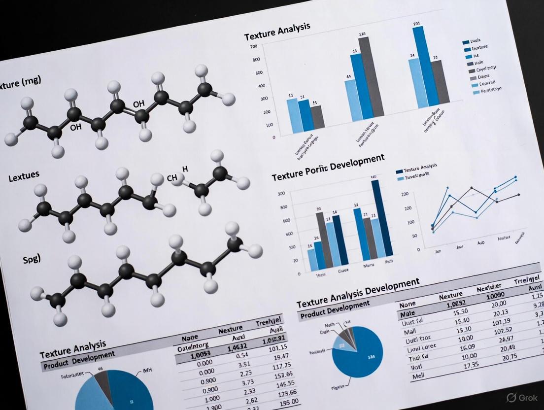
Beyond Hardness: How Texture Analysis is Revolutionizing Product Development in Pharmaceuticals and Biomedicine
This article provides a comprehensive overview of the critical role of texture analysis in modern product development, with a specific focus on pharmaceuticals and biomedical applications.
Beyond Hardness: How Texture Analysis is Revolutionizing Product Development in Pharmaceuticals and Biomedicine
Abstract
This article provides a comprehensive overview of the critical role of texture analysis in modern product development, with a specific focus on pharmaceuticals and biomedical applications. It explores the foundational principles of texture as a key quality attribute, details advanced methodological applications from tablet testing to mucoadhesion, addresses common troubleshooting and optimization challenges, and outlines robust validation and comparative frameworks. Tailored for researchers, scientists, and drug development professionals, this review synthesizes current methodologies, standards, and future-looking trends to equip practitioners with the knowledge to leverage texture analysis for developing safer, more effective, and patient-centric products.
The Texture Imperative: Defining Quality and Performance in Product Development
Texture is a critical quality attribute defined as the sensory and functional manifestation of the structural, mechanical, and surface properties of materials detected through the senses of vision, hearing, touch, and kinesthetics. In industrial product development, texture analysis transitions this subjective sensory experience into objective, quantifiable data. This quantitative approach is strategically important for ensuring product consistency, meeting stringent regulatory standards, and fulfilling consumer expectations across diverse sectors including food, pharmaceuticals, and materials science [1] [2]. The process enables researchers to correlate quantitatively measured physical properties with subjective sensory perceptions, establishing robust quality benchmarks essential for research and development (R&D) and quality control (QC) [2].
The following diagram illustrates the fundamental relationship between sensory perception and the quantitative data obtained through texture analysis.
Fundamental Measurement Techniques
The quantification of texture relies on two primary methodological approaches: mechanical testing, which measures a sample's response to applied forces, and digital image analysis, which extracts textural features from visual data.
Mechanical Texture Analysis
Mechanical texture analyzers operate by applying controlled compression or tension forces to a sample using various probe geometries. The instrument measures the sample's response, generating load versus time or load versus distance graphs [2]. These systems can perform a wide range of tests including rupture, compression, tension, penetration, and shearing, each designed to probe specific mechanical properties. The data acquisition occurs at high speeds, capturing up to 2000 data points per second to accurately record rapid fracture or adhesion events [3].
Digital Image Texture Analysis
Digital image analysis provides a non-destructive alternative for texture quantification. This approach involves computer analysis of images captured by digital cameras or smartphones to create unique 'digital texture-fingerprints' for each sample [4]. These fingerprints serve as benchmarks for monitoring production process stability and final product quality. The method is particularly valuable for analyzing complex surface structures, such as bread porosity or cell distribution in biological samples, and can be scaled for industrial deployment with minimal running costs [4] [5].
Experimental Protocols and Methodologies
Standard Mechanical Testing Protocol
The following workflow outlines the standard operating procedure for conducting mechanical texture analysis using professional instrumentation.
Step 1: Instrument Calibration and Force Verification - Calibration of both force and distance is essential to ensure measurement accuracy. This involves placing certified weights on the calibration platform to check live readings and calibrating at specific weight values as required by good laboratory practice. Position calibration allows tests to be performed in percentage strain and enables recording of sample height [3].
Step 2: Test Setup and Design - Researchers can automatically load from a library of 400+ sample projects containing pre-configured methods, macros, spreadsheet layouts, and report templates. For novel applications, tests can be built from standard arm movements (e.g., compression, penetration, TPA, tensile) using TA Settings to define speeds, distances, triggers, and other parameters. The "Testmaker" feature allows compilation of custom sequences from drag-and-drop arm movement commands for advanced multi-stage tests [3].
Step 3: Sample Preparation and Mounting - Prepare samples according to standardized dimensions and mount them securely on the testing platform. Environmental conditions (temperature, humidity) should be documented as they may affect results. For comparative studies, maintain consistent sample preparation methods across all specimens [2].
Step 4: Test Execution and Data Collection - Using the "Run a Test" window, define file names, batch IDs, operator names, probe attachments, and sample dimensions. Initiate the test to collect high-speed data (up to 2000 points/second) for fracture events or adhesive testing. Simultaneously capture multi-channel inputs from additional measurement devices such as temperature sensors or acoustic emission detectors when required [3].
Step 5: Data Analysis and Interpretation - Utilize software tools for rapid extraction of common parameters including peak force, area under the curve, gradient, and time differences. Implement macros to automate repetitive analysis steps and standardize data processing across instruments and operators. Apply advanced analysis features such as smoothing and inflection point detection for more complex research applications [3].
Step 6: Reporting and Documentation - Generate professional reports combining graphs, tables, images, logos, test settings, and batch information. Use customizable report templates to establish strict formats for presenting information to stakeholders and auditors. Export data to Laboratory Information Management Systems (LIMS) for integration with broader quality systems [3].
Digital Image Analysis Protocol
Image Acquisition - Capture high-resolution images of samples under consistent lighting conditions using digital cameras or smartphones. For 3D surface texture modeling, acquire z-stack images using super-resolution microscopy (e.g., Airyscan) for fine-scale description of shape and texture [5].
Image Processing and Surface Extraction - Convert acquired images into 3D models of the surface. For bread texture analysis, the Bread Texture Analyser application processes images to generate parameters unique to each texture and applies color mapping techniques to visualize connected regions by area size [4].
Texture Quantification - Apply mathematical functions to quantify textural properties:
- Variance: Measures how far each texture value deviates from the average, assessing heterogeneity changes [6].
- Entropy: Measures disorder or complexity of texture, reaching maximum for random textures and minimum for regular textures [6].
- Energy: Measures distribution of energy along the frequency axis over scale and orientation [6].
Spectral Decomposition Analysis - Use spectral decomposition analysis to quantify differences in surface geometry by comparing surface textures across different samples or genotypes [5].
Quantitative Data Analysis and Interpretation
Texture analysis generates diverse quantitative parameters that define material properties. The table below summarizes key measurements and their significance across different applications.
Table 1: Key Quantitative Parameters in Texture Analysis
| Parameter | Definition | Significance | Application Examples |
|---|---|---|---|
| Peak Load (Hardness) | Maximum force required to achieve specified deformation | Measures sample hardness; indicates resistance to deformation | Force to split gelatin capsules (12,813-16,585g) [2] |
| Deformation at Peak Load | Distance sample deforms before fracture/peak load | Indicates elasticity or brittleness | Gelatin capsule elasticity (4.06-5.13mm) [2] |
| Work Done | Area under force-distance curve to target point | Energy required to overcome internal bonds | Gelatin capsule bond strength (114.8-281.8mJ) [2] |
| Variance | Measure of how texture values deviate from average | Quantifies heterogeneity of structure | WSI heterogeneity assessment [6] |
| Entropy | Measure of disorder or complexity in texture | Higher values indicate more random textures | Tissue or bread structure complexity [6] |
| Energy | Distribution of energy along frequency axis | Reflects organization of structural elements | Cellular organization analysis [6] |
Statistical analysis is integral to interpreting texture data. Modern texture analysis software performs statistical calculations for multiple samples (typically up to 25 samples), providing mean values and standard deviations for critical parameters. This enables researchers to determine significant differences between product formulations and establish acceptable quality ranges [2].
The Scientist's Toolkit: Essential Research Reagents and Materials
Successful texture analysis requires specialized equipment, software, and consumables. The following table details key components of a texture analysis laboratory.
Table 2: Essential Research Reagents and Solutions for Texture Analysis
| Item | Function | Application Context |
|---|---|---|
| Texture Analyzer Instrument | Measures response to compression, tension, shearing, penetration | Universal testing system for solid and semi-solid samples [2] |
| Exponent Connect Software | Instrument control, data acquisition, analysis, and reporting | Full-featured analysis for R&D and regulated environments [3] |
| Specialized Probes | Various geometries to apply specific force types | Compression plates, penetration probes, tensile grips [2] |
| Calibration Weights | Verify and calibrate force measurement accuracy | Regular instrument calibration per GLP requirements [3] |
| Environmental Modules | Measure temperature, humidity, acoustic emission during testing | Multi-channel data acquisition for comprehensive analysis [3] |
| Sample Preparation Tools | Standardize sample dimensions and mounting | Cutting jigs, mounting fixtures for consistent preparation |
Applications in Product Development Research
Texture analysis provides critical data throughout the product development lifecycle, from initial formulation to final quality control.
Pharmaceutical Development
In pharmaceutical research, texture analysis ensures drug products meet patient expectations for handling and administration. Metered dose inhalers (MDIs) require precise compression characteristics to deliver accurate and reproducible doses. Suppositories must balance smoothness with structural integrity—being hard enough to avoid breakage during insertion yet not causing discomfort. Tablet coating integrity, mucoadhesion properties, and gelatin capsule stability are all critically evaluated using texture analysis [2].
Compatibility between drug substances and delivery components represents another crucial application. Hygroscopic fillings can absorb water from gelatin capsule shells, making them brittle and prone to breakage under mechanical strain. Texture analyzers help manufacturers identify these effects and optimize formulations accordingly [2].
Food Product Development
In food science, texture analysis drives product innovation and quality assurance, particularly as consumer preferences shift toward healthier and functional foods. Digital image analysis creates "digital texture-fingerprints" that serve as benchmarks for monitoring baking process stability and final product quality. These fingerprints efficiently inspect bread texture, evaluating porosity, density, and cell distribution with minimal effort and running costs [4].
Biomaterials and Biological Research
Texture quantification extends to biological applications where surface geometry and mechanical properties influence function. Protocols for 3D surface texture modeling and quantitative spectral decomposition analysis enable researchers to compare surface textures across genotypes, determining how genes of interest impact cellular cluster surface geometry [5].
Texture analysis represents a vital bridge between subjective sensory perception and objective quantitative data, serving as a cornerstone of modern product development research. By implementing standardized mechanical testing protocols and emerging digital image analysis techniques, researchers can generate reproducible, statistically validated texture parameters that correlate with sensory experiences. As technology advances—with integration of IoT, automation, and increasingly sophisticated analysis software—texture analysis continues to expand its applications across pharmaceutical, food, and materials science sectors. This evolution empowers researchers to develop products that consistently meet quality expectations, regulatory requirements, and consumer preferences in an increasingly competitive marketplace.
In the realm of product development, particularly for pharmaceuticals and functional foods, texture has emerged as a critical physicochemical parameter that directly influences product performance, efficacy, and user experience. Texture analysis transcends mere sensory appeal, serving as a fundamental property that governs drug release kinetics, bioavailability, and patient compliance. For researchers and drug development professionals, understanding the intricate relationship between physical properties and therapeutic outcomes is no longer optional but essential for innovating next-generation products.
The textural features of a delivery matrix—including surface area, pore volume, pore size distribution, and structural integrity—directly impact its drug adsorption capacity and release profile [7]. Beyond the molecular structure of active pharmaceutical ingredients (APIs), the physical architecture of delivery systems determines how APIs interact with biological environments, navigate physiological barriers, and ultimately reach their target sites. This whitepaper examines the fundamental principles connecting material texture to product functionality, providing technical guidance for leveraging texture analysis in advanced product development.
Texture-Property Relationships in Drug Delivery Systems
The Pore Size-Release Kinetics Correlation
Nanoporous materials represent a paradigm shift in controlled drug delivery, where textural properties can be precisely engineered to modulate release profiles. Research on nanoporous TiO₂ matrices demonstrates a clear correlation between pore dimension and drug release kinetics [7]. When these matrices were loaded with ibuprofen as a model drug, researchers observed that materials with tailored, unimodal pore distributions provided more predictable and sustained release patterns compared to those with heterogeneous pore structures.
Table 1: Correlation Between Pore Characteristics and Drug Release Performance in Nanoporous TiO₂ Matrices
| Textural Property | Impact on Drug Delivery Performance | Experimental Findings |
|---|---|---|
| Pore Size Dimension | Determines diffusion rate of drug molecules; optimal size prevents burst release | Pores sized 2-3× larger than drug molecule showed ideal sustained release [7] |
| Pore Size Distribution | Affects predictability and consistency of release kinetics | Unimodal, narrow distribution demonstrated more controlled release vs. broad distribution [7] |
| Surface Area | Influences drug loading capacity; higher surface area enables greater adsorption | Matrices with surface area >100 m²/g showed significantly improved loading efficiency [7] |
| Synthetic Control | Allows precision engineering of textural properties for specific drug molecules | Sol-gel approach enabled pore size tuning for different therapeutic molecules [7] |
Advanced Material Systems for Controlled Release
Beyond traditional matrices, advanced material systems including nanocarriers, hydrogels, and bioresponsive polymers have demonstrated how texture manipulation can enhance therapeutic precision. These systems leverage their structural properties to achieve targeted release profiles:
- Nanocarriers (liposomes, dendrimers, nanoparticles): Their small size and structural versatility enable remarkable targeting accuracy, minimizing side effects while improving therapeutic index [8].
- Hydrogels: These three-dimensional, hydrophilic polymeric networks provide platforms for minimally invasive applications while enabling sustained drug release at target sites over time [8].
- Bioresponsive polymers: These materials respond to environmental stimuli (pH, temperature, enzymatic activity) to trigger site-specific drug release, enhancing both safety and efficacy [8].
Table 2: Texture-Property Relationships in Advanced Drug Delivery Systems
| Delivery System | Key Textural Properties | Impact on Product Efficacy |
|---|---|---|
| Nanoporous Inorganic Matrices | Pore size, pore volume, surface area, surface chemistry | Sustained release, reduced initial burst, improved loading capacity [7] |
| Polymeric Nanoparticles | Particle size, degradation rate, surface functionalization | Enhanced permeability and retention, targeted delivery, reduced toxicity [8] |
| Hydrogels | Mesh size, crosslink density, swelling ratio, mechanical strength | Controlled release kinetics, tissue-like properties, injectable formulations [8] |
| Nanogels/Microgels | Stimuli-responsive swelling, deformability, water content | Adaptable drug release, improved biological barrier penetration [8] |
Experimental Protocols for Texture Analysis
Standardized Texture Analysis Method for Biological Materials
A validated texture analysis method following the American Society of Agricultural and Biological Engineers (ASABE S368.4) standard provides a robust framework for evaluating convex-shaped materials, including certain drug delivery systems and medical foods [9]. This method offers reproducible techniques for quantifying mechanical properties that correlate with product performance.
Sample Preparation Protocol
- Sample Acquisition: Obtain test materials from reliable sources, ensuring batch-to-batch consistency. Record lot numbers and storage conditions [9].
- Treatment Groups: Implement multiple processing treatments to simulate different conditions:
- Blanch/Freeze/Thaw (BFT): Basic preservation method serving as control
- BFT + Microwave (BFT+M): Represents rapid heating method
- BF + Stove-top Cooking (BF+C): Simulates conventional heating [9]
- Storage Conditions: Store samples at -15°C until analysis to maintain integrity. Thaw refrigerated (2°C) overnight prior to testing [9].
Compression and Puncture Analysis
The ASABE method outlines two complementary approaches:
- Compression Analysis: Utilizes a flat probe with sufficient surface area to fully cover the product. The probe compresses the sample against a flat plate while measuring force required to reach rupture point. This simulates molar compression during consumption [9].
- Puncture Analysis: Employs a probe smaller than the test product that pushes through the surface into the center, simulating incisor biting. Measures force required to break through the outer surface and can continue through the entire sample [9].
Texture Modification and Analysis Workflow
The following diagram illustrates the comprehensive workflow for texture analysis and modification in product development:
Texture Analysis and Modification Workflow
The Scientist's Toolkit: Essential Research Reagents and Materials
Table 3: Essential Research Reagents and Materials for Texture Analysis
| Research Material | Function/Application | Technical Specifications |
|---|---|---|
| Nanoporous TiO₂ Matrices | Drug carrier for sustained release studies | High surface area (>100 m²/g), tunable pore size (2-50 nm), sol-gel derived [7] |
| Texture Analyzer | Quantifies mechanical properties of materials | Equipped with compression plates, puncture probes, and force transducers (0.1N sensitivity) [9] |
| Hydrocolloids | Texture modifiers for controlled release systems | Includes cellulose derivatives, starches, gums; provide gelling, thickening, stabilizing properties [10] [11] |
| Polymeric Excipients | Matrix formers for controlled release dosage forms | PLGA, chitosan, HPMC; varying molecular weights, viscosity grades, and degradation rates [8] |
| Bioresponsive Polymers | Stimuli-responsive drug delivery systems | pH-sensitive (PAA), temperature-sensitive (PNIPAM), enzyme-degradable polymers [8] |
The strategic manipulation of textural properties presents unprecedented opportunities for enhancing product efficacy and patient experience in pharmaceutical development. By correlating physical parameters with performance outcomes, researchers can engineer advanced delivery systems with precision-release profiles, improved targeting capabilities, and enhanced patient compliance. The methodologies and relationships outlined in this technical guide provide a foundation for evidence-based texture design in product development pipelines.
As the field advances, the integration of artificial intelligence and machine learning approaches will further revolutionize texture optimization, enabling predictive modeling of material behavior and accelerated development of tailored therapeutic solutions [8]. Through continued refinement of texture analysis protocols and their systematic application across development cycles, researchers can harness the full potential of physical property optimization to drive innovation in drug delivery and patient-centered care.
In pharmaceutical development, the critical quality attributes (CQAs) of a dosage form are integral to its safety, efficacy, and patient compliance. Among these CQAs, textural properties—specifically hardness, friability, mucoadhesion, and disintegration—play a fundamental role in determining product performance [12] [13]. The measurement and control of these properties are essential throughout formulation design, process development, and quality control. Texture analysis has emerged as a versatile, scientifically rigorous methodology that provides quantitative data on these mechanical characteristics, enabling researchers to make informed decisions [14] [13]. This guide details the core principles, measurement techniques, and implications of these key textural properties, framing them within the essential context of modern pharmaceutical product development research.
Hardness
Definition and Role in Product Performance
Tablet hardness, often referred to as crushing strength or resistance to crushing, is defined as the force required to break a tablet when applied diametrically [15]. It is a paramount mechanical property that ensures a tablet's structural integrity during subsequent processing steps such as coating, packaging, and transportation [12] [15]. More critically, hardness directly influences the drug's bioavailability; a tablet that is too hard may disintegrate and dissolve too slowly, reducing the therapeutic dose available for absorption, while a tablet that is too soft may crumble, leading to inaccurate dosing [15].
Measurement Methodologies
Hardness is typically measured using a tablet hardness tester, which places the tablet under a bending or tensile load until it fractures [15]. Both manual and electronic testers are used, with electronic testers providing superior precision, repeatability, and data export capabilities [15]. The test involves positioning the tablet between anvils and gradually applying force until the tablet breaks, with the breaking force recorded as the hardness [15]. The units of measurement are kilograms (kg), Newtons (N), or kiloponds (KP) [15].
Table 1: Key Factors Affecting Tablet Hardness and Their Impact
| Factor Category | Specific Factor | Impact on Hardness |
|---|---|---|
| Formulation | Binder type and concentration | Increased binder concentration typically enhances hardness by improving particle-particle bonding [15]. |
| Nature and amount of API | Some APIs can influence the compression operation, leading to hardness variations [15]. | |
| Manufacturing Process | Compression force | Increased compression force generally results in harder tablets, though excessive force can cause capping or lamination [15]. |
| Granulation method (wet vs. dry) | Wet granulation typically produces harder tablets compared to dry granulation [15]. | |
| Moisture content in granules | A low moisture content (e.g., 2-4%) can act as a binder and improve hardness, while very dry granules often produce friable tablets [16]. | |
| Post-Processing | Coating (e.g., film-coating) | The application of a coating can generally contribute to an increase in overall tablet hardness [15]. |
Standard Experimental Protocol for Hardness Testing
- Instrument Calibration: Verify the calibration of the hardness tester according to the manufacturer's instructions and standard operating procedures.
- Tablet Sampling: Select a statistically appropriate number of tablets (e.g., 10 units) from a representative batch.
- Positioning: Place a single tablet standing on its rim between the two anvils of the testing apparatus.
- Force Application: Initiate the test to apply a gradually increasing force to the tablet until it fractures.
- Data Recording: Record the force (in N or KP) at which the tablet breaks.
- Analysis: Calculate the mean hardness and standard deviation for the sampled tablets. Compare the results against pre-established acceptance criteria specific to the formulation [15].
Friability
Definition and Role in Product Performance
Friability is a measure of a tablet's tendency to lose weight (chip, cap, or break) due to mechanical shock, friction, and abrasion during handling, packaging, and shipping [16]. It is a critical indicator of a tablet's physical durability and mechanical strength. Testing for friability helps ensure that tablets remain intact from the point of manufacture until they are consumed by the patient, thereby guaranteeing dose accuracy and product aesthetics [16].
Measurement Methodologies
Friability is tested using a friabilator, a device consisting of a transparent, rotating drum [16]. A pre-weighed sample of tablets is placed in the drum, which rotates at a controlled speed (typically 25 rpm), causing the tablets to repeatedly fall from a fixed height (usually 6 inches) with each revolution [16]. After a specified number of rotations (commonly 100 revolutions, or 4 minutes), the tablets are dedusted and re-weighed [16]. The percentage weight loss is calculated using the formula:
[ \text{% Friability} = \frac{(W1 - W2)}{W_1} \times 100 ]
Where ( W1 ) is the initial weight and ( W2 ) is the final weight of the tablets. The acceptance criterion is generally a loss of less than 0.5% to 1.0% [16]. If any tablet shows cracking, splitting, or breaking, the sample fails the test.
Table 2: Factors Influencing Friability Test Results
| Factor | Influence on Friability |
|---|---|
| Tablet Hardness | There is a direct correlation; tablets with higher hardness generally exhibit lower friability [15] [16]. |
| Tablet Shape | Tablets with rounded edges (e.g., convex) tend to be less friable than those with flat or sharp edges. |
| Surface Characteristics | A rough or pitted tablet surface is more prone to erosion and abrasion than a smooth one. |
| Moisture Content | An optimal, low level of moisture (2-4%) can act as a binder and reduce friability. Very dry granulations often produce more friable tablets [16]. |
| Formulation Composition | The type and proportion of binders, fillers, and lubricants significantly impact the tablet's cohesion and resistance to abrasion [16]. |
Standard Experimental Protocol for Friability Testing
- Sample Preparation: Dedust and accurately weigh a sample of approximately 6.5 g of tablets (or 10 whole tablets if the unit weight is greater than 0.65 g).
- Drum Setup: Place the tablets in the friabilator drum, ensuring it is clean and dry.
- Rotation: Set the instrument to 100 revolutions and start the test.
- Tablet Removal and Dedusting: After the test, remove the tablets and carefully remove any loose dust.
- Final Weighing: Accurately re-weigh the tablets.
- Calculation and Interpretation: Calculate the percentage weight loss. The test is typically repeated twice if the results are doubtful, and the mean of the three tests should not exceed 1.0% [16].
Mucoadhesion
Definition and Role in Product Performance
Mucoadhesion, or bioadhesion, is the ability of a dosage form or material to adhere to a mucosal surface (e.g., buccal, nasal, vaginal) for an extended period [13]. This property is a critical quality attribute for mucoadhesive drug delivery systems as it can localize the drug at the site of action, enhance drug absorption by increasing contact time, and improve patient compliance by reducing dosing frequency [12] [13]. The adhesion process involves an initial contact and wetting stage, followed by the interpenetration of polymer chains with the mucus network, and the formation of secondary chemical bonds [13].
Measurement Methodologies
Texture analyzers are widely used for the in vitro assessment of mucoadhesive strength, providing standardized and reproducible data [17] [13]. Common tests include:
- Tensile Strength Measurement: The force required to detach a dosage form (e.g., a tablet or film) from a mucosal substrate (e.g., porcine mucosa or a synthetic membrane) is measured [12] [13]. Specialized attachments like Miniature Tensile Grips or an Adhesive Indexing System are used [12].
- Shear Strength Measurement: The resistance of the adhesive bond to a lateral force is evaluated.
- Tack/Adhesiveness Measurement: The "stickiness" or initial attachment force is often measured using a spherical probe that contacts the sample and then retracts, with the negative force area in the force-time curve indicating adhesiveness [12] [17].
Table 3: Key Parameters and Methods for Mucoadhesion Testing
| Test Method | Measured Parameter | Typical Dosage Form Applications |
|---|---|---|
| Tensile Test | Peak detachment force (N), Work of adhesion (N.mm) | Buccal tablets, oral thin films, patches [12] [13]. |
| Shear Test | Time for detachment under a constant shear load | Mucoadhesive gels and patches. |
| Probe Tack Test | Adhesiveness (N.mm), Maximum adhesive force (N) | Semisolid gels, transdermal patches [12] [17]. |
Standard Experimental Protocol for Tensile Mucoadhesion Testing
- Substrate Preparation: Secure a piece of freshly excised porcine mucosa or a hydrated synthetic membrane to a stationary base of the texture analyzer using a tissue holder.
- Sample Mounting: Attach the mucoadhesive dosage form (e.g., tablet) to the upper probe or cylinder of the texture analyzer using a suitable adhesive.
- Contact: Lower the probe at a defined speed until a pre-defined contact force (e.g., 0.5 N) is achieved between the sample and the substrate. Maintain this contact for a specified dwell time (e.g., 30-300 seconds) to allow for bond formation.
- Detachment: Retract the probe upward at a constant speed until the sample is completely detached from the substrate.
- Data Analysis: The force-distance curve is analyzed to determine the peak detachment force (N) and the work of adhesion (N.mm), which is the area under the force-distance curve [13].
Disintegration
Definition and Role in Product Performance
Disintegration is the process by which a solid dosage form breaks down into smaller particles or granules upon contact with a liquid medium [18]. For immediate-release oral dosage forms, this is a crucial prerequisite for the subsequent dissolution and absorption of the active pharmaceutical ingredient (API) [18]. The disintegration time is thus a critical performance indicator, as a delay can directly impact the drug's onset of action and bioavailability [18]. While traditional pharmacopoeial tests provide a pass/fail outcome, advanced texture analysis methods offer a more mechanistic understanding and quantitative profiling of the disintegration process [18] [13].
Measurement Methodologies
- Pharmacopoeial (Compendial) Test: The standard test involves placing a tablet in a tube with a mesh bottom, which is then moved up and down in a vessel containing disintegration medium (e.g., simulated gastric fluid) at 37°C. The time for the complete disintegration of the tablet is recorded [18].
- Texture Analysis Method: A probe attached to a texture analyzer is immersed into a tablet that is submerged in a disintegration medium. The force required for penetration is measured over time. The disintegration profile (force vs. time) allows for the detection of the onset and endpoint of disintegration with high sensitivity, making it particularly useful for discriminating between fast-disintegrating formulations like orodispersible tablets [13].
Standard Experimental Protocol for Texture Analysis-Based Disintegration
- Apparatus Setup: Fit the texture analyzer with a cylindrical probe (e.g., 5 mm diameter). The Tablet Disintegration Rig is a specialized accessory for this purpose [12].
- Media and Sample Preparation: Fill a vessel with the desired disintegration medium (e.g., purified water or phosphate buffer pH 6.8) and maintain it at 37 ± 1°C. Place a single tablet in the vessel, ensuring it is fully submerged.
- Test Initiation: Start the test, which typically involves the probe moving to apply a small, constant force to the tablet surface at periodic intervals (e.g., every 5-10 seconds).
- Data Collection: The force required for the probe to penetrate the tablet is recorded as a function of time. As the tablet hydrates and disintegrates, the resistance to penetration decreases.
- Endpoint Determination: The disintegration time is identified as the point at which the resistance force drops to a near-zero or a pre-defined threshold value, indicating the loss of tablet structure [12] [13].
The Researcher's Toolkit: Experimental Workflows & Essential Materials
Understanding the logical flow of how these properties are evaluated and controlled is key to successful formulation development. The following diagrams and table summarize the core experimental workflows and reagents.
Diagram 1: Hardness & Friability Testing Workflow
Diagram 2: Mucoadhesion & Disintegration Testing Workflow
Table 4: Essential Research Reagents and Materials for Texture Analysis
| Item / Solution | Function in Experimentation |
|---|---|
| Texture Analyzer | The core instrument that applies a controlled force to a sample and measures its response, providing quantitative data on mechanical properties [12] [13]. |
| Specialized Rigs & Probes | Adapt the instrument for specific tests (e.g., Tablet Disintegration Rig, Miniature Tensile Grips, Cylinder Probes for hardness) [12]. |
| Mucosal Substrate | Biological (e.g., porcine buccal/gastric mucosa) or synthetic membranes used as a model surface for in vitro mucoadhesion testing [13]. |
| Disintegration Media | Buffered solutions (e.g., simulated gastric or intestinal fluid, pH 6.8 phosphate buffer) that mimic physiological conditions for disintegration and dissolution testing [18]. |
| Standard Disintegrants | Excipients like croscarmellose sodium (CCS), crospovidone (XPVP), and sodium starch glycolate (SSG) that are added to formulations to promote tablet breakup [18]. |
| Polymer/Binder Solutions | Solutions of polymers like HPMC, PVP, or various carbomers used as binders to increase hardness or as mucoadhesive agents to enhance adhesion [15] [13]. |
The rigorous characterization of hardness, friability, mucoadhesion, and disintegration is non-negotiable in the development of robust, effective, and patient-centric pharmaceutical products. Texture analysis provides the scientific backbone for obtaining quantitative, actionable data on these critical quality attributes. As the pharmaceutical landscape evolves with novel dosage forms and personalized medicines, the role of texture profiling will only expand, bridging the gap between formulation design, predictive performance, and successful clinical outcomes. By integrating these fundamental physical tests into the development workflow, scientists can ensure product stability, efficacy, and safety from the laboratory to the patient.
In the competitive landscapes of industries such as pharmaceuticals, food, and cosmetics, the imperative for superior quality control and product consistency has never been greater. This demand is a primary catalyst for the adoption of advanced analytical technologies, including texture analysis. For researchers and drug development professionals, objective, quantifiable data on material properties is indispensable for innovating while ensuring safety, efficacy, and compliance. Texture analyzers have thus transitioned from supportive tools to central components of the research and development (R&D) infrastructure, enabling precise measurement of physical characteristics like firmness, viscosity, elasticity, and spreadability [19]. This technical guide examines the core market drivers behind this growth and provides detailed methodologies for integrating texture analysis into robust product development protocols.
The texture analyzer market is experiencing significant expansion, driven by cross-industry demands for objectivity in quality control. The following table summarizes key quantitative market data and its direct link to quality and consistency demands.
Table 1: Texture Analyzer Market Overview and Key Drivers
| Metric | Market Data | Relation to Quality & Consistency Demand |
|---|---|---|
| U.S. Market Value (2025) | $11.04 Billion [20] | Reflects high investment in quality control infrastructure. |
| Projected U.S. Market Value (2033) | $21.29 Billion [20] | Indicates sustained and growing demand for quality assurance. |
| Compound Annual Growth Rate (CAGR) | 11.57% (2026-2033) [20] | Signals rapid adoption of objective testing technologies. |
| Global Market Estimate (2025) | $500 Million [21] | Highlights worldwide focus on quality standards. |
| Global Market CAGR | 7% (2025-2033) [21] | Underscores a persistent, global growth trend. |
| Dominant Application Segment | Food & Beverage (e.g., Fruit & Vegetable testing ~30% of market) [21] | Driven by consumer demand for consistent sensory experiences. |
| Other Key Application Segments | Meat (~25%), Flour Products (~15%), Other (~30% incl. Pharma & Cosmetics) [21] | Shows diverse need for texture control across industries. |
This growth is propelled by several interconnected factors:
- Stringent Regulatory Requirements: Regulatory bodies worldwide mandate rigorous quality control. In pharmaceuticals, adherence to FDA and EMA standards is non-negotiable for product approval and market release. Texture analysis provides the quantitative data necessary for regulatory compliance, from testing tablet hardness to ensuring the consistency of gels and ointments [20] [19].
- Consumer Expectations and Market Competition: Consumers expect products to perform identically every time. Inconsistencies in texture—such as a crumbly gluten-free cake or a too-thick ketchup—erode brand loyalty and consumer trust [22]. Objective measurement allows companies to establish a "gold standard" for each product and ensure every batch meets these specifications [22].
- The Shift to Proactive Quality Management: Industries are moving from reactive to proactive quality assurance. Trends like Real-Time Release Testing (RTRT) in pharmaceuticals leverage in-process monitoring to release batches based on data collected during manufacturing, significantly reducing cycle times [23]. Texture analysis integrated into these processes provides continuous, actionable data.
Texture Analysis in Pharmaceutical Research: Key Applications and Protocols
In pharmaceutical research, texture analysis is critical for optimizing drug formulations and ensuring patient comfort and safety. The following table outlines essential reagent and material solutions for these applications.
Table 2: Key Research Reagent Solutions in Pharmaceutical Texture Analysis
| Research Reagent / Material | Function in Texture Analysis |
|---|---|
| Hydrogel-Based Formulations | Serves as a model for testing swelling properties, crucial for drug delivery mechanisms and advanced wound care products [24]. |
| Coating Solutions for Tablets | Used to assess the durability and performance of functional or protective coatings on pills, impacting dissolution and stability [24]. |
| Adhesive Formulations | Essential for evaluating the adhesive strength of medical tapes, transdermal patches, and drug-eluting devices [24]. |
| Powder Blends for Tableting | The physical consistency of powder is a precursor to measuring the hardness and friability of finished tablets [24]. |
| Gel & Ointment Bases | Formulations are tested for consistency and viscosity to ensure correct application, stability, and drug release profile [24] [19]. |
Detailed Experimental Protocol: Tablet Hardness and Friability Testing
Objective: To quantitatively determine the mechanical strength (hardness) of a tablet and its resistance to abrasion (friability) to ensure it can withstand packaging and shipping while disintegrating appropriately in the body.
Materials and Equipment:
- Texture Analyzer (e.g., from Stable Micro Systems, SHIMADZU, AMETEK) equipped with a tablet holder or a compression platen [21] [24].
- Friability tester (e.g., a rotating drum with a defined internal curvature).
- Pre-manufactured tablets from the batch under test.
- Analytical balance.
Methodology:
- Sample Preparation: For hardness testing, select at least 10 tablets from the batch at random. For friability, deduct the total mass of a group of tablets (e.g., 20 tablets) to 0.1% of their original weight or 6.5 g, whichever is less.
- Hardness Testing:
- Calibrate the texture analyzer according to the manufacturer's instructions.
- Place a single tablet on the platform centered under the probe.
- Program the instrument to perform a compression test. The probe will descend at a defined speed (e.g., 0.5 mm/s) until the tablet fractures.
- The force (in Newtons or Kiloponds) required to cause fracture is recorded as the tablet hardness.
- Repeat for all 10 tablets and calculate the average and standard deviation [24].
- Friability Testing:
- Remove any dust from the selected tablets and record the precise combined weight (W₁).
- Place the tablets into the friability drum, which is then rotated at a standard speed (e.g., 25 rpm) for a set number of rotations (e.g., 100 rotations).
- Remove the tablets, carefully dedust them, and weigh them again (W₂).
- Calculate the percentage friability using the formula: % Friability = [(W₁ - W₂) / W₁] × 100. A maximum loss of 1.0% is typically considered acceptable for most products [24].
Data Interpretation: Hardness data ensures tablets are robust without being impervious to dissolution. Friability data directly indicates the tablet's susceptibility to chipping and breaking, which can affect dosage accuracy. This quantitative profile is vital for substantiating claims in regulatory submissions [22] [24].
Advanced and Emerging Methodologies
Beyond traditional mechanical testing, novel approaches are enhancing texture analysis.
Digital Image Texture Analysis
This non-destructive method uses computer analysis of images to assess texture. A recent study developed a Bread Texture Analyser application that creates 'digital texture-fingerprints' from images of bread crumb structure, quantifying parameters like porosity, density, and cell distribution [4]. This methodology was validated against sensory evaluation and can be scaled for industrial production monitoring. Its application in pharmaceuticals could be explored for analyzing the surface morphology of tablets or scaffolds.
Integration with Intelligent Systems
The future of texture analysis lies in its integration with broader quality management ecosystems. Cloud-based Quality Management Systems (QMS) and Laboratory Information Management Systems (LIMS) allow for seamless transfer of texture data, enabling real-time analytics and triggering automated investigations when results are out of specification [23] [25]. Furthermore, the integration of AI and machine learning enables predictive quality analytics, where historical texture data is used to forecast potential quality deviations before they occur [26] [25].
The workflow below illustrates how texture analysis is integrated into a modern, data-driven quality control system.
The demand for rigorous quality control and product consistency is a powerful, enduring market driver firmly establishing texture analysis as a cornerstone of modern product development research. The transition from subjective assessment to data-driven, objective quantification empowers researchers and pharmaceutical professionals to innovate with confidence, ensure patient safety, and navigate complex regulatory landscapes. As technologies like AI, cloud computing, and digital image analysis continue to converge with physical testing, the role of texture analysis will only deepen, enabling more intelligent, efficient, and predictive approaches to achieving perfect product consistency.
In the highly regulated pharmaceutical industry, texture analysis has emerged as an indispensable tool for ensuring product quality, safety, and efficacy. Texture, defined as the sensory and functional manifestation of the structural, mechanical, and surface properties of samples, represents a critical quality attribute (CQA) for an extensive range of pharmaceutical products [2] [27]. The measurement of both texture and mechanical properties in pharmaceutical products is driven by the imperative of product stability, consumer acceptance, and patient safety [12]. For drug development professionals, mastering the regulatory landscape surrounding texture characterization is essential for successful product development and approval.
Global regulatory agencies including the FDA and EMA require comprehensive characterization of physical properties, especially when these attributes directly influence drug performance, patient compliance, and clinical outcomes [28] [27]. This technical guide examines the intersection of texture analysis and regulatory compliance, providing scientists with methodologies and frameworks for implementing texture testing throughout the pharmaceutical development lifecycle. By establishing scientifically robust and standardized testing protocols, manufacturers can demonstrate consistent product quality while accelerating the pathway to regulatory approval.
Texture Analysis in Pharmaceutical Development
Fundamental Principles of Texture Analysis
A Texture Analyser is a mechanical instrument that quantifies physical properties by applying controlled forces to samples and measuring their responses. The system operates through a travelling arm fitted with a load cell that moves in either compression or tension modes to deform samples [29]. This instrument records force-time-distance data, which is displayed as a curve on a graph that, when analyzed, objectively quantifies textural properties [29]. Modern texture analyzers can perform diverse test types including compression, extension, cutting, extrusion, bending, and shearing, measuring properties such as fracturability, chewiness, stickiness, consistency, and springiness [29].
The versatility of texture analysis systems is enhanced through specialized probes and attachments that can be customized for specific pharmaceutical applications. These components are precision-engineered from food-grade stainless steel, aerospace-grade aluminium, or high-performance engineering plastics to ensure accuracy and reproducibility [30]. This adaptability makes texture analysis applicable across diverse pharmaceutical forms, from solid oral dosage forms to semisolid preparations and transdermal delivery systems.
Regulatory Framework and Quality Standards
Pharmaceutical manufacturers must operate within a stringent regulatory framework that mandates comprehensive product characterization. While specific pharmacopoeial standards for texture analysis methodology are still evolving, regulatory agencies require demonstration of product consistency and manufacturing control [28] [31].
Regulatory considerations for texture analysis include:
- ICH Guidelines: ICH Q6A outlines specifications for drug substances and products, requiring measurement of critical attributes using validated methods [28].
- FDA Expectations: The U.S. Food and Drug Administration expects sponsors to justify target ranges for critical physical properties and demonstrate robust control strategies [28] [27].
- Documentation Requirements: Regulatory submissions must include comprehensive texture data, method validation reports, batch analysis, and correlations to product performance [28].
Table 1: Key Regulatory Guidelines Impacting Texture Analysis
| Regulatory Body/Guideline | Key Requirements | Relevance to Texture Analysis |
|---|---|---|
| ICH Q6A | Specifications for drug substances and products | Mandates measurement of critical quality attributes |
| FDA Guidance | Control strategies for product consistency | Requires physical property characterization and control |
| GMP Standards | Current Good Manufacturing Practice | Demands validated analytical methods and documentation |
| EMA Guidelines | Quality of transdermal and topical products | Specific requirements for adhesion testing [27] |
Key Texture Parameters and Their Regulatory Significance
Critical Quality Attributes by Dosage Form
Different pharmaceutical dosage forms possess distinct critical texture attributes that directly impact their safety, efficacy, and patient acceptability. Regulatory compliance requires manufacturers to identify and control these parameters throughout the product lifecycle.
Table 2: Critical Texture Attributes by Pharmaceutical Dosage Form
| Dosage Form | Key Texture Parameters | Regulatory Significance | Testing Methods |
|---|---|---|---|
| Tablets | Hardness, friability, tensile strength, disintegration time | Ensures mechanical strength for handling yet appropriate disintegration for bioavailability [12] [31] | Diametral compression, indentation, disintegration tests [12] |
| Capsules | Rupture force, seal strength, tensile properties | Prevents premature release and ensures content integrity [12] [2] | Capsule tensile rig, puncture tests [12] |
| Transdermal Patches | Adhesive strength, tack, peel strength | Ensures proper adhesion during wear without skin damage [12] [27] | Peel tests, tack tests, shear adhesion [12] [27] |
| Semisolid Formulations | Hardness, adhesiveness, cohesiveness, spreadability | Impacts drug release, application experience, and dosing accuracy [12] [27] | Texture Profile Analysis (TPA), back extrusion, spreadability rigs [12] |
| Suppositories | Hardness, melting behavior, rupture strength | Affects patient comfort, insertion ease, and drug release [2] | Compression, penetration tests [2] |
| Metered Dose Inhalers | Actuation force, spray characteristics | Ensures consistent dosing and patient usability [2] [30] | Compression, extrusion tests [30] |
Texture Profile Analysis (TPA) in Pharmaceutical Applications
Texture Profile Analysis (TPA) represents a fundamental methodology for characterizing semisolid pharmaceutical formulations. This two-cycle compression test automatically calculates multiple texture properties that correlate with sensory perception and product performance [29] [27]. For transdermal and topical products, TPA provides critical insights into structure, spreadability, adhesion, and consistency through parameters including hardness, adhesiveness, cohesiveness, and elasticity [27]. Recent advancements in automation and multimodal analysis have significantly enhanced the precision and applicability of TPA in pharmaceutical development [27].
Experimental Protocols and Methodologies
Standardized Testing Approaches
Implementing robust, standardized testing protocols is essential for generating reproducible data that meets regulatory scrutiny. While specific methods must be adapted to each product, established methodologies provide a foundation for reliable texture analysis.
Tablet Coating Adhesion Testing
Purpose: Quantitatively measure the adhesion strength of tablet coatings to monitor batch consistency and optimize coating formulations [12].
Equipment: Texture Analyser equipped with Tablet Coating Adhesion Rig [12].
Methodology:
- Secure the coated tablet in the sample holder using double-sided tape
- Lower the spherical probe to contact the tablet surface with a trigger force of 0.05N
- Apply compression to the coating at a test speed of 0.5-1.0 mm/s
- Measure the force required to rupture the coating layer
- Calculate the adhesion strength from the peak force and contact area
Parameters Measured: Coating adhesion strength (N/mm²), failure mode (adhesive vs. cohesive) [12]
Mucoadhesive Strength Testing
Purpose: Evaluate the adhesive strength of buccal, nasal, or other mucosal drug delivery systems to ensure appropriate residence time at the application site [12].
Equipment: Texture Analyser with Adhesive Indexing System and appropriate mucosal membrane substrate [12].
Methodology:
- Secure the mucosal membrane (porcine or synthetic) to the platform
- Hydrate the membrane with artificial saliva to simulate physiological conditions
- Apply the adhesive formulation to the probe and contact the membrane with a defined force
- Maintain contact for a specified dwell time (typically 30-300 seconds)
- Withdraw the probe at a constant speed and measure the peak detachment force
Parameters Measured: Bioadhesive force (N), work of adhesion (N·mm), detachment profile [12]
Transdermal Patch Peel Adhesion Testing
Purpose: Determine the adhesive properties of transdermal delivery systems to ensure they remain in place during wear yet remove without skin damage [27].
Equipment: Texture Analyser with 180° Peel Rig [12].
Methodology:
- Apply the transdermal patch to a standardized substrate (steel plate or synthetic skin)
- Roll over the patch with a standardized weight to ensure uniform adhesion
- Clamp the free end of the patch to the peel rig
- Peel the patch at 180° angle at a constant speed of 10-300 mm/min
- Record the peel force throughout the removal process
Parameters Measured: Peel strength (N/mm), peel force consistency, adhesion failure mode [12] [27]
Experimental Workflow for Pharmaceutical Texture Analysis
The following diagram illustrates the systematic workflow for implementing texture analysis in pharmaceutical development, from method establishment to regulatory submission:
Diagram 1: Pharmaceutical Texture Analysis Workflow. This workflow outlines the systematic process from identifying Critical Quality Attributes (CQAs) through to regulatory documentation and batch release.
The Scientist's Toolkit: Essential Equipment and Reagents
Successful implementation of texture analysis requires specialized equipment and materials designed for pharmaceutical applications. The following toolkit details essential components for establishing robust texture testing capabilities.
Table 3: Essential Research Reagent Solutions for Pharmaceutical Texture Analysis
| Equipment/Reagent | Function/Application | Regulatory Relevance |
|---|---|---|
| Texture Analyser | Core instrument for applying controlled forces and measuring sample responses [29] | Provides validated, reproducible data for quality control and regulatory submissions |
| Tablet Hardness Fixtures | Measure diametral compression strength and fracture resistance of tablets [12] | Ensures tablets withstand handling while maintaining appropriate disintegration [31] |
| Powder Compaction Rigs | Assess compaction behavior and flow properties of powdered formulations [12] | Critical for ensuring content uniformity and processability in solid dosage forms |
| Mucoadhesive Testing Attachments | Evaluate bioadhesion to mucosal surfaces for buccal, nasal, and vaginal formulations [12] | Correlates with residence time and drug absorption potential |
| Peel Testing Rigs | Quantify adhesive properties of transdermal patches and wound dressings [12] [27] | Validates patient adherence and appropriate removal characteristics [27] |
| Artificial Biological Substrates | Simulate skin, mucosal, or other biological surfaces for adhesion testing [12] | Provides standardized, reproducible testing conditions mimicking in vivo performance |
| Texture Analysis Software | Control instrumentation, analyze data, and generate compliance documentation [29] [2] | Enables data integrity, audit trails, and regulatory reporting capabilities |
Advanced Applications in Novel Drug Delivery Systems
Microneedle Characterization
Texture analysis plays a pivotal role in the development of microneedle (µND) systems, an innovative transdermal platform. These devices require robust mechanical strength to ensure effective skin penetration and drug release [27]. Texture analyzers characterize critical properties including hardness, flexibility, and puncture strength, simulating forces encountered during skin penetration [27]. This analysis provides valuable insights into µND performance, ensuring safety, functionality, and patient compliance. Specific mechanical tests include fracture force measurement, insertion force analysis, and bending assessments to optimize design parameters [27].
Topical and Semisolid Formulations
For topical products including creams, ointments, and gels, texture analysis ensures consistent sensory characteristics and drug delivery performance. The spreadability and consistency of topical formulations are pivotal for therapeutic efficacy and patient compliance [12]. Texture analyzers assess these properties using specialized attachments such as spreadability rigs and back extrusion cells, ensuring uniform application and optimal drug delivery [12]. Recent advances have established correlations between instrumental measurements and sensory perception, enabling formulators to optimize patient acceptability [32] [27].
Implementation in Quality Systems
Integration with Pharmaceutical Quality by Design (QbD)
Texture analysis aligns seamlessly with the Quality by Design (QbD) framework mandated by regulatory agencies for systematic product development. Within QbD, texture parameters serve as critical quality attributes that must be monitored and controlled throughout manufacturing [31]. By establishing mathematical relationships between process parameters, material attributes, and texture outcomes, manufacturers can define a design space that ensures consistent product quality [31]. This proactive approach facilitates regulatory flexibility while reducing post-approval changes.
Lifecycle Management and Stability Testing
Texture analysis provides critical data throughout the product lifecycle, from formulation development to post-market surveillance. During stability studies, texture measurements detect changes in mechanical properties that may indicate product degradation or performance alterations [12]. Monitoring parameters such as tablet hardness, gel consistency, or adhesive strength over time provides early indicators of stability issues before chemical degradation becomes apparent [12]. This comprehensive approach supports shelf-life determination and ensures products maintain their critical quality attributes throughout their labeled shelf life.
Texture analysis represents an essential capability for pharmaceutical manufacturers operating within today's stringent regulatory environment. By implementing robust texture testing methodologies, manufacturers can demonstrate product consistency, patient-centric design, and manufacturing control – all critical elements for regulatory approval and commercial success. As novel drug delivery systems continue to evolve, texture characterization will play an increasingly vital role in bridging the gap between laboratory development and real-world product performance. Drug development professionals who master these techniques position their organizations to efficiently navigate the regulatory landscape while delivering high-quality products that meet patient needs.
From Theory to Practice: Essential Texture Analysis Methods and Cutting-Edge Applications
Texture analysis is the science of objectively measuring the physical properties of materials, quantifying how they deform, flow, or break under an applied force. In product development research, particularly in the pharmaceutical and food industries, texture analysis provides indispensable data that bridges the gap between subjective sensory perception and quantifiable mechanical properties. A Texture Analyser serves as a core instrument in this field, delivering critical, reproducible data on product performance, quality, and consistency. This objective quantification is vital for ensuring that products—from a life-saving tablet to a new food product—are not only effective but also meet stringent quality control standards and consumer expectations for texture and functionality [33] [34]. By providing this empirical evidence, the Texture Analyser becomes a cornerstone in research and development (R&D), new product development, and process optimization [33].
Fundamental Operating Principle
At its core, a Texture Analyser functions as a controlled mechanical system that deforms a sample in a precise and reproducible manner while meticulously recording its response.
- Basic Mechanism: The instrument features a motorized travelling arm that moves upward or downward to compress or stretch a test sample. This arm is fitted with a sensitive Load Cell that continuously measures the force exerted on the sample or by the sample during the test [33].
- Data Acquisition: The system records three fundamental parameters: Force, Distance, and Time. This data is typically presented as a curve on a graph, which provides a characteristic "fingerprint" of the sample's textural properties. Analysis of this force-time or force-distance curve reveals critical information about the sample's mechanical behavior, such as its hardness, fracturability, adhesiveness, and springiness [33] [35].
- Test Flexibility: The true versatility of a Texture Analyser stems from its ability to accommodate a vast array of probes and attachments. These fixtures, attached to the instrument's base and/or moving arm, enable the simulation of a wide range of real-world interactions—such as biting, spreading, cutting, or bending—thereby allowing researchers to measure all types of physical and textural properties of solid and semi-solid systems [33] [34].
Classification of Measurement Techniques
Texture analysis methodologies can be broadly classified into three distinct types, each serving a different purpose and providing a different level of information about the material being tested. The choice of technique depends on the research and development goals, the nature of the sample, and the required depth of analysis [34].
Table 1: Comparison of Texture Analysis Testing Approaches
| Testing Type | Purpose / Result Type | Advantages | Limitations / Typical Applications |
|---|---|---|---|
| Imitative | Mimics real-world handling or use; descriptive results (e.g., bite force, spreadability) | Realistic and directly consumer-relevant | Test-specific, not standardized; widely used in food, cosmetics, and packaging [34] |
| Empirical | Measures response under controlled conditions; relative results (e.g., firmness, adhesiveness) | Highly reproducible and ideal for quality control (QC) | Results are dependent on probe geometry and test conditions; used for QC and benchmarking [34] |
| Fundamental | Determines intrinsic material properties; absolute results (e.g., Young's Modulus, yield stress) | Scientifically precise and allows for comparison with literature data | Requires complex setup, precise sample preparation, and sophisticated analysis; common in R&D and material science [34] |
Core Test Types and Methodologies
Depending on the selected probe or attachment, a Texture Analyser can perform a diverse suite of test types, each designed to probe specific mechanical properties. The following table summarizes the most common test methodologies and their applications [34].
Table 2: Common Texture Analysis Test Types and Their Applications
| Test Type | What It Measures / Primary Outputs | Typical Sample Types | Common Probes / Attachments |
|---|---|---|---|
| Compression Test | Resistance to deformation; firmness, modulus, yield strength, recovery | Solids, semi-solids, foams, packaging | Flat or cylindrical probes, compression platens, Ottawa Cell |
| Penetration (Puncture) Test | Resistance to probe entry; hardness, fracture force | Gels, coatings, fruits, soft solids | Small-diameter cylinder, cone, needle, or ball probe |
| Cutting / Shearing Test | Resistance to cutting; shear force, toughness, cutting work | Meat, gels, vegetables, packaging films | Blade set, Warner-Bratzler shear, craft knife rig |
| Extrusion Test | Force to push material through an orifice; consistency, cohesiveness | Pastes, gels, semi-liquids | Back Extrusion Rig, Forward Extrusion Rig, Spreadability Rig |
| Bending/ Flexure Test | Force to bend until fracture; fracture force, flexural modulus, brittleness | Biscuits, bars, plastics, laminates | Three-point Bend Rig, Lipstick Cantilever Rig |
| Tension Test | Resistance to extension; tensile strength, elasticity | Films, adhesives, textiles, plastics | Tensile Grips |
| Adhesion Test | Force to separate surfaces; peak force, work of adhesion, peel strength | Creams, gels, adhesives | Peel Rigs, Spherical Probe |
Texture Analysis in Pharmaceutical Product Development
In the pharmaceutical industry, texture analysis has become a versatile tool for the characterization of solid oral dosage forms and other medical products, playing a critical role in ensuring drug efficacy, safety, and patient compliance [14]. The quantitative data provided by a Texture Analyser supports formulation scientists and quality assurance professionals at various stages of drug development.
Key Pharmaceutical Applications and Protocols
- Tablet Mechanical Strength: Tablet hardness and friability are critical quality attributes. A compression test using a flat-faced cylindrical probe measures the force required to fracture a tablet, indicating its ability to withstand packaging and shipping stresses. Friability can be assessed by measuring the weight loss of a tablet sample after being tumbled in a chamber, with texture analysis providing complementary data on chipping and breakage resistance [24] [14].
- Mucoadhesion Testing: For buccal, sublingual, or nasal drug delivery systems, the adhesive strength between the dosage form and the mucosal tissue is crucial. A texture analyser equipped with a synthetic membrane and a temperature-controlled chamber can measure the force required to detach a formulation, providing key data for optimizing residence time and drug absorption [14].
- Gel Consistency and Topical Formulation Spreadability: The viscosity and stability of gels, ointments, and creams influence their application and effectiveness. Back extrusion tests or spreadability rigs are used to quantify consistency, ensuring batch-to-batch uniformity and desired sensory feel upon application [24].
- Needle Penetration Force: To enhance patient comfort, the force required for a needle to penetrate tissues or simulants is measured. This protocol uses a needle probe to puncture a representative substrate, ensuring that injection devices are designed for minimal discomfort [24].
- Coating Durability: The resilience of functional or protective coatings on pills is vital for taste-masking, controlled release, or stability. A texture analyser can perform a compression or abrasion test on a coated tablet to assess the coating's resistance to cracking and peeling during handling [24].
The Scientist's Toolkit: Essential Reagents and Materials
Table 3: Key Research Reagent Solutions for Pharmaceutical Texture Analysis
| Item | Function in Experimentation |
|---|---|
| Synthetic Mucin Membranes | Simulates buccal or mucosal tissue for in vitro mucoadhesion testing of films, patches, and gels [14]. |
| Pharmaceutical Gel Simulants | Standardized substrates for measuring the penetration force of needles or the consistency of topical formulations [24]. |
| Tablet Coating Substrates | Provides a consistent surface for testing the adhesion and durability of active or protective coatings on solid dosage forms. |
| Hydrogel Formulations | Used to model and test the swelling properties and fluid absorption behavior of hydrogel-based drug delivery systems [24]. |
| Standard Reference Materials | Calibrated materials with known mechanical properties (e.g., hardness, elasticity) for instrument calibration and method validation. |
Operational Workflow and Data Analysis
Modern Texture Analysers offer multiple operational interfaces to suit different user needs and environments, from standalone control panels for quick quality checks to sophisticated software for advanced analysis [33].
- Instrument Operation: Users can operate the system via a Touchscreen Control Panel for simple tests and immediate results, a Browser Interface for remote operation from a tablet or laptop, or through feature-rich Exponent Connect software on a connected PC for full control, ultra-fast data collection (up to 2000 points per second), and comprehensive analysis capabilities [33].
- Data Interpretation: The resulting force-distance-time curve is analyzed to extract specific texture parameters. For instance, in a Texture Profile Analysis (TPA)—a two-bite compression test—key parameters like hardness, cohesiveness, springiness, gumminess, and chewiness are automatically calculated from the curve's characteristic peaks and areas [33] [34].
- Application Libraries: To aid researchers, software like Exponent Connect often includes libraries of validated test protocols for various material types, significantly reducing method development time and ensuring reliable and comparable results [34].
The Texture Analyser stands as a pillar of objective quantification in product development research. Its core principle—deforming a sample in a controlled fashion and measuring its response—provides an unparalleled window into the physical and mechanical properties of materials. From ensuring the easy spreadability of a food product to guaranteeing the mechanical integrity and performance of a pharmaceutical tablet, the data generated by this instrument is indispensable. By integrating imitative, empirical, and fundamental testing methodologies, it empowers researchers and scientists across industries to innovate with confidence, optimize processes, and maintain the highest standards of quality control, thereby fulfilling a critical role in the broader context of bringing successful, high-quality products to market.
In the rigorous world of pharmaceutical development, the mechanical integrity of solid oral dosage forms is a critical determinant of product quality, stability, and therapeutic performance. Texture analysis—the quantitative measurement of a product's physical properties—provides essential data that bridges formulation development with final product performance. For tablets, which remain the most prevalent dosage form globally, mechanical testing transcends mere quality control; it provides fundamental insights into the structural consequences of formulation choices and manufacturing processes. Within a comprehensive product development framework, understanding a tablet's response to mechanical stress directly informs its ability to withstand production, packaging, and shipping, while ensuring consistent drug release and patient compliance.
This technical guide examines three cornerstone mechanical tests—hardness, friability, and diametrical compression—that collectively form a critical part of the texture analysis arsenal for solid oral dosage forms. These tests are not merely compliance exercises but are integral to the Quality by Design (QbD) paradigm, enabling scientists to establish critical quality attributes (CQAs) and link them to critical process parameters (CPPs). The following sections provide a detailed examination of each test's principles, methodologies, and significance, supported by current market data, standardized protocols, and emerging technological innovations that are reshaping this field.
Core Principles of Key Mechanical Tests
Tablet Hardness Testing
Tablet hardness, more precisely defined as tablet crushing strength, measures the force required to diametrically break a tablet when compressed between two parallel plates. It is a direct indicator of the inter-particulate bonding forces within the compacted powder structure. The test serves multiple vital functions: it ensures the tablet possesses sufficient strength to survive subsequent processing steps such as coating, packaging, and transportation; it provides an indirect, non-destructive assessment of tensile strength; and it serves as an in-process control to monitor the consistency of the compression process. A tablet with insufficient hardness may cap, laminate, or crumble, while excessive hardness can lead to unacceptable delays in disintegration and dissolution, potentially compromising bioavailability.
Tablet Friability Testing
Friability testing quantifies the tendency of a tablet to surface-erode, dust, or fragment under the abrasive action of handling. It is a measure of a tablet's surface durability and resistance to attrition. The test involves tumbling a pre-weighed sample of tablets in a rotating drum for a fixed number of revolutions, after which the tablets are de-dusted and re-weighed to calculate the percentage weight loss. A low friability value is crucial for ensuring dose uniformity, preventing cross-contamination in packaging lines, and maintaining consumer confidence through an intact, professional appearance. It is a sensitive indicator of problems such as over-drying, insufficient binding, or poor formulation design.
Diametrical Compression Testing
The diametrical compression test, also known as the Brazilian disk test, is a fundamental method for determining the intrinsic tensile strength of brittle or quasi-brittle materials, including pharmaceutical compacts and advanced ceramic matrix composites used in specialized applications. In this test, a cylindrical disk or tablet is compressed diametrically between two platens. This loading configuration induces a tensile stress perpendicular to the loading diameter, which leads to fracture. The major advantage of this method is its ability to directly calculate the tensile strength of a material from a simple compression test, as the failure is initiated by tensile stress rather than compressive crushing. This principle is well-established in classical mechanics and has been confirmed through finite element analysis and photoelasticity studies [36]. Its application spans from fundamental research on material deformation behavior to quality control of finished dosage forms.
Table 1: Global Market Outlook for Key Tablet Testing Equipment (2025-2035)
| Equipment Type | Market Size (2025) | Projected Market Size (2035) | CAGR | Primary Growth Drivers |
|---|---|---|---|---|
| Tablet Hardness Testers | USD 268.0 Million [37] | USD 537.1 Million [37] | 7.2% [37] | Quality control, regulatory compliance, pharmaceutical industry expansion [37]. |
| Tablet Friability Testers | Information Missing | Information Missing | 6.8% [38] | Patient safety focus, rising pharmaceutical demand, stringent quality standards [38]. |
| Texture Analyzers | USD 500 Million [21] | Information Missing | 7.0% [21] | Demand for objective QC, product innovation, automation in labs [21]. |
Quantitative Market and Segment Analysis
The global market for pharmaceutical testing equipment demonstrates robust growth, reflecting the increasing emphasis on product quality and regulatory stringency. The tablet hardness tester market, a critical segment, is projected to grow from USD 268.0 million in 2025 to USD 537.1 million by 2035, reflecting a Compound Annual Growth Rate (CAGR) of 7.2% [37]. This growth is primarily fueled by the global expansion of the pharmaceutical and healthcare industries, an increasing focus on research and development for new drug formulations, and the unwavering need for precise quality control to ensure drug efficacy and safety [37].
Market segmentation reveals distinct preferences and trends. In terms of automation, manual testers are projected to hold a dominant 45.0% share of the market revenue in 2025, valued for their cost-effectiveness, simplicity, and tactile control in small to medium-scale production environments [37]. Regarding modularity, portable testers are expected to command a 50.0% share in 2025, with growth supported by the demand for versatile testing solutions that can be deployed across various laboratory and production line settings [37]. The leading end-use industry is healthcare and pharmaceuticals, projected to capture 35.0% of the market revenue in 2025, driven by stringent global regulatory standards and the critical need to ensure tablet uniformity, potency, and safety [37].
Geographically, the market dynamics are varied. The Asia-Pacific region is emerging as the most promising growth area, with countries like India and China exhibiting CAGRs of 9.0% and 9.7%, respectively, driven by rapid pharmaceutical sector expansion and strengthening quality control infrastructure [37]. Meanwhile, established markets in North America and Europe continue to exhibit steady growth, underpinned by advanced research infrastructure and rigorous regulatory frameworks [39] [37].
Table 2: Key Regional Growth Forecasts for Tablet Hardness Tester Market (2025-2035)
| Region / Country | Projected CAGR (%) | Key Growth Influences |
|---|---|---|
| China | 9.7% [37] | Rapid pharmaceutical sector expansion, strong government support, technology advancement. |
| India | 9.0% [37] | Growing generic drug manufacturing, increasing domestic production, expanding healthcare access. |
| Germany | 8.3% [37] | Presence of key players, advanced research infrastructure, high regulatory standards. |
| United States | 6.8% [37] | Large pharmaceutical industry, focus on automation and operational efficiency. |
| Japan | 5.4% [37] | Mature market with steady technological adoption and focus on precision. |
Detailed Experimental Protocols
Protocol for Tablet Hardness Testing
Principle: To determine the force required to cause a tablet to fracture diametrically, providing a measure of its crushing strength.
Equipment: A calibrated tablet hardness tester (manual, semi-automatic, or automatic). The instrument must have a calibrated force gauge and compatible jaw/anvil fixtures for the tablet size.
Procedure:
- Calibration: Verify the calibration of the hardness tester using standard weights or a reference calibrator as per the manufacturer's instructions and a predefined schedule.
- Sample Selection: Randomly select at least 10 intact tablets from a batch or process stream to ensure statistical significance.
- Instrument Setup: For automated or semi-automatic testers, set the test parameters, including the test speed (typically 1-2 mm/min) and any pre-load force. Ensure the unit of measurement (e.g., kp, N, SC) is correctly selected and documented.
- Testing:
- Place a tablet horizontally between the two anvils of the tester. For round tablets, the loading axis should be the diameter.
- Initiate the test. The instrument will apply an increasing force until the tablet fractures.
- Record the force at which the tablet breaks.
- Clear any fragments and reset the instrument for the next test.
- Repeat the process for all sampled tablets.
- Data Analysis and Interpretation:
- Calculate the mean, standard deviation, and coefficient of variation for the recorded hardness values.
- Report the results in the required units (e.g., Strong-Cobb Units (SCU), Kiloponds (kp), or Newtons (N); note: 1 kp ≈ 9.81 N).
- Compare the results against the predetermined specification limits. The coefficient of variation should typically be below 6% to indicate good manufacturing uniformity.
Protocol for Tablet Friability Testing
Principle: To assess the physical durability of uncompressed tablets by measuring the percentage weight loss after exposure to rolling and tumbling motion.
Equipment: A friabilator (a drum with an internal diameter of 286 mm and a depth of 39 mm, rotating at 25 ± 1 rpm), a precision analytical balance, and a tablet deduster.
Procedure:
- Sample Preparation: Dedust a sample of whole tablets and accurately weigh them collectively (W₁). The total sample weight should be equivalent to ~6.5 g. For tablets with an average weight of 0.65 g or less, a sample of 10 tablets is typically used.
- Test Setup: Place the tablets into the friabilator drum. Ensure the drum is clean and free of any residue from previous tests.
- Testing: Rotate the drum for 100 revolutions (which takes exactly 4 minutes at 25 rpm).
- Post-Test Handling: After 100 revolutions, remove the tablets from the drum. Remove any loose dust from the tablets using a soft brush or air stream.
- Final Weighing: Accurately re-weigh the intact tablets (W₂). Any tablets that are broken or cracked during the test are excluded from the final weight, and the test is repeated if the results are questionable.
- Calculation and Interpretation:
- Calculate the percentage friability using the formula: % Friability = [(W₁ - W₂) / W₁] × 100
- Generally, a maximum weight loss of 1.0% is considered acceptable for most pharmaceutical products, though stricter limits may be applied based on product requirements.
- A high friability value indicates a poor formulation, insufficient binder, or over-drying, which necessitates reformulation or process adjustment.
Protocol for Diametrical Compression Testing
Principle: To determine the tensile strength of a material by applying a compressive load along the diameter of a disk-shaped specimen, inducing tensile stress at its center.
Equipment: A universal testing machine (UTM) or texture analyzer with calibrated load cells, flat-faced compression platens, and software for data acquisition and analysis.
Procedure:
- Sample Preparation: Prepare specimens with well-defined geometry, typically cylindrical disks with parallel, flat faces. The specimens can be standard ceramic disks (e.g., Φ4.5 mm or Φ9 mm as used in high-temperature studies [40]) or pharmaceutical compacts.
- Instrument Setup: Mount the compression platens on the UTM. Select an appropriate load cell for the expected failure load. Set the crosshead speed (a standard rate for pharmaceuticals is 1-2 mm/min).
- Testing:
- Carefully place the disk specimen horizontally on the lower platen, ensuring it is centered.
- The upper platen descends and makes contact with the specimen.
- The load is applied continuously until the specimen fractures.
- The peak load at failure (P) is recorded.
- Data Analysis and Interpretation:
- Calculate the tensile strength (σt) using the standard equation for the Brazilian disk test: σt = 2P / (π D t) where P is the load at fracture, D is the diameter of the disk, and t is the thickness of the disk.
- For advanced analysis, particularly with brittle materials like ceramics, the data can be treated using Weibull statistics to characterize the distribution of tensile strength and the material's flaw population [40]. Digital image correlation (DIC) can also be employed to measure full-field strain and observe damage progression in real-time [40].
Visualizing Test Workflows and Relationships
The following diagrams, generated using Graphviz DOT language, illustrate the logical relationships between the core tests and their role in product development, as well as a specific advanced experimental workflow.
Diagram 1: The interrelationship between core mechanical tests and their contribution to defining critical quality attributes within a product development framework.
Diagram 2: Detailed experimental workflow for the diametrical compression test, from specimen preparation to advanced data analysis options.
Diagram 3: A novel in-line monitoring approach for tablet physical properties using CIELAB color space transformation, enabling real-time release testing [41].
The Scientist's Toolkit: Essential Research Reagents and Materials
A successful testing program relies on both robust equipment and well-characterized materials. The following table details key reagents, materials, and instruments used in the featured tests and field of study.
Table 3: Essential Research Reagents and Equipment for Mechanical Testing
| Item / Solution | Function / Role | Example Formulations / Specifications |
|---|---|---|
| α-Lactose Monohydrate | A common diluent/filler in model tablet formulations. Particle size (e.g., Fine vs. Coarse) influences compaction and mechanical strength. | Foremost 310 (fine), Tablettose 80 (coarse) [41]. |
| Microcrystalline Cellulose (MCC) | A plastically-deforming excipient that promotes strong bond formation, enhancing tablet hardness. | Emcocel 90M [41]. |
| Magnesium Stearate | A lubricant that prevents sticking to tooling but can reduce inter-particulate bonding and tablet strength if over-used. | Ligamed MF-2-V [41]. |
| Theophylline Monohydrate | A model active pharmaceutical ingredient (API) with plastic deformation behavior and UV/Vis absorption characteristics. | Used in formulation studies [41]. |
| Universal Testing Machine (UTM) | A versatile instrument capable of performing diametrical compression, flexural, and tensile tests on various materials. | Used with calibrated load cells and flat platens [40] [36]. |
| CIELAB Color Space Model | A color model that transforms spectral data into perceptual color parameters (L, a, b, C), used for in-line monitoring of physical tablet properties like porosity and tensile strength. | Applied to data from in-line UV/Vis spectroscopy [41]. |
Emerging Trends and Future Outlook
The field of mechanical testing for solid oral dosage forms is undergoing a significant transformation, driven by technological innovation and evolving regulatory expectations. A key trend is the shift from traditional end-product testing toward Real-Time Release Testing (RTRT). A groundbreaking approach involves the use of in-line UV/Vis spectroscopy combined with a transformation to the CIELAB color space to simultaneously monitor critical physical attributes like porosity and tensile strength during the manufacturing process [41]. Changes in tablet surface roughness and porosity alter the light reflection (diffuse vs. specular), which is captured as a change in the chroma value (C*), enabling non-destructive, univariate analysis for real-time quality assessment [41].
Automation and connectivity are also revolutionizing the laboratory. The integration of Internet of Things (IoT) capabilities allows for remote monitoring and data management, while AI and machine learning algorithms are being applied to predict tablet behavior and optimize formulations [42] [38]. There is a concurrent focus on developing more user-friendly interfaces and portable, ruggedized testing equipment that can be deployed flexibly across quality control environments [38] [43]. Furthermore, the market is seeing the introduction of multi-functional testers that combine friability and hardness testing in a single unit, thereby optimizing laboratory workflows and reducing the equipment footprint [43]. These advancements, framed within the context of texture analysis, provide researchers and drug development professionals with unprecedented tools to ensure the delivery of safe, effective, and high-quality pharmaceutical products to patients worldwide.
Texture analysis has emerged as a critical methodology in pharmaceutical product development, providing quantitative data essential for ensuring the efficacy, safety, and patient compliance of advanced drug delivery systems. Within the context of a broader thesis on the role of texture analysis in product development research, this technical guide focuses on three sophisticated applications: the evaluation of mucoadhesion, syringeability, and transdermal patches. These applications represent critical quality attributes (CQAs) for a growing category of complex drug products, where performance is intrinsically tied to mechanical properties [17] [27].
For researchers and drug development professionals, mastering these characterization techniques is paramount. Texture analyzers provide the standardized, reproducible data required to make informed decisions during formulation optimization, quality control, and regulatory submission processes [24] [44]. This guide details the experimental protocols, key parameters, and material considerations for each application, serving as a practical resource for implementing these techniques in a pharmaceutical development setting.
Mucoadhesion Testing
Mucoadhesive drug delivery systems are designed to adhere to mucosal membranes, enabling localized therapy, prolonged residence time, and enhanced drug bioavailability. Quantifying adhesive strength is crucial for developing effective buccal, nasal, vaginal, and gastrointestinal formulations [45].
Experimental Protocol for Mucoadhesion Testing
A standardized protocol for evaluating mucoadhesion utilizes a texture analyzer equipped with a mucoadhesion rig. The following optimized method, derived from recent studies on polymer matrices, provides a reliable framework [46] [47]:
- Equipment Setup: A texture analyzer (e.g., TA.XTplusC) fitted with a mucoadhesion test rig and a temperature-controlled vessel is used. A cylindrical probe (e.g., 7-10 mm diameter) is standard.
- Sample Preparation: Test materials (e.g., lyophilized polymer matrices, compressed disks, or films) are attached to the probe's underside using a stiff, double-sided tape or cyanoacrylate adhesive. For powders, a monolayer of particles can be applied to an adhesive-coated probe.
- Membrane Preparation: Porcine gastric or intestinal mucosa is typically used as the biological substrate. The mucosa is trimmed to a consistent thickness and secured on the platform. For standardization, artificial membranes can also be employed.
- Environmental Control: The test is conducted in a vessel containing a test medium, such as simulated gastric fluid (SGF, pH 2) or simulated intestinal fluid (SIF, pH 6.8), often with 1% w/v mucin, maintained at 37°C.
- Test Cycle:
- Approach: The probe descends at a defined speed (e.g., 1.0 mm/s) until it contacts the mucosa.
- Contact: A defined force (e.g., 0.5-1.0 N) is applied for a specified contact time (e.g., 1-5 minutes) to establish an adhesive bond.
- Withdrawal: The probe retracts at a constant speed (e.g., 0.5-1.0 mm/s), and the force required to detach the sample from the mucosa is measured.
- Data Analysis: The maximum force (N) recorded during withdrawal is the adhesive failure force. This value is often normalized to the sample's contact area (N/cm²) to calculate mucoadhesive strength [46] [47] [45].
Key Parameters and Data
The table below summarizes the optimized test parameters and representative results from a recent study on polymer matrices.
Table 1: Optimized Mucoadhesion Test Parameters and Results
| Parameter | Optimized Condition | Impact on Measurement |
|---|---|---|
| Start Speed | 10 mm/min | Ensures controlled initial contact. |
| Contact Force | 1 N | Standardizes the pressure applied to form the bond. |
| Contact Time | 60 s | Allows for sufficient polymer-mucin interaction. |
| Return Speed | 60 mm/min | Determines the rate at which adhesive strength is measured. |
| Test Medium | SIF (pH 6.8) or SGF (pH 2) | Simulates physiological environment; pH significantly influences results. |
| Polymer Type | Mucoadhesive Strength (N/cm²) | |
| Pressed Pectin Disk | >20.0 | Exhibits very high mucoadhesive strength. |
| Chitosan-based Matrix | Variable (pH-dependent) | Strong adhesion in specific pH environments. |
| HPMC-based Capsule | Variable (pH-dependent) | Swelling behavior affects adhesion. |
Workflow Visualization
The following diagram illustrates the key steps and decision points in the mucoadhesion testing protocol.
Syringeability Testing
Syringeability refers to the force required to initiate and maintain plunger movement in a syringe, which is a critical performance attribute for prefilled syringes (PFS) and drug-device combination products. Excessive break-loose or glide forces can lead to patient discomfort, dosing inaccuracies, and product failure [44] [48].
Experimental Protocol for Syringeability Testing
The assessment of syringeability is conducted using a universal testing machine (UTM) or a texture analyzer, following standardized methods outlined in ISO 11040-4 Annex E [44] [48]:
- Equipment Setup: A UTM with a calibrated load cell (e.g., 200 N capacity) is used. The syringe is firmly mounted in a universal fixture, and the plunger is aligned with the machine's crosshead.
- Test Execution: The crosshead moves downward at a constant speed (e.g., 50 mm/min for a 1 mL syringe) to depress the plunger for a specified distance (e.g., 18.2 mm). The force versus displacement profile is recorded throughout the plunger travel.
- Data Analysis: Key metrics are automatically derived from the force-displacement curve:
- Break-Loose Force (First Fmax): The maximum force recorded at the beginning of the test (typically 0-2 mm of travel), required to overcome static friction and initiate plunger movement.
- Glide Force (Fav): The average force required to maintain continuous plunger movement after initiation (e.g., from 2-18.2 mm of travel).
- Maximum Glide Force (Fmax) and Minimum Glide Force (Fmin): The peak and trough forces observed during the glide phase [44].
Key Parameters and Data
The table below summarizes typical force values and key influencing factors for syringeability, based on case studies with prefilled syringes.
Table 2: Syringeability Force Parameters and Influencing Factors
| Parameter | Typical Value/Description | Significance |
|---|---|---|
| Break-Loose Force | ~4 N (for a 1 mL syringe) | High force indicates difficulty initiating injection. |
| Average Glide Force | ~1.5 N (for a 1 mL syringe) | High force indicates difficult injection; variability suggests inconsistency. |
| Plunger Speed | 50 mm/min (per ISO 11040-4) | Standardized speed for reproducible results. |
| Influencing Factor | Impact on Syringeability | |
| Barrel Siliconization | Inadequate siliconization increases glide force. | |
| Elastomer Formulation | The stopper's composition directly affects friction. | |
| Solution Viscosity | Higher viscosity increases both break-loose and glide forces. | |
| Fill Line & Plunger Height | Manufacturing consistency is critical for low variability. |
Transdermal Patch Characterization
Adhesion is a CQA for transdermal delivery systems (TDS), directly impacting drug delivery, safety, and patient compliance. Texture analysis provides in vitro methods to quantitatively assess adhesive properties, supplementing subjective in vivo evaluations [49] [17] [27].
Experimental Protocol for Adhesion Testing
A multimodal approach is recommended for comprehensive characterization of TDS, incorporating traditional mechanical tests and advanced imaging techniques [49].
- Probe Tack Test: This is the primary method for quantifying adhesive strength.
- Equipment Setup: A texture analyzer with a flat-faced cylindrical probe (e.g., 5-10 mm diameter).
- Test Execution: The probe approaches the adhesive surface of the patch at a constant speed, applies a defined contact force for a brief time, and then retracts at a constant speed.
- Data Analysis: The maximum force required to detach the probe from the adhesive is recorded as the tack force.
- Complementary Techniques:
- Interferometry: Used to analyze the surface topography and roughness of the adhesive layer. Changes after tampering or aging can be quantified.
- Infrared (IR) Thermography: Evaluates structural and adhesive deficiencies by detecting variations in heat distribution across the patch surface.
- In Vitro Permeation Test (IVPT): Using Franz diffusion cells, this test correlates adhesive performance with drug flux, which is crucial for efficacy [49].
Key Parameters and Data
The table below outlines the core mechanical tests and key parameters for transdermal patch evaluation.
Table 3: Transdermal Patch Adhesion Test Methods and Parameters
| Test Method | Measured Parameters | Application & Significance |
|---|---|---|
| Probe Tack Test | Tack Force (N) | Quantifies initial "stickiness" and bond formation. |
| Peel Adhesion Test | Peel Force (N/cm) | Measures force to remove a patch from a substrate at an angle. |
| Shear Strength Test | Holding Time (min) | Assesses cohesive strength and resistance to sliding. |
| Interferometry | Surface Roughness (µm) | Detects physical changes in adhesive related to performance. |
| IR Thermography | Heat Distribution | Identifies areas of poor contact or adhesive failure. |
Workflow Visualization
The following diagram illustrates the multimodal approach to characterizing transdermal patch adhesion.
The Scientist's Toolkit: Essential Research Reagent Solutions
Successful implementation of these advanced texture analysis applications requires specific materials and instruments. The following table details key solutions for researchers in this field.
Table 4: Essential Research Reagent Solutions for Texture Analysis
| Item | Function/Application | Examples & Notes |
|---|---|---|
| Texture Analyzer | Core instrument for applying and measuring force/displacement. | Stable Micro Systems TA.XTplusC; Zwick Roell UTM. |
| Mucoadhesion Test Rig | Holds tissue/membrane and allows temperature-controlled fluid bath for in-vivo-like conditions. | Often includes specific probes (cylindrical, conical) for different sample types. |
| Porcine Mucosa | Biological substrate for mucoadhesion testing. | Requires consistent harvesting, trimming, and storage. |
| Simulated Biological Fluids | Media to mimic physiological environments (e.g., gastric, intestinal). | SGF (pH 2), SIF (pH 6.8), often with added mucin (1% w/v). |
| Universal Syringe Holder | Fixture to securely hold syringes of various sizes during testing. | Ensures alignment and prevents slippage during plunger movement. |
| Standard Test Probes | Interfaces for performing tack, peel, and compression tests on patches and semisolids. | Flat-faced cylinders for tack; acrylic rings for peel. |
| Artificial Membranes | Standardized substrates for adhesion testing (e.g., steel for patches). | Simplifies sample prep and improves reproducibility vs. biological tissue. |
| Strat-M Membrane | Synthetic skin model for in vitro permeation testing (IVPT). | Used in Franz diffusion cells to predict drug flux. |
Texture analysis provides an indispensable toolkit for the quantitative characterization of advanced pharmaceutical dosage forms. As detailed in this guide, the rigorous evaluation of mucoadhesion, syringeability, and transdermal patch adhesion is fundamental to designing products that are not only effective but also reliable and user-friendly. The standardized protocols and multimodal approaches outlined here empower researchers and drug development professionals to bridge the gap between laboratory data and real-world product performance. By integrating these texture analysis methods throughout the product development lifecycle, from initial formulation to quality control, the industry can continue to advance the design of safer, more efficient, and patient-centric drug delivery systems.
Texture analysis is emerging as a pivotal discipline in advanced pharmaceutical development, providing critical quantitative insights into the physical properties of drug products that directly influence their safety, efficacy, and manufacturability. As the industry advances toward more sophisticated therapeutic modalities—including personalized 3D-printed medications, complex biologics, and ingestible smart pills—the role of texture characterization has expanded beyond traditional quality control to become fundamental to product design and performance. In 3D-printed pharmaceuticals, texture analysis guides the optimization of filament properties and layer adhesion to ensure structural integrity and predictable drug release profiles [50]. For biologic formulations, it enables precise characterization of particle aggregation and viscosity to maintain stability and bioactivity [51]. In smart pills, texture parameters inform the design of sensor-compatible coatings and gastrointestinal transit behavior [52]. This technical guide examines the methodologies, experimental protocols, and applications of texture analysis across these three innovative domains, providing drug development professionals with comprehensive frameworks for implementing these techniques in research and development pipelines.
Texture Analysis in 3D-Printed Drug Development
Technical Applications and Methodologies
Three-dimensional printing technology represents a paradigm shift from conventional mass production to additive manufacturing of pharmaceuticals, enabling unprecedented customization of dosage forms [50]. Texture analysis provides critical data throughout the 3D pharmaceutical printing workflow, from raw material characterization to final product validation. The technology enables the creation of intricate structures and geometries impossible with conventional techniques, facilitating advanced drug delivery systems with sophisticated release profiles including staggered or delayed release [50].
Table 1: Quantitative Texture Parameters in 3D-Printed Pharmaceutical Development
| Texture Parameter | Application in 3D Printing | Target Range | Impact on Drug Performance |
|---|---|---|---|
| Filament Tensile Strength | Fused Deposition Modeling (FDM) | 2.5-4.5 MPa | Prevents nozzle clogging, ensures continuous flow |
| Layer Adhesion Force | Multi-layer printing | >1.8 N/mm² | Maintains structural integrity of complex geometries |
| Surface Roughness (Ra) | Post-printing quality control | <15 μm | Controls drug release rate, enhances patient acceptability |
| Tablet Hardness | Personalized dosage forms | 4-12 kP | Prevents breakage, allows swallowing without affecting dissolution |
| Bioink Viscosity | Semi-solid extrusion (SSE) | 5-50 Pa·s | Ensures shape fidelity while maintaining printability |
Public perspectives on 3D-printed medications reveal strong emphasis on usability features including child- and elderly-friendly forms, visual differentiation, and time-indicator markings [53]. Texture analysis directly addresses these requirements by enabling quantification of mouthfeel, swallowability, and mechanical stability of customized dosage forms. Furthermore, texture parameters provide critical quality control metrics for the regulatory compliance of 3D-printed medications, where consistency must be demonstrated despite the personalized nature of production [50].
Experimental Protocol: Texture Analysis for FDM-Printed Tablets
Objective: To evaluate the mechanical properties and structural integrity of fused deposition modeling (FDM) 3D-printed tablets using texture analysis instrumentation.
Materials and Equipment:
- Texture analyzer with 50kg load cell
- 3-mm cylindrical probe
- FDM 3D printer with pharmaceutical-grade nozzle
- Hydrophobic polymer filaments (HPMC, PVA)
- Controlled humidity chamber (50% RH)
- Digital calipers
Methodology:
- Specimen Preparation: Print tablet specimens (10mm diameter, 4mm height) using standardized G-code parameters: layer height 0.1mm, infill density 95%, printing temperature 190°C.
- Conditioning: Condition specimens at 22°C±2°C and 50%±5% RH for 24 hours prior to testing.
- Texture Analyzer Setup: Configure the texture analyzer with the following parameters:
- Test type: Compression
- Pre-test speed: 1.0 mm/s
- Test speed: 0.5 mm/s
- Post-test speed: 10.0 mm/s
- Target mode: Strain
- Strain: 50%
- Trigger force: 5.0 g
- Mechanical Testing: Position specimen centrally under the probe. Initiate test to measure:
- Peak force (hardness)
- Area under curve (energy to fracture)
- Distance to fracture (brittleness)
- Elastic recovery
- Data Analysis: Calculate mean values from n=10 replicates. Perform statistical analysis using one-way ANOVA with post-hoc Tukey test (p<0.05).
This protocol enables formulators to optimize printing parameters and polymer compositions to achieve target texture profiles that correlate with consumer preferences for personalized medications [53].
Advanced Texture Characterization of Biologics and Particulate Systems
Particle Analysis in Biopharmaceutical Development
The complexity of modern biologics—including therapeutic proteins, vaccines, lipid nanoparticles (LNPs), and cell-based medicinal products (CBMPs)—has necessitated advanced texture and particle analysis techniques that span nanometer to micrometer size ranges [51]. Unlike small molecules, where texture primarily concerns bulk properties, biologics require characterization of both "wanted" particles (such as viruses and LNPs) and "unwanted" particles (like protein aggregates) that can compromise safety and efficacy.
Table 2: Particle Analysis Techniques for Biologic Formulations
| Technique | Size Range | Application in Biologics | Limitations |
|---|---|---|---|
| Background Membrane Imaging (BMI) | >2 μm | High-contrast imaging of visible and subvisible particles | Limited to particles above 2μm |
| Size-Exclusion Chromatography (SEC) | 0.1-10 μm | Oligomer analysis for protein therapeutics | Limited robustness in QC applications |
| Light Obscuration (LO) | 1-100 μm | Subvisible particle analysis in release testing | Limited information on particle identity |
| Turbidity Measurements | Submicron | Detection of particles causing reversible self-association | Qualitative, requires complementary methods |
| Fluorescence Membrane Microscopy | >2 μm | Enhanced detection of biologically relevant particles | Specialized equipment requirements |
The analytical approach must evolve with the product's development stage. Early development favors methods with low sample consumption, high throughput, and automation, while late-stage development and release testing require robustness, statistical relevance, and ease of use in regulated environments [51].
Experimental Protocol: Subvisible Particle Analysis via BMI
Objective: To implement Background Membrane Imaging (BMI) for detection and characterization of subvisible particles in protein therapeutic formulations.
Materials and Equipment:
- Automated BMI system with 96-well plate platform
- Membrane filters (0.45μm pore size)
- Reference standards: silicone oil, cellulose, protein aggregates
- Microsyringe (100μL)
- Fluorescence imaging module
Methodology:
- Sample Preparation: Dilute protein formulation to target concentration in appropriate buffer. Centrifuge at 2000g for 5 minutes to remove large aggregates.
- Background Imaging: Load membrane filter plate into BMI instrument. Capture background image of pristine membrane using standardized illumination.
- Filtration and Particle Capture: Apply 50μL sample to membrane filter using vacuum filtration (5psi). Wash with 100μL buffer to remove soluble proteins.
- Sample Imaging: Re-image membrane with captured particles using identical parameters to background capture.
- Image Processing:
- Align background and sample images precisely
- Subtract background on pixel-by-pixel basis
- Apply particle detection algorithm (minimum 3-pixel cluster)
- Classify particles by size, shape, and fluorescence intensity
- Data Analysis: Report particle count per mL, size distribution, and morphology classification. Compare against pre-established thresholds for product quality.
This protocol's strength lies in its background subtraction capability, which eliminates membrane texture interference and enhances particle detection contrast by 10x compared to liquid measurements [51].
Texture Considerations in Smart Pills and Ingestible Electronics
Material Science and Performance Requirements
Smart pills, or ingestible sensors, represent a convergence of pharmaceutical science and digital technology, creating unique texture analysis challenges that span traditional solid dosage form characterization and electronic component integrity. These devices must maintain mechanical stability through the gastrointestinal tract while enabling reliable operation of embedded sensors, cameras, and transmitters [52].
The growth of the smart pills market is driven by demand for less invasive diagnostics and solutions for medication non-adherence, with key players including Medtronic, Olympus Corporation, and CapsoVision [52]. These companies employ strategies like partnerships and technology integration to enhance their market presence, with a focus on improving sensor technologies and wireless communication systems.
Table 3: Texture and Material Requirements for Smart Pill Components
| Component | Material Considerations | Texture/Mechancial Parameters | Performance Relationship |
|---|---|---|---|
| Capsule Shell | pH-dependent polymers | Disintegration time: <45 min in intestinal fluid | Protects electronics until target location |
| Internal Structure | Medical-grade silicones | Shore A hardness: 40-60 | Cushions electronic components from GI motility |
| Sensing Surfaces | Polished medical ceramics | Surface roughness: Ra <0.8 μm | Prevents biofilm adhesion, ensures clear imaging |
| Sealing Interfaces | Thermal-bonded plastics | Peel strength: >15 N/cm | Maintains integrity in hydrated environment |
| Camera Window | Optical-grade acrylic | Light transmission: >92% | Ensures image clarity for diagnostics |
Smart pills face significant challenges including data privacy concerns, high costs, and regulatory complexities [52]. Texture analysis contributes to addressing technical hurdles such as ensuring reliable data transmission through body tissue and extending device battery life by optimizing material selection and structural integrity.
Integrated Experimental Workflows and Data Analysis
Cross-Technology Experimental Design
The following workflow diagram illustrates an integrated approach to pharmaceutical texture analysis across the three innovation domains discussed in this guide:
Diagram 1: Integrated workflow for pharmaceutical texture analysis across development stages.
The Scientist's Toolkit: Essential Research Reagents and Materials
Table 4: Key Research Reagent Solutions for Pharmaceutical Texture Analysis
| Reagent/Material | Function in Texture Analysis | Application Specifics |
|---|---|---|
| Pharmaceutical Polymer Filaments | Base material for FDM 3D printing | HPMC, PVA with tailored melt viscosity; enables personalized dosage forms [50] |
| Protein Stabilizer Cocktails | Prevent aggregation in biologic formulations | Sucrose, trehalose, surfactants; maintain particle stability during analysis [51] |
| GI-Mimetic Fluids | Simulate gastrointestinal environment | FaSSGF/FeSSIF buffers; test smart pill integrity and performance [52] |
| Reference Standard Particles | Calibrate particle analysis instruments | Silicone oil, cellulose, polystyrene; validate BMI and light obscuration methods [51] |
| Bioink Formulations | Enable semi-solid extrusion printing | Alginate, gelatin-based with controlled rheology; create complex drug release profiles [50] |
Texture analysis has evolved from a quality control checkpoint to an indispensable tool guiding the development of next-generation pharmaceuticals. As 3D printing, biologics, and digital pills continue to transform drug delivery, the role of sophisticated texture characterization will only expand in importance. Future developments will likely include the integration of artificial intelligence for predictive texture modeling, standardized protocols for regulatory acceptance, and miniaturized texture analysis tools for point-of-care manufacturing settings. The ongoing challenge for researchers will be to develop increasingly sensitive yet high-throughput methods that can keep pace with pharmaceutical innovation while ensuring patient safety, treatment efficacy, and manufacturing consistency across all therapeutic modalities.
Within pharmaceutical product development research, texture analysis has emerged as a pivotal tool for quantifying the critical physical attributes that underpin product performance, stability, and efficacy. This technical guide explores its specific application through a case study on capsule shell integrity—a fundamental quality attribute for solid dosage forms. Gelatin capsules, the second most widely used pharmaceutical form after tablets, offer significant patient compliance advantages, including ease of swallowing and the ability to mask unpleasant tastes [54]. However, their integrity is dynamically challenged by interactions with the fill formulation and environmental storage conditions [54]. This case study demonstrates how texture analysis provides the quantitative, mechanical data essential for guiding formulation development, predicting stability issues, and ensuring drug product quality throughout the shelf life, thereby embodying the critical role of physical characterization in modern pharmaceutical research and development.
Capsule Shell Integrity: Fundamentals and Challenges
The shell of a soft gelatin capsule (SGC) is a dynamic system primarily composed of gelatin (40-45%), water, and plasticizers, with potential additives like opacifiers and colorants [54]. Gelatin, a protein derived from collagen hydrolysis, forms a thermoreversible triple-helix structure in water, which is responsible for its mechanical and functional properties [54]. The integrity of this shell is paramount for protecting the active pharmaceutical ingredient (API), ensuring accurate dosing, and controlling drug release.
Several factors can critically compromise shell integrity, leading to stability failures, which can be systematically quantified using texture analysis:
- Moisture Content Changes: The gelatin shell is highly sensitive to humidity. Hygroscopic fillings can absorb water from the shell, making it brittle and prone to fracture under mechanical stress [55]. Conversely, high external humidity can cause over-softening and sticking [54].
- Cross-linking: API substances containing reactive aldehydes can form cross-links with lysine residues in the gelatin. This process stiffens the polymer network, reducing the shell's solubility and potentially leading to incomplete drug release in the gastrointestinal tract [55].
- Component Migration: During storage, physical migration of components can occur between the liquid or semisolid fill and the shell. This can plasticize or destabilize the shell structure, altering its mechanical properties [54].
- Manufacturing and Storage Conditions: Factors such as temperature, humidity, and exposure to ultraviolet (UV) light during production and storage can profoundly impact the gelatin's mechanical behavior and promote undesirable physical and chemical interactions [54].
The Role of Texture Analysis in Quantifying Mechanical Properties
Texture analysis instruments objectively measure the response of a sample to controlled forces such as compression, tension, and penetration. In pharmaceutical development, they provide quantitative data that correlates with subjective product attributes and critical quality metrics [2]. For capsule shells, these instruments are indispensable for measuring the key mechanical properties that define integrity.
Table 1: Key Mechanical Properties of Capsule Shells Measured via Texture Analysis
| Mechanical Property | Description | Significance for Capsule Integrity & Stability |
|---|---|---|
| Hardness/Peak Load | The maximum force required to rupture the capsule shell [2]. | Indicates overall shell strength and resistance to breakage during handling and packaging. Insufficient hardness may lead to premature rupture. |
| Elasticity/Deformation at Peak Load | The distance the shell deforms before rupture [2]. | A measure of flexibility and brittleness. Low elasticity suggests a brittle shell that may fracture under minor stress. |
| Work Done (to Rupture) | The total energy required to break the capsule, calculated as the area under the force-distance curve [2]. | Reflects the overall toughness of the shell, integrating both strength and elasticity. |
| Adhesive Strength | The force required to separate the capsule shell from another surface or, internally, the force holding the capsule seam together [24]. | Critical for ensuring the capsule remains sealed after manufacturing and does not leak its fill. |
The following workflow outlines a standardized experimental protocol for assessing capsule shell integrity using a texture analyzer:
Diagram 1: Texture analysis workflow for capsule integrity testing.
Experimental Protocol: Tensile Test for Shell Integrity
This protocol details the method for quantifying the mechanical strength of gelatin capsules using a tension test, which simulates the stresses that could cause a capsule to split open.
- Objective: To measure the force, deformation, and energy required to rupture a gelatin capsule shell under tensile stress.
- Equipment:
- Procedure:
- Sample Preparation: Condition capsules at a standard relative humidity (e.g., 45-55% RH) and temperature for at least 24 hours before testing to ensure consistent and representative moisture content [55].
- Instrument Setup: Mount the tension fixture to the texture analyzer. The fixture typically consists of two clamps that hold the cap and body of the capsule. Calibrate the instrument according to the manufacturer's instructions.
- Method Definition: Program the texture analyzer with the test parameters. A typical test method would include:
- Test Type: Tension.
- Pre-test Speed: 1.0 mm/s.
- Test Speed: 0.5 mm/s.
- Target Distance: 15 mm (or until rupture) [2].
- Trigger Force: 5 g (to initiate data collection upon contact).
- Execution: Mount a capsule in the fixture, ensuring it is held securely at both ends. Initiate the test. The texture analyzer will pull the capsule apart at the defined constant speed while recording the force and distance.
- Data Analysis: From the resulting force-distance graph, extract the following quantitative parameters [2]:
- Peak Load (g or N): The maximum force recorded, indicating shell hardness.
- Deformation at Peak Load (mm): The extension at the point of rupture, indicating elasticity.
- Work Done (mJ): The area under the curve from the test start to the rupture point, indicating overall toughness.
- Application: This test can be used to compare different shell formulations, assess the impact of various fill formulations on shell integrity, or monitor shell properties under different storage conditions.
Table 2: Example Quantitative Data from a Tensile Test on Gelatin Capsules (n=10)
| Capsule Sample | Mean Peak Load (g ± SD) | Mean Deformation at Peak Load (mm ± SD) | Mean Work Done (mJ ± SD) |
|---|---|---|---|
| Green Gelatin Capsule | 16585 ± 163 | 5.13 ± 0.44 | 281.8 ± 91.2 |
| White Gelatin Capsule | 12813 ± 239 | 4.06 ± 0.38 | 114.8 ± 37.9 |
Source: Adapted from [2]
The data in Table 2 demonstrates how texture analysis quantifies clear differences between two capsule types. The green capsules are significantly harder (higher peak load), more elastic (greater deformation), and tougher (more than double the work done) than the white capsules [2].
Linking Shell Integrity to Drug Product Stability
The mechanical properties of the capsule shell are not merely physical attributes; they are directly linked to the chemical stability and bioavailability of the encapsulated drug. A compromised shell can lead to several critical failure modes.
The relationship between measurable mechanical properties and subsequent stability outcomes can be visualized as a failure pathway:
Diagram 2: Stability failure pathway for defective capsule shells.
- Altered Dissolution and Bioavailability: Cross-linking, a key stability challenge, stiffens the gelatin structure and reduces its solubility. This can delay or prevent the capsule from dissolving in the gastrointestinal tract, thereby impairing drug release and absorption [54] [55]. Texture analysis can track increases in shell hardness and elasticity that signal the onset of cross-linking before it becomes visible.
- Physical Leakage and Dose Accuracy: A shell that has become brittle due to moisture loss or has weak seal strength is prone to cracking or splitting during handling and transport. This can lead to leakage of the fill material, resulting in inaccurate dosing and potential product loss [2].
- Protection from Environmental Factors: The gelatin shell acts as a primary barrier against atmospheric oxygen and moisture. A breach in this barrier exposes the API to conditions that can accelerate hydrolytic and oxidative degradation pathways, leading to a loss of potency and the formation of potentially harmful impurities [57]. For instance, a study on light-sensitive D-LSD utilized a liquid-filled capsule formulation specifically designed to protect the API from photodegradation, highlighting the protective function of the shell [58].
Advanced Applications and Complementary Analytical Techniques
While texture analysis provides crucial mechanical data, a stability-guided formulation approach integrates it with other analytical techniques to build a comprehensive understanding.
- Thermal Analysis: Differential Scanning Calorimetry (DSC) is used to measure the melting temperature (T~m~) of the gelatin. Changes in T~m~ can indicate alterations in the gelatin's triple-helix structure, such as those caused by cross-linking or plasticization [54].
- Stability Modeling: Predictive modeling frameworks are being developed to simulate moisture uptake and drug degradation in packaged products. These models integrate the water vapor transmission rate of the packaging and the moisture sorption properties of the drug product to predict shelf-life, aiding in the rational design of packaging to protect capsule integrity [57].
- Formulation Optimization: Texture analysis is used proactively in development to screen excipients and shell compositions. For example, it can quantify the effects of different plasticizers (e.g., glycerin, propylene glycol) on shell flexibility and the impact of different gelatin sources (bovine, porcine, piscine) on mechanical strength [54].
Table 3: The Scientist's Toolkit for Capsule Shell Integrity Research
| Tool / Reagent | Function in Research |
|---|---|
| Texture Analyzer | Core instrument for quantifying mechanical properties (hardness, elasticity, adhesive strength) of the shell under controlled conditions [24] [2]. |
| Tension Fixture | Specialized accessory for the texture analyzer designed to hold and pull apart capsules for tensile strength measurement [2]. |
| Gelatin (Type A/B/E) | The primary polymer for shell formation. Different types (acid-treated, alkali-treated, enzymatic) offer varying gel strengths and physicochemical properties [54]. |
| Plasticizers (e.g., Glycerin, Sorbitol) | Excipients added to the shell formulation to impart flexibility and prevent brittleness by reducing the glass transition temperature of gelatin [54]. |
| Dynamic Mechanical Analysis (DMA) | A technique used to characterize the viscoelastic properties of the gelatin film over a range of temperatures and frequencies, providing insight into its structural stability [54]. |
| Differential Scanning Calorimetry (DSC) | A thermal analysis technique used to study gelatin phase transitions (melting temperature) and detect changes in protein structure due to cross-linking or interactions [54]. |
This case study underscores that capsule shell integrity is a critical quality attribute inextricably linked to drug stability and performance. Texture analysis serves as a powerful, quantitative tool within the product development research arsenal, enabling scientists to move beyond subjective assessment. By providing reliable data on key mechanical properties, it facilitates a deep understanding of how formulation variables and storage conditions impact product quality. The integration of texture analysis with thermal and stability modeling techniques embodies a modern, quality-by-design approach to pharmaceutical development. This methodology allows for the proactive identification and mitigation of risks, the optimization of formulations for robustness, and ultimately, the assurance of delivering safe, effective, and stable drug products to patients.
Navigating Challenges: Strategies for Optimizing Texture Analysis Protocols and Data
Texture analysis is an indispensable tool in product development research, providing critical quantitative data on the physical properties of materials ranging from pharmaceuticals to food products. The global texture analyzer market, a vital segment within the testing industry, is experiencing robust growth with a projected compound annual growth rate (CAGR) of 7% from 2025 to 2033, reaching an estimated value of $21.29 billion by 2033 [20] [21]. This growth is fueled by increasing demands for objective quality control, product development innovation, and stringent regulatory compliance across industries. However, researchers and development professionals face significant operational challenges in implementing these technologies, primarily centered on substantial financial investment requirements and the necessity for specialized technical expertise. This technical guide examines these critical barriers within the context of pharmaceutical product development and provides structured frameworks for navigating equipment selection and methodology implementation while maximizing research return on investment.
The Financial Landscape: Quantifying Texture Analysis Investment
The texture analyzer market presents researchers with a stratified range of equipment options characterized by significant cost variations corresponding to performance capabilities and analytical sophistication. Financial outlays represent one of the most substantial barriers to entry and scaling for research organizations.
Market Context and Financial Barriers
The global texture analyzer market demonstrates strong growth patterns, with North America maintaining leadership due to developed research infrastructure and high adoption rates [20]. The market's concentration around several key players, including Stable Micro Systems, AMETEK, and SHIMADZU—who collectively account for approximately 40% of the global market—influences both pricing structures and technological innovation pathways [21]. Market analysis identifies high equipment costs as a primary restraint, particularly limiting access for small and medium enterprises (SMEs) and academic research facilities with constrained capital equipment budgets [20].
Equipment Cost Structure and Capability Analysis
Texture analyzer instrumentation spans a broad spectrum of technical capabilities with corresponding financial commitments. The following table summarizes key performance specifications and application alignment for major instrument categories available in the research marketplace.
Table 1: Texture Analyzer Equipment Comparison and Capability Matrix
| Model Category | Maximum Force Capacity | Data Collection Rate | Key Research Applications | Cost Factor & Target User |
|---|---|---|---|---|
| Entry-Level/QC (e.g., TA.XTExpressC) | 10 kg⋅f (100 N) [59] [60] | 500 pps [59] [60] | Basic quality control, routine texture profiling, educational use [60] | Low; Suitable for budget-conscious labs and standard protocol implementation [20] |
| Universal Single Column (e.g., TA.XTplusC) | 50 kg⋅f (500 N) [59] | 2000 pps [59] | Pharmaceutical tablet testing, gel consistency, medical device compression [24] | Medium; Most popular choice for diverse product testing [59] |
| Enhanced Capacity (e.g., TA.XTplus100C) | 100 kg⋅f (1 kN) [59] | 2000 pps [59] | Hard product testing, high tensile strength materials, elongation measurement [59] | High; Specialized applications requiring higher force parameters |
| Heavy-Duty Twin Column (e.g., TA.HDplusC) | 750 kg⋅f (7.5 kN) [59] | 2000 pps [59] | Advanced materials testing, large samples, uncertain future testing requirements [59] | Premium; Research institutions with diverse, unpredictable sample types |
Beyond initial acquisition costs, researchers must account for significant ancillary investments in specialized fixtures and probes, software licensing fees, maintenance contracts, and operator training programs [21]. The total cost of ownership typically extends 25-40% beyond the base instrument price over a five-year operational horizon. Additionally, the rapid pace of technological innovation creates obsolescence pressure, potentially necessitating equipment upgrades within 5-7 years to maintain methodological relevance [20].
Expertise Requirements: Operational Competence in Texture Analysis
The sophisticated nature of modern texture analysis systems demands multidimensional expertise spanning instrument operation, experimental design, data interpretation, and maintenance protocols. The specialized knowledge requirement constitutes a significant implementation barrier alongside financial considerations.
Core Competency Domains
Successful texture analysis implementation requires researchers to develop proficiency across three interconnected competency domains:
- Technical Instrument Operation: Mastering complex instrument parameters including test speed, distance resolution, force calibration, and data collection rates. Plus-series instruments, for example, offer micron-level distance resolution (0.0005mm-0.001mm) and high-speed data collection (2000 points per second), requiring sophisticated understanding of measurement principles [59].
- Methodology Development and Optimization: Designing appropriate testing protocols that generate reproducible, clinically relevant data. This includes selecting proper fixtures, establishing test parameters that simulate real-world conditions, and validating methods for regulatory submission [24].
- Data Interpretation and Contextualization: Transforming raw force-time-distance data into meaningful product insights. Researchers must distinguish between fundamental, empirical, and imitative testing approaches and interpret complex texture profiles [60].
The Expertise Gap in Research Environments
The concentration of technical knowledge presents particular challenges in academic and pharmaceutical research environments characterized by personnel turnover through graduate programs and postdoctoral positions. This creates cyclical expertise depletion requiring continuous training investment [20]. Additionally, the complexity of certain applications and interpretation challenges with novel pharmaceutical formulations can produce data inconsistencies if not managed by experienced personnel [21]. The specialized knowledge requirement often creates operational dependencies on limited subject matter experts within research organizations, potentially creating single points of failure in critical development timelines.
Experimental Framework: Pharmaceutical Application Protocols
Texture analysis provides critical quantitative data across multiple pharmaceutical development domains. The following section details standardized experimental methodologies for key applications, with particular emphasis on technical execution parameters and analytical outcomes.
Tablet Hardness and Friability Testing
Tablet mechanical strength represents a critical quality attribute affecting product stability, packaging requirements, and dissolution performance.
Table 2: Research Reagent Solutions for Pharmaceutical Texture Analysis
| Material/Component | Function in Experimental Protocol | Technical Specifications |
|---|---|---|
| Texture Analyzer Plus Series | Primary measurement instrument | 50kg⋅f capacity, 2000 pps data collection, Exponent Connect software [59] |
| Tablet Hardness Fixture | Secures tablet during compression testing | Compatible with standard tablet shapes and sizes, calibrated anvil surfaces |
| Friability Roller | Simulates abrasion during packaging and shipping | Standardized drum dimensions, controlled rotation speed |
| Reference Standard Materials | Method validation and calibration | Certified hardness standards, controlled composition samples |
Experimental Protocol:
- Sample Preparation: Select minimum of 10 tablets from single production batch. Condition at controlled temperature (22±2°C) and relative humidity (45±5%) for 24 hours [24].
- Instrument Calibration: Verify force cell calibration using certified weights. Confirm testing speed and distance parameters.
- Fixture Selection: Install tablet hardness fixture with appropriate anvil geometry for tablet shape.
- Test Parameters: Set compression speed to 0.5-1.0 mm/s with pre-test speed of 1.0 mm/s and post-test speed of 10.0 mm/s. Trigger force: 0.05N. Target deformation: 70-80% of tablet diameter [24].
- Data Collection: Execute minimum of 10 replicates. Record peak force (hardness), deformation distance, and work of compression.
- Analysis: Calculate mean hardness and standard deviation. Plot force-time curves to identify structural failures.
Diagram 1: Tablet Testing Workflow
Gel Consistency and Ointment Rheology
Semi-solid pharmaceutical formulations require precise characterization of flow properties and structural stability.
Experimental Protocol:
- Sample Conditioning: Equilibrate gel samples at standardized temperature (32±0.5°C for dermatological applications) for minimum 2 hours prior to testing.
- Fixture Selection: Install appropriate probe geometry (typically 30-45mm diameter cylindrical probe) and container fixture.
- Method Selection: Program penetration test or continuous compression protocol based on desired data outcomes.
- Test Parameters: Set penetration depth to 30-40% of sample height. Testing speed: 1-2 mm/s. Trigger force: 0.01N.
- Data Collection: Execute minimum of 6 replicates per batch. Record firmness (peak force), adhesiveness (negative force area), and cohesiveness.
- Analysis: Calculate spreadability index for ointments (Work = Force × Distance). Compare viscoelastic recovery profiles.
Needle Penetration Force Measurement
Patient comfort during injection represents a critical quality attribute for parenteral products and delivery systems.
Experimental Protocol:
- Substrate Preparation: Prepare simulated skin substrate (silicone membranes of standardized thickness) or excised animal tissue.
- Fixture Configuration: Install needle fixture in upper probe holder. Secure substrate in lower fixture.
- Test Parameters: Set approach speed: 10 mm/s. Penetration speed: 1-50 mm/s (protocol dependent). Target depth: 5-10mm post-penetration.
- Data Collection: Execute minimum of 15 replicates per needle type/size. Record peak penetration force, roughness force (force variation during penetration), and withdrawal force.
- Analysis: Calculate mean penetration force and compare against comfort thresholds (typically <1.0N for subcutaneous administration).
Diagram 2: Implementation Success Factors
Strategic Implementation: Navigating Cost and Expertise Challenges
Pharmaceutical research organizations can employ several strategic approaches to mitigate the dual challenges of high equipment costs and specialized expertise requirements.
Cost Management Strategies
- Phased Implementation: Begin with entry-level systems (TA.XTExpressC) for fundamental testing needs, upgrading to advanced platforms as research requirements evolve [59] [60].
- Collaborative Resource Sharing: Establish multi-departmental or inter-institutional equipment sharing programs to distribute acquisition and maintenance costs while increasing utilization rates.
- Lifecycle Cost Analysis: Evaluate total cost of ownership beyond initial purchase price, including maintenance contracts, accessory requirements, and anticipated upgrade pathways.
- Strategic Vendor Partnerships: Develop relationships with manufacturers offering flexible financing options, demonstration programs, and trade-in policies for equipment refresh cycles.
Expertise Development Frameworks
- Structured Training Programs: Implement tiered training curricula addressing operator fundamentals, methodological optimization, and advanced data interpretation techniques.
- Knowledge Documentation Systems: Develop standardized operating procedures, method templates, and troubleshooting guides to preserve institutional knowledge.
- Cross-Functional Team Structures: Distribute texture analysis expertise across research teams to prevent single-point knowledge dependencies.
- External Expertise Leveraging: Utilize manufacturer application support services and contract research organizations for specialized method development and validation.
Future Directions: Emerging Trends and Technology Evolution
The texture analysis landscape continues to evolve with several emerging trends potentially impacting both cost structures and expertise requirements in pharmaceutical research.
- Miniaturization and Portability: Development of compact, portable instruments for on-site testing may reduce equipment costs while creating new application opportunities [21].
- Automation and Throughput Enhancement: Automated sample handling systems address expertise gaps by reducing operator intervention and increasing testing consistency [59].
- Advanced Data Analytics: Integration of artificial intelligence and machine learning algorithms promises to streamline data interpretation, potentially reducing analytical expertise requirements [21].
- Cross-Industry Technology Transfer: Adaptation of texture analysis protocols from food and materials science domains may accelerate pharmaceutical method development while distributing development costs across sectors.
Texture analysis represents a powerful methodology for quantifying critical physical properties in pharmaceutical development, yet successful implementation requires careful navigation of significant financial and expertise barriers. By understanding the stratified equipment landscape, implementing standardized experimental protocols, and developing strategic approaches to resource allocation and knowledge management, research organizations can effectively leverage texture analysis to advance product development objectives. The continuing evolution of instrumentation and analytical approaches promises to enhance accessibility while maintaining the methodological rigor required for pharmaceutical applications.
Texture analysis serves as a critical bridge between subjective product experience and objective, quantifiable data in product development research. For researchers and drug development professionals, establishing consistent testing protocols across diverse product lines represents a significant scientific challenge. The fundamental premise of texture analysis lies in transforming subjective sensory perceptions into measurable mechanical properties—hardness, chewiness, stickiness, brittleness, and elasticity—through controlled, repeatable methods [61]. This transformation enables direct comparison across batches, formulations, and storage conditions, providing essential data for quality control, product development, and shelf-life studies [61].
The pursuit of standardization faces inherent tensions between methodological consistency and product diversity. While standardized processes improve efficiency, reduce costs, and minimize errors [62], diverse products often possess unique characteristics that resist uniform testing approaches. This whitepaper examines the key technical hurdles in developing universal protocols and provides evidence-based strategies to overcome them, with particular emphasis on applications within pharmaceutical and consumer health product development.
Technical Hurdles in Protocol Standardization
Instrumental and Measurement Variability
Texture analysis instruments themselves introduce potential variability that must be controlled through rigorous standardization. Three core components contribute significantly to measurement consistency:
Load Cell Capacity: The force measurement system must be matched to product characteristics. Low-capacity load cells (5–10 kg) suit gummies or soft gels, while high-capacity cells (50–100 kg) are necessary for hard candies or certain solid dosage forms [61]. Using inappropriate capacity ranges compromises data accuracy.
Drive System Control: The crosshead motor must maintain precise speeds throughout testing. For viscoelastic products like hydrogels or semisolids, test speed critically affects results—faster tests make samples appear harder [61]. Even minor variations in descent rate can significantly impact measured texture parameters.
Probe Selection: Probes and fixtures must simulate real-world application while maintaining reproducibility. Cylinder probes measure gel hardness, cone/needle probes puncture soft centers, blade sets perform shear tests on semisolids, and 3-point bend rigs evaluate the fracturability of solid dosage forms [61]. Inconsistent probe geometry or surface characteristics directly introduces measurement error.
Table 1: Texture Analyzer Components and Standardization Requirements
| Component | Standardization Challenge | Impact on Results | Control Strategy |
|---|---|---|---|
| Load Cell | Capacity mismatch with product force requirements | Inaccurate hardness measurements; insufficient resolution or overloading | Match capacity to expected force range; regular calibration |
| Drive System | Inconsistent crosshead speed | Alters apparent viscoelastic properties | Validate speed accuracy monthly; maintain constant testing speed |
| Probes/Fixtures | Geometric variations, surface wear | Changes stress distribution, affecting hardness and adhesion measurements | Regular inspection; standardized cleaning protocols; certified geometry |
| Data Acquisition | Variable sampling rates | Missed peak forces or key events in force-distance curves | Fixed sampling rate; anti-aliasing filters; trigger force optimization |
Sample Preparation and Environmental Factors
Standardizing sample preparation presents particular challenges when products differ in physical form, composition, or sensitivity to environmental conditions. Natural variability in biological materials or excipients introduces inherent inconsistency that must be managed through statistical approaches and careful protocol design [63].
Key factors requiring strict control include:
Temperature Sensitivity: Pharmaceutical gels and semisolids exhibit significant rheological changes with minor temperature fluctuations. For temperature-sensitive products like suppositories or fat-based formulations, testing at a constant, specified temperature is essential [63]. Even ±2°C variation can substantially alter the mechanical properties of many dosage forms.
Moisture Content: Hydration levels dramatically affect mechanical and fracture properties. Plant-derived materials can lose approximately 5% moisture per minute when exposed to air [63]. This rapid change necessitates minimized air exposure, sealed specimens, or controlled humidity environments during testing.
Size and Shape Effects: Specimens that are too small yield different results from larger ones due to the "size effect" [63]. For reliable results, samples must exceed critical dimensions where size effects become negligible. Shape determines internal stress distribution during testing, directly impacting fracture properties.
Structural Orientation: Anisotropic materials (those with direction-dependent properties) require consistent orientation during testing. Manufactured films may have a rolling direction, while fibrous materials demonstrate different mechanical properties along versus across fibers [63].
Table 2: Sample Preparation Variables and Control Methods
| Variable | Standardization Challenge | Control Methods | Research Impact |
|---|---|---|---|
| Temperature | Ambient fluctuations altering material properties | Temperature-controlled chambers; equilibration protocols; thermal monitoring | Ensures rheological properties are accurately measured |
| Moisture Content | Rapid loss/gain affecting mechanical behavior | Sealed samples; controlled humidity; minimal air exposure | Maintains consistent sample state throughout testing |
| Dimensional Consistency | "Size effect" creating non-comparable data | Cutting templates; molds; minimum size thresholds | Eliminates dimensional artifacts from mechanical measurements |
| Structural Defects | Natural variations or manufacturing artifacts | Visual inspection; exclusion criteria; bulk testing approaches | Reduces variability from sample imperfections |
| Test Timing | Property changes over time after preparation | Fixed testing schedule; immediate testing after preparation | Prevents aging effects from influencing results |
Analytical and Data Interpretation Challenges
Beyond physical testing, standardization hurdles extend to data analysis and interpretation. The force-distance curve generated during testing contains multiple parameters that require consistent definition and measurement across product types [61]. Without standardized analytical approaches, identical samples can yield different conclusions.
Medical imaging research demonstrates how analytical variations affect texture analysis results. One study found that acquisition parameters significantly influenced texture features, with changes in magnet strength, flip angle, and scanner platform all producing statistically significant differences in results [64]. This highlights that without controlling for these variables, multi-center studies or even longitudinal single-center studies become unreliable.
Furthermore, data preprocessing decisions dramatically impact outcomes. Studies utilizing texture analysis of CT scans have demonstrated that normalization techniques, partial volume artifact correction, and segmentation methods all introduce variability that must be controlled through standardized protocols [64] [65]. The development of machine learning pipelines for quantitative texture analysis further emphasizes the need for standardized feature selection and analytical workflows to ensure reproducible results [66].
Standardized Experimental Protocols
Protocol Development Workflow
Developing standardized texture testing protocols requires a systematic approach that acknowledges product diversity while establishing consistent methodological frameworks. The following workflow provides a structured pathway for protocol development:
Protocol Development Workflow
Texture Profile Analysis (TPA) Protocol
Texture Profile Analysis (TPA) represents one of the most widely standardized methods for comprehensive texture characterization, particularly applicable to pharmaceutical gels, soft chews, and semisolid dosage forms.
Objective: To simulate two bites and extract multiple texture parameters from a single test, providing a complete texture profile [61].
Materials and Equipment:
- Texture analyzer with calibrated load cell appropriate for expected force range
- Cylindrical probe (typically 25-75mm diameter, depending on sample size)
- Flat, rigid base platform
- Temperature control chamber if testing temperature-sensitive materials
- Sharp cutting device for sample preparation (e.g., twin-blade cutter)
Sample Preparation:
- Prepare samples with uniform geometry (typically cylinders or cubes)
- For anisotropic materials, maintain consistent orientation during testing
- Equilibrate samples to standard testing temperature (e.g., 20°C or as specified)
- For hydrating materials, seal loosely with appropriate film to prevent moisture loss
- Record exact dimensions (diameter and height) for cross-sectional area calculation
Testing Parameters:
- Test Type: Two-cycle compression
- Target Strain: 25-75% of original height (must be consistent across samples)
- Test Speed: 1-2 mm/s (must be identical for both cycles)
- Time Between Cycles: 3-5 seconds (allows partial recovery)
- Trigger Force: 0.05N (sufficient to detect contact without pre-compression)
- Data Acquisition Rate: Minimum 200 points per second
Data Analysis: Calculate the following parameters from the resulting force-time curve:
- Hardness: Peak force during first compression cycle
- Cohesiveness: Area under second compression divided by area under first compression
- Springiness: Distance recovered between first and second compression
- Adhesiveness: Negative force area during probe withdrawal
- Chewiness (for semisolids): Hardness × Cohesiveness × Springiness
Validation Requirements:
- Perform on reference materials with known properties
- Establish coefficient of variation thresholds for repeatability (typically <10%)
- Document all deviations from standard protocol
Three-Point Bend Test Protocol
For solid dosage forms, brittle materials, and certain packaging components, the three-point bend test provides standardized assessment of fracturability and stiffness.
Objective: To determine the breaking strength and deformation characteristics of solid, brittle products [61].
Materials and Equipment:
- Texture analyzer with appropriate load cell capacity
- Three-point bend rig with specified support span
- Samples with consistent rectangular geometry
- Calibration weights for system verification
Sample Preparation:
- Prepare samples with consistent rectangular dimensions
- Measure and record width, height, and span length precisely
- Ensure parallel surfaces and right-angle edges
- Condition samples at standard temperature and humidity
Testing Parameters:
- Support Span: Typically 10-40mm (depending on sample stiffness)
- Test Speed: 1-3 mm/s (must be consistent across tests)
- Trigger Force: 0.1N
- Data Acquisition: Minimum 200 points per second
- Test End Point: 90% force drop from peak (indicates fracture)
Data Analysis:
- Fracture Force: Maximum force before break
- Stiffness: Slope of the linear portion of force-deformation curve
- Deformation at Break: Distance at fracture point
- Work to Fracture: Area under force-deformation curve to fracture point
Implementation Framework
Strategic Standardization Approach
Implementing standardized texture protocols across diverse products requires a balanced approach that maintains consistency where beneficial while allowing necessary flexibility. Industry leaders successfully address this through several key strategies:
Modular Protocol Design: Create standardized testing modules for common texture attributes (hardness, adhesiveness, fracturability) that can be combined according to product characteristics. This maintains consistency in core measurements while accommodating product diversity [62].
Late Differentiation: Introduce product-specific variations as late as possible in the testing process [67]. For example, use identical sample preparation and environmental controls across all products, only varying in the final test type or parameters.
Cross-Product Calibration: Establish reference materials that span expected texture ranges, enabling normalization of results across different testing setups or product categories [63].
Flexible Quality Management: Implement uniform quality management protocols with defined tolerance ranges that accommodate natural product variations while maintaining overall standards [62].
Validation and Compliance Framework
Standardized protocols require rigorous validation to ensure reliability across diverse products:
Protocol Validation Workflow
Essential Research Reagent Solutions
Successful implementation of standardized texture analysis requires specific tools and materials. The following table details essential components for establishing consistent testing protocols:
Table 3: Essential Research Reagent Solutions for Texture Analysis
| Item | Function | Standardization Benefit |
|---|---|---|
| Reference Materials (e.g., certified gels, standard polymers) | System calibration and validation | Enables cross-instrument comparison and longitudinal consistency |
| Twin-Blade Sample Preparation Tool | Creating uniform sample dimensions | Eliminates dimensional variability in prepared samples |
| Temperature Control Chamber | Maintaining constant test temperature | Controls thermal effects on material properties |
| Standardized Probes and Fixtures | Applying controlled mechanical forces | Ensures consistent stress application across tests |
| Texture Analysis Software with Standard Methods | Data acquisition and parameter calculation | Standardizes analysis approach and parameter definitions |
| Humidity Control System | Regulating moisture during testing and storage | Prevents hydration changes from affecting results |
| Calibration Weight Set | Verifying load cell accuracy | Maintains measurement traceability to standards |
Developing consistent texture testing protocols across diverse products remains challenging yet achievable through systematic standardization approaches. The key lies in balancing methodological rigor with necessary flexibility—standardizing where possible while acknowledging inherent product diversity. By implementing structured protocol development workflows, rigorous validation frameworks, and appropriate control strategies, researchers can generate reliable, comparable texture data that supports robust product development decisions.
Successful standardization ultimately strengthens research outcomes, enhances manufacturing consistency, and builds consumer trust through predictable product performance. As texture analysis continues evolving with advances in machine learning and quantitative imaging [65] [66], the fundamental importance of standardized protocols will only grow, ensuring that technological progress builds upon a foundation of methodological rigor and reproducible measurement science.
Force-distance curve analysis using Atomic Force Microscopy (AFM) and Texture Analyzers has emerged as a critical methodology for quantifying material properties at micro- and nanoscales. This technical guide examines the intricate process of interpreting complex force-distance data within the context of product development research, particularly for pharmaceutical and biomaterial applications. We present standardized protocols for data acquisition, processing, and multi-parameter analysis that enable researchers to extract meaningful mechanical properties from complex datasets. The comprehensive framework outlined herein facilitates the transformation of raw force-distance measurements into scientifically valid and industrially applicable material characteristics, thereby advancing the role of quantitative texture analysis in research and development pipelines across multiple industries.
Force-distance (F-D) curve measurement represents a fundamental technique in materials characterization, enabling the quantitative assessment of mechanical properties through controlled probe-sample interactions. Unlike typical imaging measurements that scan horizontally across sample surfaces, force-distance measurements rely exclusively on vertical movement of the probe toward and away from the sample [68]. This methodology allows precise measurement of interaction forces as a function of probe displacement, generating characteristic curves that encode rich information about material behavior under controlled mechanical stress. In product development research, particularly in pharmaceutical formulation and drug delivery system design, the ability to accurately interpret these curves is paramount for understanding structure-function relationships in complex materials.
The global texture analyzer market, valued at approximately $500 million in 2025 with a projected compound annual growth rate of 7% through 2033, reflects the increasing adoption of these techniques across research and quality control sectors [21]. This growth is driven by escalating demand for objective quality control metrics and the critical need to correlate material mechanical properties with product performance characteristics. For research scientists and drug development professionals, mastering force-distance curve interpretation provides unprecedented insights into nanomechanical properties, adhesion behavior, and viscoelastic characteristics that directly influence product stability, bioavailability, and manufacturing process optimization.
Fundamentals of Force-Distance Curves
Core Principles and Physical Significance
Force-distance curves graphically represent the relationship between the position of a piezoelectric actuator (distance) and the resulting deflection of a cantilever (force) as it interacts with a sample surface. These curves are typically characterized by approach and retraction cycles, each revealing distinct material properties. The approach curve primarily provides information about sample stiffness through indentation behavior, while the retraction curve captures adhesion phenomena between the probe and sample [68]. The precise shape and features of these curves are influenced by multiple factors including cantilever spring constant, probe geometry, environmental conditions, and specific material properties of the sample being tested.
The fundamental physical principles governing force-distance relationships stem from Hooke's Law (F = -k×), where force (F) is proportional to cantilever deflection (×) through the spring constant (k), combined with various contact mechanics models that describe tip-sample interactions. For researchers in drug development, understanding these principles is essential for designing meaningful experiments that can discriminate between different formulation characteristics, monitor stability changes, or predict in vivo performance based on nanomechanical profiling.
Standard Curve Features and Characteristics
A schematic force-distance curve exhibits several distinct regions, each corresponding to specific physical interactions:
- Non-contact region: The initial portion where the probe is sufficiently far from the surface that no detectable forces act between them, typically manifesting as a flat baseline.
- Repulsive contact region: Characterized by a linear slope where the probe is in contact with the sample and experiences repulsive forces; the slope provides information about sample stiffness.
- Adhesion region: During retraction, this portion reveals attractive forces between the probe and sample, often displaying a distinctive "pull-off" event that quantifies adhesion strength.
Table 1: Characteristic Features of Force-Distance Curves and Their Interpretations
| Curve Feature | Physical Significance | Research Application |
|---|---|---|
| Slope of contact region | Sample stiffness | Polymer cross-linking density, gel strength |
| Adhesion force magnitude | Work of adhesion | Bioadhesion measurement, coating uniformity |
| Curve hysteresis | Energy dissipation | Viscoelastic characterization |
| Multiple pull-off events | Discrete bond rupture | Receptor-ligand interaction studies |
| Break-through point | Layer penetration | Bilayer characterization, film integrity |
Complex curve shapes often provide the most valuable information for advanced applications. For instance, discontinuities in the approach curve may indicate breakthrough events through superficial layers [68], while multiple pull-off events during retraction can reveal sequential rupture of individual molecular bonds in biophysical studies [68]. These characteristic features form the foundational vocabulary for interpreting the more complex multi-parameter outcomes discussed in subsequent sections.
Experimental Design and Methodologies
Instrumentation and Probe Selection
Proper experimental design begins with selecting appropriate instrumentation and probes matched to specific research objectives. Atomic Force Microscopes for nanoscale measurements and Texture Analyzers for macroscopic testing operate on similar fundamental principles but address different scale regimes. The choice between these systems depends on the required spatial resolution, force range, and specific material properties of interest.
Probe selection represents one of the most critical factors influencing data quality and interpretation. The two primary parameters to consider are the spring constant of the cantilever and the geometry of the probe tip [68]. For nanoindentation measurements, the spring constant should be comparable to the sample stiffness to ensure sufficient deflection sensitivity while avoiding excessive deformation. For soft biological samples or pharmaceutical gels, cantilevers with spring constants in the range of 0.01-1 N/m are typically appropriate, while stiffer materials may require constants of 1-50 N/m. For adhesion measurements (force spectroscopy), highly flexible cantilevers (0.001-0.1 N/m) are preferred to maximize sensitivity to weak interaction forces.
Table 2: Probe Selection Guidelines for Different Research Applications
| Application | Recommended Spring Constant | Optimal Tip Geometry | Key Considerations |
|---|---|---|---|
| Nanoindentation of hydrogels | 0.05-0.5 N/m | Spherical colloidal probe (2-10 μm) | Well-defined geometry for Hertz model fitting |
| Cellular mechanics | 0.01-0.1 N/m | Sharp pyramidal tip | High spatial resolution for mapping |
| Polymer adhesion | 0.1-1 N/m | Functionalized colloidal probe | Chemical specificity through modification |
| Pharmaceutical powder compaction | 1-10 N/m | Flat punch | Uniform stress distribution |
| Thin film characterization | 0.1-5 N/m | Conical or pyramidal tip | Controlled penetration depth |
Tip geometry significantly influences data interpretation, particularly for nanoindentation experiments where contact mechanics models assume specific tip shapes. Spherical colloidal probes provide well-defined geometry for Hertz model analysis, while sharp pyramidal or conical tips are more appropriate for Sneddon's model applications [68]. For heterogeneous samples where averaging across larger areas is desirable, larger micron-scale spherical probes can provide more representative mechanical property measurements.
Calibration Protocols and Quality Control
Robust calibration procedures are essential for generating quantitatively accurate force-distance data. Two critical parameters must be calibrated for each experimental session: the optical lever sensitivity and the cantilever spring constant [68].
The optical lever sensitivity, expressed in nm/V, quantifies how the photodetector responds to cantilever bending and is determined by measuring a force curve on an incompressible reference surface (such as clean silicon or glass). The slope of the resulting force curve in the contact region provides the sensitivity factor, which converts raw deflection signals (in volts) to physical displacement (in nanometers). This calibration must be repeated whenever the laser alignment is adjusted or if different cantilevers are used.
Cantilever spring constant calibration can be performed using several methods:
- Thermal noise method: Analyzes the power spectral density of thermally driven cantilever oscillations in fluid or air.
- Sader method: Combines knowledge of cantilever dimensions with measured resonant frequency and quality factor.
- Laser vibrometer: Provides highly accurate calibration but requires specialized equipment.
Rigorous quality control measures should include regular verification using reference materials with known mechanical properties, such as poly dimethyl siloxane (PDMS) elastomers of defined stiffness. Additionally, baseline stability assessments should be conducted to identify and correct for thermal drift or laser interference effects that can compromise data integrity.
Standardized Testing Procedures
Consistent sample preparation and testing protocols are fundamental for obtaining reproducible, scientifically valid results. The following standardized procedure applies to most force-distance measurement applications:
- Sample mounting: Securely immobilize samples to prevent movement during testing. For soft materials, ensure proper support without inducing excessive pre-stress.
- Environmental control: Maintain consistent temperature and humidity conditions throughout testing to minimize drift and environmental effects on material properties.
- Engage parameters: Set approach parameters (speed, setpoint) to ensure gentle initial contact without excessive force.
- Force curve acquisition: Define trigger threshold, maximum load force, approach/retraction speeds, and dwell time based on material characteristics.
- Spatial mapping: For heterogeneous samples, establish grid patterns with appropriate point density to resolve features of interest.
Approach and retraction speeds must be optimized for specific material types. For viscoelastic materials, varying these speeds provides insights into rate-dependent mechanical behavior. Typical speeds range from 0.1-10 μm/s for soft biological samples to 10-100 μm/s for synthetic polymers and pharmaceutical solids.
Data Processing and Analysis Frameworks
Preprocessing and Data Cleaning
Raw force-distance data requires several preprocessing steps before quantitative analysis can be performed. The initial calibration converts deflection voltages to nanometers and then to force values (in newtons) using the previously determined sensitivity and spring constant [68]. Simultaneously, piezo displacement must be converted to actual tip-sample separation distance by accounting for cantilever bending.
A common artifact in force-distance curves is a tilted baseline, where the non-contact portion of the curve deviates from the expected horizontal line [68]. This artifact can result from laser interference, poor sample grounding, thermal drift, or long-range probe-sample forces. Baseline correction through linear fitting restores the proper horizontal baseline, typically set to zero force in the non-contact region. Additional preprocessing may include filtering to reduce high-frequency noise and identifying approach-retraction cycle pairs for subsequent analysis.
Outlier identification and removal constitute a critical step in data cleaning. Curves exhibiting unusual transient events, such as those caused by debris passing through the laser path or sudden instrumental artifacts, should be excluded from analysis [68]. Automated algorithms can flag curves with excessive noise, irregular shapes, or unexpected discontinuities that do not correspond to genuine material properties.
Multi-Parameter Extraction and Quantification
The analysis of preprocessed force-distance curves enables extraction of multiple quantitative parameters that describe various aspects of material behavior. These parameters can be categorized into stiffness-related properties derived from the approach curve and adhesion-related properties from the retraction curve.
Table 3: Key Parameters Extracted from Force-Distance Curves
| Parameter | Definition | Calculation Method | Research Significance |
|---|---|---|---|
| Young's Modulus | Material stiffness | Hertz or Sneddon model fitting to approach curve | Quantification of mechanical strength |
| Adhesion Force | Maximum attractive force | Minimum value in retract curve | Bioadhesion, surface energy |
| Work of Adhesion | Energy required for separation | Area under retraction curve | Comprehensive adhesion characterization |
| Deformation at Failure | Sample indentation at rupture | Difference between piezo and tip position | Brittleness/ductility assessment |
| Stiffness Gradient | Rate of stiffness change | Derivative of force-indentation relationship | Material homogeneity |
For nanoindentation measurements, the critical first analytical step is identifying the contact point—the precise position where the probe first establishes contact with the sample surface [68]. For soft or heterogeneous materials, this determination can be challenging but is essential for accurate modulus calculation. Subsequent fitting of the indentation curve using appropriate contact mechanics models (Hertz model for spherical tips, Sneddon model for conical/pyramidal tips) yields the Young's modulus, requiring additional inputs such as the Poisson's ratio of the material and precise probe geometry.
Adhesion analysis focuses on the retraction curve, where the minimum force value (greatest negative deflection) corresponds to the maximum adhesion force. The area between the retraction curve and the baseline represents the work of adhesion, providing a more comprehensive measure of interfacial interactions than peak force alone. For complex adhesion profiles featuring multiple discrete rupture events, additional parameters such as rupture length and individual event forces provide insights into interaction mechanics.
Advanced Analysis Techniques
Force mapping or "force volume" imaging involves collecting arrays of force-distance curves across sample surfaces, generating spatial maps of mechanical properties [68]. This technique is particularly valuable for heterogeneous materials such as pharmaceutical composites, biological tissues, or multi-component polymer systems. Analysis of these datasets requires specialized statistical approaches to identify correlations between mechanical properties and spatial position, often employing clustering algorithms to classify regions with distinct mechanical signatures.
For complex biological interactions, such as receptor-ligand binding or cellular adhesion, advanced statistical analysis of multiple force curves enables the determination of binding probabilities, interaction energies, and kinetic parameters. These analyses often require hundreds of individual measurements to achieve statistical significance, with results represented as histograms of adhesion forces or bond rupture lengths that reveal underlying biophysical mechanisms.
Time-dependent mechanical properties can be extracted through analysis of creep and stress relaxation behavior during the dwell portion of force curves, where the probe maintains constant indentation depth or applied force. These analyses provide insights into viscoelastic parameters such as relaxation time spectra and creep compliance, which are critical for understanding the performance of pharmaceutical gels and biological materials under sustained loading.
Applications in Product Development Research
Pharmaceutical Formulation Development
In pharmaceutical research, force-distance curve analysis has become an indispensable tool for rational formulation design and optimization. The mechanical properties of drug delivery systems significantly influence processing behavior, stability, and drug release profiles. Nanoindentation measurements on pharmaceutical tablets provide quantitative data on hardness, elasticity, and brittleness that correlate with compaction behavior and tabletability during manufacturing [69]. These measurements guide the selection of appropriate excipients and processing parameters to achieve desired mechanical characteristics while maintaining bioavailability.
Adhesion force measurements play a critical role in optimizing coated dosage forms, where interfacial interactions between drug layers and functional coatings determine product performance. Quantifying these interactions enables formulators to predict coating integrity, prevent delamination, and control drug release kinetics. Additionally, micromechanical characterization of individual particles within composite formulations helps identify component distribution and interfacial adhesion, facilitating the development of more robust and predictable drug delivery systems.
Biomaterial and Tissue Engineering Applications
In biomaterial development, force-distance spectroscopy provides essential insights into cell-material interactions that dictate biocompatibility and integration. Measurements of adhesion forces between functionalized probes and material surfaces mimic in vivo interactions, enabling predictive assessment of host responses. For tissue engineering scaffolds, spatial mapping of mechanical properties ensures appropriate mechanical cues for cellular attachment, proliferation, and differentiation, closely matching the native tissue environment.
The ability to measure mechanical properties at cellular and subcellular scales has revolutionized the understanding of structure-function relationships in biological systems. Investigations of drug-induced changes in cellular mechanics provide insights into mechanisms of action and potential cytotoxic effects. Similarly, characterization of extracellular matrix components guides the design of biomimetic scaffolds with tailored mechanical properties for specific regenerative medicine applications.
Food and Consumer Product Development
Texture analysis represents a critical quality attribute in food product development, where consumer acceptance heavily depends on mechanical properties such as hardness, cohesiveness, and adhesiveness [69]. Instrumental texture measurement using force-distance curves correlates with sensory evaluation panels, providing objective, quantitative data for product optimization and quality control. These measurements guide processing parameter selection and ingredient formulation to achieve desired textural attributes while maintaining product stability and shelf life.
The systematic review of factors affecting textural perception in older adults highlights the importance of tailored mechanical properties in food products for specific populations [70]. Age-associated changes in oral physiology necessitate modifications to food texture to ensure adequate nutritional intake and prevent malnutrition. Force-distance measurements enable precise characterization of these texture-modified foods, facilitating the development of products that balance sensory appeal with appropriate mechanical properties for vulnerable populations.
Visualization and Data Interpretation Tools
Experimental Workflow Diagram
Force-Distance Curve Interpretation Diagram
The Scientist's Toolkit: Essential Research Reagents and Materials
Table 4: Key Research Reagent Solutions for Force-Distance Experiments
| Material/Reagent | Function | Application Notes | Quality Control Parameters |
|---|---|---|---|
| Functionalized Colloidal Probes | Specific interaction measurement | Covalent attachment of ligands, antibodies, or polymers | Surface density quantification, binding activity verification |
| Reference Elastomers | System calibration and validation | PDMS sheets with certified Young's modulus | Batch consistency, storage stability, surface cleanliness |
| Cell Culture Media | Maintenance of physiological conditions during biological measurements | pH buffering, osmolarity matching, nutrient supply | Sterility, composition certification, endotoxin testing |
| Surface Modification Reagents | Probe and sample surface functionalization | Silane chemistry, thiol-gold interactions, bio-conjugation | Purity, reaction efficiency, monolayer characterization |
| Viscosity Standard fluids | Environmental calibration and drag force quantification | Glycerol-water mixtures of known viscosity | Temperature stability, Newtonian behavior verification |
| Cleaning and Decontamination Solutions | Surface preparation and contamination removal | Piranha solution, UV-ozone treatment, plasma cleaning | Purity, concentration verification, safety protocols |
The interpretation of force-distance curves and their multi-parameter outcomes represents a sophisticated analytical capability that significantly enhances product development research across pharmaceutical, biomaterial, and consumer goods sectors. Through standardized experimental methodologies, rigorous calibration protocols, and advanced data processing frameworks, researchers can extract quantitatively reliable mechanical and adhesion properties that inform formulation design, process optimization, and quality control. The integration of these techniques into research and development pipelines provides critical insights into structure-function relationships that ultimately accelerate innovation and improve product performance. As texture analysis technologies continue to evolve with enhancements in automation, data analysis capabilities, and spatial resolution, their role in advancing product development research will undoubtedly expand, offering increasingly powerful tools for materials characterization and optimization.
In the competitive landscape of pharmaceutical development, the success of novel formulations and advanced drug delivery systems hinges not only on their therapeutic efficacy but also on their physical and mechanical properties. Texture analysis has emerged as an indispensable tool in product development research, providing quantitative, objective data that bridges the gap between laboratory formulation and real-world product performance. This technical guide explores the adaptation of texture analysis methodologies for characterizing innovative drug delivery platforms, with a focus on meeting the challenges presented by next-generation pharmaceuticals.
The evolution of drug delivery systems, particularly in the topical and transdermal space, is driving the need for more sophisticated characterization techniques. As formulations become more complex—incorporating elements like stimuli-responsive drug release, personalized dosing, and advanced penetration enhancement—traditional quality control measures are no longer sufficient [71]. Texture analysis provides the critical data needed to optimize these parameters, ensuring that innovative designs translate into safe, effective, and patient-acceptable products.
Texture Analysis Fundamentals and Instrumentation
Core Principles and Operational Mechanisms
A texture analyzer is a texture measurement system that moves in either an up or down direction to compress or stretch a sample. The instrument's traveling arm, fitted with a load cell, records the force response of the sample to the imposed deformation. This force, distance, and time data is collected and typically presented as a curve on a graph, which, when analyzed, provides a precise measurement of the sample's textural properties [29].
The versatility of texture analyzers stems from their ability to accommodate a wide range of probes and attachments that can be affixed to the instrument base and/or arm. Depending on the selected probe, the equipment can perform compression, extension, cutting, extruding, bending, and shearing tests, measuring properties such as fracturability, chewiness, stickiness, consistency, and springiness [29]. This flexibility allows researchers to simulate virtually any physical interaction a product might encounter during manufacturing, shipping, storage, or consumer use.
Key Measurement Techniques: Texture Profile Analysis (TPA)
Originally developed for food texture measurement, Texture Profile Analysis (TPA) has been successfully adapted for pharmaceutical applications. TPA involves subjecting a sample to a two-cycle compression test, which generates a characteristic curve from which multiple texture parameters can be derived [17]. This method provides critical insights into the structure and sensory attributes of semisolid formulations through parameters such as hardness, adhesiveness, cohesiveness, and elasticity [17] [27].
For transdermal and topical products (TTPs), TPA has proven particularly valuable for characterizing complex semisolid formulations such as creams, gels, ointments, and pastes, each with unique mechanical properties that influence their application and performance [17]. The technique allows researchers to quantitatively assess properties that directly impact patient compliance and therapeutic outcomes, including spreadability, adhesion, and consistency [17] [27].
Application to Advanced Drug Delivery Systems
Transdermal and Topical Formulations
Transdermal and topical products (TTPs) represent a growing segment of the pharmaceutical market due to their advantages in localized treatment with minimal systemic side effects [71]. For these formulations, texture analysis provides essential data on critical quality attributes (CQAs) that directly impact product performance and patient acceptance.
Adhesion testing is particularly crucial for transdermal delivery systems (TDS), commonly known as patches. The adhesive properties of these systems, influenced by the viscoelasticity, surface energy, and wetting characteristics of pressure-sensitive adhesives (PSAs), can be assessed through standardized in vitro methods including peel, tack, and shear tests using texture analyzer systems [17] [27]. While in vivo assessments remain subjective, these in vitro tests enable standardized and reproducible evaluations, ensuring reliable comparisons across products and bridging gaps between laboratory and real-world performance [17].
For semisolid formulations like creams and gels, TPA provides insights into attributes that affect consumer acceptance and application ease. Spreadability can be evaluated by applying a controlled force to a sample and measuring the extent of spreading, while tackiness is assessed by measuring adhesive forces between the formulation and application surface [17].
Table 1: Key Texture Parameters for Topical and Transdermal Formulations
| Formulation Type | Critical Texture Parameters | Significance in Product Performance |
|---|---|---|
| Transdermal Patches | Peel adhesion, tack, shear strength | Ensures secure adhesion without skin damage or residue |
| Creams | Hardness, adhesiveness, spreadability | Affects application ease and absorption profile |
| Gels | Hardness, cohesiveness, elasticity | Influences release characteristics and sensory feel |
| Ointments | Hardness, adhesiveness, cohesiveness | Determines spreadability and occlusive properties |
| Pastes | Hardness, stickiness, firmness | Impacts protective barrier formation and removal |
Microneedle Technology Characterization
Microneedles (µNDs) represent one of the most promising innovations in transdermal drug delivery, enabling painless penetration of the stratum corneum barrier to access deeper skin layers [17]. These microscopic devices require robust mechanical strength to ensure effective skin penetration and controlled drug release, making texture analysis an essential component of their development process.
Texture analysis plays a pivotal role in characterizing critical µND properties including hardness, flexibility, and puncture strength by simulating forces encountered during skin penetration [17] [27]. This analysis provides valuable insights into µND performance, ensuring safety, functionality, and patient compliance. By quantifying parameters such as compressive strength, shear resistance, and deformation behavior, texture analyzers help bridge the gap between material properties and functional performance, providing a reliable framework for optimizing µND designs to ensure compatibility with the dynamic biomechanical properties of the skin [17].
The mechanical characterization of µNDs is particularly crucial as these devices must be strong enough to penetrate the skin without fracturing, yet ideally biodegradable or dissolvable for certain applications. TA systems enable researchers to simulate real-world insertion forces and evaluate mechanical robustness during this critical phase of drug delivery.
Emerging Applications in Novel Delivery Platforms
Beyond conventional formulations, texture analysis is adapting to characterize increasingly sophisticated drug delivery platforms. Stimuli-responsive systems that react to temperature, pH, or enzymes represent a growing area of innovation in topical drug delivery [71]. These smart systems ensure on-demand drug release based on the skin's condition or disease environment, improving efficacy while reducing side effects.
For these advanced systems, texture analysis provides methodologies to quantify changes in mechanical properties in response to specific triggers. For example, thermo-responsive hydrogels can be characterized for changes in hardness or elasticity at different temperatures, while pH-sensitive polymers can be evaluated for mechanical changes under varying pH conditions that simulate different skin environments or disease states.
Additionally, with the trend toward personalized formulations based on skin type, genetic factors, and specific therapeutic needs [71], texture analysis offers the objective measurement capabilities necessary to ensure consistency and quality in customized preparations.
Experimental Protocols and Methodologies
Standardized Testing Protocols for Pharmaceutical Formulations
To ensure reproducible and meaningful results, standardized testing protocols are essential in texture analysis. The following section details specific methodologies adapted for novel drug delivery systems.
Texture Profile Analysis (TPA) for Semisolid Formulations
Objective: To characterize the mechanical and sensory properties of semisolid formulations (creams, gels, ointments) through a two-cycle compression test.
Equipment: Texture analyzer equipped with a flat cylindrical probe (typically 20-50mm diameter), temperature control chamber (if testing thermo-responsive formulations).
Methodology:
- Sample Preparation: Condition formulation at standard temperature (e.g., 25°C) for at least 24 hours before testing. Fill a standard container (e.g., 50ml beaker) uniformly, avoiding air incorporation.
- Instrument Parameters:
- Pre-test speed: 1.0 mm/s
- Test speed: 0.5-2.0 mm/s (depending on formulation viscosity)
- Post-test speed: 1.0 mm/s
- Target deformation: 30-40% of original height
- Time between compressions: 5 seconds
- Trigger force: 0.05N [17]
- Data Collection: Perform minimum of 5-6 replicates per formulation.
- Key Parameters Calculated:
- Hardness: Maximum force during first compression cycle
- Adhesiveness: Negative force area representing work necessary to overcome attractive forces between sample and probe
- Cohesiveness: Ratio of areas under the second and first compression cycles
- Springiness: Distance the sample recovers between the first and second compressions [17]
Adhesion Testing for Transdermal Delivery Systems
Objective: To evaluate the adhesive properties of transdermal patches through peel, tack, and shear tests.
Equipment: Texture analyzer with appropriate grips, standardized substrate (e.g., stainless steel or synthetic skin), controlled environment chamber.
Peel Adhesion Test:
- Sample Preparation: Apply patch to standardized substrate using a defined pressure (e.g., 2.5kg roller) and dwell time (typically 1 minute).
- Test Parameters:
- Peel angle: 90° or 180°
- Test speed: 300 mm/min
- Test distance: 50-100mm
- Measurement: Average peel force required to remove patch from substrate [17].
Probe Tack Test:
- Methodology: Bring cylindrical probe (typically 5mm diameter) into contact with adhesive surface with defined force (e.g., 0.5N) and contact time (1 second).
- Withdrawal: Retract probe at constant speed (e.g., 1mm/s).
- Measurement: Maximum force required to separate probe from adhesive [17].
Table 2: Standard Testing Parameters for Transdermal System Adhesion Evaluation
| Test Type | Sample Preparation | Test Speed | Key Measured Parameters | Acceptance Criteria |
|---|---|---|---|---|
| Peel Adhesion | Applied to substrate with 2.5kg roller, 1 min dwell | 300 mm/min | Average peel force (N) | Sufficient adhesion without skin damage |
| Probe Tack | Minimal preparation, uniform surface | 0.5-1.0 mm/s | Maximum detachment force (N) | Immediate adhesion with light pressure |
| Shear Strength | Applied to vertical panel with standard area | Static load | Time to failure (minutes) | Maintains adhesion under physiological movement |
Mechanical Strength Testing for Microneedle Arrays
Objective: To evaluate the mechanical integrity of microneedle arrays under forces simulating skin insertion.
Equipment: Texture analyzer with flat plate probe or customized fixture, force transducer with appropriate capacity (typically 0-500N).
Compression Test:
- Sample Preparation: Mount µND array on stable platform.
- Test Parameters:
- Pre-test speed: 0.1-1.0 mm/s
- Test speed: 0.5 mm/s
- Target force: Sufficient to cause fracture or complete insertion
- Data acquisition rate: ≥500 points per second [17]
- Measurement: Force at which fracture occurs (if applicable), deformation characteristics.
Fracture Force Test:
- Methodology: Apply force to individual microneedles using a small probe tip.
- Parameters:
- Test speed: 0.1 mm/s
- Trigger force: 0.01N
- Measurement: Force required to fracture individual microneedles; statistical analysis of multiple needles (n≥10) [17].
Advanced Characterization Techniques
Texture Analysis of Dried Droplets for Quality Control
An innovative application of texture analysis in pharmaceutical quality control involves the characterization of patterns in dried droplets. This methodology offers a rapid screening technique for detecting impurities and concentration variations in drug solutions [72].
Protocol:
- Sample Preparation: Mix drug solution with 5% NaCl solution (10 wt%) to promote aggregation.
- Droplet Deposition: Place 2µL droplets onto pre-cleaned glass slides.
- Evaporation Conditions: Maintain substrate temperature at 63°C with 25% relative humidity to accelerate drying and pattern formation.
- Image Acquisition: Capture droplet patterns using optical microscopy (4x-10x magnification).
- Texture Analysis:
- Extract physical features (stain diameter, surface area)
- Apply Gray Level Co-occurrence Matrix (GLCM) for pattern quantification
- Classify using statistical pattern recognition [72]
This technique has demonstrated 95% accuracy in identifying medicines with 30% water dilution and 99% accuracy in detecting drugs with 10% other substances, providing a valuable complementary quality control method [72].
Data Interpretation and Analysis
Correlation of Texture Parameters with Product Performance
The effective application of texture analysis in formulation development requires establishing meaningful correlations between measured parameters and product performance. The following relationships are particularly significant:
Spreadability and Patient Compliance: Formulations with optimal spreadability values (typically measured as work required to spread a sample) demonstrate higher patient compliance in topical treatments, as excessively high spreadability forces lead to difficult application, while low values may cause dripping or inaccurate dosing [17].
Adhesive Properties and Wear Time: For transdermal patches, peel adhesion values between 0.1-0.3 N/mm generally correlate with secure adhesion without causing skin damage or leaving residue upon removal [17]. Shear strength measurements predict performance under physiological movement conditions.
Microneedle Fracture Force and Skin Penetration: µND arrays requiring 0.1-0.5N per needle typically provide reliable skin penetration without fracture, though optimal values vary based on needle geometry and material composition [17].
Table 3: Texture Parameter Correlations with Formulation Performance
| Texture Parameter | Measurement Technique | Target Ranges | Impact on Product Performance |
|---|---|---|---|
| Hardness | TPA | Product-specific optimum | Affects dispensing and application force |
| Adhesiveness | TPA, Tack Test | Balanced for secure adhesion | Influences tissue contact and drug absorption |
| Cohesiveness | TPA | 0.4-0.6 (dimensionless) | Affects structural integrity and spreadability |
| Spreadability | Extrusion/Compression | Minimum for uniform application | Impacts dose accuracy and patient experience |
| Fracture Force | Compression Test | >0.1N per microneedle | Ensures skin penetration without breakage |
Multivariate Analysis for Formulation Optimization
Advanced texture analysis incorporates multivariate statistical approaches to optimize multiple texture parameters simultaneously. Experimental design methodologies enable researchers to understand the complex relationships between formulation variables (e.g., polymer concentration, plasticizer type, cross-linking density) and resulting mechanical properties.
Response surface methodology (RSM) has proven particularly valuable for identifying optimal formulation conditions that balance sometimes competing texture attributes, such as achieving sufficient hardness while maintaining appropriate elasticity in hydrogel-based systems.
The Scientist's Toolkit: Essential Research Reagents and Materials
Table 4: Key Research Reagent Solutions for Texture Analysis Studies
| Reagent/Material | Function in Texture Analysis | Application Examples |
|---|---|---|
| Standardized Substrates | Provides consistent surface for adhesion testing | Stainless steel panels for patch testing, synthetic skin models |
| Texture Probes | Interface for applying specific deformations | Cylindrical probes for TPA, tensile grips for film testing |
| Calibration Weights | Ensures measurement accuracy across force range | Regular verification of load cell precision |
| Temperature Control Units | Maintains specified temperature during testing | Evaluation of thermo-responsive systems |
| Reference Standards | Quality control and method validation | Certified materials with known texture properties |
| Sample Containers | Standardized geometry for reproducible testing | Cylindrical vessels for semisolid testing |
| Data Analysis Software | Extracts parameters from force-time curves | Exponent Connect, proprietary texture analysis packages |
Visualization of Experimental Workflows
Texture Analysis Methodology Selection
Advanced Characterization Workflow for Novel Formulations
Texture analysis has evolved from a quality control tool to an essential component of the pharmaceutical development process, particularly for innovative drug delivery systems. The ability to quantitatively measure critical textural attributes enables researchers to optimize formulations with precision, predict in vivo performance, and ensure patient acceptance. As drug delivery systems continue to advance—incorporating elements of personalized medicine, smart release mechanisms, and minimally invasive administration—texture analysis methodologies must similarly evolve to meet these new characterization challenges.
Future developments in the field will likely include greater integration of artificial intelligence for pattern recognition in complex texture data, improved correlation between in vitro measurements and clinical performance, and standardized protocols for emerging delivery platforms. Furthermore, the trend toward miniaturized testing compatible with high-throughput screening approaches will accelerate formulation development cycles. By adapting texture analysis methodologies to keep pace with pharmaceutical innovation, researchers can ensure that novel drug delivery systems not only demonstrate therapeutic efficacy but also possess the optimal mechanical properties for clinical success and patient compliance.
Leveraging AI and Machine Learning for Predictive Texture Modeling and Process Control
Texture, as a critical quality attribute, plays a fundamental role in determining product efficacy, stability, and consumer acceptance across industries ranging from pharmaceuticals to food science. In the broader context of product development research, texture analysis has traditionally relied on physical testing methods that are often destructive, offline, and insufficient for capturing the complex, multi-dimensional nature of texture properties. The emergence of artificial intelligence (AI) and machine learning (ML) technologies has revolutionized this field, enabling predictive modeling of texture attributes and their real-time integration into process control systems. This paradigm shift allows researchers to move from reactive quality assurance to proactive quality by design, fundamentally transforming product development workflows.
The integration of AI into texture analysis represents a significant advancement in the broader thesis that texture serves as a crucial indicator of product performance and quality. By leveraging machine learning algorithms and advanced sensor technologies, researchers can now quantify and model texture attributes with unprecedented precision, establishing predictive relationships between process parameters and final product texture. This technical guide explores the core methodologies, applications, and implementation frameworks for leveraging AI and ML in predictive texture modeling and process control, with particular emphasis on pharmaceutical and advanced manufacturing applications.
Core Concepts of Texture Analysis in Product Development
Texture analysis in product development research encompasses the quantitative measurement and interpretation of mechanical, structural, and surface properties of materials. In pharmaceutical development, texture characteristics influence critical quality attributes including drug release profiles, stability, and bioavailability. Traditional texture analysis methods have relied heavily on instrumental texture analyzers that apply force-deformation measurements to quantify parameters such as hardness, cohesiveness, adhesiveness, and elasticity [21].
The evolution of AI-enhanced texture analysis has introduced two transformative capabilities: first, the ability to extract and analyze sub-visual texture features from digital images and sensor data that exceed human perceptual limits; and second, the capacity to model complex, non-linear relationships between material compositions, process parameters, and resulting texture attributes. Modern texture analysis systems now integrate computer vision, spectroscopic sensors, and AI algorithms to create comprehensive digital profiles of material texture that can be tracked throughout the development lifecycle [73].
In the context of a broader product development research framework, texture serves as a critical bridge between formulation parameters, manufacturing processes, and final product performance. AI-driven texture analysis enables researchers to establish quantitative structure-texture relationships (QSTR) that parallel the established concept of quantitative structure-activity relationships (QSAR) in drug discovery, creating a predictive framework for product development [74].
AI and Machine Learning Approaches for Texture Modeling
Machine Learning Algorithms for Texture Classification and Prediction
Multiple machine learning approaches have demonstrated efficacy in texture modeling applications, each with distinct strengths for particular data characteristics and research objectives.
Table 1: Machine Learning Algorithms for Texture Analysis
| Algorithm Category | Specific Methods | Texture Applications | Advantages |
|---|---|---|---|
| Supervised Learning | Support Vector Machines (SVM), Random Forest, Decision Trees | Classification of histological subtypes in medical imaging [75], quality grading in manufacturing | High accuracy with labeled datasets, interpretable decision boundaries |
| Deep Learning | Convolutional Neural Networks (CNN), Long Short-Term Memory (LSTM) networks | Image-based texture analysis, temporal texture evolution modeling | Automated feature extraction, handles complex nonlinear relationships |
| Ensemble Methods | Random Forest, Multiple Kernel Learning | Predictive modeling of texture based on process parameters [74] | Improved accuracy, reduced overfitting through model combination |
Random Forest classifiers have demonstrated particular efficacy in texture-based classification tasks, achieving accuracy rates of 91% in distinguishing between histological subtypes of invasive breast cancer using MRI-based texture features [75]. Similarly, Support Vector Machines (SVM) have shown robust performance in texture classification applications, with studies reporting accuracy between 77.79%–81.94% for grading breast invasive ductal carcinoma using texture analysis of MRI images [75].
Feature Extraction Methods for Texture Quantification
The transformation of raw sensor data into quantifiable texture features represents a critical step in AI-driven texture modeling. Feature extraction techniques convert unstructured data into mathematical representations that machine learning algorithms can process.
- First-Order Texture Features: These statistical descriptors are derived from the histogram of intensity values within a region of interest, including mean intensity, standard deviation, skewness, and kurtosis. They describe the distribution of values but not spatial relationships [75].
- Second-Order Texture Features: Calculated from gray-level co-occurrence matrices (GLCM), these features quantify the spatial relationship between pixel intensities, including contrast, correlation, energy, and homogeneity [75].
- Higher-Order Texture Features: These include features derived from run-length matrices (RLM) and gradient-based operators that capture more complex texture patterns and structural arrangements.
In cancer research, texture analysis of [18F]FDG PET/CT images has successfully identified predictive features for metastasis development, with key discriminative features including shape sphericity, gray level run length matrixrun length non-uniformity (GLRLMRLNU), Total Lesion Glycolysis (TLG), and Metabolic Tumor Volume (MTV) [76].
AI-Driven Process Control Systems
Integration of Texture Models into Control Systems
The implementation of AI-driven process control systems enables real-time adjustment of manufacturing parameters based on predictive texture models, creating closed-loop optimization frameworks that maintain product quality within specified tolerances. These systems typically incorporate multiple technological components:
- Computer Vision Systems: High-resolution cameras combined with deep learning algorithms enable non-contact, real-time texture monitoring on production lines. Modern systems can detect sub-visual defects and texture variations at speeds far exceeding human capabilities [73] [77].
- Sensor Integration Networks: Multi-sensor systems capture complementary data streams including thermal, spectral, and mechanical measurements that correlate with texture attributes, providing a comprehensive process signature [73].
- AI Agents for Autonomous Control: Advanced implementations employ AI agents that can interpret texture model outputs and execute control actions with minimal human intervention. Studies have documented >50% improvement in engineering efficiency and >10% decrease in Mean Time to Repair (MTTR) through implementation of such systems [78].
Architectural Framework for AI-Enhanced Process Control
A robust architectural framework for AI-driven process control integrates multiple computational and physical components into a cohesive system:
Diagram 1: AI Process Control Architecture
This architectural framework enables continuous process optimization through real-time texture monitoring and predictive control. The integration of digital twin technology allows for simulation-based optimization before implementing physical process adjustments, while AI agents enable autonomous decision-making for process control [78].
Experimental Protocols and Methodologies
Protocol for Medical Image Texture Analysis in Cancer Classification
The following detailed methodology outlines the experimental protocol for ML-based classification of histological subtypes using texture features, as demonstrated in breast cancer research [75]:
Data Acquisition and Image Segmentation
- Acquire T1-weighted pre-contrast MRI images from established medical archives (e.g., Cancer Imaging Archive)
- Perform image segmentation using specialized software (e.g., 3D Slicer) to delineate regions of interest
- Generate a comprehensive dataset with balanced representation across classes (e.g., 2444 total slices: 1890 IDC, 554 ILC)
Texture Feature Extraction
- Utilize computational platforms (e.g., MATLAB) for automated feature extraction
- Extract first-order statistical features (mean, median, skewness) from grey-level histograms
- Calculate second-order texture features from Gray Level Co-occurrence Matrices (GLCM)
- Implement standardized feature extraction algorithms (e.g., LIFEx program) for reproducibility
Feature Selection and Dataset Balancing
- Apply statistical feature selection methods (e.g., ANOVA F-test) to identify most discriminative features
- Address dataset imbalance using synthetic minority oversampling technique (SMOTE)
- Partition data into training (80%) and testing (20%) subsets with maintained class distribution
Model Training and Validation
- Implement multiple ML algorithms (Random Forest, SVM, Decision Trees, etc.) for comparative performance assessment
- Utilize cross-validation techniques (e.g., k-fold) during training to optimize hyperparameters
- Evaluate final model performance on held-out test set using accuracy, AUC, and confusion matrices
This protocol achieved a classification accuracy of 91% using Random Forest classifier on the original dataset and 87% on the SMOTE-balanced dataset for distinguishing invasive ductal carcinoma from invasive lobular carcinoma [75].
Protocol for AI-Driven Quality Control in Manufacturing
A structured methodology for implementing AI-based quality control with texture analysis capabilities:
System Configuration and Data Collection
- Install high-resolution cameras and appropriate lighting for consistent image acquisition
- Implement IoT sensors for complementary process data (temperature, pressure, vibration)
- Collect historical production data with associated quality metrics for supervised learning
Computer Vision System Implementation
- Develop annotated image datasets with examples of acceptable and defective textures
- Train Convolutional Neural Networks (CNNs) for defect detection and texture classification
- Validate system performance against human inspectors with statistical significance testing
Real-Time Monitoring and Control Integration
- Deploy trained models to edge computing devices for low-latency inference
- Establish control logic linking texture classification outcomes to process adjustments
- Implement feedback loops for continuous model improvement and adaptation
Companies implementing such systems have reported 30% reduction in defect rates and significant improvements in operational efficiency [73].
Table 2: Research Reagent Solutions for AI-Enhanced Texture Analysis
| Tool/Category | Specific Examples | Function/Application | Technical Specifications |
|---|---|---|---|
| Texture Analysis Instruments | Stable Micro Systems, AMETEK, SHIMADZU [21] | Quantitative physical texture measurement | Software-driven systems with advanced data analysis capabilities |
| Image Analysis Software | 3D Slicer, MATLAB, LIFEx [76] [75] | Medical image segmentation and texture feature extraction | Support for DICOM format, GLCM feature calculation |
| Machine Learning Frameworks | Scikit-learn, TensorFlow, PyTorch | Implementation of ML algorithms for texture classification | Support for CNN, Random Forest, SVM algorithms |
| Data Processing Tools | Python Pandas, NumPy, R | Data cleaning, feature engineering, and statistical analysis | Integration with ML pipelines, handling of large datasets |
| Sensor Technologies | High-resolution cameras, IoT sensors, spectrometers | Real-time data acquisition for process monitoring | Integration with computer vision systems |
The global texture analyzer market, valued at approximately $500 million in 2025, reflects the growing importance of these tools in research and quality control applications across industries [21]. The market is projected to grow at a compound annual growth rate (CAGR) of 7% from 2025 to 2033, driven by increasing demand for objective quality control and product development innovation [21].
Implementation Framework and Process Integration
Strategic Implementation Roadmap
Successful implementation of AI-driven texture modeling and process control requires a structured approach across multiple organizational dimensions:
Data Infrastructure Assessment
- Evaluate existing data collection systems and identify gaps in texture-relevant data capture
- Establish data governance protocols ensuring quality, consistency, and accessibility
- Implement data storage solutions accommodating large-volume image and sensor data
Model Development and Validation
- Begin with pilot projects targeting specific, high-impact texture quality attributes
- Apply appropriate machine learning algorithms based on data characteristics and project objectives
- Establish rigorous validation protocols comparing AI system performance against gold-standard methods
System Integration and Scaling
- Develop APIs for seamless integration between AI texture models and existing process control systems
- Implement gradual rollout with parallel operation alongside legacy systems
- Establish continuous monitoring systems to track model performance and detect drift
Research indicates that organizations following structured implementation frameworks achieve forecast accuracy improvements of 10-50% compared to conventional approaches, with positive ROI typically realized within 12-18 months [79].
Cross-Industry Application Framework
The application of AI-driven texture modeling extends across multiple industries, with specialized implementations:
Diagram 2: Cross-Industry Texture Applications
Future Directions and Emerging Trends
The field of AI-enhanced texture modeling and process control continues to evolve rapidly, with several emerging trends shaping future research directions:
Generative AI for Texture Synthesis: Emerging applications of Generative AI and Large Language Models (LLMs) show promise in linking multiple AI models across domains, enabling information sharing and reinforcement learning across traditional AI siloes [78]. This approach addresses intellectual property protection concerns while facilitating knowledge transfer between development teams.
Causal AI Integration: The next frontier in AI-driven texture modeling incorporates causal inference alongside correlation-based pattern recognition, enabling systems to not only predict texture outcomes but also evaluate the impact of specific process interventions [79].
Edge AI Implementation: Deployment of lightweight AI models directly on production equipment enables real-time texture analysis and process control with minimal latency, supporting time-critical manufacturing applications.
Multi-Modal Sensor Fusion: Advanced integration of complementary sensor data (visual, spectral, thermal) creates comprehensive digital texture profiles that capture material properties beyond surface characteristics.
Self-Optimizing Systems: Implementation of reinforcement learning approaches enables autonomous process optimization based on texture quality targets, creating self-improving manufacturing systems that require minimal human intervention.
Industry projections indicate that by 2025, 50% of manufacturers will rely on AI-driven insights for quality control, representing a significant shift toward AI-powered solutions in industrial applications [77]. The continued advancement of AI technologies for texture analysis promises to further transform product development research, enabling more efficient, predictive, and quality-focused development paradigms across multiple industries.
Ensuring Excellence: Validation Frameworks and Comparative Analysis for Robust QA/QC
Correlating Instrumental Data with Sensory and In-Vivo Performance (IVIVC)
The development of effective pharmaceutical products, particularly semisolid dosage forms and transdermal delivery systems, relies on rigorously characterizing their physical and mechanical properties. Texture analysis has emerged as a pivotal analytical technique within the Quality by Design (QbD) framework, providing quantitative data that bridges the gap between a product's mechanical characteristics, its sensory attributes, and its in-vivo performance [17] [80]. Establishing a predictive In Vitro/In Vivo Correlation (IVIVC) is a primary goal in modern drug development, as it creates a mathematical model that relates an in vitro property, such as drug dissolution or a textural parameter, to a relevant in vivo response, such as drug absorption [81]. This correlation is invaluable for reducing development timelines, optimizing formulations, and supporting regulatory submissions for biowaivers, ultimately ensuring that products are not only stable and manufacturable but also efficacious and acceptable to patients [80] [81].
Theoretical Foundations of IVIVC
An IVIVC model is a predictive mathematical tool that describes the relationship between a biological property produced by a dosage form and a physicochemical property of the same product. The U.S. Food and Drug Administration (FDA) provides clear guidance on the development, evaluation, and application of IVIVC, primarily for extended-release oral dosage forms, though the principles are applicable to other drug delivery systems [81].
Levels of IVIVC
The three primary levels of IVIVC, as defined by regulatory authorities, differ in their complexity and predictive power. The following table summarizes their key characteristics [81].
Table 1: Levels of In Vitro/In Vivo Correlation (IVIVC)
| Aspect | Level A | Level B | Level C |
|---|---|---|---|
| Definition | A point-to-point correlation between the in vitro dissolution/release and the in vivo input rate (e.g., absorption rate). | Uses statistical moment analysis; compares the mean in vitro dissolution time to the mean in vivo residence or absorption time. | A single point correlation relating one dissolution time point (e.g., t50%) to one pharmacokinetic parameter (e.g., Cmax or AUC). |
| Predictive Value | High – Predicts the entire in vivo plasma concentration-time profile. | Moderate – Does not reflect the actual shape of the plasma concentration curve. | Low – Does not predict the full pharmacokinetic profile. |
| Regulatory Acceptance | Most preferred by the FDA; can support biowaivers for major formulation and process changes. | Less robust; usually requires additional in vivo data for regulatory decisions. | Least rigorous; generally not sufficient for biowaivers but useful for early development. |
| Use Case / Notes | Requires data from at least two formulations with different release rates (e.g., slow, medium, fast). | Not considered suitable for setting quality control dissolution specifications. | Often used in early development; multiple Level C correlations can be developed to strengthen the model. |
Level A is the most comprehensive and is the primary model sought for regulatory submissions, as it allows for the most meaningful predictions of in vivo performance based on in vitro data [81].
Texture Analysis as a Source of In-Vitro Data
Texture analysis involves measuring the mechanical properties of a product by applying a controlled force and measuring its response. In pharmaceuticals, this provides critical, quantitative data on physical attributes that are directly linked to product performance, stability, and user sensory perception [17] [80].
Key Texture Parameters and Their Significance
Texture Profile Analysis (TPA), a technique adapted from food science, is particularly valuable for characterizing semisolid formulations like creams, gels, and ointments. It involves a double-compression test that simulates the action of product application and use. The key parameters derived from TPA and other texture tests are summarized below [17] [80].
Table 2: Key Texture Analysis Parameters and Their Pharmaceutical Relevance
| Parameter | Definition | Pharmaceutical Relevance & Correlation |
|---|---|---|
| Hardness | The peak force during the first compression. | Indicates product firmness; correlates with sensory "stiffness" and ease of removal from a container. |
| Adhesiveness | The work necessary to overcome the attractive forces between the product's surface and another surface (e.g., skin or probe). | Critical for bioadhesive systems (e.g., patches, gels); correlates with "tackiness" and residence time at the application site [17]. |
| Cohesiveness | A measure of how well the product withstands a second deformation relative to the first. | Relates to the structural integrity and "smoothness" of the product during rubbing; a cohesive product feels uniform and non-grainy. |
| Spreadability | The force or work required to spread a product. | Directly impacts patient compliance and sensory "ease of application"; low spreadability forces are often preferred. |
| Elasticity | The degree to which a deformed product returns to its original state after the deforming force is removed. | Influences the product's "feel" upon application and its ability to maintain contact with irregular surfaces like skin. |
For transdermal patches, adhesion is a Critical Quality Attribute (CQA) assessed through peel, tack, and shear tests using a texture analyzer. These tests provide standardized, reproducible evaluations of patch performance, ensuring they adhere properly without causing irritation upon removal [17]. For microneedles (µNDs), texture analysis is indispensable for measuring mechanical strength, puncture force, and flexibility, which are essential for ensuring the needles effectively penetrate the skin without breaking [17].
Methodologies for Establishing Correlation
Establishing a robust IVIVC requires a systematic, multi-step approach that integrates data from multiple sources.
Experimental Workflow for IVIVC Development
The following diagram outlines the logical workflow and key decision points in developing and validating an IVIVC model that incorporates texture analysis data.
Detailed Experimental Protocols
Protocol for Texture Profile Analysis (TPA) of a Semisolid Cream/Gel
This protocol provides a standardized method for characterizing the mechanical properties of semisolid formulations using a Texture Analyser [17] [80].
- Objective: To determine the hardness, adhesiveness, cohesiveness, and elasticity of a topical formulation.
- Equipment:
- Texture Analyser (e.g., from Stable Micro Systems) equipped with a 5 kg load cell.
- Cylinder Probe (e.g., P/25 or P/35) or a Flat-Ended Plate.
- Temperature-Controlled Container for the sample.
- Sample Preparation:
- Prepare a representative batch of the formulation.
- Fill the sample container uniformly, ensuring no air bubbles are trapped.
- Condition the sample at a controlled temperature (e.g., 32°C ± 0.5°C to simulate skin temperature) for at least 60 minutes before testing.
- Test Parameters:
- Test Mode: Compression
- Pre-Test Speed: 1.0 mm/s
- Test Speed: 0.5 mm/s
- Post-Test Speed: 1.0 mm/s
- Target Mode: Strain (e.g., compress sample to 40% of its original height)
- Trigger Force: 5.0 g
- Time between Two Compressions: 5 seconds
- Data Analysis: The software will generate a force-time curve from which the TPA parameters are automatically calculated. Hardness is the peak force of the first compression. Adhesiveness is the negative force area of the first compression. Cohesiveness is the ratio of the area under the second compression to the area under the first compression.
Protocol for Adhesion (Peel) Testing of a Transdermal Patch
- Objective: To measure the force required to peel an adhesive patch from a substrate at a specific angle and speed [17] [80].
- Equipment:
- Texture Analyser.
- 180° or 90° Peel Rig.
- Standard substrate (e.g., polished steel plate, or synthetic skin like VITRO-SKIN).
- Sample Preparation:
- Cut patch samples to a standard width (e.g., 25 mm).
- Apply the patch to the clean substrate, using a standardized roller to apply a consistent pressure (e.g., 2 kg roller passed back and forth 3 times).
- Allow the adhesive to dwell on the substrate for a specified time (e.g., 15-60 minutes) before testing.
- Test Parameters:
- Test Mode: Peel
- Pre-Test Speed: 1.0 mm/s
- Test Speed: 10.0 mm/s
- Distance: 100 mm
- Trigger Force: 0.1 N
- Data Analysis: The average force over the tested distance is reported as the peel adhesion strength. This quantitative data can be correlated with in-vivo patch adhesion performance and skin irritation.
The Scientist's Toolkit: Essential Reagents and Materials
Successful texture analysis and IVIVC development depend on specialized equipment and materials. The following table details key research solutions used in this field [80].
Table 3: Essential Research Reagent Solutions for Texture Analysis and IVIVC
| Item / Reagent | Function & Application |
|---|---|
| Texture Analyser | The core instrument that applies controlled forces and measures material responses. Used for TPA, adhesion, penetration, and extrusion tests on a wide range of pharmaceutical products [80]. |
| Cylinder Probes (e.g., P/5, P/25) | Used for puncture/compression tests on tablets, capsules, and gels to measure hardness, rupture force, and gel strength [80]. |
| Spreadability Rig (e.g., A/SPR) | A specialized attachment that simulates the spreading action to quantitatively assess the spreadability of creams, ointments, and gels, a critical sensory attribute [80]. |
| Adhesive Indexing System / 180° Peel Rig | Used for determining the peel strength of transdermal patches and adhesive dressings from substrates like steel or synthetic skin, providing data on tack and adhesion stability [80]. |
| Tablet Disintegration Rig | Provides a quantitative means to establish the onset, rate, and time of disintegration of fast-dissolving tablets or wafers, linking physical breakdown to drug release [80]. |
| Synthetic Skin (e.g., VITRO-SKIN) | A biologically relevant substrate that mimics the surface energy, texture, and pH of human skin. Used as a standardized substrate for testing transdermal patch adhesion and topical product application. |
| Powder Flow Analyser | Measures flow properties of powdered APIs and excipients, such as cohesion, caking, and flow speed dependence, which are critical for content uniformity and manufacturing [80]. |
| Stable Micro Systems' Exponent Software | The software suite that controls the Texture Analyser, acquires data, and performs automated calculation of key texture parameters, ensuring reproducibility and ease of analysis. |
Data Analysis, Visualization, and Model Validation
Data Visualization for Correlation Analysis
Effective data visualization is critical for interpreting the complex relationships between texture parameters and in-vivo performance. The following graph illustrates a conceptual Level A IVIVC, where the in-vitro release profile of a drug from a formulation, influenced by its mechanical properties, is directly correlated with the in-vivo absorption profile.
Statistical and Modeling Approaches
The mathematical relationship established during model development (Step D3 in the workflow) can take various forms. Linear and nonlinear regression models are commonly used. For more complex relationships, techniques like multiple linear regression may be employed if multiple texture parameters (e.g., hardness and adhesiveness) collectively influence the in-vivo response [82] [81]. Advanced modeling software, such as GraphPad Prism, is widely used in scientific research for this purpose due to its comprehensive suite of regression and statistical tools [82].
Furthermore, the industry is increasingly exploring the integration of Physiologically Based Pharmacokinetic (PBPK) modeling and Artificial Intelligence (AI) to enhance the predictive power of IVIVCs. These tools can analyze complex datasets to uncover patterns and improve prediction accuracy, accounting for physiological variables and formulation characteristics [81].
Model Validation
Once a model is developed, its predictability must be evaluated. This is typically done by using the model to predict the in-vivo performance of a new formulation that was not used to build the model. The prediction error (%PE) for key pharmacokinetic parameters like Cmax and AUC is calculated. According to FDA guidance, an average %PE of ≤10% establishes the model's predictability, while a %PE of 10-20% indicates inconclusive predictability and may require further data. A %PE of >20% generally means the model is not predictive [81].
The integration of texture analysis into the IVIVC paradigm represents a powerful strategy for rational pharmaceutical development. By providing quantitative, reproducible data on critical mechanical properties, texture analysis moves beyond subjective sensory descriptions and creates a solid foundation for predicting in-vivo performance. A successfully validated IVIVC model, built upon robust instrumental data, empowers scientists to optimize formulations with confidence, secure regulatory approval more efficiently via biowaivers, and ensure that the final product delivers the intended therapeutic benefit while meeting patient expectations for sensory attributes. As modeling technologies and analytical techniques continue to advance, the role of texture analysis in bridging the in-vitro/in-vivo gap will only become more pronounced, accelerating the development of next-generation drug products.
Within product development research, texture analysis serves as a critical bridge between formulation and consumer acceptance, providing objective quantification of sensory properties that determine product quality and consistency. Establishing robust validation protocols for these analytical methods is fundamental to ensuring that measurement data is reliable, comparable, and meaningful. Validation through the core principles of repeatability (intra-assay precision), reproducibility (inter-assay precision), and sensitivity (ability to detect differences) transforms subjective texture perception into validated, actionable data. This technical guide provides researchers and scientists with a structured framework for developing these essential validation protocols, enabling the generation of high-quality data that can confidently guide product development decisions from initial concept to final quality control.
Core Principles of Method Validation
The validation of a texture analysis method rests on three foundational pillars. These principles assess the method's reliability and detection capabilities under defined conditions.
Repeatability refers to the precision of a method under the same operating conditions over a short interval of time. It is assessed when the same analyst uses the same instrument and the same sample preparation protocol to repeatedly test identical samples. A highly repeatable method will yield a low coefficient of variation (CV%) among these replicate measurements, indicating minimal random variation within a single laboratory session [63].
Reproducibility evaluates the precision of the method under varied conditions, such as between different analysts, different instruments, different days, or different laboratories. It is a more stringent measure of method robustness, ensuring that results are consistent and transferable across the broader research and development environment. Successful demonstration of reproducibility indicates that the method is sufficiently rugged and well-documented to be deployed beyond its development setting [83].
Sensitivity defines the ability of a method to detect statistically significant differences in the texture property of interest when a small change occurs in the product. A sensitive method can distinguish between samples with subtle variations in formulation or processing, which is crucial for identifying critical control parameters and optimizing product attributes. Sensitivity is often expressed as the minimum detectable difference or the signal-to-noise ratio of the measurement system.
Table 1: Key Metrics for Validation Principles
| Validation Principle | Primary Question | Typical Experimental Approach | Key Statistical Metric |
|---|---|---|---|
| Repeatability | How consistent are the results when everything is kept the same? | Multiple measurements of a homogeneous sample by one analyst in one session. | Standard Deviation (SD), Coefficient of Variation (CV%) |
| Reproducibility | How consistent are the results under changing conditions? | Measurements of the same sample by different analysts, on different instruments, or on different days. | Inter-class Correlation Coefficient (ICC), ANOVA components |
| Sensitivity | What is the smallest real difference the method can detect? | Testing samples with known, incremental variations in the property of interest. | Minimum Detectable Difference (MDD), Signal-to-Noise Ratio |
Foundational Step: Optimizing Sample Preparation
The accuracy of any texture analysis begins long before the instrument is activated; it is fundamentally rooted in rigorous sample preparation. Inconsistent preparation introduces significant variability that no analytical protocol can later correct. The following guidelines are critical for achieving the reproducible sample state required for method validation [63].
Standardization of Sample Dimensions and Geometry
The size and shape of a test specimen directly determine the distribution of stresses within the material during testing, directly impacting its fracture properties and the resulting force data [63]. For processed materials with consistent shapes, such as biscuits or tablets, the inherent consistency can be sufficient. However, for natural products like meat, fruits, and vegetables, which have inherent variability, it is essential to create reproducible geometric shapes. The use of templates, moulds, or precision cutting tools (e.g., a twin-blade sample preparation tool) to create standardized cylinders or cubes is highly recommended. Research indicates that a small difference in dimensions, for instance, presenting an 11mm x 11mm cube instead of a 10mm x 10mm one, can result in a 10% increase in cross-sectional area, potentially leading to a 20% higher force result before any actual sample variation is even considered [63].
Control of Environmental and Material Conditions
The mechanical and fracture properties of most biological and food materials are highly sensitive to environmental conditions.
- Moisture Content: Materials like fruit flesh, meat, and bakery products can lose moisture rapidly upon exposure to air, significantly altering their textural properties within minutes. To minimize this variability, samples should be sealed loosely in film, tested in a constant humidity environment, or tested within a very short, standardized timeframe after preparation [63].
- Temperature: Temperature profoundly influences the rheological properties of samples. It can affect the stiffness of plant and animal tissues and the glass transition of materials like pasta and snacks. For temperature-sensitive products like gels and fats, temperature control before and during testing is often mandatory. This is especially critical for frozen products, where minor temperature fluctuations can alter ice crystal size and cause significant mechanical damage, leading to large variations in results [63].
Consideration of Product Heterogeneity
The approach to sample preparation must be tailored to the nature of the product.
- Natural vs. Formulated Products: Native/natural foods (e.g., fruits, meat, vegetables) possess inherent structural variability. Testing one strawberry and then another from the same batch can yield surprisingly different results. For these products, "bulk" testing, where a certain weight or number of pieces is tested in one single test, is frequently recommended to provide an averaging effect. In contrast, formulated foods (e.g., bread, cheese, margarine) are manufactured from ingredients, and their texture is a direct result of processing and formulation, allowing for more direct control and typically lower inherent variability [63].
- Structural Orientation (Anisotropy): Many materials are anisotropic, meaning their mechanical properties vary according to the direction of the applied force. A classic example is meat, which fractures much more easily between its fibers than across them. For such materials, the orientation of the test specimen must be meticulously controlled and kept consistent across all replicate tests [63].
Detailed Experimental Protocol: Texture Profile Analysis (TPA)
Texture Profile Analysis (TPA) is a classic double-compression test that imitates the action of chewing, providing a multi-parameter profile of a product's textural properties. It is widely used for characterizing a vast range of products, from cultured meat to baked goods and gels [84]. The following is a detailed protocol for performing TPA, suitable for inclusion in a validation study.
Sample Preparation
- Shape and Size: Using a sharp punch or cutter, shape the sample into cylindrical probes. A common diameter is 8 mm, but this should be optimized for the product [84].
- Thickness Standardization: To ensure uniform thickness, use a precision template (e.g., a methacrylate plate with a cylindrical hole) and a microtome blade. Insert the cylindrical piece into the hole and slide the blade across the plate to trim the sample to the exact desired thickness [84].
- Conditioning: Remove samples from refrigeration and allow them to equilibrate to room temperature for a standardized time (e.g., 1 hour) before testing to minimize the impact of temperature variation on mechanical properties [84].
Instrument Setup and Test Parameters
- Equipment: A universal testing machine or a Texture Analyser fitted with a 50 N load cell is appropriate for many food products [29] [84].
- Probe Selection: A flat plate or a large-diameter cylindrical probe is typically used to perform a uniaxial compression test.
- Parameter Definition: The following parameters must be pre-defined and held constant for all measurements in a validation study:
- Test Speed: The speed of the probe's descent (e.g., 1 mm/s).
- Target Strain: The percentage deformation to which the sample is compressed (e.g., 50% or 75% of its original height).
- Pause Time: The time between the two compression cycles (e.g., 5 seconds).
- Trigger Force: The auto-start force (e.g., 0.1 N).
Data Acquisition and Analysis
The instrument's software records force, time, and distance data throughout the two compression cycles. From the resulting force-time curve, several key texture parameters are automatically calculated, as detailed in the table below [84].
Table 2: Key Parameters from Texture Profile Analysis (TPA)
| TPA Parameter | Definition | Calculation | Textural Correlation |
|---|---|---|---|
| Hardness | The peak force during the first compression cycle. | F1 (N) | The force required to achieve a given deformation. |
| Springiness | The degree to which the sample returns to its original height after the first compression. | t2 / t1 (Ratio of times) | Elastic recovery. |
| Cohesiveness | The ratio of the work done during the second compression to the work done during the first compression. | (Area A5 + A6) / (Area A3 + A4) | The internal strength of the sample's structure. |
| Chewiness | The energy required to masticate a solid sample to a state ready for swallowing. | Hardness × Cohesiveness × Springiness | Relevant for solid products. |
| Resilience | How well the sample recovers its shape during the first compression cycle's decompression. | Area A3 / Area A4 | Instantaneous elasticity. |
| Young's Modulus | The stiffness of the material, calculated from the initial slope of the force-deformation curve. | Slope of the linear region (Stress/Strain) | Intrinsic material stiffness. |
The Scientist's Toolkit: Essential Research Reagents and Materials
A standardized set of tools and materials is fundamental to executing reproducible texture analysis. The following table details key items required for preparing and testing samples, as referenced in the experimental protocols.
Table 3: Essential Materials for Texture Analysis Validation
| Item Category | Specific Example | Critical Function in Validation |
|---|---|---|
| Sample Preparation Tools | Twin-Blade Sample Cutter, Microtome, Template/Punch [63] [84] | Ensures geometrically identical test specimens, critical for reducing variability in stress distribution and fracture properties. |
| Texture Analyser | Stable Micro Systems TA.XT Series or equivalent [29] | The core instrument that applies controlled deformation and measures the sample's force response. Must be regularly calibrated. |
| Probes & Attachments | Flat Plate Plunger (for TPA), Warner-Bratzler Blade (for cutting) [29] [84] | The interface with the sample; different probes are required to measure different physical properties (compression, shear, etc.). |
| Load Cell | 50 N capacity (for most food products) [84] | The sensor that measures the force; selecting a cell with an appropriate capacity ensures measurement accuracy and precision. |
| Temperature Control Unit | Temperature-Controlled Chamber or Peltier Cabinet [63] | Maintains samples at a constant temperature before and during testing, which is vital for thermally sensitive materials like fats and gels. |
| Data Acquisition Software | Exponent Connect or equivalent [29] | Controls the instrument, collects force-time-distance data at high speeds (e.g., 2000 pps), and performs initial data analysis. |
Quantifying Repeatability, Reproducibility, and Sensitivity
Once data is collected from controlled experiments, statistical analysis is applied to quantify the three pillars of the validation protocol.
Data Collection Design
To collectively assess repeatability and reproducibility, a nested (hierarchical) experimental design is employed. For example, two analysts (Analyst A and Analyst B) each prepare and test the same homogeneous sample batch in triplicate on the same day (intra-day repeatability). This entire process is then repeated on a second day (inter-day reproducibility). This design allows for the variance components to be partitioned and analyzed.
Statistical Analysis and Acceptance Criteria
The data generated from the nested design is analyzed using Analysis of Variance (ANOVA) to isolate the sources of variation (e.g., between analysts, between days, within replicates). The following table outlines the core calculations and proposed criteria for a validated method.
Table 4: Statistical Assessment of Validation Metrics
| Validation Metric | Statistical Calculation | Target Acceptance Criteria |
|---|---|---|
| Repeatability | Coefficient of Variation (CV%) within an analyst's replicates on the same day. | CV% < 5-10% (product-dependent). Lower is better. |
| Inter-Analyst Reproducibility | Inter-class Correlation Coefficient (ICC) or comparison of means via t-test. | ICC > 0.9; No significant difference (p > 0.05) between analysts. |
| Inter-Day Reproducibility | Coefficient of Variation (CV%) across all measurements on different days. | CV% < 10-15%. Demonstrates method ruggedness. |
| Sensitivity | Calculation of the Minimum Detectable Difference (MDD) based on the standard deviation and a chosen power level. | MDD should be smaller than the texturally relevant difference for the product. |
Texture analysis provides an objective, quantitative method for characterizing the physical and mechanical properties of products, which is critical for ensuring quality, performance, and consumer satisfaction. In pharmaceutical development, this translates to directly measurable critical quality attributes (CQAs) such as tablet hardness, gel spreadability, or transdermal patch adhesion [24] [17]. By employing instrumental texture analysis, researchers and scientists can replace subjective, human sensory evaluation with reproducible, numerical data. This facilitates robust formulation development, precise quality control, and the successful creation of novel drug delivery systems, from 3D-printed tablets to microneedles [24] [17].
The core instrument in this field is the texture analyzer, a universal testing machine configured for product-specific measurements. Its operation involves applying a controlled force to a product sample via a selected probe and recording the resulting force, time, and distance relationships [84] [17]. Selecting the appropriate methodology—encompassing the probe, test type, and analysis parameters—is therefore not a mere procedural step but a fundamental strategic decision that directly impacts the relevance and reliability of the data for a given product's development context.
Foundational Principles and Key Parameters
The mechanical properties of products are typically determined through two complementary analytical approaches: Texture Profile Analysis (TPA) and fundamental mechanical tests.
Texture Profile Analysis (TPA)
TPA is a double-compression test that simulates the action of chewing, providing a multi-parameter "profile" of a product's textural properties [84]. It is particularly valuable for semi-solid formulations like creams, gels, and ointments, as it quantifies attributes that correlate with sensory perception during application [17]. The primary parameters derived from a TPA force-time curve are detailed in the table below.
Table 1: Key Parameters Measured in Texture Profile Analysis (TPA)
| Parameter | Technical Definition | Significance in Product Development |
|---|---|---|
| Hardness | The peak force during the first compression cycle [84]. | Indicates resistance to deformation; critical for dose uniformity in tablets and spreadability of creams [24] [17]. |
| Cohesiveness | The ratio of the area under the second compression cycle to the area under the first cycle (A2/A1) [84]. | Measures the internal bond strength of the product's structure; impacts integrity and mouthfeel [84]. |
| Springiness | The ratio of the time to achieve peak force in the second cycle to the time in the first cycle (t2/t1) [84]. | Describes the degree to which a product returns to its original shape after deformation [84]. |
| Adhesiveness | The work necessary to overcome the attractive forces between the product's surface and the probe surface [17]. | Essential for transdermal patches and bioadhesive gels to ensure proper contact and drug delivery [24] [17]. |
Fundamental Mechanical Tests
Beyond TPA, specific tests are designed to measure individual properties. These often provide data that can be translated into fundamental material properties like Young's Modulus (elasticity) or Shear Modulus [84].
- Compression/Extrusion Tests: Used to evaluate firmness, brittleness, and spreadability. Probes like cylindrical or conical are used for solid and semi-solid products, respectively [24] [85].
- Tensile/Pull Tests: Measure the force required to pull two surfaces apart, directly quantifying the adhesive strength of tapes, patches, and films [24].
- Puncture/Penetration Tests: Assess the hardness of gels or the force required for a needle or microneedle array to penetrate a simulant material or tissue [24] [17].
- Shear/Cutting Tests: Utilize fixtures like the Warner-Bratzler blade to measure the force to cut through a sample, indicative of toughness [84].
The following diagram illustrates the standard experimental workflow for establishing a texture analysis methodology, from sample preparation to data interpretation.
Diagram 1: Standard Workflow for Texture Analysis Method Development
A Guide to Probe and Parameter Selection
Selecting the correct probe and test parameters is the most critical step in method development. The choice is dictated by the product's physical form and the specific CQA under investigation.
Table 2: Guide to Probe Selection and Application for Different Product Forms
| Product Form | Recommended Probe / Fixture | Primary Measured Parameters | Typical Application & Rationale |
|---|---|---|---|
| Tablets & Hard Capsules | Cylindrical Probe (for compression) | Hardness, Fracturability, Young's Modulus [24] | Measures resistance to crushing (tablet hardness tester); ensures mechanical strength for packaging and shipping [24]. |
| Semi-solids (Creams, Gels) | Cone Probe, Back Extrusion Rig | Firmness, Consistency, Cohesiveness, Spreadability [17] | Simulates product behavior during application from a jar or tube; assesses stability and sensory feel [17]. |
| Transdermal Patches & Adhesives | Peel Rig, Adhesive Probe | Peel Force, Tack, Shear Adhesion [24] [17] | Quantifies adhesive performance to ensure it sticks securely to skin but removes without trauma [17]. |
| Medical Devices (e.g., Syringes) | Custom Fixture (e.g., vial, membrane) | Plunger Force, Glide Force, Puncture Force [24] | Ensures user-friendliness and consistent delivery of liquid formulations (e.g., injectables) [24]. |
| Microneedles | Flat Plate Cylinder, Polyurethane Simulant | Fracture Force, Insertion Force [17] | Evaluates mechanical strength to penetrate the skin's stratum corneum without breaking, ensuring effective drug delivery [17]. |
| Soft Gel Capsules & Biologics | Spherical Probe, Compression Plate | Burst Strength, Elasticity, Deformation [24] [84] | Assesses integrity and flexibility of delicate structures under stress during handling and storage [24]. |
The decision-making process for selecting the optimal probe and test type based on a product's properties and target attributes can be visualized as a logical pathway.
Diagram 2: Probe and Test Selection Decision Pathway
Detailed Experimental Protocols
To ensure reproducibility and data integrity, a meticulously defined experimental protocol is essential. The following section outlines standardized methodologies for key tests in pharmaceutical development.
Protocol for Texture Profile Analysis (TPA) of a Hydrogel
This protocol is adapted from methods used to characterize semisolid formulations and cultured meat analogs, demonstrating the transferability of TPA across R&D fields [84] [17].
- Objective: To quantify the mechanical texture properties (hardness, cohesiveness, springiness) of a hydrogel formulation.
- Sample Preparation:
- Prepare a batch of the hydrogel and allow it to set under controlled conditions (e.g., 25°C for 24 hours).
- Using a cork borer or custom cutter, prepare a minimum of six (n≥6) cylindrical samples (e.g., 10mm diameter, 10mm height) [84].
- Ensure samples are uniform and free of air bubbles. Test at a consistent temperature (e.g., 20±1°C).
- Equipment Setup:
- Texture Analyzer: Equipped with a 50 N load cell.
- Probe: A flat-faced cylindrical acrylic or aluminum probe (e.g., 35-50mm diameter), larger than the sample to ensure full compression.
- Test Parameters:
- Data Analysis:
- From the resulting force-time curve, calculate hardness (F1), cohesiveness (Area2/Area1), springiness (T2/T1), and adhesiveness (negative force area after first compression) using the instrument's software [84].
Protocol for Mechanical Strength Testing of a Microneedle Array
This protocol is critical for ensuring microneedles (µNDs) possess the mechanical strength to penetrate the skin barrier without fracture [17].
- Objective: To determine the axial compression force required to induce fracture in a microneedle array.
- Sample Preparation:
- Mount the µND array on a rigid, flat base attached to the texture analyzer's base plate. Ensure it is perfectly level.
- If using a skin simulant, a layer of polyurethane film or similar material should be placed over the base.
- Equipment Setup:
- Texture Analyzer: Equipped with a high-resolution load cell (e.g., 100 N).
- Probe: A flat-faced, rigid stainless-steel plate probe large enough to cover the entire µND array.
- Test Parameters:
- Pre-test speed: 0.5 mm/s
- Test speed: 0.1 mm/s (to simulate slow, controlled insertion)
- Post-test speed: 10.0 mm/s
- Target distance/force: A distance sufficient to compress the needles fully, or until a sharp drop in force (indicating fracture) is observed.
- Trigger force: 0.01 N.
- Data Analysis:
- The fracture force is identified as the peak force (N) on the force-displacement curve before a significant force drop.
- The mechanical strength is reported as force per needle (Total Fracture Force / Number of Needles in the array) or as force per unit area [17].
The Scientist's Toolkit: Essential Research Reagents and Materials
Successful texture analysis requires more than just the analyzer. The table below lists key materials and reagents essential for preparing and conducting rigorous experiments.
Table 3: Essential Research Reagents and Materials for Texture Analysis
| Item Name | Function/Application | Technical Notes |
|---|---|---|
| Universal Testing Machine (Texture Analyzer) | Core instrument for applying controlled force/displacement and recording data [84]. | Should be equipped with a calibrated load cell appropriate for the expected force range (e.g., 50N for gels, 500N for tablets). |
| Cylindrical & Cone Probes | Performing compression, penetration, and TPA tests on a wide range of products [24] [17]. | Typically made of stainless steel or acrylic. Size and geometry must be selected based on the sample. |
| Peel Rig & Tensile Grips | Measuring the adhesive properties of transdermal patches and films [17]. | Allows for 90° or 180° peel tests, as well as tensile tests on films. |
| Polyurethane Film or Porcine Skin | Simulating human skin for penetration and adhesion tests of transdermal systems and microneedles [17]. | Provides a standardized, reproducible, and ethical substrate for in vitro testing. |
| Standard Reference Materials | Calibrating and verifying the performance of the texture analyzer. | Includes certified weights for force calibration and standardized materials (e.g., elastic polymers) for method validation. |
| Sample Preparation Tools | Creating uniform and consistent test specimens [84]. | Includes cork borers, custom-made cutters, microtome blades, and templates to ensure identical sample geometry. |
Application in Pharmaceutical Product Development
Texture analysis methodologies are pivotal across the entire pharmaceutical development lifecycle, from initial formulation to final quality control.
- Formulation Optimization: Texture analysis provides quantitative data to guide the selection of excipients and manufacturing processes. For example, it can determine the optimal concentration of a gelling agent to achieve the desired hardness and spreadability of a topical product, or identify the best binder for a tablet formulation to ensure adequate hardness without compromising dissolution [24] [17].
- Novel Drug Delivery Systems: For advanced products like 3D-printed drugs, texture analyzers ensure the structural integrity of complex geometries. In microneedle development, they are indispensable for measuring the puncture strength required to penetrate the stratum corneum without fracture, a critical factor for patient safety and efficacy [24] [17].
- Quality Control and Stability: Once product specifications are set, texture analysis serves as a rapid, objective QC check. It can monitor batch-to-batch consistency and assess shelf-life by tracking textural changes (e.g., softening of a gel or hardening of a tablet) over time under various storage conditions [24] [85].
The strategic selection of probes, parameters, and methodologies in texture analysis is a cornerstone of modern, data-driven product development. By moving beyond subjective assessment to quantitative measurement, researchers can precisely engineer product attributes that correlate directly with performance, stability, and user experience. As the pharmaceutical industry continues to innovate with complex formulations and novel delivery platforms, the rigorous application of these comparative instrumental methodologies will be paramount in ensuring that new products are not only effective but also reliable and acceptable to patients.
Texture Analysis represents a critical, objective methodology in product development research for quantifying the mechanical and physical properties of materials. In the competitive landscape of pharmaceuticals and material science, it provides a scientific framework for benchmarking products against competitors, ensuring consistency, and driving innovation. By translating subjective sensory experiences into quantifiable data, texture analysis allows researchers and drug development professionals to perform precise product matching and identify clear pathways for product improvement. This technical guide delves into the application of texture analysis as a pivotal tool for establishing product superiority and achieving market differentiation.
The core principle of texture analysis involves measuring a product's response to forces that simulate real-world handling, manufacturing, and consumption. The data generated provides insights into properties such as hardness, fracturability, chewiness, gumminess, adhesiveness, and cohesiveness. In pharmaceutical development, this is paramount for optimizing drug delivery systems, ensuring manufacturing quality control, and enhancing patient compliance. The process moves product development beyond subjective comparisons, creating a robust, data-driven strategy for matching or exceeding competitor offerings.
Market Context and Growth Drivers
The expanding adoption of texture analysis is reflected in the robust growth of the global market. This growth is fueled by the increasing emphasis on quality control and product standardization across key industries.
Table 1: Global Texture Analyser Market Outlook
| Metric | Value in 2025 | Projected Value in 2033 | CAGR |
|---|---|---|---|
| Market Size | $12.06 billion [86] | $22.01 billion [86] | 10.55% [86] |
| U.S. Market Size | $11.04 billion [20] | $21.29 billion [20] | 11.57% [20] |
| Micro Texture Analyzer Market Size | - | $550 million by 2032 [87] | 6.8% [87] |
This market expansion is primarily driven by several key factors:
- Stringent Quality Control: Rising consumer and regulatory demands for high-quality, consistent, and safe products compel manufacturers to invest in advanced analytical instruments [20] [87] [86].
- Technological Advancements: The development of more sophisticated, automated, and user-friendly analyzers with enhanced data connectivity and IoT capabilities is broadening their application [86].
- R&D and Innovation: Increased research and development activities, particularly in pharmaceuticals for novel drug delivery systems (e.g., 3D-printed drugs, biodegradable implants), necessitate precise material characterization [24] [87].
Key Texture Parameters for Pharmaceutical Benchmarking
For pharmaceutical scientists, specific mechanical parameters are critical for evaluating product performance. These parameters, measured with a Texture Analyser, provide direct correlations to product quality and patient experience.
Table 2: Key Measured Parameters for Pharmaceutical Product Benchmarking
| Parameter | Definition | Significance in Product Benchmarking |
|---|---|---|
| Hardness | The peak force during the first compression cycle (first bite). | Indicates tablet integrity, dose uniformity, and ease of swallowing [24]. |
| Friability | The tendency of a tablet to chip, break, or crumble during handling. | Measures resistance to abrasion and ensures structural stability during packaging and transport [24]. |
| Adhesiveness | The work necessary to overcome the attractive forces between the surface of the product and the surface of the probe (or oral tissues). | Crucial for buccal or sublingual films; high adhesiveness can improve drug residence time and bioavailability [24]. |
| Cohesiveness | The extent to which a material can be deformed before it ruptures. | Relates to the structural integrity of a gel or semi-solid and its behavior under stress [88]. |
| Springback / Elasticity | The degree to which a material returns to its original shape after deformation. | Important for hydrogels and implants; indicates recovery and long-term performance at the application site [88]. |
| Tear Resistance | Measure of the ability of materials to resist tearing. | A key parameter for evaluating transdermal patches and soft gel capsules [24] [88]. |
Understanding these parameters allows for a direct, head-to-head comparison with competitor products. For instance, benchmarking the hardness and friability of a new tablet formulation against a market leader ensures equivalent mechanical strength. Similarly, comparing the adhesiveness of a newly developed oral film against an established product provides a target for patient acceptability and efficacy.
Experimental Protocols for Product Matching
A successful benchmarking study relies on standardized, repeatable experimental protocols. The following methodologies are essential for comparing internal development samples with competitor products.
Tablet Hardness and Friability Testing
Objective: To quantify and compare the mechanical strength and durability of tablets.
- Apparatus: Texture Analyser equipped with a calibrated load cell and a tablet hardness test fixture (often a compression plate versus a base).
- Method:
- Place a single tablet on the base of the fixture.
- The probe (compression plate) descends at a defined test speed (e.g., 0.5 mm/s) to compress the tablet diametrically.
- The test continues until the tablet fractures.
- Data Analysis: The maximum force (N) recorded before fracture is the hardness. To compare with a competitor, the average hardness of multiple batches should be statistically matched. Friability is often inferred from the fracture pattern or tested separately in a friabilator, with the Texture Analyser providing complementary data on chipping resistance [24].
Gel Consistency and Spreadability
Objective: To evaluate the viscosity, stability, and application feel of semi-solid formulations like gels and ointments.
- Apparatus: Texture Analyser with a backward extrusion rig or a compression platen.
- Method (Backward Extrusion):
- Fill a cylindrical container with the gel sample.
- A disc probe (slightly smaller than the container diameter) descends into the sample at a fixed speed, forcing the material to flow upward through the annular gap ("backward extrusion").
- The force required to extrude the material is measured throughout the test cycle.
- Data Analysis: The consistency is indicated by the positive force peak during compression. The viscosity can be correlated to the resistance force. Spreadability can be quantified as the work required to extrude the material, allowing for direct comparison with competitor products for sensory attributes [24] [88].
Needle Penetration Force for Injectables
Objective: To assess the sharpness of a needle and the force required for penetration, which is directly related to patient comfort.
- Apparatus: Texture Analyser with a needle fixture and a simulated skin substrate (e.g., synthetic rubber membrane).
- Method:
- Mount the needle or syringe securely on the moving arm of the analyzer.
- Position the substrate on the base.
- The needle descends at a controlled speed to penetrate the substrate.
- Data Analysis: The maximum force (N) recorded during penetration is the key metric. Benchmarking involves testing a competitor's needle under identical conditions to establish a target penetration force for a new product, ensuring equivalent or improved patient comfort [24].
Adhesive Strength for Patches and Transdermal Systems
Objective: To measure the force required to detach an adhesive patch from a substrate.
- Apparatus: Texture Analyser with a tensile grip set or a probe designed for peel tests.
- Method (90° or 180° Peel Test):
- Apply a patch to a standard substrate (e.g., stainless steel or simulated skin).
- Secure one end of the patch to the upper grip and the substrate to the base.
- The analyzer pulls the patch away at a defined angle and speed.
- Data Analysis: The average force (N) required to peel the patch is its adhesive strength. This is critical for ensuring a new transdermal delivery system remains in place as effectively as the market leader without causing damage upon removal [24] [88].
Diagram 1: Texture Analysis Benchmarking Workflow for Product Matching.
The Researcher's Toolkit: Essential Materials and Reagents
A well-equipped laboratory is fundamental for executing a rigorous texture analysis benchmarking program. The following table details the key solutions and materials required.
Table 3: Essential Research Reagent Solutions for Texture Analysis
| Item / Solution | Function in Benchmarking Experiments |
|---|---|
| Texture Analyser | The core instrument that applies a controlled force to a sample and measures its response. Key specifications include load cell capacity, flexibility in fixtures, and data acquisition software [24] [88]. |
| Load Cell | An electro-mechanical device within the analyzer that records the force response of the sample. Different capacities (e.g., 5kg, 50kg) are needed for different products, from soft gels to hard tablets [88]. |
| Calibration Weights | Used to verify the accuracy and precision of the load cell measurements, ensuring data integrity for reliable competitor comparison [88]. |
| Test Fixtures (Probes & Rigs) | Specialized attachments that define the test type (e.g., compression plates, extrusion cells, needle fixtures, tensile grips). The choice of fixture is critical for simulating the correct application stress [88]. |
| Standardized Substrates | Synthetic skin/membranes for penetration tests, stainless steel plates for adhesion tests. Standardization ensures that comparisons with competitor products are fair and reproducible. |
| Data Analysis Software | Software provided with the instrument that is used to capture force-time/distance graphs and extract key parameters (e.g., hardness, adhesiveness, cohesiveness) for statistical analysis [86]. |
Strategic Implementation for Product Improvement
The ultimate goal of benchmarking is not just to match competitors but to surpass them. Texture analysis data provides a clear, quantitative roadmap for achieving product improvement.
From Data to Design Improvement
The flow of information from texture analysis to a superior product design can be visualized as a continuous cycle of measurement and refinement.
Diagram 2: Product Improvement Cycle Driven by Texture Analysis.
Targeting Specific Product Attributes
- Enhancing Patient Comfort: If a competitor's subcutaneous injection requires a higher needle penetration force, the improvement strategy could involve sourcing sharper needles or reformulating the drug solution to reduce viscosity. The Texture Analyser provides the data to validate these changes [24].
- Optimizing Drug Release Profiles: For extended-release formulations, the hardness and swelling properties of a tablet are critical. If a competitor's product shows an ideal release profile linked to a specific hardness, developers can use this target to fine-tune their own compression forces and polymer blends [24].
- Improving Product Stability: By conducting creep tests or stress relaxation tests on hydrogels, developers can benchmark the long-term structural stability of their product against a competitor. Improvements can be made by cross-linking agents or polymer concentrations to enhance mechanical integrity over time [88].
Texture analysis is an indispensable, powerful tool in the arsenal of modern product development researchers. It transforms the subjective art of product comparison into an objective, data-driven science. By implementing the rigorous experimental protocols and strategic frameworks outlined in this guide, scientists and drug development professionals can effectively benchmark their products against competitors, identify precise areas for enhancement, and systematically engineer superior products that win in the marketplace. The quantitative data generated not only guides R&D but also provides compelling evidence for regulatory submissions and market claims, solidifying the critical role of texture analysis in advanced product development research.
Statistical Analysis and Data Handling for Quality Assurance and Regulatory Submission
In the context of product development research, particularly for pharmaceuticals and medical products, texture analysis provides critical quantitative data on material properties. The effective management, analysis, and presentation of this data are fundamental to ensuring product quality and achieving regulatory compliance. Regulatory agencies including the U.S. Food and Drug Administration (FDA), the European Medicines Agency (EMA), and the Pharmaceuticals and Medical Devices Agency (PMDA) in Japan are redefining expectations for biometric data, real-world evidence, and adaptive trial designs [89]. These agencies mandate that data must be structured, reproducible, and fully compliant with evolving global frameworks like the ICH E6(R3) Good Clinical Practice (GCP) guideline [89]. The role of statistical programming has therefore become central to regulatory strategy, as the way data is managed and presented directly affects submission outcomes and approval success.
Statistical analysis transforms raw texture data into evidence that demonstrates a product's safety, efficacy, and quality. This process requires not only accuracy but also foresight, adaptability, and a deep understanding of compliance frameworks to ensure seamless approvals [89]. For researchers and drug development professionals, mastering the intersection of data analysis and regulatory requirements is no longer optional but a critical component of successful product development.
Regulatory Landscape and Data Requirements
The regulatory landscape for submissions is characterized by an increasing demand for standardization and transparency. Regulatory compliance is no longer confined to a dedicated department but is a shared responsibility across all functions handling clinical trial data [89]. Agencies scrutinize the entire data lifecycle, from collection and transformation to analysis and reporting, expecting data to be audit-ready at all times [89].
Key Regulatory Shifts
- Expanding Scope: Regulations now extend beyond clinical trials to include post-market surveillance and real-world evidence (RWE), which are central to regulatory reviews [89].
- Globalized Frameworks: Regulatory frameworks are becoming more aligned across the FDA, EMA, PMDA, and other major agencies, requiring companies to develop multi-region compliance strategies [89].
- Standardized Data Formats: Regulatory agencies expect biometric data to be structured and submitted in standardized formats. The CDISC standards, including the Study Data Tabulation Model (SDTM) and Analysis Data Model (ADaM), are critical for ensuring compliance across global submissions [89]. Submissions that fail to meet these standards risk delays, additional queries, or outright rejection.
Data from Texture Analysis in Product Development
Texture Analysers are pivotal instruments in pharmaceutical and medical product R&D, providing quantitative data on the mechanical properties of formulations and devices [24]. The data generated is essential for optimizing product performance, ensuring consistency, and enhancing patient experience. The global texture analyzer market, valued at hundreds of millions of dollars, is experiencing robust growth, fueled by the rising need for objective quality control and stringent regulatory compliance [21].
The following table summarizes key texture analysis methods and their applications in pharmaceutical development:
Table 1: Key Texture Analysis Methods in Pharmaceutical Development
| Application Area | Measured Parameter | Function in Product Development | Typical Product Examples |
|---|---|---|---|
| Solid Dosage Forms | Tablet Hardness & Friability | Determines structural integrity and resistance to chipping/breakage during handling and packaging [24]. | Tablets, Caplets |
| Semi-Solid Formulations | Gel Consistency & Viscosity | Assesses the stability, spreadability, and application feel of gels and ointments [24]. | Topical Gels, Creams, Ointments |
| Medical Devices | Needle Penetration Force | Quantifies the force required for a needle to penetrate tissue, directly related to patient comfort [24]. | Syringes, Auto-injectors |
| Advanced Delivery Systems | Swelling Properties | Evaluates the fluid absorption and swelling behavior of hydrogel-based products and drug delivery mechanisms [24]. | Hydrogel Implants, Controlled-release Tablets |
| Packaging & Integrity | Soft Gel Capsule Integrity | Ensures capsules possess the necessary strength and flexibility without being brittle [24]. | Soft Gelatin Capsules |
| Device Functionality | Syringe Plunger Force | Assesses the force required to depress a syringe plunger, ensuring user-friendliness [24]. | Pre-filled Syringes, Injection Pens |
Experimental Protocols for Texture Analysis
To ensure data is reliable and reproducible, standardized experimental protocols for texture analysis must be followed. Below are detailed methodologies for key tests.
Tablet Hardness and Friability Testing
- Objective: To determine the mechanical strength of tablets and their resistance to abrasion.
- Equipment: Texture Analyser (e.g., from Stable Micro Systems) equipped with a tablet hardness tester and a friabilator.
- Procedure:
- Sample Preparation: Select a representative sample of at least 20 tablets. For hardness testing, use intact tablets. For friability, pre-dust the tablets.
- Hardness Test: Place a single tablet horizontally in the hardness tester. Apply force via a plunger until the tablet fractures. Record the force required (typically in kiloponds or Newtons).
- Friability Test: Weigh the tablet sample collectively (W₁). Place them in the friabilator, which rotates at 25 rpm for 100 revolutions. Remove the tablets, dedust, and reweigh (W₂).
- Data Analysis: Calculate the average hardness and standard deviation. Calculate the percentage friability as:
[(W₁ - W₂) / W₁] * 100. A maximum loss of 1.0% is generally acceptable for most products.
Gel Consistency and Viscosity Analysis
- Objective: To assess the viscosity and structural stability of medical gels and ointments.
- Equipment: Texture Analyser fitted with a back extrusion rig or a conical probe.
- Procedure:
- Sample Preparation: Fill a standard container uniformly with the gel sample, avoiding air bubbles.
- Test Setup: Select the appropriate probe (e.g., 35mm diameter acrylic disc) and set the test conditions (e.g., pre-test speed: 1.0 mm/s, test speed: 2.0 mm/s, post-test speed: 10.0 mm/s, distance: 30mm).
- Test Execution: The probe compresses the gel and then extrudes it upwards through the annular gap. The force-distance curve is recorded.
- Data Analysis: Key parameters extracted include the Extrusion Force (peak force), which indicates consistency/viscosity, and the Area under the Curve, which represents the work of extrusion, related to product stability.
Needle Penetration Force Measurement
- Objective: To quantify the force required for a needle to penetrate a simulated tissue or membrane, ensuring patient comfort.
- Equipment: Texture Analyser with a tensile grip and a universal needle fixture.
- Procedure:
- Sample Preparation: A representative membrane (e.g., synthetic skin or medical-grade rubber) is secured in a fixture. The needle under test is attached to the upper arm of the analyzer.
- Test Setup: Define the test parameters, including the speed of penetration and the depth of penetration.
- Test Execution: The needle is driven downward at a constant speed to penetrate the membrane. The force is recorded throughout the penetration cycle.
- Data Analysis: The maximum peak force during penetration is the key metric. Lower forces indicate a sharper needle and a less painful injection.
Data Workflow and Statistical Pathway
The journey from raw data acquisition to regulatory submission follows a rigorous, structured pathway. The diagram below outlines the key stages of this workflow, emphasizing the critical role of statistical programming and data handling in ensuring quality and compliance.
Data Workflow from Acquisition to Submission
This workflow ensures data integrity and audit readiness at every stage. A critical parallel process is the experimental texture analysis itself, which feeds into this primary data workflow. The following diagram details the specific steps for conducting a texture analysis test and integrating its results.
Texture Analysis Experimental Workflow
The Scientist's Toolkit: Essential Research Reagents and Materials
To execute the experimental protocols and generate high-quality, reproducible data, a standardized set of materials and tools is required. The following table details key components of the research toolkit for texture analysis in a pharmaceutical context.
Table 2: Essential Research Toolkit for Pharmaceutical Texture Analysis
| Tool/Reagent | Function & Importance |
|---|---|
| Texture Analyser | The core instrument (e.g., from Stable Micro Systems, AMETEK, or SHIMADZU) that applies force to a sample and measures its response. It is critical for generating objective, quantitative texture data [21] [24]. |
| Specialized Probes & Fixtures | Includes cylinder probes, penetration probes, extrusion rigs, and tensile grips. These attachments define the type of test (e.g., compression, penetration, extrusion) and must be selected to match the product and property being analyzed [24]. |
| Standard Reference Materials | Certified materials with known properties (e.g., standard hardness blocks, calibrated weights). Used for the regular calibration and verification of the Texture Analyser to ensure measurement accuracy and data validity. |
| CDISC-Compliant Software | Statistical software (e.g., SAS, R) and systems configured to generate analysis datasets and statistical outputs that comply with CDISC standards (SDTM, ADaM). This is non-negotiable for regulatory submissions to major agencies [89]. |
| Data Validation Scripts | Automated scripts and programs used to check dataset integrity, identify outliers, and ensure that data transformations are accurate and reproducible, maintaining audit readiness [89]. |
Statistical Programming for Regulatory Compliance
Statistical programming is the bridge between raw texture data and a successful regulatory submission. Its impact on compliance is manifested in several key areas:
- Structuring Data for Submissions: Statistical programmers apply CDISC standards (SDTM and ADaM) to ensure data is standardized, traceable, and submission-ready. Misaligned data structures are a primary cause of regulatory delays and queries [89].
- Supporting Clinical Study Reports (CSRs): Programmers generate the Tables, Listings, and Figures (TLFs) that form the statistical backbone of CSRs. Accuracy here is non-negotiable, as inconsistencies can trigger regulatory queries [89].
- Integrating Real-World Evidence (RWE): With up to 75% of drug approvals now incorporating RWE, statistical programmers are essential for integrating data from electronic health records and patient registries into submission packages [89].
- Anticipating Regulatory Queries: Senior programmers work proactively to structure datasets flexibly, allowing for additional analyses to be conducted efficiently in response to agency requests, thereby reducing submission timeline disruptions [89].
- Maintaining Audit Readiness: Beyond the final submission, statistical programming teams ensure that every dataset is fully validated, traceable, and ready for audit at any point during a trial, minimizing regulatory risk [89].
Key roles driving this success include Lead Statistical Programmers who oversee regulatory alignment, Senior Statistical Programmers who produce validated code, and Statistical Programmers who execute the programming tasks for CSRs and submissions [89]. The convergence of technical skill and regulatory knowledge in these roles is critical for navigating the modern compliance landscape.
Conclusion
Texture analysis has evolved from a niche tool to a cornerstone of robust product development, particularly in pharmaceuticals, where it directly impacts efficacy, safety, and patient compliance. By mastering its foundational principles, methodological applications, and optimization strategies, researchers can effectively navigate development challenges. The future of texture analysis is intrinsically linked to technological advancement, with AI and machine learning poised to enable real-time property prediction and control during manufacturing. For the biomedical field, embracing these sophisticated, data-driven texture characterization methods will be paramount for innovating next-generation therapies, from personalized medicines to advanced drug delivery systems, ensuring they meet the highest standards of quality and performance.
References
- 1. The Strategic Importance of Texture Analysis in Pet Food ... [https://www.eurofinsus.com/food-testing/resources/beyond-taste-the-strategic-importance-of-texture-analysis-in-pet-food-development-and-testing/]
- 2. Use of Texture Analyzer on Pharmaceutical Products [https://www.labunlimited.com/texture-analyzer-and-pharmaceutical-products]
- 3. Exponent Connect – Software for texture analysis ... [https://www.stablemicrosystems.com/texture-analysis/software/]
- 4. Digital image analysis to assess the texture of bread products [https://www.sciencedirect.com/science/article/pii/S2772502225007528]
- 5. Protocol for 3D surface texture modeling and quantitative ... [https://www.sciencedirect.com/science/article/pii/S2666166724002132]
- 6. Texture Quantification [https://bio-protocol.org/exchange/minidetail?id=3304269&type=30]
- 7. Effect of textural properties on the drug delivery behaviour ... [https://www.sciencedirect.com/science/article/abs/pii/S1387181110003719]
- 8. Revolutionizing Drug Delivery: The Impact of Advanced ... [https://pmc.ncbi.nlm.nih.gov/articles/PMC11944361/]
- 9. Quantitative texture analysis comparison of three legumes [https://www.frontiersin.org/journals/plant-science/articles/10.3389/fpls.2023.1208295/full]
- 10. Food Texture Market Growth Outlook, Development ... [https://www.linkedin.com/pulse/food-texture-market-growth-outlook-development-challenges-bxcfc/]
- 11. Food Texture Market Size 2024-2028 [https://www.technavio.com/report/food-texture-market-industry-analysis]
- 12. Pharmaceutical Product Texture Tests [https://www.stablemicrosystems.com/applications/pharmaceutical-and-medical-products/]
- 13. A versatile tool for pharmaceutical evaluation of solid oral ... [https://www.sciencedirect.com/science/article/abs/pii/S0378517323003368]
- 14. a versatile tool for pharmaceutical evaluation of solid oral ... [https://pubmed.ncbi.nlm.nih.gov/37019322/]
- 15. Hardness Testing: Basic Principles and Methods [https://www.teledynelabs.com/products/physical/tablet-hardness-testing/what-is-a-hardness-test]
- 16. Friability Test in the Pharmaceutical Industry - Ensuring [https://www.torontech.com/articles/friability-test-in-the-pharmaceutical-industry/]
- 17. Advances in Characterization of Transdermal and Topical ... [https://link.springer.com/article/10.1208/s12249-025-03148-x]
- 18. A Review of Disintegration Mechanisms and Measurement ... [https://pmc.ncbi.nlm.nih.gov/articles/PMC5382187/]
- 19. Texture Analyzer in the Real World: 5 Uses You'll Actually ... [https://www.linkedin.com/pulse/texture-analyzer-real-world-5-uses-youll-actually-rau5f/]
- 20. United States Texture Analyser Market Size 2025 Report [https://www.linkedin.com/pulse/united-states-texture-analyser-market-size-2025-report-fivff/]
- 21. Texture Analyzer Strategic Insights: Analysis 2025 and ... [https://www.archivemarketresearch.com/reports/texture-analyzer-442909]
- 22. Our Texture Analyser is a valuable employee - Case study [https://stablemicrosystems.com/blog/2025/case-study-how-our-texture-analyser-became-our-most-valuable-employee/]
- 23. 2025 Quality and Compliance Trends [https://clarkstonconsulting.com/insights/2025-quality-and-compliance-trends/]
- 24. Pharma: the application of texture analysis on the latest ... [https://www.stablemicrosystems.com/blog/2024/pharma-the-application-of-texture-analysis-on-the-latest-developments/]
- 25. 2025 Quality Management Trends [https://www.isotracker.com/blog/2025-quality-management-trends/]
- 26. Quality Management Trends and Predictions for 2025 [https://www.qualityze.com/blogs/quality-management-trends-2024]
- 27. Advances in Characterization of Transdermal and Topical ... [https://pubmed.ncbi.nlm.nih.gov/40461867/]
- 28. Pharmaceutical Particle Size Analysis: Methods ... [https://www.labmanager.com/pharmaceutical-particle-size-analysis-methods-significance-and-regulatory-considerations-33989]
- 29. How a Texture Analyser works [https://www.stablemicrosystems.com/texture-analysis/how-a-texture-analyser-works/]
- 30. Texture Analyser | Probes and Attachments [https://www.masontechnology.ie/blog/choosing-the-right-texture-analyser-probes-and-attachments/]
- 31. a versatile tool for pharmaceutical evaluation of solid oral ... [https://www.pharmaexcipients.com/news/solid-oral-dosage-forms-texture-analysis/]
- 32. A Guide to Best Practice in Sensory Analysis ... [https://pmc.ncbi.nlm.nih.gov/articles/PMC10535428/]
- 33. How a Texture | Stable Micro Systems Analyser works [https://www.stablemicrosystems.com/HowATextureAnalyserWorks.html]
- 34. Types of testing | Texture Analyser methods [https://www.stablemicrosystems.com/test-types/]
- 35. How a texture | Mecmesin | analyzer works Texture Analysis [https://www.textureanalyzers.com/how-texture-analyzer-works]
- 36. Fractographic Analysis of Ceramic Materials Tested Using ... [https://www.nist.gov/publications/fractographic-analysis-ceramic-materials-tested-using-diametral-compression-test]
- 37. Tablet Hardness Testers Market | Global Market Analysis ... [https://www.futuremarketinsights.com/reports/tablet-hardness-testers-market]
- 38. Tablet Friability Tester Market Report: Trends, Forecast and ... [https://www.researchandmarkets.com/reports/6059961/tablet-friability-tester-market-report-trends?srsltid=AfmBOooSdDQ9_ph62mFuYpLJTccN7dGZfkCDEKeMusU9IOSWDA0i50sO]
- 39. Tablet Hardness Testers Market Report 2025 (Global Edition) [https://www.cognitivemarketresearch.com/tablet-hardness-testers-market-report]
- 40. High temperature interlaminar tensile strength of a SiCf/SiC ... [https://www.sciencedirect.com/science/article/pii/S0955221925006326]
- 41. Utilization of CIELAB Color Space - PMC - PubMed Central [https://pmc.ncbi.nlm.nih.gov/articles/PMC11944359/]
- 42. How Tablet Friability Testers Works — In One Simple Flow ... [https://www.linkedin.com/pulse/how-tablet-friability-testers-works-one-simple-flow-iu4qf/]
- 43. Tablet Friability Testing Equipment Market Insights and ... [https://www.linkedin.com/pulse/tablet-friability-testing-equipment-market-insights-forecast-vyfhc/]
- 44. Essential Performance-Requirement Assessment of ... [https://www.bioprocessintl.com/fill-finish/essential-performance-requirement-assessment-of-prefilled-syringes-break-loose-and-gliding-force-measurement-and-analysis]
- 45. The measurement of mucoadhesion/bioadhesion [https://www.stablemicrosystems.com/blog/2022/the-measurement-of-mucoadhesionbioadhesion/]
- 46. Optimization of a mucoadhesion testing protocol using a ... [https://pubmed.ncbi.nlm.nih.gov/40865839/]
- 47. and pectin-based lyophilized polymer matrices and enteric- ... [https://www.sciencedirect.com/science/article/pii/S0141813025077025]
- 48. Syringeability Test - Syringe Functionality Testing [https://www.pharmacopoeiatest.com/syringeability-test/]
- 49. Multimodal in vitro characterization techniques for ... [https://www.sciencedirect.com/science/article/abs/pii/S0939641125001602]
- 50. The Future of Medicine: How 3D Printing Is Transforming ... [https://pmc.ncbi.nlm.nih.gov/articles/PMC11946218/]
- 51. Why Today's Biologics Need Smarter Particle Analysis [https://www.halolabs.com/blog/beyond-impurities-why-todays-biologics-need-smarter-particle-analysis/]
- 52. Smart Pills Company Evaluation Report 2025 [https://finance.yahoo.com/news/smart-pills-company-evaluation-report-142500533.html]
- 53. Public perspectives on 3D printed medications: Insight for ... [https://www.sciencedirect.com/science/article/pii/S0928098725001800]
- 54. Shell Formulation in Soft Gelatin Capsules [https://pmc.ncbi.nlm.nih.gov/articles/PMC11468233/]
- 55. Measuring The Integrity Of Hard Gelatin Capsules Using ... [https://www.bioprocessonline.com/doc/measuring-the-integrity-of-hard-gelatin-capsules-using-texture-analysis-0001]
- 56. Pharmaceuticals | Mecmesin - Texture Analysis [https://www.textureanalyzers.com/pharmaceuticals]
- 57. Predictive Modeling of Drug Product Stability in ... [https://pmc.ncbi.nlm.nih.gov/articles/PMC12473751/]
- 58. Stability-Guided Formulation of a Light-Sensitive D-LSD ... [https://pmc.ncbi.nlm.nih.gov/articles/PMC12196330/]
- 59. Compare Texture Analysers [https://www.stablemicrosystems.com/CompareTextureAnalysers.html]
- 60. Texture Analyzer for QC-Based Texture Analysis [https://www.azom.com/equipment-details.aspx?EquipID=7074]
- 61. The Ultimate Guide to Texture Analysis [https://www.jymachinetech.com/the-ultimate-guide-to-texture-analysis-technical-principles-2025/]
- 62. How to Standardize in a Diverse Product Factory? [https://www.intelligenceindustrielle.com/en/blog/comment-standardiser-dans-une-usine-de-produits-varies]
- 63. How to optimise your texture analysis sample preparation [https://www.stablemicrosystems.com/blog/2025/texture-measurement-how-to-optimise-your-sample-preparation-for-consistent-results/]
- 64. Quantitative variations in texture analysis features ... [https://pmc.ncbi.nlm.nih.gov/articles/PMC6236836/]
- 65. Impact of quantitative CT texture analysis on the outcome ... [https://www.sciencedirect.com/science/article/pii/S2212137424000964]
- 66. Quantitative texture analysis using machine learning for ... [https://respiratory-research.biomedcentral.com/articles/10.1186/s12931-024-03004-9]
- 67. 4 Top Ways to Manage Global Challenges Through ... [https://icma.com/2022/12/14/4-top-ways-to-manage-global-challenges-through-standardization]
- 68. Measuring and Analyzing Force-Distance Curves with AFM [https://www.afmworkshop.com/newsletter/measuring-and-analyzing-force-distance-curves-with-atomic-force-microscopy?srsltid=AfmBOoqZLKR6NHFkyEfNKKcR6_7KScvvBDIRG4sCQ_v0Jt3lph0pnYlN]
- 69. Defining your test when using a Texture Analyser [https://www.stablemicrosystems.com/blog/2025/understanding-how-a-texture-analyser-works-defining-your-test/]
- 70. A systematic review of the factors affecting textural ... [https://www.sciencedirect.com/science/article/pii/S0195666325003551]
- 71. Topical Drug Delivery Trends 2025 [https://tiogaresearch.com/topical-drug-delivery-trends-2025-advanced-formulation-science-and-smart-delivery-systems/]
- 72. Texture Analysis of Dried Droplets for the Quality Control of ... [https://pmc.ncbi.nlm.nih.gov/articles/PMC8231125/]
- 73. AI Quality Control Intelligence Guide 2025 [https://www.rapidinnovation.io/post/ai-agent-quality-control-intelligence]
- 74. A review on machine learning approaches and trends in ... [https://www.sciencedirect.com/science/article/pii/S2001037021003421]
- 75. Machine learning-based classification of histological ... [https://www.nature.com/articles/s41598-025-23498-7]
- 76. Machine Learning and Texture Analysis of [18F]FDG PET ... [https://pmc.ncbi.nlm.nih.gov/articles/PMC10968471/]
- 77. How AI Transforms Quality Control in Modern ... [https://www.datategy.net/2024/11/25/how-ai-transforms-quality-control-in-modern-manufacturing/]
- 78. 2025 - The Evolution of AI Applications for Process Control ... [https://www.pdf.com/resources/2025-the-evolution-of-ai-applications-for-process-control-at-semicon-taiwan/]
- 79. AI Forecasting Models: Everything You Need to Know in ... [https://nwai.co/ai-forecasting-models-everything-you-need-to-know-in-2025/]
- 80. Pharmaceutical Product Texture Tests | Stable Micro Systems [https://www.stablemicrosystems.com/PharmaceuticalAndMedicalProductTesting.html]
- 81. CMC Perspectives of In Vitro / In Vivo Correlation (IVIVC) ... [https://premier-research.com/perspectives/cmc-perspectives-of-in-vitro-in-vivo-correlation-ivivc-in-drug-development/]
- 82. Prism [https://www.graphpad.com/features]
- 83. Preferred Attribute Elicitation (PAE) in the Sensory Descriptive ... [https://pmc.ncbi.nlm.nih.gov/articles/PMC12079308/]
- 84. Application of texture analysis methods for the ... [https://www.nature.com/articles/s41598-022-07785-1]
- 85. Snack products: exploring texture analysis in the latest ... [https://www.stablemicrosystems.com/blog/2024/snack-products-the-application-of-texture-analysis-on-the-latest-developments/]
- 86. Texture Analyser Market Revenue Report | Global Forecast ... [https://www.linkedin.com/pulse/texture-analyser-market-revenue-report-global-forecast-analysis-9dfce/]
- 87. Micro Texture Analyzer Market Report [https://dataintelo.com/report/micro-texture-analyzer-market]
- 88. Texture Analysis and Materials Testing Terminology [https://www.stablemicrosystems.com/glossary.html]
- 89. Statistical Programming Roles Shaping Regulatory ... [https://www.warmanobrien.com/resources/blog/statistical-programming-roles-shaping-regulatory-strategies/]