Advanced Methods for Detecting Food Allergens and Contaminants: A 2025 Guide for Research and Development
This article provides a comprehensive analysis of the current landscape and future directions in food allergen and contaminant detection, tailored for researchers, scientists, and drug development professionals.
Advanced Methods for Detecting Food Allergens and Contaminants: A 2025 Guide for Research and Development
Abstract
This article provides a comprehensive analysis of the current landscape and future directions in food allergen and contaminant detection, tailored for researchers, scientists, and drug development professionals. It covers foundational knowledge on key biological, chemical, and physical hazards, explores the principles and real-world applications of both conventional and emerging analytical methodologies, addresses critical challenges in method optimization and regulatory compliance, and offers a comparative evaluation of technology performance. The synthesis of these core intents aims to inform R&D strategy, facilitate the adoption of innovative portable and green analytical techniques, and highlight the growing convergence of food safety with clinical and biomedical research, particularly in understanding allergenicity and mitigating health risks.
Understanding the Landscape: A Primer on Food Allergens and Contaminants for Researchers
The integrity of the global food supply is continuously challenged by a spectrum of chemical threats that pose significant risks to public health and economic stability. For researchers and drug development professionals, a precise understanding of these hazards—food allergens, environmental chemical contaminants, and economically motivated adulterants—is fundamental to developing effective detection and mitigation strategies. These three categories represent distinct challenges: allergens involve specific proteins that trigger immune responses in sensitized individuals; environmental contaminants persist from pollution or natural sources into the food chain; and adulterants are deliberately introduced for economic gain [1] [2] [3]. Framed within a broader thesis on detection methodologies, this document provides detailed application notes and experimental protocols to support advanced research into identifying and quantifying these hazards. The subsequent sections will delineate the defining characteristics of each threat, summarize current and emerging analytical techniques in a structured format, and provide granular laboratory protocols for the most pivotal detection methods.
Threat Characterization and Definitions
The "threat spectrum" in food safety encompasses diverse agents with unique origins, motivations, and health impacts. A clear taxonomic distinction is essential for directing appropriate analytical and regulatory responses.
Food Allergens: These are naturally occurring proteins that can provoke an immunoglobulin E (IgE)-mediated immune response in sensitive individuals. The reactions can range from mild hives to life-threatening anaphylaxis [4]. In the United States, the Federal Food, Drug, and Cosmetic Act identifies nine "major" food allergens: milk, eggs, fish, Crustacean shellfish, tree nuts, peanuts, wheat, soybeans, and sesame, with the latter being added by the FASTER Act in 2023 [4] [5]. The primary concern in manufacturing is cross-contact, where allergens are inadvertently transferred to products not intended to contain them, making reliable detection critical for accurate labeling and consumer protection [4].
Environmental Chemical Contaminants: This category includes substances that enter the food supply unintentionally from environmental sources or from certain agricultural and industrial processes. They are not added deliberately. The U.S. Food and Drug Administration (FDA) monitors a range of such contaminants, with a particular focus on toxic elements like arsenic, lead, cadmium, and mercury through its "Closer to Zero" initiative, which aims to reduce exposure in vulnerable populations [1]. Other environmental contaminants include perchlorate, radionuclides, dioxins, and per- and polyfluoroalkyl substances (PFAS) [1]. Exposure to these elements, especially during active brain development, is associated with potential neurological harm and other systemic toxicities [1].
Economically Motivated Adulterants (EMA): Also termed "food fraud," EMA occurs when substances are intentionally omitted, substituted, or added to a food to increase its apparent value or reduce its cost of production, for economic gain [2] [3]. This practice is particularly insidious as the adulterants can be non-traditional and chosen specifically to evade routine quality control checks. Examples include the substitution of high-value fish species with lower-cost varieties, the dilution of olive oil with cheaper oils, and the infamous 2007 and 2013 incidents involving melamine in milk and horsemeat in beef products, respectively [2] [6]. The health consequences can be severe, including cancer, liver, kidney, and cardiovascular diseases [2].
Table 1: Characterization of the Food Threat Spectrum
| Threat Category | Definition & Origin | Primary Motivation | Common Examples | Key Health Concerns |
|---|---|---|---|---|
| Food Allergens | Naturally occurring proteins causing immune response [4] | Unintentional cross-contact during processing [4] | Milk, Egg, Peanut, Tree Nuts, Soy, Wheat, Shellfish, Fish, Sesame [5] | Allergic reactions (hives, anaphylaxis) [4] |
| Environmental Contaminants | Substances from environment/pollution entering food [1] | Unintentional contamination | Arsenic, Lead, Cadmium, Mercury, Dioxins, PFAS [1] | Neurological damage, cancer, organ failure [1] |
| Economically Motivated Adulterants | Deliberate substitution, addition or dilution for profit [2] [3] | Economic gain | Melamine in milk, horsemeat in beef, Sudan Red dye in spices [2] | Organ failure, cancer, decreased immunity [2] |
Analytical Methodologies for Detection
The detection of food threats relies on a diverse array of analytical techniques, selected based on the nature of the target analyte (e.g., protein, DNA, or specific chemical molecule), the food matrix, and the required sensitivity. The following section and corresponding table summarize the core methodologies.
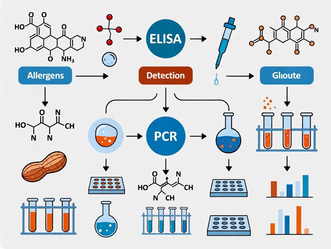
Protein-Based Detection (Allergens): The workhorse for allergen detection is the Enzyme-Linked Immunosorbent Assay (ELISA), which uses antibodies to specifically target and quantify allergenic proteins. This method is clinically relevant as it directly measures the molecule responsible for the allergic reaction [7]. A key limitation is that food processing (e.g., heating) can denature proteins, altering their structure and potentially leading to false negatives if the antibody cannot recognize the deformed protein [7]. Liquid Chromatography-Tandem Mass Spectrometry (LC-MS/MS) is an emerging, highly sensitive technique that detects signature peptides from allergenic proteins. While powerful, it currently lacks standardized methods and is more costly than ELISA [7].
DNA-Based Detection (Adulteration & Allergens): The Polymerase Chain Reaction (PCR) is a highly specific technique that amplifies unique DNA sequences to identify the biological source of an allergen or an adulterant (e.g., a specific meat species) [2] [7]. It is particularly useful when protein-based methods fail due to extreme processing conditions. However, since it detects DNA and not the allergenic protein itself, a positive PCR result does not always correlate with the presence of the protein at a level that would cause an allergic reaction [7].
Chromatography & Spectroscopy (Contaminants & Adulterants): These techniques form the backbone of chemical analysis. Gas Chromatography (GC) and Liquid Chromatography (LC), often coupled with mass spectrometry (MS), are used to separate, identify, and quantify a vast range of chemical contaminants and adulterants, from pesticide residues and mycotoxins to melamine and unauthorized dyes [2] [8] [6]. Mass Spectrometry platforms like MALDI-TOF MS (Matrix-Assisted Laser Desorption/Ionization – Time of Flight Mass Spectrometry) are gaining traction for their ability to rapidly generate unique protein or peptide "fingerprints" that can authenticate food species and detect adulteration in a high-throughput manner [6].
Table 2: Summary of Key Analytical Methods for Food Threat Detection
| Method | Principle | Primary Applications | Advantages | Limitations |
|---|---|---|---|---|
| ELISA [7] | Antibody-antigen binding for protein detection | Allergen testing, specific protein quantification | Quantitative, high throughput, clinically relevant (targets protein) | Protein denaturation from processing can cause false negatives |
| PCR [2] [7] | Amplification of species-specific DNA sequences | Species adulteration, allergen source identification | Highly specific, works on highly processed foods | Qualitative/semi-quantitative; detects DNA, not allergenic protein |
| LC-MS/MS [8] [7] | Separation by LC, identification by tandem MS | Allergen peptides, chemical contaminants, mycotoxins | Highly sensitive and specific, multi-target capability | High cost, complex data analysis, no standardized allergen methods |
| GC-MS [2] [6] | Separation by GC, identification by MS | Volatile organic compounds, pesticides, fatty acids | Excellent separation power, robust compound libraries | Requires volatile or derivatized samples |
| MALDI-TOF MS [6] | Ionized molecule "fingerprinting" based on mass/charge | Microbial ID, food speciation, protein profiling | High-speed, high-throughput, minimal sample prep | Semi-quantitative, requires robust reference databases |
Integrated Workflow for Threat Detection
The following diagram outlines a generalized decision-making workflow for selecting an appropriate detection method based on the suspected hazard and food matrix, a critical first step in analytical protocol design.
Detailed Experimental Protocols
Protocol: Detection of Allergenic Proteins via ELISA
This protocol describes the quantitative detection of a specific allergenic protein (e.g., from peanut or milk) in a solid food matrix using a commercial sandwich ELISA kit [7].
4.1.1 Research Reagent Solutions
Table 3: Essential Reagents for Allergen ELISA
| Reagent/Material | Function | Notes for Researchers |
|---|---|---|
| Commercial ELISA Kit | Provides pre-coated plates, antibodies, standards, and buffers. | Select a kit validated for your target allergen (e.g., peanut, egg). |
| Sample Diluent Buffer | Extracts protein from the food matrix and provides a compatible medium for the assay. | Composition is often proprietary; use the kit-provided buffer. |
| Blocking Buffer | Typically 1-5% BSA or non-fat dry milk in PBS. | Prevents non-specific binding of antibodies to the plate or sample components. |
| Wash Buffer | Usually PBS or Tris-based with a detergent (e.g., Tween-20). | Removes unbound reagents and reduces background signal. |
| Enzyme Substrate | Tetramethylbenzidine (TMB) is common. | Produces a colorimetric signal proportional to the amount of captured allergen. |
| Stop Solution | 1-2 M Sulfuric Acid or HCl. | Halts the enzyme-substrate reaction, stabilizing the signal for measurement. |
4.1.2 Step-by-Step Procedure
- Sample Preparation: Homogenize 1 g of the solid food sample with 10 mL of the provided extraction buffer. Vortex vigorously for 2 minutes and then centrifuge at 4,500 x g for 10 minutes at room temperature. Collect the supernatant for analysis. For complex matrices, filtration or defatting may be required.
- Standard Curve Preparation: Reconstitute the protein standard and prepare a serial dilution in the provided diluent to create a concentration series covering the kit's dynamic range (e.g., 0, 1, 5, 10, 25, 50 ng/mL).
- Assay Incubation: Add 50-100 µL of standards, prepared samples, and controls to the antibody-coated microwells. Incubate for a specified time (e.g., 60 minutes at room temperature) to allow the allergenic protein to bind.
- Washing: Decant the liquid from the wells and wash 3-5 times with 300 µL of wash buffer, ensuring complete removal of liquid between washes.
- Detection Antibody Incubation: Add the enzyme-conjugated detection antibody to each well. Incubate for a specified time (e.g., 60 minutes at room temperature). Wash again as in Step 4.
- Signal Development: Add the enzyme substrate solution (e.g., TMB) to each well. Incubate in the dark for 10-20 minutes until color develops.
- Reaction Stopping & Reading: Add the stop solution to each well. The blue color will turn yellow. Read the absorbance immediately at 450 nm using a microplate reader.
- Data Analysis: Generate a standard curve by plotting the mean absorbance of each standard against its concentration. Use the curve to interpolate the concentration of the allergenic protein in the test samples.
4.1.3 Validation Notes
- Spike Recovery: To validate the method for a new matrix, perform a spike recovery test. Add a known amount of the pure allergen to a blank sample, process it through the entire protocol, and calculate the percentage of the allergen recovered. Recovery of 80-120% is typically acceptable [7].
- Cross-Reactivity: Be aware of potential cross-reactivity. For example, a walnut test might cross-react with pecan due to biological similarity. The kit manufacturer's information should be consulted [7].
Protocol: Detection of Meat Speciation Adulteration via PCR
This protocol is designed to detect the adulteration of a declared meat (e.g., beef) with an undeclared species (e.g., horse or pork) using DNA extraction and species-specific PCR [2] [6].
4.2.1 Research Reagent Solutions
Table 4: Essential Reagents for Meat Speciation PCR
| Reagent/Material | Function | Notes for Researchers |
|---|---|---|
| DNA Extraction Kit | Isolates high-quality genomic DNA from the meat matrix. | Silica-membrane based kits are common. |
| Species-Specific Primers | Short DNA sequences that bind to and amplify a unique region of the target species' DNA. | Must be designed for high specificity (e.g., targeting mitochondrial cytochrome b gene). |
| PCR Master Mix | Contains Taq DNA polymerase, dNTPs, MgCl₂, and reaction buffer. | Provides all components necessary for the DNA amplification reaction. |
| Agarose | Polysaccharide used to create a gel for electrophoresis. | Typically used at 1.5-2.0% in TAE buffer. |
| Gel Stain | Ethidium bromide or safer alternatives (e.g., SYBR Safe). | Intercalates with DNA, allowing visualization under UV light. |
| DNA Molecular Weight Ladder | A mix of DNA fragments of known sizes. | Essential for confirming the size of the amplified PCR product. |
4.2.2 Step-by-Step Procedure
- DNA Extraction: Following the manufacturer's protocol for the DNA extraction kit, isolate genomic DNA from approximately 25 mg of the raw or cooked meat sample. Quantify the DNA purity and concentration using a spectrophotometer (A260/A280 ratio ~1.8 is ideal).
- PCR Reaction Setup: Prepare a 25 µL reaction mixture containing:
- 12.5 µL of PCR Master Mix
- 1.0 µL each of forward and reverse species-specific primers (10 µM stock)
- 50-100 ng of template DNA
- Nuclease-free water to 25 µL.
- Thermal Cycling: Place the tubes in a thermal cycler and run a program such as:
- Initial Denaturation: 95°C for 5 minutes.
- 35-40 Cycles of:
- Denaturation: 95°C for 30 seconds.
- Annealing: (Primer-specific temperature, e.g., 60°C) for 30 seconds.
- Extension: 72°C for 1 minute.
- Final Extension: 72°C for 7 minutes.
- Hold: 4°C.
- Gel Electrophoresis: Prepare a 1.5% agarose gel in 1x TAE buffer with a fluorescent nucleic acid stain. Mix 5-10 µL of each PCR product with a loading dye and load into the gel wells alongside a DNA ladder. Run the gel at 100-120 V for 30-40 minutes.
- Visualization & Analysis: Visualize the gel under UV light. The presence of a band at the expected molecular weight (e.g., 200 base pairs) confirms the presence of the target species. The absence of a band indicates the species is not present above the detection limit of the assay.
Protocol: Analysis of Chemical Contaminants via LC-MS/MS
This protocol provides a generalized framework for the sensitive detection and quantification of chemical contaminants, such as pesticide residues or mycotoxins, using Liquid Chromatography-Tandem Mass Spectrometry [8].
4.3.1 Research Reagent Solutions
Table 5: Essential Reagents for LC-MS/MS Analysis of Contaminants
| Reagent/Material | Function | Notes for Researchers |
|---|---|---|
| Extraction Solvents | Acetonitrile, Methanol, Acidified Acetonitrile. | Extracts the target analyte from the food matrix. Solvent choice is analyte-dependent. |
| QuEChERS Extraction Salts | MgSO₄, NaCl, buffering salts. | Used in a quick, easy, cheap, effective, rugged, and safe (QuEChERS) sample prep to induce partitioning. |
| Dispersive SPE Sorbents | PSA, C18, GCB. | Removes co-extracted matrix interferents like fatty acids, sugars, and pigments during clean-up. |
| LC Mobile Phases | Water and Methanol/Acetonitrile, often with modifiers (e.g., Formic Acid, Ammonium Acetate). | Carries the sample through the chromatographic column, separating analytes. |
| Analytical Standards | Pure certified reference standards of the target analytes. | Essential for creating a calibration curve and for positive identification based on retention time and mass spectra. |
4.3.2 Step-by-Step Procedure
- Sample Preparation & Extraction: Homogenize the food sample. Weigh 10 g into a 50 mL centrifuge tube. Add 10 mL of acetonitrile (1% formic acid) and shake vigorously for 1 minute. Add a pre-packaged QuEChERS salt mixture (e.g., containing 4 g MgSO₄, 1 g NaCl), shake immediately and vigorously for another minute, and then centrifuge.
- Extract Clean-up (dSPE): Transfer 1 mL of the upper acetonitrile layer to a 2 mL dSPE tube containing sorbents (e.g., 150 mg MgSO₄, 25 mg PSA). Vortex for 30 seconds and centrifuge. The clean extract is then transferred to an autosampler vial, potentially after dilution or concentration.
- Liquid Chromatography (LC): Inject an aliquot (e.g., 5 µL) of the cleaned extract onto the LC system. Use a reversed-phase C18 column and a gradient elution program. A typical gradient might run from 95% water/5% methanol to 5% water/95% methanol over 10-15 minutes, effectively separating the analytes.
- Tandem Mass Spectrometry (MS/MS) Detection: The eluting analytes are ionized (typically using Electrospray Ionization - ESI) and enter the mass spectrometer. The instrument is operated in Multiple Reaction Monitoring (MRM) mode, where a specific precursor ion for each analyte is selected and fragmented, and a specific product ion is monitored. This provides high selectivity and sensitivity.
- Data Analysis: Using the instrument software, integrate the peak areas for each analyte and its corresponding internal standard. Generate a calibration curve using the analyzed standards. Quantify the contaminants in the samples by interpolating from this curve. Positive identification requires the sample peak to have the same retention time and MRM transition ratio as the standard.
The Scientist's Toolkit
A robust research program requires a suite of reliable reagents, standards, and analytical instruments. The following table details key solutions for the featured experiments.
Table 6: Research Reagent Solutions for Food Threat Detection
| Tool Category | Specific Examples | Research Function |
|---|---|---|
| Immunoassay Reagents | Commercial ELISA Kits, Anti-peanut/Anti-milk monoclonal antibodies, Protein standards (e.g., β-lactoglobulin, Ara h1) | Target-specific protein quantification for allergen detection and risk assessment [7]. |
| Molecular Biology Kits | DNA Extraction Kits (silica-membrane), Taq DNA Polymerase, dNTPs, Species-specific Primer/Probe Sets | Genetic material isolation and amplification for species identification and adulteration detection [2] [7]. |
| Chromatography Standards | Certified Pesticide/Mycotoxin Reference Standards, Stable Isotope-Labeled Internal Standards (e.g., ¹³C-labeled), Fatty Acid Methyl Esters (FAMEs) | Calibration, precise quantification, and quality control in chromatographic methods like GC-MS and LC-MS/MS [8]. |
| Sample Preparation | QuEChERS Extraction Kits, Solid-Phase Extraction (SPE) Cartridges (C18, Florisil, NH₂), Solvents (HPLC/MS-grade Acetonitrile, Methanol) | Efficient extraction and clean-up of analytes from complex food matrices to reduce interference and protect instrumentation [8]. |
| Mass Spectrometry | MALDI Matrices (e.g., α-Cyano-4-hydroxycinnamic acid), LC-MS/MS and GC-MS Systems, High-Resolution Mass Spectrometers (HRMS) | Provides definitive analyte identification and characterization through accurate mass measurement and structural elucidation [8] [6]. |
Navigating the complex threat spectrum of food allergens, chemical contaminants, and adulterants demands a sophisticated, multi-pronged analytical approach. As detailed in these application notes, the researcher's arsenal ranges from the antibody-based specificity of ELISA and the genetic precision of PCR to the separation power and definitive identification offered by chromatographic and mass spectrometric techniques. The choice of method is critically dependent on the nature of the threat and the food matrix, a decision-making process that is greatly aided by the workflows and protocols provided. The ongoing development of these methodologies, including the refinement of high-throughput techniques like MALDI-TOF MS and the establishment of standardized LC-MS/MS protocols for allergens, represents the forefront of food safety research. By implementing and validating these detailed protocols, scientists and drug development professionals can contribute significantly to the protection of public health and the assurance of food authenticity.
In 2025, global food safety regulatory bodies are implementing significant updates that directly impact research and development in food allergen and contaminant detection. The U.S. Food and Drug Administration (FDA), European Food Safety Authority (EFSA), and Codex Alimentarius Commission have aligned their priorities toward enhancing pre-market safety evaluations, strengthening post-market surveillance, and addressing emerging chemical risks. These developments reflect a concerted global effort to integrate advanced scientific methodologies, including New Approach Methodologies (NAMs) and artificial intelligence, into regulatory frameworks. For researchers focused on detection methods, understanding these evolving priorities is crucial for developing compliant, effective, and innovative analytical solutions.
Major Regulatory Bodies and Their 2025 Focus Areas
Table 1: Key Regulatory Priorities for 2025
| Regulatory Body | Primary 2025 Focus Areas | Key Updates & Deadlines |
|---|---|---|
| U.S. FDA (Human Foods Program) | Food Chemical Safety, Allergen Labeling, Microbiological Safety, Nutrition | Final guidance on food allergen thresholds (Sep 2025); Post-market assessment framework update; Closer to Zero action levels for contaminants [4] [9]. |
| European Food Safety Authority (EFSA) | Novel Food Applications, Food Allergen Assessment | Updated Novel Food guidance effective February 2025; Enhanced data requirements for novel food safety assessments [10] [11]. |
| Codex Alimentarius (FAO/WHO) | Food Additives, Contaminant Limits, Food Hygiene | New standards for lead in spices (2.5 mg/kg) and culinary herbs (2.0 mg/kg); Revised code of practice for aflatoxins in peanuts [12] [13]. |
Detailed Analysis of 2025 Regulatory Priorities
U.S. Food and Drug Administration (FDA)
The FDA's Human Foods Program has been reorganized to centralize risk management, with food chemical safety representing a cornerstone of its 2025 deliverables [9]. A critical initiative is the advancement of the "Closer to Zero" action levels for environmental contaminants like lead, cadmium, and arsenic in foods intended for infants and young children [9]. This includes targeting the issuance of final guidance on action levels for lead, which will establish enforceable thresholds and drive the need for more sensitive detection methodologies in infant formula and baby food [9].
Regarding allergens, the FDA is prioritizing the evaluation of non-listed food allergens (those beyond the nine major allergens) based on a structured assessment framework finalized in January 2025 [4] [14]. This guidance outlines the scientific factors for evaluating the public health importance of emerging allergens, which will inform future labeling and manufacturing requirements [14]. Furthermore, the FDA is actively developing AI-driven tools like the Warp Intelligent Learning Engine (WILEE) for post-market signal detection and surveillance of the food supply, representing a significant shift toward data-driven risk prioritization [9].
European Food Safety Authority (EFSA)
EFSA's updated guidance on Novel Food applications, effective February 2025, mandates more detailed scientific requirements for market authorization [10]. Applicants must now provide comprehensive data on the novel food's composition, production process, stability, and anticipated intake, with heightened emphasis on allergenicity potential and nutritional safety [10]. This reflects EFSA's commitment to a precautionary principle, ensuring that innovative foods entering the EU market are not nutritionally disadvantageous and are safe for consumption, including for sensitive individuals [10].
The guidance also strongly encourages the use of validated alternative methods to minimize animal testing, in alignment with the EU's broader strategic goal to phase out in-vivo studies [10]. This directive challenges researchers to develop robust in-vitro or in-silico models for predicting protein allergenicity, a rapidly evolving field in food safety science.
Codex Alimentarius Commission
The Codex Alimentarius Commission, at its 48th session in November 2025, adopted several standards with direct implications for contaminant monitoring and control [12]. Notably, it set Maximum Levels (MLs) for lead in dried cinnamon (2.5 mg/kg) and dried culinary herbs (2.0 mg/kg), addressing the neurotoxic effects of lead exposure [12]. These internationally recognized standards are critical for harmonizing global trade and protecting consumer health, providing a benchmark for national regulatory frameworks.
Additionally, the Commission revised the Code of Practice (CXC 55-2004) for preventing aflatoxin contamination in peanuts, integrating new scientific information on optimal harvesting stages and the effect of roasting processes in reducing aflatoxin levels [12]. This provides a critical framework for managing these potent carcinogens throughout the supply chain.
Emerging Detection Technologies and Methodologies
The regulatory focus on lower contaminant thresholds and unlisted allergens is accelerating the adoption of highly sensitive and multiplexed detection technologies.
Table 2: Emerging Technologies in Allergen and Contaminant Detection
| Technology | Principle | Key Applications | Sensitivity/LOD |
|---|---|---|---|
| Mass Spectrometry (MS) | Detection of proteotypic peptides via multiple reaction monitoring (MRM) | Simultaneous quantification of specific allergenic proteins (e.g., Ara h 3/6 in peanut, Bos d 5 in milk) [15] | High sensitivity and specificity across complex food matrices [15] |
| AI-Enhanced Hyperspectral Imaging (HSI) | Computer vision and machine learning analysis of spectral signatures | Non-destructive, real-time allergen detection without altering food integrity [15] | Varies by analyte and model training |
| Multiplexed Immunoassays | Simultaneous binding of multiple antibodies to specific protein targets | High-throughput screening for multiple allergens in a single sample run [15] | As low as 0.01 ng/mL for specific allergens [15] |
| ATP-based Sanitation Verification | Cloud-based integration of adenosine triphosphate readings with allergen data | Real-time sanitation verification and predictive risk management in production facilities [15] | Provides indirect monitoring of hygiene control |
Experimental Protocol: Multiplex Mass Spectrometry for Allergen Detection
This protocol details the simultaneous detection and quantification of key allergenic proteins from peanut, milk, and egg in a baked goods matrix using liquid chromatography-tandem mass spectrometry (LC-MS/MS) [15].
1. Sample Preparation:
- Homogenization: Precisely weigh 2 g of the ground food sample into a 50 mL centrifuge tube.
- Protein Extraction: Add 10 mL of extraction buffer (50 mM ammonium bicarbonate, pH 8.0) and 10 µL of 1 M dithiothreitol (DTT). Vortex vigorously for 1 minute and incubate at 60°C for 30 minutes with shaking.
- Alkylation: Cool to room temperature. Add 40 µL of 1 M iodoacetamide (IAA), vortex, and incubate in the dark for 30 minutes.
- Digestion: Add 100 µL of 0.1 µg/µL trypsin solution. Incubate at 37°C for 4 hours with continuous shaking.
- Clean-up: Acidify the digest with 1% formic acid and purify using a C18 solid-phase extraction (SPE) cartridge. Elute peptides with 60% acetonitrile/0.1% formic acid and dry under a gentle nitrogen stream.
2. LC-MS/MS Analysis:
- Chromatography: Reconstitute the dried peptides in 100 µL of 2% acetonitrile/0.1% formic acid. Inject 10 µL onto a reverse-phase C18 column (2.1 mm x 150 mm, 1.7 µm particle size). Use a gradient of 2% to 40% solvent B (0.1% formic acid in acetonitrile) over 25 minutes.
- Mass Spectrometry: Operate the mass spectrometer in positive electrospray ionization (ESI+) mode with scheduled Multiple Reaction Monitoring (MRM). Monitor specific precursor ion > product ion transitions for proteotypic peptides of Ara h 3 (peptide: LDNLNQNLR; transition: 530.3 -> 801.4), Bos d 5 (peptide: VLVLDTDYK; transition: 539.8 -> 853.4), and Gal d 1 (peptide: GGLEPINFQTAADQAR; transition: 886.4 -> 1135.6).
3. Data Quantification:
- Prepare a calibration curve using certified reference materials of the purified allergenic proteins.
- Use stable isotope-labeled versions of the target peptides as internal standards.
- Quantify based on the ratio of the analyte peak area to the internal standard peak area, interpolated from the linear calibration curve.
The Scientist's Toolkit: Key Research Reagent Solutions
Table 3: Essential Reagents for Advanced Food Safety Research
| Research Reagent / Material | Function & Application | Justification for Use |
|---|---|---|
| Certified Reference Materials (CRMs) | Calibration and validation of analytical methods for contaminants and allergens [15]. | Essential for meeting the traceability and accuracy demands of new regulatory thresholds (e.g., Codex lead MLs) [12]. |
| Stable Isotope-Labeled Peptides | Internal standards for multiplex MS-based allergen quantification [15]. | Correct for matrix effects and ionization efficiency variations, ensuring precise and reproducible results. |
| Proteotypic Peptide Standards | Target enrichment for specific allergenic proteins (e.g., Ara h 3, Bos d 5) [15]. | Enable highly specific detection and avoid cross-reactivity, crucial for monitoring undeclared allergens. |
| Monoclonal Antibody Panels | Development of immunoassays for emerging and major food allergens. | Provide the specificity required for detecting individual allergenic proteins in complex food matrices. |
| PFAS Analytical Standards | Quantifying per- and polyfluoroalkyl substances in food [9]. | Critical for FDA's research initiative to better understand PFAS exposure from food [9]. |
Regulatory Workflow and Research Integration
Navigating the updated regulatory landscape requires a systematic approach from method development to compliance. The following diagram outlines the integrated workflow for validating a new detection method against 2025 regulatory criteria, from initial risk assessment to final application.
The 2025 regulatory priorities of the FDA, EFSA, and Codex Alimentarius present both challenges and opportunities for researchers in food allergen and contaminant detection. The overarching trends point toward stricter contaminant limits, a broader scope of regulated allergens, and an increased reliance on sophisticated, non-targeted analytical techniques. Success in this evolving landscape will depend on the widespread adoption of advanced technologies like multiplex mass spectrometry and AI-enhanced imaging, coupled with robust, validated experimental protocols. By aligning research and development with these global regulatory directions, scientists can actively contribute to a safer, more transparent, and innovative food supply.
The Rising Burden of Food Allergies and Its Implications for Public Health Research
Food allergy is a significant and growing public health problem, estimated to affect at least 1 in 10 adults and 1 in 13 children in developed nations [16]. This pathological immune system reaction, triggered by the ingestion of allergenic proteins, has far-reaching effects on individual well-being, family dynamics, and healthcare systems. The global food supply chain's complexity, coupled with the potential for severe and sometimes life-threatening reactions, necessitates advanced research into reliable detection methodologies [17] [18]. The burden of foodborne diseases, including those caused by allergens, is substantial, with unsafe food causing an estimated 600 million illnesses and 420,000 deaths annually worldwide [18]. This application note frames the rising burden of food allergies within the critical context of detecting food allergens and contaminants, providing structured data, detailed experimental protocols, and visual workflows to support public health research and safety initiatives.
The Public Health and Economic Burden
The impact of food allergies extends beyond immediate health reactions, creating significant nutritional, psychological, and economic challenges. Children with food allergy are at risk for inadequate nutrient intake, poor growth, feeding difficulties, and anxiety [16]. The financial burden is considerable, with one systematic review reporting mean household-level out-of-pocket costs of $3,339 and opportunity costs of $4,881 [17]. At a societal level, the total productivity loss associated with foodborne diseases in low- and middle-income countries is estimated at US$ 95.2 billion per year, with an annual cost of treating these illnesses estimated at US$ 15 billion [18].
Table 1: Major Food Allergens and Their Prevalence Characteristics
| Allergen Category | Examples | Persistence Profile | Key Regulatory Status |
|---|---|---|---|
| Peanut | - | Typically persistent [16] | Major allergen (FALCPA) [4] |
| Tree Nuts | Almonds, Pecans, Walnuts [4] | Typically persistent [16] | Major allergen (FALCPA) [4] |
| Seafood | Fish, Crustacean shellfish (e.g., Crab, Lobster) [4] | Typically persistent [16] | Major allergen (FALCPA) [4] |
| Milk | Casein, Beta-lactoglobulin [19] | Often transient [16] | Major allergen (FALCPA) [4] |
| Egg | - | Often transient [16] | Major allergen (FALCPA) [4] |
| Sesame | - | - | 9th major allergen (FASTER Act, 2023) [4] |
Analytical Frameworks for Allergen Detection
Effective management and prevention of food-allergic reactions rely on the accurate detection and quantification of allergenic proteins in food products. However, several factors complicate this analysis, including the composition of food extracts, the impact of food processing on proteins, and the inherent variability of immunological responses [17]. To ensure consistency, Health Canada's Allergen Methods Committee (AMC) recommends using the Bicinchoninic Acid Assay (BCA test) with Bovine Serum Albumin (BSA) as a common reference for quantifying protein content in calibrators, despite variations in allergen protein behavior [19].
Table 2: Comparison of Major Allergen Detection Methodologies
| Methodology | Principle | Key Advantages | Key Limitations | Best Suited For |
|---|---|---|---|---|
| Immunoassays (ELISA) | Antigen-antibody binding detected enzymatically [20]. | High specificity and sensitivity; commercially available kits [17]. | May detect denatured proteins; affected by food matrix [17]. | Routine detection of specific, preselected allergens [17] [19]. |
| DNA-Based Methods (PCR) | Amplification of species-specific DNA sequences [20]. | High specificity; useful for processed foods where protein is denatured [17]. | Indirect (DNA presence ≠ protein presence); not for all foods [17]. | Detecting preselected species contamination (e.g., speciation) [17]. |
| Mass Spectrometry | Separation and identification based on mass-to-charge ratio [21] [20]. | High specificity and multiplexing capability; can detect multiple allergens [17]. | Expensive equipment; complex data analysis; requires expertise [17]. | Confirmatory analysis and untargeted discovery of allergens [17]. |
| Multiplex Allergen Microarray | IgE-binding inhibition on a biochip with many allergens [17]. | Multiplexed; identifies many known/unknown IgE-binding proteins in one test [17]. | Relatively new; limited widespread use and validation [17]. | Comprehensive allergen profiling and discovery [17]. |
Detailed Experimental Protocols
Protocol: Multiplex Allergen Microarray-Based Inhibition Assay (SPHIAa)
This protocol is adapted for identifying and profiling IgE-binding proteins in a food sample using a multiplex biochip [17].
1. Reagent and Sample Preparation
- Food Extract Preparation: Homogenize the food sample. Extract proteins using a high-salt or high-pH buffer to improve the recovery of allergenic proteins [17]. Determine protein concentration using a standardized method like the BCA assay [19].
- Control Solutions: Prepare a positive control (e.g., a known allergenic food extract) and a negative control (extraction buffer alone).
2. Inhibition Assay Setup
- Incubation: In a tube, pre-incubate a constant volume of a pooled human serum sample (from individuals allergic to the target food) with serial dilutions of the prepared food extract for 30-60 minutes at room temperature. This allows allergens from the food sample to bind to serum IgE.
- Biochip Application: Apply the pre-incubated mixture onto the multiplex allergen microarray biochip (e.g., ISAC, FABER), which contains immobilized allergenic proteins.
- Binding and Washing: Incubate the biochip according to manufacturer specifications. Any free, unbound IgE from the serum that recognizes the immobilized allergens on the chip will bind. Wash the biochip thoroughly to remove unbound materials.
3. Detection and Analysis
- Labeling: Add a fluorescently labelled anti-human IgE antibody to the biochip and incubate.
- Signal Detection: Wash the biochip again and scan it with a microarray scanner to detect fluorescence signals.
- Data Interpretation: A reduced fluorescence signal at a specific spot on the biochip indicates that the food extract contained the corresponding allergen, which inhibited the binding of serum IgE to the immobilized allergen. The signal reduction is proportional to the allergen's concentration in the food sample.
Protocol: Validation of Allergen Detection Methods as per Health Canada AMC Guidelines
This protocol outlines the evaluation process for quantitative allergen detection methods intended for compliance activities [19].
1. Define Evaluation Parameters
- Reference Material: Select a well-characterized, representative material for the allergenic food commodity (e.g., defatted hazelnut flour).
- Matrices: Select appropriate food matrices for evaluation based on common allergen presence.
- Fortification Levels: Prepare samples at three key levels: a blank (no added allergen), 2x the method's Limit of Quantification (LOQ), and 5x LOQ.
2. Inter-laboratory Testing
- Laboratory Participation: A minimum of three AMC-affiliated laboratories, plus the method developer's laboratory, should participate in a full evaluation.
- Sample Analysis: Each laboratory analyzes the fortified and blank samples with a predefined number of replicates. The results, expressed in µg of protein per gram of commodity, are reported centrally.
3. Performance Assessment
- Data Analysis: Calculate key performance metrics including repeatability, reproducibility, recovery, and linearity.
- Compendium Reporting: The generated data is compiled into a report for the web-based Compendium of Methodologies, allowing users to assess the method's suitability for their needs without implying regulatory endorsement.
Research Reagent Solutions
Table 3: Essential Reagents and Materials for Allergen Research
| Reagent/Material | Function/Application | Example/Note |
|---|---|---|
| Reference Materials | Serves as a common ground for method evaluation and calibration; essential for quantitative accuracy [19]. | Defatted hazelnut flour; well-characterized and reproducible [19]. |
| Protein Standard (BSA) | Reference standard for quantifying total protein content in allergen extracts using the BCA assay [19]. | Bovine Serum Albumin (BSA) [19]. |
| High-Salt/High-pH Buffers | Extraction buffers to improve the solubility and recovery of allergenic proteins from complex food matrices [17]. | Phosphate-buffered saline (PBS) with high salt concentration [17]. |
| Pooled Allergic Human Sera | Critical reagent for immunoassays and inhibition studies to detect IgE-binding proteins [17]. | Sera from multiple individuals with confirmed allergy to the target food [17]. |
| Multiplex Allergen Biochip | Solid-phase support for simultaneous detection of numerous IgE reactivities in a single sample [17]. | ISAC or FABER test chips [17]. |
| Fluorescent Anti-IgE Antibody | Detection antibody for visualizing IgE binding in microarray and other fluorescent immunoassays [17]. | Fluorescein or Cy3-labeled antibody [17]. |
Visualization of Workflows
Food Allergen Detection Research Pathway
Multiplex Microarray Inhibition Assay Workflow
The rising global burden of food allergies demands robust, sensitive, and multifaceted research approaches to protect public health. Accurate detection of food allergens is complicated by variable thresholds, the effects of food processing, and complex matrices. This application note has detailed the public health context, presented comparative analytical data, and provided detailed protocols for advanced methods like the multiplex allergen microarray. The ongoing development of international standards, reference materials, and validated methods, as coordinated by bodies like the Allergen Methods Committee, is crucial for harmonizing global efforts [19]. Future research must continue to refine these detection methodologies, explore the impact of cofactors on reactivity, and integrate new technologies like biosensors and artificial intelligence to enhance food safety and improve the quality of life for allergic individuals [16] [20].
Food allergen detection presents significant challenges that are profoundly influenced by the composition and processing of the food matrix itself. Complex matrices can interfere with analytical techniques, leading to potential false negatives that pose serious risks to consumer safety. For researchers and scientists in drug development and food safety, understanding these matrix effects is crucial for developing accurate detection methodologies. This application note details the primary food matrices of concern, provides optimized experimental protocols for allergen extraction, and outlines advanced analytical techniques for detecting allergens and contaminants in these challenging systems. The protocols are framed within the context of a broader research thesis on advancing detection methods for food allergens and contaminants, addressing current gaps in standardization and recovery rates.
Key Challenging Food Matrices in Allergen Analysis
The structural complexity and compositional variability of certain food matrices significantly complicate allergen detection and quantification. The table below summarizes the primary challenging matrices, their key interferents, and the associated analytical complications.
Table 1: Key Food Matrices of Concern in Allergen Detection
| Food Matrix Category | Specific Examples | Key Interfering Compounds/Properties | Impact on Allergen Detection |
|---|---|---|---|
| Plant-Based Alternative Proteins | Plant-based meats, dairy alternatives, insect proteins, fungal proteins | Protein cross-reactivity, high polyphenol content, novel protein structures | Masks target allergens, creates false positives in immunoassays, requires new reference materials and methods [22]. |
| Thermally Processed Foods | Baked biscuits, roasted nuts, pasteurized dairy, ultra-processed foods | Protein denaturation, Maillard reaction products, protein aggregation | Alters antibody-binding epitopes, reduces extractability, lowers analytical recovery [23]. |
| High-Fat/High- Polyphenol Matrices | Chocolate desserts, nut spreads, dark cocoa products | Fats, tannins, polyphenols from cocoa | Binds to and precipitates allergenic proteins, quenches assay signals, necessitates specialized extraction buffers [23]. |
| Complex Multi-Ingredient Systems | Bakery products, confectionery items, sauces, dressings | Multiple allergen sources (e.g., wheat, eggs, milk, nuts, soy), emulsifiers, stabilizers | Creates high risk of cross-contact, introduces multiple interferents, complicates method development and validation [22]. |
The rise of alternative proteins, including plant-based, insect, and fermentation-derived ingredients, introduces novel allergenic proteins and potential cross-reactivity with known allergens, demanding specialized testing beyond standard immunoassays [22]. Furthermore, food processing methods such as heat treatment, enzymatic hydrolysis, and fermentation can modify protein structures, affecting their detectability by conventional antibody-based tests [22].
Detailed Experimental Protocols
Optimized Allergen Extraction from Challenging Matrices
Efficient and reproducible extraction of allergenic proteins is the most critical step for accurate detection, particularly from complex, processed matrices. The following protocol is optimized for simultaneous extraction of multiple clinically relevant allergens.
Research Reagent Solutions
Table 2: Essential Reagents for Allergen Extraction Optimization
| Reagent/Solution | Function/Principle | Key Considerations |
|---|---|---|
| Extraction Buffer D(50 mM Carbonate-Bicarbonate, 10% Fish Gelatine, pH 9.6) | Alkaline pH and fish gelatine help solubilize proteins and minimize binding to matrix components, improving recovery from baked and complex matrices [23]. | Ideal for general use across multiple allergen types. Fish gelatine is preferred over mammalian gelatine to avoid cross-reactivity. |
| Extraction Buffer J(PBS, 2% Tween-20, 1 M NaCl, 10% Fish Gelatine, 1% PVP, pH 7.4) | Detergent (Tween), high salt, and PVP are critical for disrupting hydrophobic interactions and binding polyphenols in chocolate and high-fat matrices [23]. | Superior for challenging matrices rich in polyphenols (e.g., chocolate, certain spices). |
| Fish Gelatine | Acts as a protein blocking agent, reducing non-specific binding of allergens to tube walls and food particulates [23]. | Reduces surface adsorption, a significant source of protein loss. |
| Polyvinylpyrrolidone (PVP) | Binds and precipitates polyphenols and tannins, preventing them from interfering with proteins and assay antibodies [23]. | Essential for recovering allergens from cocoa, berries, and other polyphenol-rich foods. |
| Sodium Dodecyl Sulfate (SDS) | Ionic detergent that denatures proteins and disrupts strong protein-lipid and protein-protein interactions [23]. | Use with caution as it may denature epitopes recognized by some antibodies. |
Sample Preparation and Extraction Workflow
The following diagram illustrates the complete workflow for preparing and analyzing incurred food samples for allergen content.
Materials and Equipment:
- Prepared incurred food matrices (e.g., chocolate dessert, biscuit dough)
- Selected extraction buffers (D and J from Table 1)
- Analytical balance
- Vortex mixer
- Orbital incubator (e.g., Stuart SI500)
- Centrifuge capable of maintaining 4°C
- Micropipettes and sterile tips
Procedure:
- Sample Homogenization: Begin with a finely ground and well-mixed sample to ensure representativeness.
- Weighing: Precisely weigh 1.0 ± 0.05 g of the homogenized sample into a 15 mL centrifuge tube.
- Buffer Addition: Add 10 mL of the pre-warmed (60°C) selected extraction buffer, ensuring a 1:10 sample-to-buffer ratio.
- Initial Mixing: Vortex the mixture vigorously for 30 seconds to ensure complete suspension of the sample.
- Incubation: Incubate the sample in an orbital incubator at 60°C for 15 minutes with shaking set to 175 rpm. This elevated temperature aids in protein solubilization.
- Clarification: Centrifuge the samples at 1250 rcf for 20 minutes at 4°C to pellet insoluble debris.
- Supernatant Collection: Carefully collect the clarified supernatant from the middle of the tube, avoiding the surface lipid layer and the bottom pellet. Proceed immediately to analysis or store at -20°C.
Validation Notes: This optimized protocol typically achieves recoveries of 50-150% for 14 major food allergens from most matrices. However, matrices containing chocolate or subjected to intense thermal processing may still yield lower recoveries, necessitating method-specific validation [23].
Advanced Analytical Detection Techniques
Following extraction, selecting an appropriate detection method is vital. The choice depends on the required sensitivity, specificity, and the need for multiplexing.
Allergen-Specific Multiplex Immunoassay (e.g., MARIA)
Multiplex arrays allow for the simultaneous quantification of multiple specific allergens in a single sample, saving time and sample volume.
Procedure:
- Bead Coupling: Covalently couple allergen-specific monoclonal antibodies to distinct magnetic bead regions.
- Assay Setup: Mix the bead set with sample extracts and allergen standards in a microplate well.
- Incubation: Incubate with shaking to allow the formation of bead-allergen complexes.
- Detection: Add a biotinylated detection antibody followed by streptavidin-phycoerythrin.
- Analysis: Analyze the plate using a multiplex reader (e.g., Luminex). The median fluorescence intensity is proportional to the allergen concentration, calculated against a standard curve.
Advantages: This method provides high-throughput, multiplexed analysis with improved standardization and reporting clarity by targeting clinically relevant proteins like Ara h 3/6 (peanut) and Bos d 5 (milk) [15] [23].
Liquid Chromatography with Tandem Mass Spectrometry (LC-MS/MS)
LC-MS/MS offers high specificity and is particularly useful for detecting allergens in processed foods where proteins are denatured or for confirming results from immunoassays.
Procedure:
- Protein Digestion: Digest the extracted protein sample with trypsin to generate peptide fragments.
- Chromatographic Separation: Inject the peptides onto a reverse-phase UHPLC column for separation.
- Mass Spectrometry Analysis: Analyze eluting peptides using a triple-quadrupole mass spectrometer in Multiple Reaction Monitoring (MRM) mode.
- Quantification: Monitor specific proteotypic peptide ions unique to the target allergen. Quantify by comparing the peak areas of the target peptides to those of a stable isotope-labeled internal standard.
Advantages: LC-MS/MS can achieve detection limits as low as 0.01 ng/mL, provides high specificity by targeting marker peptides, and is less susceptible to antibody cross-reactivity issues [15] [22]. It is the preferred method for quantifying specific proteins in complex matrices where antibody-based methods may fail.
Emerging Technologies and Future Directions
The field of food allergen detection is rapidly evolving with the integration of artificial intelligence and novel biosensing technologies.
- AI-Enhanced Testing: Machine learning models are being developed to predict the allergenicity of new ingredients before they enter the supply chain. Furthermore, AI is combined with non-destructive techniques like Hyperspectral Imaging (HSI) and Fourier Transform Infrared (FTIR) spectroscopy for real-time, in-line allergen detection without altering food integrity [15].
- Biosensors and Portable Kits: Advanced biosensor technologies using gold nanoparticles and graphene-based transducers are achieving femtomolar detection limits. These platforms are paving the way for point-of-use testing solutions, including smartphone-integrated devices, which could enable monitoring at various points in the supply chain [22].
- Regulatory Shifts: The FDA's recent activities, including a virtual public meeting on food allergen thresholds and the issuance of Edition 5 guidance on allergen labeling, signal a move towards more standardized, science-based regulatory frameworks [4] [24]. The "Make America Healthy Again" (MAHA) strategy also highlights increased scrutiny on food additives and chemicals, which may influence allergen management practices [24].
Accurate detection of food allergens in challenging matrices such as plant-based alternatives and processed foods requires a meticulous, multi-faceted approach. The optimized extraction protocols detailed herein, utilizing specialized buffers containing fish gelatine and PVP, are critical for achieving reliable recovery rates. Subsequent analysis by allergen-specific multiplex immunoassays or confirmatory LC-MS/MS provides the sensitivity and specificity required for both research and regulatory compliance. As the food landscape continues to evolve with novel ingredients and processing technologies, so too must the analytical methods, with a clear trend towards the integration of AI, biosensors, and harmonized international standards to ensure public safety.
From Bench to Batch: Analytical Techniques for Allergen and Contaminant Detection
In the ongoing effort to ensure global food safety, the precise detection of allergens and contaminants remains a paramount challenge for researchers, scientists, and drug development professionals. Food allergies, which are adverse immune responses to specific food proteins, affect millions of individuals worldwide, with prevalence rates rising annually [25]. The accurate detection and quantification of biological and chemical contaminants in complex food matrices are critical for protecting public health, ensuring regulatory compliance, and maintaining consumer trust. This document details the established, conventional methodologies that form the backbone of modern food safety analysis: chromatography, enzyme-linked immunosorbent assay (ELISA), and polymerase chain reaction (PCR)-based methods. These techniques provide the sensitivity, specificity, and robustness required for definitive identification and measurement of target analytes, from pathogenic bacteria and mycotoxins to undeclared allergenic proteins [7] [26] [27].
Each of the three conventional workhorses offers distinct advantages based on the analyte of interest and the specific requirements of the analysis. ELISA is a quantitative immunological method that leverages the specificity of antibody-antigen interactions, typically targeting specific allergenic proteins, which are the molecules that directly cause adverse reactions in sensitized individuals [7]. PCR-based methods, in contrast, are molecular techniques that target the DNA of the organism containing the allergenic protein, providing indirect evidence of its potential presence [7]. Chromatography, particularly when coupled with mass spectrometry (e.g., LC-MS/MS), is a powerful analytical chemistry technique that separates complex mixtures and can identify and quantify specific proteins or peptides based on their mass, offering high specificity and the ability to analyze multiple allergens simultaneously [26] [28].
The table below provides a quantitative comparison of these core techniques.
Table 1: Quantitative Comparison of Conventional Food Allergen and Contaminant Detection Methods
| Method | Typical Analytes | Principle | Detection Limit | Throughput | Quantitative Ability | Key Advantage |
|---|---|---|---|---|---|---|
| ELISA | Specific proteins (e.g., casein, tropomyosin) [7] | Antibody-antigen binding with enzymatic signal generation [26] | Variable; specific kits can reach ppm/ppb levels [26] | Moderate to High | Quantitative results expressed in protein [7] | High clinical relevance as it targets the reactive protein [7] |
| PCR-Based | Species-specific DNA sequences [7] | Amplification of target DNA sequences [28] | High (e.g., dPCR for low-level contaminants) [28] | High (esp. multiplex qPCR) [28] | Qualitative or Quantitative (qPCR, dPCR) [28] | High sensitivity and specificity for DNA; resistant to food matrix effects in some cases [7] |
| Chromatography (LC-MS/MS) | Proteins, peptides, chemical contaminants [26] | Physical separation followed by mass-based detection [28] | High (e.g., trace levels below ppb) [28] | Moderate | Quantitative | Can analyze multiple allergens/contaminants at once (multiplexing) [26] |
Detailed Experimental Protocols
Protocol for Allergen Detection via Sandwich ELISA
Sandwich ELISA is a highly specific and quantitative method widely used for detecting proteins like β-lactoglobulin in milk or tropomyosin in shellfish [26]. The following protocol outlines the key steps.
Workflow Overview: Sandwich ELISA
Materials:
- Microtiter Plates: Coated with a capture antibody specific to the target allergen (e.g., mouse antiglycinin monoclonal antibody for soy) [26].
- Blocking Buffer: Typically a protein-based solution like Bovine Serum Albumin (BSA) or non-fat dry milk to prevent non-specific binding [26].
- Allergen Standards: A series of known concentrations of the purified allergen for generating a standard curve.
- Test Samples: Food extracts prepared in an appropriate extraction buffer.
- Detection Antibody: An antibody specific to a different epitope on the target allergen, conjugated to an enzyme (e.g., Horseradish Peroxidase - HRP) [26].
- Enzyme Substrate: A chromogenic substrate (e.g., TMB for HRP) that produces a color change upon reaction with the enzyme.
- Stop Solution: An acid (e.g., sulfuric acid) to terminate the enzyme-substrate reaction.
- Microplate Reader: An instrument capable of measuring the absorbance of the solution in each well.
Procedure:
- Coating and Blocking: The wells of a microtiter plate are pre-coated with the capture antibody. Any remaining protein-binding sites are then blocked with a blocking buffer to minimize non-specific background signal [26].
- Incubation with Sample: Add the prepared test samples and allergen standards to the respective wells. Incubate to allow the target allergen (antigen) to bind to the immobilized capture antibody, forming an antigen-antibody complex.
- Detection Antibody Binding: After washing to remove unbound materials, add the enzyme-conjugated detection antibody. This antibody binds to the captured allergen, forming a "sandwich" [26].
- Signal Development and Detection: Following another wash step to remove unbound detection antibody, add the enzyme substrate. The enzyme catalyzes a reaction that produces a colored product. The reaction is stopped after a fixed time, and the absorbance is measured with a microplate reader [26].
- Quantification: The absorbance of the standards is used to generate a standard curve. The concentration of the allergen in the test samples is interpolated from this curve.
Protocol for Pathogen Detection via Quantitative PCR (qPCR)
qPCR allows for the simultaneous amplification and quantification of specific DNA sequences, making it ideal for detecting and quantifying foodborne pathogens like Salmonella or Listeria [28].
Workflow Overview: Quantitative PCR (qPCR)
Materials:
- DNA Extraction Kit: For isolating and purifying genomic DNA from the food matrix.
- Species-Specific Primers and Probes: Short, synthetic oligonucleotides designed to bind exclusively to a unique DNA sequence of the target organism. Probes are typically labeled with a fluorophore and a quencher.
- qPCR Master Mix: A pre-mixed solution containing heat-stable DNA polymerase, dNTPs, MgCl₂, and reaction buffers.
- qPCR Instrument: A thermal cycler equipped with a optical detection system to monitor fluorescence in real-time.
Procedure:
- DNA Extraction: Extract and purify total DNA from the food sample. The efficiency of this step is critical for the final quantitative result [28].
- Reaction Setup: Prepare the qPCR reaction mix by combining the extracted DNA, master mix, and species-specific primers and probes.
- Amplification and Real-Time Detection: Load the reaction plates into the qPCR instrument. The instrument runs a series of temperature cycles (denaturation, annealing, extension). During each cycle, the probe is cleaved, separating the fluorophore from the quencher and generating a fluorescent signal proportional to the amount of amplified DNA [28].
- Data Analysis: The instrument software plots fluorescence against cycle number. The cycle threshold (Ct) is determined for each sample, which is the cycle number at which fluorescence exceeds a background level. The Ct value is inversely proportional to the starting quantity of the target DNA. Quantification is achieved by comparing the Ct values of unknown samples to a standard curve generated from samples with known DNA concentrations [28].
Protocol for Multi-Allergen Detection via LC-MS/MS
Liquid Chromatography-Tandem Mass Spectrometry (LC-MS/MS) is a confirmatory technique that is gaining acceptance for the quantitative detection of multiple allergens in complex food samples [26].
Workflow Overview: LC-MS/MS for Allergen Detection
Materials:
- Extraction Buffer: A solution to efficiently solubilize proteins from the food matrix.
- Digestion Enzyme: Typically trypsin, which cleaves proteins at specific amino acid residues to generate peptides.
- Signature Peptides: Unique peptide sequences that serve as markers for the specific allergenic protein [26].
- Liquid Chromatography System: For separating the complex peptide mixture based on hydrophobicity.
- Tandem Mass Spectrometer: Equipped with an electrospray ionization (ESI) source and a triple quadrupole mass analyzer for highly selective and sensitive detection.
Procedure:
- Protein Extraction and Digestion: Proteins are extracted from the food sample. The extracted proteins are then digested with trypsin to generate a mixture of peptides [26].
- Chromatographic Separation: The peptide mixture is injected into the LC system, where peptides are separated based on their chemical properties as they pass through a chromatographic column.
- Ionization and Mass Analysis: The eluting peptides are ionized via electrospray ionization and enter the first quadrupole (Q1) of the mass spectrometer, which selects ions of a specific mass-to-charge ratio (m/z) corresponding to a target signature peptide. These selected precursor ions are then fragmented in a collision cell (second quadrupole), and the resulting product ions are analyzed in the third quadrupole (Q2) [28].
- Identification and Quantification: The unique pattern of fragment ions (transition ions) serves as a definitive "fingerprint" for the target peptide and its parent protein. Quantification is achieved by comparing the signal intensity of the transition ions in the sample to that of a calibration curve prepared from known concentrations of the signature peptide or protein standard [26].
Research Reagent Solutions
The successful application of these conventional methods relies on a suite of specific, high-quality reagents. The following table details essential materials and their functions.
Table 2: Essential Research Reagents for Food Allergen and Contaminant Analysis
| Reagent / Material | Function | Application Examples |
|---|---|---|
| Capture & Detection Antibodies | Bind specifically to target allergenic proteins (antigens) with high affinity [26]. | Monoclonal mouse anti-glycinin for soy detection; polyclonal rabbit anti-tropomyosin for shellfish [26]. |
| Species-Specific Primers & Probes | Initiate and report the amplification of a unique DNA sequence from a target organism via PCR [28]. | Primers for Salmonella virulence genes; TaqMan probes for peanut DNA (Ara h 1-6) [28]. |
| Signature Peptides | Unique peptide sequences that serve as analytical markers for specific allergenic proteins in LC-MS/MS [26]. | Peptides from Ara h 3 and Ara h 6 for peanut; peptides from casein for milk [26]. |
| Chromatography Columns | Stationary phases for separating complex mixtures of peptides or chemicals before detection [28]. | C18 reversed-phase columns for peptide separation in LC-MS/MS [28]. |
| Enzyme-Substrate Systems | Generate a measurable signal (e.g., color, fluorescence) proportional to the amount of detected analyte in ELISA [26]. | Horseradish Peroxidase (HRP) with TMB substrate; Alkaline Phosphatase (AP) with pNPP substrate [26]. |
Chromatography, ELISA, and PCR-based methods collectively provide a powerful and complementary toolkit for addressing the complex challenges of food allergen and contaminant analysis. While emerging technologies promise future advancements in speed and data integration, these conventional workhorses remain indispensable for their proven reliability, quantitative rigor, and well-understood protocols. The choice of method depends critically on the analytical question: ELISA for direct, clinically relevant protein quantification; PCR for highly sensitive and specific detection of biological sources; and LC-MS/MS for definitive, multi-analyte confirmation. A thorough understanding of the principles, applications, and detailed methodologies of these techniques, as outlined in this document, is fundamental for researchers and scientists dedicated to ensuring food safety and protecting public health.
The global food system faces mounting challenges in ensuring food safety, driven by increasing allergic sensitivities among consumers and the persistent threat of microbial contamination. The precision of food safety management directly impacts public health, regulatory compliance, and economic stability through reduced recall incidents. This application note details three transformative technological paradigms—biosensors, CRISPR-based systems, and Whole Genome Sequencing (WGS)—that are redefining detection capabilities for food allergens and contaminants. These methodologies enable unprecedented sensitivity, specificity, and operational speed, moving food safety analysis from reactive testing to proactive risk management. The content is structured to provide researchers, scientists, and drug development professionals with actionable technical protocols, comparative performance data, and implementation frameworks applicable to both industrial and research settings.
Advanced Biosensor Platforms for Allergen Detection
Biosensor technology represents a frontier in rapid, on-site food allergen detection. These systems translate molecular recognition events into quantifiable electrical or optical signals, facilitating real-time monitoring.
Electrochemical biosensors, the current vanguard in this category, function by detecting changes in electrical properties upon allergen-antibody or allergen-aptamer binding. The iEAT2 (integrated Exogenous Allergen Test 2) system exemplifies this technology. It is a compact, electrochemical sensing platform designed for the simultaneous detection of multiple allergens [29]. Its operation hinges on immunoreactions occurring on the surface of electrode chips, where binding events alter the interfacial electron transfer, generating a measurable current signal proportional to allergen concentration.
A distinct class of AI-enhanced non-destructive diagnostics, including Hyperspectral Imaging (HSI) and Fourier Transform Infrared (FTIR) spectroscopy, leverages machine learning to identify allergen signatures without altering food integrity [15]. These systems capture spectral data from food samples, which AI models then analyze against trained libraries to detect and quantify contaminating allergens.
Performance Metrics and Applications
The table below summarizes the quantified performance of emerging biosensor platforms as reported in recent literature.
Table 1: Performance Metrics of Advanced Biosensor Platforms for Allergen Detection
| Technology | Detection Principle | Key Allergens Detected | Reported Sensitivity | Assay Time | Key Advantage |
|---|---|---|---|---|---|
| iEAT2 Sensor | Electrochemical | Gliadin, Ara h 1, Ovalbumin | Below established allergic reaction thresholds [29] | < 15 minutes [29] | Multiplexing & portability |
| AI-Enhanced HSI/FTIR | Spectral Imaging & Machine Learning | Not Specified (Model Dependent) | Not Specified | Real-time [15] | Non-destructive analysis |
| Mass Spectrometry | Detection of Proteotypic Peptides | Peanut (Ara h 3, Ara h 6), Milk (Bos d 5), Egg (Gal d 1, Gal d 2) [15] | As low as 0.01 ng/mL [15] | Varies with sample prep | High specificity for protein markers |
Experimental Protocol: Multiplexed Allergen Detection with an Electrochemical Biosensor
Application: Simultaneous on-site detection of gliadin, Ara h 1, and ovalbumin in processed food products.
Workflow Overview: The following diagram illustrates the key steps in the biosensor protocol.
Materials:
- iEAT2 Device: Compact potentiostat with a multi-electrode array chip [29].
- Reagent Kit: Includes extraction buffers, antibody-conjugated magnetic beads, and washing solutions.
- Torsion Grinder: For uniform homogenization of solid food samples [29].
- Positive Controls: Purified standards of gliadin, Ara h 1, and ovalbumin.
Procedure:
- Sample Preparation: Weigh 1 g of homogenized food sample. Add 10 mL of proprietary extraction buffer and mix thoroughly for 2 minutes using a vortex mixer. Centrifuge at 5,000 x g for 5 minutes to pellet debris. Collect the supernatant [29].
- Sample Loading: Pipette 100 µL of the extracted supernatant into the designated well of the iEAT2 electrode chip.
- Incubation and Binding: Allow the chip to incubate at room temperature for 10 minutes. During this period, target allergenic proteins bind specifically to capture antibodies immobilized on the electrode surfaces.
- Washing: Automatically rinse the electrode array with a wash buffer to remove unbound materials and reduce non-specific signal.
- Electrochemical Measurement: The device applies a voltage sweep and measures the resulting current (e.g., via amperometry) at each of the 16 independent electrodes. The current signal is directly correlated with the concentration of the captured allergen [29].
- Data Analysis: The device's software converts the electrical signals into allergen concentrations (e.g., ppm) using an internal calibration curve and displays the results for all three allergens simultaneously.
CRISPR/Cas Systems for Pathogen and Contaminant Detection
The CRISPR (Clustered Regularly Interspaced Short Palindromic Repeats) and associated Cas proteins system has been repurposed from a gene-editing tool into a powerful diagnostic platform for nucleic acid detection, offering exceptional specificity and single-base resolution [30].
The core mechanism relies on a Cas enzyme (e.g., Cas12, Cas13) programmed by a guide RNA (crRNA) to find a specific DNA or RNA sequence from a pathogen. Upon target recognition, the Cas enzyme becomes activated and unleashes a "collateral cleavage" or "trans-cleavage" activity, non-specifically cutting nearby reporter molecules [30] [31]. This collateral activity amplifies the detection signal.
- Cas12a: Targets DNA and exhibits collateral cleavage of single-stranded DNA (ssDNA) reporters [31].
- Cas13a: Targets RNA and exhibits collateral cleavage of single-stranded RNA (ssRNA) reporters [31].
This system can be coupled with Functional Nucleic Acids (FNAs) like aptamers to detect non-nucleic acid targets, such as small molecules or toxins. The aptamer undergoes a structural change upon binding its target, which can then trigger the release of a short DNA activator that initiates the CRISPR/Cas reaction [30].
Experimental Protocol: DetectingSalmonella entericavia CRISPR-Cas12a
Application: Sensitive and specific detection of Salmonella enterica in food homogenates.
Workflow Overview: The following diagram illustrates the multi-stage amplification and detection process.
Materials:
- Cas12a Enzyme: Recombinant protein.
- Custom crRNA: Designed against a unique sequence in the Salmonella invA gene.
- ssDNA Reporter Probe: Quenched fluorescent oligonucleotide (e.g., FAM-TTATT-BHQ1).
- Isothermal Amplification Kit: Recombinase Polymerase Amplification (RPA) kit.
- Fluorometer or Lateral Flow Dipstick: For endpoint or real-time signal detection.
Procedure:
- Sample Preparation and DNA Extraction: Enrich the food sample in buffered peptone water. Extract genomic DNA from 1 mL of enriched culture using a commercial bacterial DNA extraction kit. Elute in 50 µL of nuclease-free water.
- Target Pre-amplification (RPA): Prepare a 50 µL RPA reaction mix according to the manufacturer's instructions. Use primers targeting a ~200 bp region of the Salmonella invA gene. Incubate the reaction at 39°C for 15-20 minutes to amplify the target DNA.
- CRISPR/Cas12a Detection:
- Prepare a 20 µL reaction mix containing: 100 nM Cas12a, 50 nM crRNA, 500 nM of ssDNA reporter probe, and 1x reaction buffer.
- Add 5 µL of the RPA amplification product to the CRISPR reaction mix.
- Incubate at 37°C for 10-15 minutes.
- Signal Visualization:
- Fluorometric Method: Measure fluorescence in real-time or at endpoint. A significant increase in fluorescence over a negative control indicates the presence of Salmonella.
- Lateral Flow Method:
Research Reagent Solutions for CRISPR-Based Detection
Table 2: Essential Reagents for CRISPR/Cas Food Safety Assays
| Reagent / Material | Function | Example / Note |
|---|---|---|
| Cas Enzyme | Core nuclease for target recognition and signal generation. | Cas12a for DNA targets (e.g., bacteria), Cas13a for RNA targets (e.g., viruses) [30] [31]. |
| crRNA | Guides Cas enzyme to the specific target sequence. | Must be designed against a conserved and unique region of the pathogen's genome [31]. |
| Reporter Probe | Molecule cleaved to generate a detectable signal. | Fluorescent ssDNA for Cas12a (FAM-TTATT-BHQ1); Fluorescent ssRNA for Cas13a [31]. |
| Isothermal Amplification Kit | Pre-amplifies target nucleic acids to enhance sensitivity. | Recombinase Polymerase Amplification (RPA) or LAMP kits [30]. |
| Lateral Flow Dipstick | For simple, equipment-free visual readout. | Interprets results as visible bands, ideal for field use [30]. |
Whole Genome Sequencing (WGS) for Outbreak Investigation and Surveillance
WGS is a high-resolution molecular typing method that determines the complete DNA sequence of a pathogen's genome. It has become the gold standard for foodborne outbreak investigation and microbial surveillance [32] [33].
Unlike traditional methods that may only examine a few genetic markers, WGS provides the full genetic blueprint of a microorganism. Bioinformatic tools then compare single-nucleotide polymorphisms (SNPs) across the genomes of bacterial isolates from sick patients, food products, and production environments. Isolates with highly similar or indistinguishable genomes are considered part of the same outbreak cluster, enabling precise source attribution [32]. Furthermore, WGS can simultaneously identify virulence genes and predict antimicrobial resistance (AMR) profiles, offering a comprehensive risk assessment from a single test [32] [33].
Experimental Protocol: WGS for Source Tracking ofListeria monocytogenes
Application: High-resolution genetic comparison of Listeria monocytogenes isolates to confirm or refute their relatedness in a suspected outbreak.
Materials:
- Bacterial Isolates: Pure cultures from patient clinical samples, food, and environmental swabs.
- DNA Extraction Kit: For high-molecular-weight, high-purity genomic DNA.
- Next-Generation Sequencing (NGS) Platform: e.g., Illumina NovaSeq.
- Bioinformatics Software: Tools for genome assembly (e.g., SPAdes), phylogenetic analysis (e.g., RAxML), and gene finding (e.g., ABRicate).
Procedure:
- Isolate Culturing and DNA Extraction: Sub-culture pure isolates on appropriate agar plates. Extract genomic DNA, quantifying it via fluorometry and ensuring high purity (A260/A280 ~1.8-2.0).
- Library Preparation and Sequencing: Fragment the genomic DNA to a target size. Attach sequencing adapters and barcodes (unique for each sample) to create sequencing libraries. Pool libraries and perform shotgun sequencing on an NGS platform to generate high-coverage (>50x) whole genome data.
- Bioinformatic Analysis:
- Quality Control & Assembly: Filter raw sequencing reads for quality and adapter content. Assemble the cleaned reads into contiguous sequences (contigs) to reconstruct the genome.
- Core Genome Multilocus Sequence Typing (cgMLST): Compare the isolates by analyzing the sequences of thousands of core genes. A difference of ≤7 alleles between isolates strongly suggests they are part of the same outbreak cluster.
- Phylogenetic Tree Construction: Build a tree to visualize the genetic relatedness of all isolates, providing a clear graphical representation of the outbreak strain and its closest matches.
- Data Reporting and Integration: Report the genetic distance (allelic differences) between isolates. Integrate WGS findings with epidemiological trace-back data to pinpoint the exact source of contamination.
The selection of an appropriate detection technology depends on the application's specific requirements for sensitivity, speed, information depth, and cost.
Table 3: Comparative Analysis of Emerging High-Sensitivity Detection Techniques
| Parameter | Biosensors (e.g., iEAT2) | CRISPR/Cas Systems | Whole Genome Sequencing (WGS) |
|---|---|---|---|
| Primary Application | Allergen and small molecule detection [29] | Pathogen and toxin detection [30] | Pathogen strain typing & outbreak溯源 [32] |
| Key Advantage | Speed and portability for on-site use | High specificity and single-base resolution | Ultimate resolution for source attribution |
| Typical Time-to-Result | Minutes (< 15 min) [29] | < 1 hour [34] | Days to weeks |
| Sensitivity | ppm to ppb levels [29] | aM (attomolar) levels [31] | Single organism (after culture) |
| Information Depth | Presence/Absence or concentration of a specific target | Presence/Absence of a specific genetic target or marker | Entire genetic makeup of the organism |
| Infrastructure Needs | Low (Portable device) | Low to Moderate (Heating block, simple reader) | High (NGS sequencer, high-performance computing) |
In conclusion, the landscape of food safety testing is being reshaped by these high-sensitivity techniques. Biosensors offer unparalleled speed for allergen monitoring at the point of need. CRISPR-based diagnostics provide a rapid, precise, and potentially field-deployable platform for pathogen detection. Whole Genome Sequencing delivers the definitive resolution required for complex outbreak investigations and proactive surveillance. The synergistic application of these technologies across the food supply chain promises a more predictive, preventive, and precise food safety ecosystem, ultimately enhancing public health protection on a global scale.
Within food safety research, the precise detection of allergens and contaminants is paramount for protecting public health. This document details practical application notes and standardized protocols for three critical domains: quality control in manufacturing, label verification for regulatory compliance, and supply chain monitoring for traceability. Framed within a broader research thesis on detection methods, the content provides actionable procedures and data for scientists and drug development professionals to implement and refine in laboratory and industrial settings. The focus is on applying established and emerging analytical techniques to mitigate the risks posed by major food allergens and physical contaminants, supported by quantitative data and step-by-step experimental workflows.
Quality Control: Allergen Detection in Manufacturing
Proactive quality control (QC) in food manufacturing is a reactive process focused on identifying and addressing defects in finished products [35]. It is a critical component of a wider Quality Assurance (QA) system that focuses on preventing defects and hazards [35]. For allergens, QC involves systematic monitoring and testing to prevent cross-contact and ensure accurate labeling.
Key Allergen Detection Analytical Techniques
The selection of an analytical technique depends on the research or testing objective, the food matrix, and the required sensitivity. The following table summarizes the primary methods used for allergen detection.
Table 1: Key Analytical Techniques for Allergen Detection in Food
| Technique | Principle | Key Applications | Sensitivity | Advantages | Limitations |
|---|---|---|---|---|---|
| Enzyme-Linked Immunosorbent Assay (ELISA) [36] [37] | Antibody-based detection of allergenic proteins. | Routine screening of raw materials and finished products for specific allergenic proteins (e.g., peanut, milk). | Parts per million (ppm) [37] | High specificity and sensitivity; cost-effective; high throughput; regulatory acceptance [37]. | Can be compromised by food matrix interference; may detect proteins that are denatured in processing. |
| Polymerase Chain Reaction (PCR) [36] [37] | Amplification of allergen-specific DNA sequences. | Detection in highly processed foods where proteins may be denatured; confirmation of allergen presence. | High sensitivity for DNA | Effective for processed foods; high specificity. | Detects genetic material, not the allergenic protein itself; cannot assess allergenicity [36]. |
| Liquid Chromatography-Tandem Mass Spectrometry (LC-MS/MS) [38] | Separation and identification of protein-derived peptide markers. | Multi-allergen screening in a single analysis; confirmatory method. | High sensitivity and specificity | Detects multiple allergens simultaneously; high reliability; fewer false negatives [38]. | Complex sample preparation; requires specialized equipment and expertise. |
Experimental Protocol: ELISA-Based Allergen Testing
This protocol outlines the steps for using an ELISA kit to detect a specific allergen (e.g., peanut) in a processed food product, based on standard industry practice [36] [37].
1. Research Reagent Solutions & Materials:
Table 2: Essential Materials for ELISA-Based Allergen Detection
| Item | Function |
|---|---|
| Commercial ELISA Kit | Provides pre-coated plates, antibodies, buffers, and standards for a specific allergen. |
| Food Sample | The test material, representative of the production batch. |
| Microplate Reader | Instrument to measure the absorbance (optical density) of the solution in the ELISA plate wells. |
| Pipettes and Tips | For accurate and precise liquid handling. |
| Sample Extraction Buffer | To extract the target allergenic protein from the complex food matrix. |
| Centrifuge | To clarify the sample extract after processing. |
2. Step-by-Step Workflow:
- Sample Preparation: Homogenize the food sample. Precisely weigh a sub-sample and mix it with the provided extraction buffer. Vortex and incubate as per kit instructions. Centrifuge to sediment particulate matter, collecting the supernatant for analysis.
- Antibody Binding: Pipette the extracted sample, positive controls, and a series of allergen standards of known concentration into the wells of the ELISA plate, which are pre-coated with a capture antibody. Incubate to allow the target allergen protein to bind.
- Washing: Wash the plate multiple times with a wash buffer to remove unbound proteins and other matrix components.
- Secondary Antibody Addition: Add an enzyme-linked detection antibody that specifically binds to the captured allergen. Incubate and then wash again to remove any unbound detection antibody.
- Substrate Reaction: Add a enzyme substrate solution to the wells. The enzyme catalyzes a reaction that produces a colored product.
- Signal Measurement & Quantification: Stop the reaction and measure the intensity of the color (absorbance) using a microplate reader. The intensity is proportional to the amount of allergen present in the well. Generate a standard curve from the known standards and use it to calculate the allergen concentration in the unknown sample.
3. Workflow Diagram:
Label Verification: Ensuring Regulatory Compliance
Label inspection has evolved from manual checks to automated, AI-powered systems that ensure accuracy and regulatory compliance, directly preventing recalls due to undeclared allergens [39].
Key Components of an Automated Label Inspection System
Modern systems integrate hardware and software to verify every aspect of a label at high speed.
Table 3: Core Components of Automated Label Inspection Systems
| System Component | Sub-Type | Function | Performance Metric |
|---|---|---|---|
| Hardware | High-resolution area/line scan cameras | Captures detailed images of every label on the production line. | Operate at speeds >1,000 products/minute [39]. |
| Software & Algorithms | Optical Character Recognition (OCR) & Verification (OCV) | Reads and verifies alphanumeric text (e.g., ingredient lists, expiration dates). | Ensures accuracy of critical allergen information. |
| Barcode Scanning & Grading | Scans and grades linear and 2D barcodes against industry standards (e.g., GS1). | Ensures traceability and inventory accuracy. | |
| Pattern Matching & Edge Detection | Verifies label positioning, artwork integrity, and detects physical defects. | Identifies wrinkles, tears, and print quality issues. | |
| AI & Machine Learning | Diagnostic Analytics | Identifies patterns of recurring defects for proactive process optimization. | Shifts focus from defect detection to defect prevention. |
Protocol: Automated Allergen Declaration Verification
This protocol describes the process for using an automated vision system to verify the presence and accuracy of allergen declarations on product labels.
1. Research Reagent Solutions & Materials:
Table 4: Essential Systems for Automated Label Verification
| Item | Function |
|---|---|
| Automated Inspection System | Integrated unit comprising cameras, lighting, and processing computer. |
| High-Resolution Industrial Cameras | To capture clear images of each label for analysis. |
| Inspection Software Suite | Contains OCR, OCV, and pattern matching algorithms to analyze label data. |
| Master Label Database | The "golden record" of approved label artwork and text for comparison. |
2. Step-by-Step Workflow:
- System Setup & Calibration: Install the vision system over the production line after packaging. Calibrate the lighting and camera focus to ensure consistent image quality. Load the master label data for the product SKU into the inspection software.
- Image Acquisition & Processing: As each labeled product passes the inspection point, the camera captures a high-resolution image. The software pre-processes the image to correct for orientation and lighting variations.
- Data Verification: The software executes a series of checks in real-time:
- OCR/OCV: Reads the ingredient list and verifies that the text matches the master data, specifically checking for the presence of required allergen statements (e.g., "Contains: Milk, Soy").
- Artwork Inspection: Checks that the allergen warning is in the correct location and format.
- Barcode Verification: Ensures the barcode is scannable and links to the correct product information in the traceability system.
- Accept/Reject Decision & Feedback: Based on the verification results, the system signals a pass or fail. Non-conforming products are automatically rejected from the line. The system logs all defects and provides real-time feedback to operators for process adjustment.
3. Workflow Diagram:
Supply Chain Monitoring: Integrated Traceability from Source to Shelf
Effective allergen management requires a holistic approach across the entire supply chain, from raw material sourcing to finished product distribution. This involves rigorous supplier management, traceability systems, and monitoring at key nodes.
Key Components of Supply Chain Monitoring
Table 5: Critical Controls for Allergen Management in the Supply Chain
| Supply Chain Stage | Monitoring Activity | QC Technique | Purpose |
|---|---|---|---|
| Raw Material Sourcing [40] | Supplier approval and raw material inspection. | Certificate of Analysis (CoA) review; onsite audits; raw material testing (ELISA). | Verify that ingredients are free from undeclared allergens before entering the production facility. |
| Production & Processing [40] [35] | Hygiene control and in-process monitoring. | ATP swabbing; lateral flow allergen tests; environmental monitoring. | Validate cleaning efficacy and prevent cross-contact during manufacturing. |
| Finished Product & Distribution [35] | Finished product testing and traceability. | Microbiological/chemical testing; batch number tracking; shelf-life verification. | Verify final product safety and enable rapid, targeted recalls if needed. |
Protocol: Allergen Monitoring in the Supply Chain
This protocol provides a framework for establishing a comprehensive allergen monitoring program across the supply chain.
1. Research Reagent Solutions & Materials:
Table 6: Essential Materials for Supply Chain Monitoring
| Item | Function |
|---|---|
| Supplier Audit Checklist | Standardized tool to assess supplier's allergen control programs. |
| Lateral Flow Devices (Rapid Tests) | For on-site, rapid verification of allergen presence on equipment or in samples. |
| Electronic Traceability System | Software to log and track batch numbers, CoAs, and test results from source to customer. |
| ATP Monitoring System | To verify the general hygiene and cleaning efficacy of surfaces before production. |
2. Step-by-Step Workflow:
- Supplier Qualification: Define detailed raw material specifications, including allergen requirements [40]. Conduct audits of potential suppliers to evaluate their allergen control practices before approval.
- Incoming Raw Material Inspection: Upon receipt, verify supplier CoAs and inspect raw materials. Perform periodic confirmatory testing using ELISA or PCR on incoming shipments to validate the absence of cross-contamination [40] [36].
- In-Process Monitoring & Cleaning Validation: During production, use rapid tests (e.g., lateral flow devices) to monitor for allergen cross-contact on shared equipment [36]. After cleaning, swab equipment surfaces and test with ELISA or lateral flow devices to validate that allergen residues have been reduced to acceptable levels.
- Finished Product Verification & Traceability: Test finished products according to a risk-based schedule. Ensure every product unit is marked with a unique batch code. Maintain a robust traceability system that links finished products back to their raw material batches, enabling rapid identification and recall of affected products should a contamination event occur [40] [35].
3. Workflow Diagram:
The Rise of Portable and Rapid Tests for On-Site Screening in Manufacturing and Retail
The global landscape for food safety testing is undergoing a significant transformation, driven by technological innovation and increasing regulatory pressures. The rapid food safety testing market, projected to grow from $19.66 billion in 2025 to $31.22 billion by 2030 at a Compound Annual Growth Rate (CAGR) of 9.7%, underscores the critical importance of advanced detection methodologies [41]. This expansion parallels growth in the broader rapid test kit market, which encompasses both healthcare and food safety applications and was valued at $40.75 billion in 2024, with expectations to reach $80.07 billion by 2032 [42]. This remarkable growth is fueled by an increasing consumer focus on food safety, stringent regulatory standards, and the rising demand for convenience and packaged foods that carry inherent contamination risks through their complex supply chains [41].
Within this broader context, portable screening technologies have emerged as transformative tools for enabling real-time decision-making at critical control points in both manufacturing and retail environments. The integration of digitalization and artificial intelligence represents a paradigm shift from traditional laboratory-bound testing to agile, on-site detection systems that provide results within minutes rather than days [43]. These technologies are particularly vital for addressing the "big three" allergen hazards identified in food service: hidden allergens, cross-contamination, and poor communication [44]. The food allergen testing market specifically is projected to more than double over the next decade, climbing from approximately $970 million in 2025 to over $2 billion by 2035, reflecting the heightened focus on allergen detection as a critical component of food safety protocols [45].
Table 1: Global Market Overview for Rapid Testing Technologies (2025-2030)
| Market Segment | 2024/2025 Market Value | Projected Value | CAGR | Primary Growth Drivers |
|---|---|---|---|---|
| Rapid Food Safety Testing | $19.66 billion (2025) | $31.22 billion (2030) | 9.7% [41] | Stringent regulations, rising foodborne illnesses, demand for packaged foods [41] |
| Food Allergen Testing | $970 million (2025) | $2 billion (2035) | - | Increased consumer awareness, regulatory tightening, complex supply chains [45] |
| Overall Rapid Test Kit Market | $40.75 billion (2024) | $80.07 billion (2032) | 8.81% [42] | Point-of-care demand, technological advances, preventive healthcare focus [42] |
Key Technologies and Research Reagent Solutions
The technological ecosystem for portable and rapid testing has evolved significantly, offering researchers and food safety professionals a diverse toolkit for on-site contaminant detection. These technologies vary in their detection principles, sensitivity, and appropriate application contexts, allowing for strategic selection based on specific testing requirements.
Core Detection Technologies
Immunoassay-based techniques, particularly Enzyme-Linked Immunosorbent Assay (ELISA) and Lateral Flow Devices (LFDs), dominate the portable testing landscape due to their reliability and relatively simple implementation. ELISA detects specific proteins with high sensitivity and is considered cost-effective for routine testing of common allergens like gluten, peanut, and egg [45]. LFDs operate on a similar antigen-antibody reaction principle but provide yes/no results in minutes, making them ideal for quick line checks and mobile testing teams [45]. PCR-based technologies have gained prominence for their exceptional sensitivity and accuracy, particularly valuable for detecting allergens even in heat-treated or highly processed foods where protein structures may be denatured [45]. Biosensors and microfluidic devices represent the cutting edge of portable testing, functioning similarly to blood glucose meters with minimal training requirements [45]. These emerging platforms are increasingly integrated with AI and machine learning capabilities that enhance pattern recognition across test results, enabling predictive analytics and more accurate interpretation of complex data [43].
Research Reagent Solutions
Table 2: Essential Research Reagents and Materials for Rapid Food Safety Testing
| Reagent/Material | Function | Application Examples |
|---|---|---|
| Antibodies (Monoclonal/Polyclonal) | Target capture and detection in immunoassays | ELISA for gluten, peanut, egg allergens; Lateral Flow Devices [45] |
| DNA Primers and Probes | Amplification and detection of allergen-specific genetic markers | PCR-based detection of allergens in processed foods [45] |
| Enzymes and Substrates | Signal generation in enzymatic detection systems | Horseradish peroxidase in ELISA; chromogenic substrates [45] |
| Extraction Buffers and Lysis Reagents | Sample preparation and target release | Efficient extraction of allergens from complex food matrices [45] [46] |
| Nanoparticles (Gold, Fluorescent) | Signal amplification and detection | Lateral flow assay visual readout; enhanced sensitivity in biosensors [47] |
| Positive/Negative Control Materials | Quality assurance and validation | Verification of test performance for each batch [45] |
| Membrane Matrices (Nitrocellulose) | Reaction platform for capillary flow | Lateral flow test strips [45] |
Application Notes: Implementation in Manufacturing and Retail
Manufacturing Environment Protocols
In manufacturing facilities, rapid testing technologies serve as critical tools for preventive contamination control and quality assurance. The complex production environment, with shared equipment and continuous operation, necessitates robust testing protocols at multiple control points. Leading food manufacturers like Nestlé and Tyson Foods have implemented comprehensive rapid testing programs that employ technologies such as PCR and immunoassays to ensure product safety before distribution [41]. These implementations have demonstrated measurable outcomes, including a 30% reduction in recall incidents and a 20% decrease in testing turnaround times according to industry metrics [46].
A particularly effective approach in manufacturing settings is the layered testing protocol, which combines different technologies based on their strengths and applications. This strategy might include ATP monitoring for rapid hygiene verification complemented by allergen-specific ELISA or PCR testing for definitive contamination assessment [45]. This integrated approach enables manufacturers to establish a comprehensive food safety net that addresses both general sanitation and specific allergen risks.
Diagram 1: Layered Allergen Testing in Manufacturing
Retail and Food Service Applications
In retail and food service environments, the challenges and testing requirements differ significantly from manufacturing settings. These establishments face unique hurdles including frequent menu changes, limited staff training, and direct consumer interaction, necessitating simpler and more immediate testing solutions. The primary hazards in these settings have been identified as hidden allergens, cross-contamination, and communication breakdowns between staff and customers with allergies [44].
Digital tools, including smartphone applications and rapid allergen detection devices, are emerging as valuable solutions for these environments, providing real-time support for both staff and diners [44]. Lateral Flow Devices are particularly valuable in retail settings due to their simplicity and rapid results, functioning similarly to home pregnancy tests and providing yes/no readings within minutes [45]. These tests enable kitchen staff to quickly verify surface cleanliness and ingredient safety before food preparation, significantly reducing the risk of allergen exposure.
For retail environments with higher risk products, such as specialty bakeries or prepared foods sections, a hybrid approach combining rapid screening with periodic laboratory confirmation offers an optimal balance of practicality and safety. This might involve weekly ELISA testing of high-risk ingredients combined with daily LFD checks of preparation surfaces and finished products. This strategy aligns with industry data indicating that over 70% of food companies now conduct routine allergen testing, resulting in an approximate 25% reduction in allergen-related recalls [46].
Experimental Protocols and Methodologies
Protocol: Multiplex Allergen Detection Using Lateral Flow Immunoassay
Principle: This protocol describes a multiplex lateral flow immunoassay for the simultaneous detection of multiple allergenic proteins (e.g., peanut, milk, egg) from a single food sample. The assay leverages antibody-antigen interactions on a nitrocellulose membrane with colloidal gold nanoparticles as the detection system [45].
Materials and Reagents:
- Multiplex lateral flow test strips (commercially available or developed in-house)
- Extraction buffer (phosphate-buffered saline with Tween 20, pH 7.4)
- Sample homogenizer or blender
- Positive controls for each target allergen
- Negative control (allergen-free matrix)
- Timer
- Optional: portable strip reader for quantitative analysis
Procedure:
- Sample Preparation: Homogenize 5g of food sample with 25mL of extraction buffer for 2 minutes. Allow coarse particles to settle or centrifuge at 3000g for 3 minutes.
- Test Setup: Label test strips for each sample and controls. Place strips on a flat, non-absorbent surface.
- Sample Application: Pipette 100μL of sample supernatant onto the sample pad of the test strip.
- Development: Allow the test to develop for 15 minutes at room temperature.
- Result Interpretation: Visually inspect test lines in the detection window:
- Control line must appear for valid test
- Test lines appear at positions corresponding to detected allergens
- Compare intensity to reference card for semi-quantitative analysis
- Documentation: Record results, including any faint test lines. Take photographs if necessary for documentation.
Validation Parameters:
- Limit of Detection: Validate against reference materials for each allergen (typically 1-10 ppm)
- Specificity: Confirm no cross-reactivity with non-target allergens
- Repeatability: Coefficient of variation <15% for replicate testing
Protocol: Surface Hygiene and Allergen Control Monitoring Using ATP System
Principle: Adenosine Triphosphate (ATP) monitoring provides rapid assessment of surface cleanliness by detecting residual organic matter. While not allergen-specific, ATP systems serve as an effective preliminary screening tool to verify cleaning efficacy before allergen-specific testing [45].
Materials and Reagents:
- ATP monitoring system (e.g., Hygiena EnSURE Touch)
- ATP surface swabs
- Calibration check standards
- Data logging application (optional)
Procedure:
- Pre-Cleaning Assessment:
- Select representative sampling sites (3-5 points per equipment piece)
- Swab defined area (typically 10x10cm) using standardized pressure and pattern
- Activate swab in monitoring system and record RLU (Relative Light Units) values
- Document pre-cleaning results
Cleaning Intervention:
- Execute standardized cleaning protocol for the equipment
- Use validated allergen removal cleaning procedures
Post-Cleaning Verification:
- Sample identical locations using fresh ATP swabs
- Record RLU values
- Compare to established thresholds:
- Pass: <50 RLU (manufacturing), <100 RLU (retail)
- Caution: 50-150 RLU (manufacturing), 100-250 RLU (retail)
- Fail: >150 RLU (manufacturing), >250 RLU (retail)
Escalation Protocol:
- For failed results: Re-clean and retest
- If repeated failures occur: Implement allergen-specific testing (LFD or ELISA)
- Document all actions and results for audit purposes
Data Interpretation and Trend Analysis:
- Compile RLU values over time to identify sanitation performance trends
- Use statistical process control to establish baseline performance
- Investigate and address out-of-trend results
Diagram 2: Rapid Test Technology Integration
Future Perspectives and Research Directions
The field of portable and rapid testing continues to evolve with several promising technological developments on the horizon. Artificial intelligence and machine learning are poised to revolutionize result interpretation and risk prediction, with systems like the Applm AI-based protein modeling platform already demonstrating capability to accurately predict allergenicity of new compounds before they enter the food supply [45]. The integration of whole genome sequencing (WGS) into food safety management, while currently primarily a laboratory-based technique, is expected to influence portable testing through the development of targeted detection methods based on WGS-derived biomarkers [43].
Biosensor technology is advancing toward greater sensitivity and multiplexing capability, with researchers developing platforms capable of detecting multiple allergens simultaneously from minimal sample volumes [45]. These systems are increasingly incorporating Internet of Things (IoT) connectivity, enabling seamless data transmission to centralized monitoring systems and facilitating real-time decision-making across distributed manufacturing and retail operations [43]. The growing emphasis on sustainability is also driving innovation in test kit design, with manufacturers exploring renewable materials and reduced packaging waste without compromising test performance [43].
The convergence of these technologies points toward a future where integrated food safety systems provide comprehensive protection throughout the food supply chain. Blockchain technology is being implemented to enhance traceability and transparency, creating immutable records of testing results that travel with food products from manufacturer to consumer [43]. This holistic approach, combining advanced detection technologies with digital record-keeping and predictive analytics, represents the next frontier in food safety management—shifting the paradigm from reactive testing to proactive prevention of contamination incidents.
Table 3: Emerging Technologies and Their Potential Impact on Rapid Food Safety Testing
| Technology | Current Stage | Potential Applications | Time to Widespread Adoption |
|---|---|---|---|
| AI-Based Predictive Analytics | Early Implementation | Forecasting contamination risks, optimizing testing schedules | 2-3 years [41] [43] |
| Portable Biosensors with Connectivity | Commercialization | Real-time results transmitted to cloud for immediate action | 1-2 years [45] |
| Multiplex Allergen Panels | Expanding | Simultaneous detection of 10+ allergens from single sample | Currently available [45] |
| Whole Genome Sequencing Integration | Research Phase | Enhanced precision in pathogen identification and tracking | 3-5 years [43] |
| Blockchain for Test Result Traceability | Early Adoption | Immutable record of safety verification throughout supply chain | 2-4 years [43] |
| Sustainable Test Kit Designs | Development | Reduced environmental impact through renewable materials | 1-3 years [43] |
Navigating Analytical Challenges: Optimization and Compliance Strategies
Overcoming Hurdles in Sample Preparation and Complex Food Matrices
The accurate detection of food allergens and contaminants is paramount for public health and regulatory compliance. However, the analysis is consistently challenged by the inherent complexity of food matrices, which can interfere with analytical procedures, and the difficulties in preparing representative samples where target analytes are both stable and accessible [48]. Effective sample preparation is therefore a critical first step, designed to isolate, purify, and concentrate analytes of interest from a complex mixture of fats, proteins, carbohydrates, and other constituents [49]. This document outlines detailed application notes and protocols for modern techniques that overcome these hurdles, with a specific focus on methods suited for allergen and contaminant research. The content is structured to provide researchers with actionable methodologies, supported by quantitative data and visual workflows, to enhance the reliability and reproducibility of their analytical results.
Modern Sample Preparation Techniques
Traditional sample preparation methods often involve large volumes of solvents, extended processing times, and multiple steps that can lead to analyte loss or degradation. Contemporary approaches aim to streamline these processes, making them more efficient, environmentally friendly, and compatible with downstream analytical instrumentation like liquid chromatography-mass spectrometry (LC-MS) or immunoassays.
Pressurized Liquid Extraction (PLE)
Pressurized Liquid Extraction (PLE), also known as accelerated solvent extraction, utilizes liquid solvents at elevated temperatures and pressures to rapidly and efficiently extract analytes from solid or semi-solid food samples [49].
Key Advantages:
- Efficiency: High temperature increases the solubility and diffusion rates of analytes, while high pressure keeps the solvent in a liquid state above its boiling point, leading to faster extraction times (typically 5-20 minutes) [49].
- Automation: The process is fully automated, allowing for sequential extraction of multiple samples with high reproducibility.
- Reduced Solvent Consumption: Uses significantly less solvent (often 10-50 mL) compared to traditional techniques like Soxhlet extraction.
Experimental Protocol:
- Sample Preparation: Homogenize the food sample and mix it with a dispersant agent (e.g., diatomaceous earth) to prevent aggregation and ensure uniform solvent flow.
- Cell Loading: Pre-weigh the sample mixture and pack it into a stainless-steel extraction cell.
- Parameter Setting: Program the PLE system with the following optimized parameters:
- Temperature: 40-200°C (optimize based on analyte stability).
- Pressure: 500-2000 psi.
- Solvent: Select based on analyte polarity (e.g., methanol-water or acetonitrile-water mixtures for allergens; hexane for non-polar contaminants).
- Static Time: 5-15 minutes.
- Flush Volume: 40-60% of cell volume.
- Purge Time: 60-120 seconds with inert gas (N₂).
- Extraction: Initiate the cycle. The cell is filled with solvent, heated and pressurized, held statically, and then purged into a collection vial.
- Post-Processing: The collected extract may require evaporation, reconstitution in a compatible solvent, and filtration prior to analysis.
Supercritical Fluid Extraction (SFE)
Supercritical Fluid Extraction (SFE) uses supercritical fluids, most commonly CO₂, which exhibits liquid-like solvating power and gas-like diffusivity and viscosity [49].
Key Advantages:
- Selectivity: Solvation power can be fine-tuned by adjusting pressure and temperature.
- Clean Extracts: Produces minimal organic solvent waste, resulting in cleaner extracts that require less clean-up.
- Gentle on Analytes: CO₂ is inert and the process occurs at low temperatures, making it suitable for thermolabile compounds.
Experimental Protocol:
- Sample Preparation: Homogenize and dry the sample to a low moisture content (<10%) to prevent ice formation and system blockage.
- Cell Loading: Load the sample into a high-pressure extraction vessel.
- Parameter Setting: Configure the SFE system:
- Pressure: 100-500 bar.
- Temperature: 40-80°C.
- CO₂ Flow Rate: 1-4 mL/min.
- Modifier: Add a polar modifier (e.g., 5-20% methanol) to the CO₂ to enhance extraction of mid-to-high polarity analytes like proteins.
- Extraction Time: 10-60 minutes (dynamic flow mode).
- Extraction and Collection: The supercritical fluid passes through the sample, solubilizes the target analytes, and is then depressurized into a collection vessel containing a trapping solvent or onto a solid-phase cartridge.
Emerging Solvents and Supporting Techniques
Deep Eutectic Solvents (DES): These are novel, biodegradable solvents formed from a hydrogen bond donor and acceptor. They are low-cost, have low toxicity, and can be tailored for specific extraction applications, offering a green alternative to conventional organic solvents [49].
In Silico Modeling: Computational tools are increasingly used to predict the behavior of solvents and analytes within complex food matrices, helping to optimize extraction parameters virtually before conducting wet-lab experiments, thus saving time and resources [49].
Quantitative Comparison of Extraction Techniques
The selection of an appropriate sample preparation technique depends on multiple factors, including the nature of the analyte, the food matrix, and the requirements for throughput and sensitivity. The table below provides a structured comparison of the key techniques discussed.
Table 1: Quantitative Comparison of Modern Sample Preparation Techniques for Complex Food Matrices
| Technique | Optimal Solvent System | Typical Temperature Range | Typical Pressure Range | Extraction Time | Relative Solvent Consumption | Key Applications in Food Analysis |
|---|---|---|---|---|---|---|
| Pressurized Liquid Extraction (PLE) | Organic/Water mixtures (e.g., Methanol-Water, Acetonitrile) | 40-200 °C | 500-2000 psi | 10-30 min | Medium | Allergen protein extraction, contaminant analysis (mycotoxins, pesticides) from solid foods [49]. |
| Supercritical Fluid Extraction (SFE) | Supercritical CO₂ (with/without modifiers) | 40-80 °C | 100-500 bar | 15-60 min | Very Low | Extraction of fats/oils, non-polar contaminants; can be tuned for polar analytes with modifiers [49]. |
| Gas-Expanded Liquid Extraction (GXL) | CO₂-expanded liquids (e.g., Ethanol, Water) | 25-60 °C | 50-100 bar | 20-40 min | Low | A "green" intermediate technique between liquids and supercritical fluids, suitable for heat-sensitive compounds [49]. |
Experimental Workflow for Allergen Detection in a Complex Matrix
The following diagram and protocol describe a generalized, yet detailed, workflow for detecting allergenic proteins in a processed food product (e.g., detecting peanut allergen in a chocolate cookie), integrating the sample preparation techniques discussed above.
Diagram 1: Sample preparation workflow for allergen detection.
Detailed Protocol: Allergen Protein Extraction from a Baked Good
Aim: To extract, purify, and concentrate allergenic proteins from a complex, high-fat and high-sugar matrix for subsequent detection by LC-MS/MS.
Materials and Reagents:
- Homogenized food sample (e.g., chocolate cookie)
- n-Hexane or petroleum ether (for defatting)
- Extraction buffer (e.g., Phosphate Buffered Saline, PBS, or a commercial extraction cocktail containing reducing and denaturing agents)
- Organic solvents (Acetone, Methanol)
- Centrifuge and tubes
- Vortex mixer
- Ultrasonic bath
- SpeedVac or nitrogen evaporator
- 0.22 µm syringe filter
Procedure:
- Lipid Removal (Defatting):
- Weigh 2 g of homogenized sample into a 50 mL centrifuge tube.
- Add 20 mL of n-hexane, vortex vigorously for 1 minute.
- Centrifuge at 4,000 x g for 10 minutes at 4°C.
- Carefully decant and discard the organic (upper) layer.
- Repeat the defatting process twice.
- Air-dry the defatted pellet under a fume hood to evaporate residual solvent.
Protein Extraction & Solubilization:
- To the defatted pellet, add 10 mL of pre-chilled extraction buffer.
- Vortex for 2 minutes, then sonicate in an ice bath for 15 minutes.
- Place the tube on a rocking platform or rotator for 1-2 hours at 4°C for continuous extraction.
- Centrifuge at 10,000 x g for 20 minutes at 4°C to pellet insoluble debris.
- Collect the supernatant (crude protein extract) into a new tube.
Clean-up & Purification (Protein Precipitation):
- Add 4 volumes of ice-cold acetone (e.g., 4 mL acetone to 1 mL supernatant) to the crude extract.
- Incubate at -20°C for a minimum of 4 hours, or preferably overnight.
- Centrifuge at 12,000 x g for 15 minutes at 4°C. A protein pellet should be visible.
- Decant and discard the supernatant without disturbing the pellet.
- Wash the pellet with 1 mL of ice-cold methanol (or 80% acetone) and centrifuge again for 5 minutes. Discard the wash.
Concentration & Reconstitution:
- Air-dry the protein pellet for 5-10 minutes to evaporate residual solvent.
- Reconstitute the pellet in 100-500 µL of a buffer compatible with your downstream analysis (e.g., 50 mM ammonium bicarbonate for LC-MS).
- Vortex and briefly sonicate to fully resuspend the proteins.
- Centrifuge briefly to remove any insoluble material and filter the solution through a 0.22 µm membrane.
- The sample is now ready for allergen-specific detection (e.g., via enzymatic digestion followed by LC-MS/MS).
The Scientist's Toolkit: Key Research Reagent Solutions
Successful analysis requires a suite of reliable reagents and materials. The following table details essential items for sample preparation in food allergen and contaminant research.
Table 2: Essential Research Reagents and Materials for Food Allergen and Contaminant Analysis
| Reagent/Material | Function & Application | Key Considerations |
|---|---|---|
| Extraction Buffers (PBS, Urea, Commercial Kits) | Solubilizes and stabilizes target proteins from the food matrix. Prevents proteolysis and maintains protein integrity during extraction. | Composition (presence of detergents, reducing agents) must be compatible with both the extraction technique and the downstream analytical method (e.g., MS-friendly buffers). |
| Immunoaffinity Columns (IAC) | Highly selective clean-up tool. Uses immobilized antibodies to capture specific allergens or contaminants (e.g., mycotoxins), removing interfering matrix components. | Critical for achieving low detection limits in complex matrices. Requires careful elution conditions to release the bound analyte without damaging the column. |
| Enzymes for Digestion (Trypsin) | Proteolytic enzyme used in bottom-up proteomics. Digests extracted proteins into predictable peptides for identification and quantification by LC-MS/MS. | Requires high purity (sequencing grade) to avoid autolysis. Digestion efficiency (time, temperature, enzyme-to-substrate ratio) must be optimized. |
| Solid-Phase Extraction (SPE) Sorbents | A versatile clean-up technique. Removes salts, lipids, pigments, and other impurities based on hydrophobic, hydrophilic, or ion-exchange interactions. | Sorbent chemistry (C18, HLB, Ion-exchange) must be selected based on the physicochemical properties of the target analyte(s). |
| Stable Isotope-Labeled Internal Standards (SILIS) | Synthetic peptides or proteins with heavy isotopes (e.g., ¹³C, ¹⁵N) added to the sample prior to digestion. Corrects for analyte loss during preparation and ion suppression in MS. | Essential for achieving accurate and precise quantification. Should be identical in behavior to the native analyte throughout the sample preparation process. |
Optimizing DNA-Based Methods for Efficiency and Reliability in Allergen Detection
For individuals with food allergies, strict avoidance of allergenic foods is the primary management strategy, making reliable detection methods crucial for accurate food labeling and consumer safety [50]. DNA-based detection methods, particularly those utilizing the polymerase chain reaction (PCR), provide a powerful alternative to protein-based immunoassays, especially for processed foods where protein structures may be denatured but DNA remains amplifiable [50] [51]. The reliability of these methods in complex food matrices depends on several factors, including DNA extraction efficiency, amplification target selection, and adaptation to food processing effects. This application note outlines optimized protocols and strategic approaches for enhancing the efficiency and reliability of DNA-based allergen detection, providing researchers with standardized methodologies for accurate allergen monitoring in food products.
Current DNA-Based Detection Methods
DNA-based methods detect the genetic material of allergenic ingredients rather than the allergenic proteins themselves. While this provides advantages in stability and sensitivity, it also presents unique challenges for quantification.
PCR and Its Advanced Derivatives
Conventional PCR forms the foundation of DNA-based allergen detection, amplifying specific DNA sequences to detectable levels. However, several advanced derivatives have been developed to address specific detection needs:
- Real-time quantitative PCR (qPCR): Enables both detection and quantification of allergen DNA through real-time monitoring of amplification, providing high sensitivity with detection limits reaching the femtogram level [52].
- Digital PCR (dPCR): Provides absolute quantification of DNA targets without requiring standard curves, offering enhanced precision for low-abundance allergens and recently demonstrating superior sensitivity for detecting sesame allergens [50].
- Multiplex PCR: Allows simultaneous detection of multiple allergens in a single reaction, improving efficiency for comprehensive allergen screening programs [52].
- Loop-mediated isothermal amplification (LAMP): Provides rapid amplification without thermal cycling, facilitating development of portable detection devices for field use [50].
The table below compares the key characteristics of these PCR-based methods:
Table 1: Comparison of PCR-Based Allergen Detection Methods
| Method | Time Requirement | Relative Cost | Accuracy/Quantification Capability | Key Applications |
|---|---|---|---|---|
| Conventional PCR | Relatively fast (few hours) | Low | Lower; qualitative only | Genotyping, presence/absence detection [52] |
| qPCR | Faster with real-time monitoring | Higher | High; precise quantification | Gene expression, viral load, allergen quantification [52] |
| Multiplex PCR | Time-consuming optimization | Higher | Moderate to high; multi-target detection | Pathogen detection, multi-allergen screening [52] |
| Digital PCR | Longer analysis process | High | High; absolute quantification | Rare mutation detection, absolute quantification [50] [52] |
| LAMP | Rapid (≤60 minutes) | Low to moderate | Moderate; qualitative/semi-quantitative | Rapid screening, point-of-care testing [50] |
Emerging DNA-Based Technologies
Beyond PCR, several innovative approaches are advancing the field of allergen detection:
- CRISPR-Cas Systems: These gene-editing technologies have been adapted for diagnostic applications, offering specific nucleic acid detection with potential for rapid allergen identification [52].
- DNA Barcoding: This technique utilizes short, standardized genetic regions to identify species presence in complex food products, particularly useful for detecting unexpected allergenic ingredients [52].
- Next-Generation Sequencing (NGS): Provides comprehensive analysis of all DNA in a sample, enabling untargeted detection of potential allergens without prior knowledge of specific ingredients [52].
Optimization Strategies for Reliable Detection
DNA Extraction and Quality Assessment
The reliability of any DNA-based detection method depends heavily on DNA extraction efficiency and quality. The cetyltrimethyl ammonium bromide (CTAB)-based method has proven effective for various food matrices, including baked goods [51]. Key optimization considerations include:
- Inhibitor Removal: Food matrices often contain polysaccharides, polyphenols, and fats that inhibit PCR amplification. Additional purification steps, such as chloroform extraction and CTAB precipitation, significantly improve DNA quality [51].
- Quality Assessment: UV spectrophotometry (A260/A280 ratio) provides DNA purity indication, while agarose gel electrophoresis visualizes DNA integrity, particularly important for processed samples [51]. -Processed Food Adaptation: For highly processed foods, using smaller sample sizes (100mg) and additional grinding steps improves extraction efficiency from degraded templates [51].
Primer Design and Target Selection
Strategic primer design significantly enhances detection reliability across various food processing conditions:
- Amplicon Length Optimization: Studies demonstrate that shorter amplicons (200-300 bp) consistently amplify from processed foods where longer fragments fail, making them essential for detecting allergens in baked or thermally processed products [51].
- Multi-Copy Targets: Selecting multi-copy genes (e.g., mitochondrial DNA) or high-copy number genomic regions improves sensitivity, especially crucial for detecting low-abundance allergens [51].
- Species-Specific Sequences: Designing primers against unique genetic signatures of allergenic species prevents cross-reactivity with non-target ingredients [51] [52].
- Multi-Target Approach: Simultaneously targeting several allergen genes (e.g., HMW-GS and LMW-GS for wheat; Zea m 14, Zea m 8, and zein for maize) provides redundant detection channels, improving overall assay reliability [51].
Table 2: Effect of Processing Conditions on DNA Detection Sensitivity
| Processing Factor | Impact on DNA Detection | Optimization Strategy |
|---|---|---|
| Temperature | DNA fragmentation increases with temperature | Shorter amplicon targets (200-300 bp) [51] |
| Processing Time | DNA degradation correlates with duration | Multi-copy gene targets [51] |
| Food Matrix Complexity | PCR inhibition varies by matrix | Additional purification steps, internal controls [51] |
| pH Extremes | DNA depurination and degradation | Rapid extraction, neutralization buffers [50] |
| Mechanical Stress | DNA shearing | Gentle extraction protocols [50] |
Analytical Validation
Proper validation ensures method reliability across different matrices and processing conditions:
- Limit of Detection (LOD): Establish the lowest detectable amount of target DNA for each allergen, typically requiring dilution series in relevant food matrices [51].
- Specificity Testing: Verify absence of cross-reactivity with non-target species, particularly closely related commodities [51] [52].
- Robustness Assessment: Evaluate method performance across different processing conditions (time, temperature, pH) to establish operational boundaries [51].
- Reference Materials: Use incurred samples with known allergen concentrations rather than spiked samples for more accurate validation, as this better represents real-world matrix effects [50].
Experimental Protocols
DNA Extraction from Processed Foods
Protocol: CTAB-Based DNA Extraction for Allergen Detection
Principle: This method effectively isolates high-quality DNA from various food matrices while removing PCR inhibitors through CTAB precipitation and chloroform extraction.
Reagents and Solutions:
- CTAB extraction buffer (2% CTAB, 1.4 M NaCl, 20 mM EDTA, 100 mM Tris-HCl, pH 8.0)
- Proteinase K (20 mg/mL)
- RNase A (10 mg/mL)
- Chloroform:isoamyl alcohol (24:1)
- CTAB precipitation solution (1% CTAB, 50 mM NaCl)
- Isopropanol
- 70% ethanol
- TE buffer (10 mM Tris-HCl, 1 mM EDTA, pH 8.0)
Procedure:
- Sample Preparation: Homogenize 100 mg of food sample using an electric grinder at 5000 rpm for 2 minutes to achieve flour-like consistency [51].
- Cell Lysis: Incubate sample with 1 mL CTAB extraction buffer and 10 μL Proteinase K at 65°C for 60 minutes with occasional mixing.
- RNA Removal: Add 5 μL RNase A and incubate at 37°C for 15 minutes.
- Chloroform Extraction: Add equal volume chloroform:isoamyl alcohol, mix thoroughly, and centrifuge at 12,000 × g for 10 minutes. Transfer aqueous phase to new tube.
- CTAB Precipitation: Mix with 0.6 volumes CTAB precipitation solution, incubate at 65°C for 30 minutes, and centrifuge at 12,000 × g for 10 minutes. Discard supernatant.
- DNA Resuspension: Dissolve pellet in 350 μL NaCl (1.2 M) by incubating at 65°C for 10 minutes.
- Second Chloroform Extraction: Add equal volume chloroform:isoamyl alcohol, mix, centrifuge, and transfer aqueous phase.
- Isopropanol Precipitation: Add 0.6 volumes isopropanol, mix gently, and centrifuge at 12,000 × g for 15 minutes. Discard supernatant.
- Wash: Wash DNA pellet with 500 μL 70% ethanol, centrifuge at 12,000 × g for 5 minutes, and air-dry pellet.
- Resuspension: Dissolve DNA in 100 μL TE buffer or sterile deionized water [51].
Quality Control:
- Assess DNA concentration and purity using NanoDrop spectrophotometer (A260/A280 ratio of ~1.8 indicates pure DNA) [51].
- Verify DNA integrity by agarose gel electrophoresis (1% gel) [51].
PCR Amplification of Allergen Genes
Protocol: Detection of Wheat and Maize Allergen Genes
Principle: This optimized PCR protocol targets specific allergen genes in wheat (HMW-GS, LMW-GS) and maize (Zea m 14, Zea m 8, zein) with demonstrated reliability even in processed foods.
Reaction Components:
- Template DNA (10-100 ng)
- PCR buffer (1× final concentration)
- dNTPs (200 μM each)
- Magnesium chloride (1.5-2.5 mM)
- Forward and reverse primers (0.2-0.5 μM each)
- DNA polymerase (0.5-1.0 unit)
- Nuclease-free water to final volume
Primer Sequences: Design primers to generate 200-300 bp amplicons targeting:
- Wheat HMW glutenin and LMW glutenin genes
- Maize Zea m 14, Zea m 8, and zein genes [51]
Thermal Cycling Conditions:
- Initial denaturation: 95°C for 3-5 minutes
- 35-40 cycles of:
- Denaturation: 95°C for 30 seconds
- Annealing: Primer-specific temperature (55-65°C) for 30 seconds
- Extension: 72°C for 30-60 seconds
- Final extension: 72°C for 5-7 minutes [51]
Analysis:
- Separate PCR products by agarose gel electrophoresis (1.5-2.0%)
- Visualize using ethidium bromide or SYBR Safe staining
- Confirm expected amplicon sizes by comparison with DNA ladder
Diagram 1: Workflow for DNA-Based Allergen Detection (76 characters)
The Scientist's Toolkit: Essential Research Reagents
Table 3: Essential Reagents for DNA-Based Allergen Detection
| Reagent/Category | Specific Examples | Function/Purpose |
|---|---|---|
| DNA Extraction | CTAB buffer, Proteinase K, RNase A, Chloroform:Isoamyl alcohol | Cellular lysis, protein digestion, RNA removal, and purification of genomic DNA from complex food matrices [51] |
| PCR Amplification | Thermostable DNA polymerase, dNTPs, MgCl₂, Species-specific primers | Enzymatic amplification of target allergen gene sequences with high specificity [51] |
| Detection Systems | SYBR Green, TaqMan probes, Agarose, Ethidium bromide | Visualization and quantification of amplified DNA products [52] |
| Quality Control | DNA molecular weight markers, Positive control DNA, PCR inhibition controls | Verification of assay performance, size determination, and identification of potential inhibition [51] |
| Specialized Kits | Commercial DNA extraction kits, qPCR master mixes, Digital PCR reagents | Standardized, optimized protocols for consistent performance across laboratories [50] |
Optimizing DNA-based methods for allergen detection requires a comprehensive approach addressing DNA extraction, target selection, and amplification efficiency. The protocols and strategies outlined here provide researchers with validated methodologies for reliable detection of food allergens, even in highly processed products. As food supply chains become increasingly complex, these DNA-based techniques offer critical tools for ensuring accurate food labeling and protecting allergic consumers. Future developments will likely focus on multiplexed detection platforms, rapid screening methods, and integration with biosensor technologies to further enhance monitoring capabilities throughout the food production chain.
Implementing HACCP and Allergen Control Plans to Prevent Cross-Contact
For researchers and scientists in food safety and drug development, controlling allergenic hazards is a critical challenge that intersects with broader contaminant detection research. The inadvertent transfer of food allergens, a process known as cross-contact, poses significant risks to consumer health and represents a substantial liability for food manufacturers, accounting for approximately 34.1% of U.S. food recalls [37]. Unlike microbiological contamination, allergenic hazards can trigger severe reactions at trace concentrations, necessitating control plans grounded in highly sensitive detection methodologies.
The Hazard Analysis and Critical Control Points (HACCP) system provides a science-based, preventive framework for controlling such hazards, extending from raw material production through consumption of finished products [53]. When integrated with targeted allergen control plans, HACCP forms a robust defense against cross-contact, leveraging advanced analytical techniques including immunoassays, mass spectrometry, and nucleic acid-based detection to validate control measures [50] [37]. This article details the implementation of these integrated plans within the context of modern food allergen research, providing structured protocols, data comparisons, and analytical workflows for scientific and industrial applications.
Regulatory and Scientific Context of Major Food Allergens
Effective control plans begin with precise identification of target allergens. Regulatory frameworks globally define specific priority allergens, though these lists are subject to revision based on emerging epidemiological and clinical data. In the United States, the Food and Drug Administration (FDA) identifies nine major food allergens, including peanuts, tree nuts, dairy, soy, wheat, eggs, fish, Crustacean shellfish, and sesame [54] [37]. Recent updates to FDA guidance reflect an evolving scientific understanding; for instance, the interpretation of "milk" now includes milk from goats, sheep, and other ruminants, while "eggs" includes those from ducks, geese, quail, and other fowl [54].
Conversely, some substances have been de-listed based on updated evidence. Coconut, for example, is no longer considered a major food allergen under the revised FDA guidance and should not be declared in a "Contains" statement, though it must still be listed in the ingredient list by its common or usual name [54]. Several other tree nuts, including beech nut, butternut, chestnut, chinquapin, ginkgo nut, and lychee nut, have also been removed from the mandatory allergen list [54]. This dynamic regulatory landscape underscores the importance for researchers to maintain current knowledge of allergen classifications that inform hazard analysis.
Integrating Allergen Controls into a HACCP Framework
The HACCP system is built upon seven established principles that provide a logical framework for identifying and controlling food safety hazards [53]. For allergen control, this systematic approach is paramount.
The Seven HACCP Principles
- Conduct a Hazard Analysis: Identify potential biological, chemical (including allergens), and physical hazards.
- Determine Critical Control Points (CCPs): Identify points where control can be applied to prevent, eliminate, or reduce a food safety hazard to an acceptable level.
- Establish Critical Limits: Set maximum/minimum values (e.g., time, temperature, concentration) to control each CCP.
- Establish Monitoring Procedures: Plan sequences of observations or measurements to assess whether CCPs are under control.
- Establish Corrective Actions: Define steps to take when a deviation occurs at a CCP.
- Establish Verification Procedures: Confirm that the HACCP system is working effectively (e.g., through record review, calibration, and testing).
- Establish Record-Keeping and Documentation: Maintain documentation demonstrating the HACCP system is operating under control [53].
Establishing Allergen-Related Prerequisite Programs
Before applying HACCP principles, robust Prerequisite Programs (PRPs) must be established to provide the foundational hygienic environment necessary for effective allergen control. These programs are not CCPs but are essential for managing the operational conditions that could lead to cross-contact [53]. Key PRPs for allergen control include:
- Sanitation Standard Operating Procedures (SSOPs): Detailed protocols for cleaning and sanitizing food contact surfaces, utensils, and equipment to remove allergen residues [55] [56].
- Color-Coded Equipment: Using dedicated, distinctly colored equipment (e.g., cutting boards, knives, utensils) for specific allergens or allergen-free zones to prevent inadvertent cross-contact [55].
- Segregated Storage: Storing allergenic ingredients and products separately from non-allergenic foods in clearly labeled, sealed containers, with raw materials placed on lower shelves to prevent drips [55] [56].
- Staff Training: Comprehensive education for all employees on allergen awareness, proper handling procedures, and the serious health consequences of cross-contact [55] [56].
- Supplier Management: Verifying that ingredient suppliers have adequate allergen control programs and providing accurate allergen information [56].
Identifying Allergen Critical Control Points (CCPs)
Within a process flow, true CCPs for allergens are points where control is essential to prevent, eliminate, or reduce allergen cross-contact to an acceptable level. Common CCPs and their control measures include:
- Ingredient Receipt and Storage: Verification of allergen labeling on incoming ingredients and immediate segregation into designated storage areas.
- Dedicated Processing Lines or Scheduling: Physically separating production lines for allergenic and non-allergenic products or running them at different times with rigorous cleaning in between.
- Labeling and Packaging Verification: Automated or manual checks to ensure final product labels accurately declare all intended allergens and that packaging is correct to prevent misbranding.
The following workflow illustrates the logical decision process for integrating allergen controls within a HACCP plan, from initial hazard analysis through verification:
Analytical Methods for Allergen Detection and Verification
Verification of allergen control measures relies on analytical techniques capable of detecting trace amounts of allergenic proteins or their genetic markers. The choice of method depends on the food matrix, the specific allergen, required sensitivity, and whether the test is for monitoring surfaces or quantifying allergens in finished products.
Comparison of Major Detection Methodologies
The following table summarizes the key characteristics of mainstream allergen detection methods, highlighting their utility in a HACCP verification context [15] [50] [37].
Table 1: Comparison of Food Allergen Detection Methods for Verification and Monitoring
| Method Type | Principle | Target | Sensitivity | Advantages | Limitations | Typical Application in HACCP Verification |
|---|---|---|---|---|---|---|
| ELISA [50] [37] | Antibody-antigen binding with enzymatic signal generation | Specific allergenic proteins (e.g., Ara h 1 in peanut, Bos d 5 in milk) | 1-10 ppm (mg/kg) [37] | High specificity and sensitivity; cost-effective; high throughput; regulatory acceptance | Protein denaturation during processing may affect detection; antibody cross-reactivity | Quantitative verification of finished products and ingredients; routine batch monitoring |
| PCR [50] [37] | Amplification of allergen-specific DNA sequences | Species-specific DNA segments | ~10 ppm (mg/kg) or lower [50] | Effective for processed foods where proteins are denatured; high specificity | Indirect method (does not detect protein itself); unable to distinguish allergenic from non-allergenic components of same species | Confirmatory testing in complex/heated matrices; investigating potential cross-contact |
| Lateral Flow Immunoassay (LFIA) [50] | Immuno-chromatographic rapid test | Specific allergenic proteins | Varies; generally less sensitive than lab ELISA | Rapid (10-20 minutes); portable for on-site use; simple operation | Semi-quantitative/qualitative; lower sensitivity and specificity vs. lab ELISA | Rapid sanitation verification (swab tests) on food contact surfaces; preliminary screening |
| Mass Spectrometry (MS) [15] [50] | Detection and quantification of proteotypic peptides | Unique peptide markers from allergenic proteins | 0.01-1 ppm (mg/kg) [15] | High specificity and multiplexing capability; can detect multiple allergens simultaneously; high precision | High cost; requires skilled operators; complex sample preparation | High-resolution confirmation of allergen presence; method development and validation |
| Biosensors [50] | Signal transduction from antibody or aptamer binding | Specific proteins or peptides | Varies; can be very high | Potential for real-time monitoring; high sensitivity; portability | Often in development stage; can be costly and complex | Emerging tool for potential at-line monitoring and rapid diagnostics |
Detailed Experimental Protocol: ELISA for Allergen Verification
Method: Enzyme-Linked Immunosorbent Assay (ELISA) Application: Quantitative detection of a specific allergenic protein (e.g., peanut Ara h 1, milk casein) in environmental swab samples (sanitation verification) or in finished food products [50] [37].
Materials and Equipment
- Commercial ELISA kit specific to the target allergen
- Microplate reader (absorbance)
- Micropipettes and tips
- Distilled or deionized water
- Incubator or controlled temperature block
- Sample extraction solution (typically provided in kit)
- Test tubes or microcentrifuge tubes
- Plasticware for sample preparation
Procedure
Sample Preparation:
- For Food Samples: Homogenize a representative sample. Precisely weigh a sub-sample and extract allergenic protein using the provided extraction buffer according to kit instructions (typically involves shaking or blending followed by centrifugation to clarify).
- For Surface Swabs: Use a sterile swab moistened with extraction buffer to sample a defined surface area (e.g., 100 cm²). Place the swab in a tube containing a known volume of extraction buffer and vortex to elute residues.
Assay Procedure:
- Dispense standards, controls, and prepared samples into designated wells of the antibody-coated microplate.
- Add enzyme-conjugated antibody to each well. Incubate according to kit protocol (e.g., 60 minutes at room temperature) to allow for sandwich complex formation.
- Wash the plate thoroughly with wash buffer to remove unbound components.
- Add enzyme substrate solution to each well. Incubate in the dark for the specified time (e.g., 15-30 minutes) for color development.
- Stop the reaction by adding stop solution.
Measurement and Analysis:
- Measure the absorbance of each well at the specified wavelength (e.g., 450 nm) using a microplate reader within 15 minutes of stopping the reaction.
- Generate a standard curve from the absorbance values of the known standards.
- Interpolate the concentration of the allergen in the samples from the standard curve. Apply any dilution factors to calculate the final concentration in the original sample or on the swabbed surface.
Research Reagent Solutions for Allergen Detection
Selecting appropriate reagents is fundamental to the accuracy and reliability of allergen detection methods. The following table outlines key reagents and their functions in the featured analytical protocols.
Table 2: Key Research Reagents for Food Allergen Detection
| Reagent / Material | Function / Principle | Application Examples |
|---|---|---|
| Allergen-Specific Antibodies (Monoclonal/Polyclonal) | Primary capture and detection agents that bind specifically to epitopes on target allergenic proteins. | ELISA, Lateral Flow Immunoassays (LFIA), Immunosensors [50] [37] |
| Enzyme Conjugates (e.g., HRP, ALP) | Enzymes linked to detection antibodies; catalyze colorimetric, chemiluminescent, or fluorescent signal generation for quantification. | ELISA, Enhanced LFIA [50] |
| Proteotypic Peptide Standards (Stable Isotope-Labeled) | Synthetic internal standards with identical amino acid sequence to target allergen peptides, but heavier mass; enable precise quantification by mass spectrometry. | Liquid Chromatography-Mass Spectrometry (LC-MS/MS) [50] |
| Allergen-Specific Primers and Probes | Short, single-stranded DNA sequences designed to bind to and amplify unique genomic regions of the allergenic food source. | Real-time PCR (qPCR), Digital PCR (dPCR) [50] |
| Magnetic Beads (Functionalized) | Solid-phase support coated with antibodies or DNA probes; used to isolate and concentrate target analytes from complex food matrices. | Immunomagnetic separation-MS, Aptamer-based assays [50] |
Strategic Workflow for Allergen Detection Method Selection
Choosing the optimal detection method requires a systematic evaluation of the research or control objective, the sample matrix, and required performance characteristics. The following decision pathway guides researchers through this selection process:
Implementing HACCP and allergen control plans represents a convergence of systematic process management and cutting-edge analytical science. The rigorous framework of HACCP, when bolstered by targeted prerequisite programs and validated by precise detection methods such as ELISA, PCR, and mass spectrometry, provides a formidable defense against the risk of allergen cross-contact. For researchers and scientists, the ongoing development of more sensitive, rapid, and multiplexed detection technologies will continue to enhance the ability to verify control measures and protect public health. As regulatory standards evolve and global supply chains become more complex, the integration of these disciplines will remain a critical frontier in food safety and allergen research.
Integrating Digital Tools and AI for Predictive Risk Management and Data Aggregation
The management of food allergens represents a critical public health challenge, affecting nearly 33 million individuals in the United States alone and requiring robust detection and prevention strategies to prevent life-threatening anaphylactic reactions [57]. Within this context, predictive risk management—the proactive identification and mitigation of potential hazards before they cause harm—is undergoing a revolutionary transformation through the integration of artificial intelligence (AI) and advanced digital tools [58]. This paradigm shift moves beyond traditional reactive methods, such as ELISA and PCR, which while reliable, can be time-consuming and limited in scope [15]. Emerging technologies now enable greater precision diagnostics, real-time monitoring, and data-driven risk management, thereby enhancing consumer safety, improving regulatory compliance, and reducing the economic impact of food recalls [15] [58]. These Application Notes detail the experimental protocols and data aggregation methodologies underpinning this innovative framework, providing researchers and drug development professionals with the tools to advance detection capabilities for food allergens and contaminants.
Predictive Risk Management Framework
Predictive risk assessment constitutes a proactive approach to identifying and mitigating potential food allergen threats before they can cause harm. This framework leverages advanced analytics, machine learning, and historical data to evaluate patterns and forecast risks, enabling organizations to address issues early, strengthen defensive protocols, and allocate resources effectively [58].
Core Components of the Predictive Framework
A robust predictive risk assessment framework for allergen management is built upon several interconnected components:
- Data Integration and Aggregation: The foundation lies in combining information from diverse internal and external sources, including production logs, endpoint testing results, environmental monitoring data, and global allergen threat feeds. This consolidated view reveals trends and hidden weaknesses that isolated data sets cannot show [58].
- Machine Learning and Artificial Intelligence: Machine learning models process millions of data points to uncover meaningful patterns indicative of potential contamination. These systems continuously learn from new incidents and user behavior, refining their predictive accuracy and identifying subtle anomalies that signal emerging risks [58].
- Real-Time Monitoring and Alerting: Continuous oversight of production environments and supply chains ensures that organizations are not blindsided by sudden deviations. These systems immediately generate alerts upon detecting suspicious events, drastically reducing the window for potential allergen exposure [58].
- Risk Scoring and Prioritization: Predictive tools assign quantitative scores to identified risks based on their potential impact and probability. This allows research and quality control teams to focus resources and interventions on the most critical vulnerabilities, ensuring efficient and effective risk mitigation [58].
Signaling Pathway for AI-Driven Allergen Risk Prediction
The following diagram illustrates the integrated data flow and decision-making pathway within a predictive AI system for allergen management.
Data Aggregation and AI Technologies
The efficacy of predictive risk management is contingent upon the sophisticated aggregation of heterogeneous data and the application of specialized AI technologies. This multi-dimensional approach facilitates a nuanced understanding of allergen risks across the entire food production lifecycle.
Data Integration Architecture
Effective predictive analytics depends on the consolidation of data from disparate sources into a unified analytical environment. The table below summarizes the critical data streams and their contributions to allergen risk prediction.
Table 1: Data Sources for Allergen Risk Prediction
| Data Category | Specific Sources | Risk Prediction Application |
|---|---|---|
| Internal Data | Production logs, Electronic Health Records (EHR), Laboratory results (e.g., ELISA, PCR), Sanitation verification reports | Identifies historical contamination patterns and correlates production parameters with allergen detection events [58]. |
| External Data | Global allergen incident reports, Regulatory databases (global labelling regulations), Scientific literature on novel allergens, Supplier certification data | Provides context on emerging allergens and compliance requirements, enabling proactive adjustments to detection protocols [15] [58]. |
| Real-Time Sensor Data | Hyperspectral Imaging (HSI) streams, Fourier Transform Infrared (FTIR) spectroscopy readings, ATP meter readings, Environmental monitors | Enables immediate detection of allergen contamination on processing surfaces and within product matrices, triggering real-time interventions [15]. |
AI Technologies for Allergen Detection and Prediction
AI-enhanced detection technologies are overcoming the limitations of traditional methods by providing non-destructive, highly sensitive, and scalable solutions.
- AI-Enhanced Non-Destructive Testing: Technologies such as Hyperspectral Imaging (HSI), Fourier Transform Infrared (FTIR) spectroscopy, and Computer Vision (CV), when combined with machine learning, allow for the non-destructive, real-time detection of allergens without altering food integrity. Furthermore, AI models can predict the allergenicity of novel ingredients before they enter the supply chain, fundamentally improving safety-by-design approaches [15] [57].
- High-Sensitivity Analytical Techniques: Mass spectrometry, particularly when coupled with multiplexed immunoassays, can simultaneously quantify specific allergenic proteins (e.g., Ara h 3 and Ara h 6 in peanuts; Bos d 5 in milk) with detection limits as low as 0.01 ng/mL. This offers unparalleled sensitivity and specificity across all key allergens featured in global labelling regulations [15].
- Cloud-Based Sanitation Verification: Adenosine Triphosphate (ATP) meters, traditionally used for hygiene monitoring, are now integrated into cloud-based allergen control systems. These platforms combine ATP readings with microbial and allergen data to generate visualized heat maps and trend analyses across facility surfaces. This supports predictive sanitation management, informs automated sample scheduling, and provides centralized dashboards for compliance auditing [15].
Experimental Protocols
This section provides detailed methodologies for implementing key AI-driven and traditional techniques in food allergen research. These protocols are designed to be reproducible and to generate high-quality, reliable data for risk assessment.
Protocol 1: AI-Assisted Non-Destructive Allergen Detection using Hyperspectral Imaging (HSI)
1.0 Purpose: To detect and quantify surface allergen contamination on food processing equipment and raw materials without destroying the sample.
2.0 Scope: Applicable to grains, flours, fruits, meats, and processed foods.
3.0 Principle: Hyperspectral Imaging (HSI) captures spatial and spectral information across a wide range of wavelengths. Machine learning models are trained to recognize the unique spectral signatures of allergenic proteins against different food backgrounds [15] [57].
4.0 Materials and Equipment:
- Hyperspectral imaging camera (NIR or SWIR range)
- Computer with ML analysis software (e.g., Python with scikit-learn or TensorFlow)
- Sample stage with consistent, controlled lighting
- Calibration standards (e.g., Spectralon)
- Samples with known allergen concentrations for model training
5.0 Procedure:
- System Calibration: Perform dark current and white reference calibration using standard procedures.
- Data Acquisition: Capture hyperspectral cubes of training samples with known, varying concentrations of the target allergen (e.g., peanut powder on a stainless-steel surface or mixed into flour).
- Model Training:
- Extract spectral signatures from the regions of interest (ROIs) corresponding to allergen contamination.
- Pre-process spectra (e.g., Savitzky-Golay smoothing, Standard Normal Variate normalization).
- Train a classification (e.g., Support Vector Machine) or regression model (e.g., Partial Least Squares Regression) to identify the allergen and predict its concentration.
- Validation: Validate the model using a separate set of samples not included in the training set. Calculate accuracy, precision, recall, and limit of detection (LOD).
- Deployment: Use the trained model to analyze unknown samples in real-time on the production line.
6.0 Data Analysis:
- The primary output is a prediction map visualizing the spatial distribution and concentration of the allergen on the sample surface.
- Report the LOD and LOQ (Limit of Quantification) for the model against validated methods like ELISA.
Protocol 2: Multiplexed Allergen Quantification using Liquid Chromatography-Mass Spectrometry (LC-MS/MS)
1.0 Purpose: To simultaneously detect and quantify multiple specific allergenic proteins in a complex food matrix with high sensitivity and specificity.
2.0 Scope: Used for definitive confirmation and quantification of allergens in finished products and ingredients.
3.0 Principle: Proteins are extracted from the food matrix, digested with trypsin, and the resulting proteotypic peptides are separated by liquid chromatography and analyzed by tandem mass spectrometry. Detection and quantification are based on unique peptide markers for each allergen [15].
4.0 Materials and Equipment:
- LC-MS/MS system with electrospray ionization (ESI)
- C18 reversed-phase UHPLC column
- Trypsin (proteomics grade)
- Urea, Dithiothreitol (DTT), Iodoacetamide (IAA), Ammonium bicarbonate
- Stable isotope-labeled peptide internal standards for each target allergen
- Solvents: Water, Acetonitrile (LC-MS grade), Formic acid
5.0 Procedure:
- Protein Extraction: Homogenize 1 g of food sample in 10 mL of extraction buffer (e.g., 50 mM ammonium bicarbonate with 1% SDS).
- Protein Reduction and Alkylation:
- Add DTT to 10 mM and incubate at 60°C for 30 min.
- Cool, add IAA to 20 mM, and incubate in the dark for 30 min.
- Protein Digestion:
- Dilute SDS concentration to <0.1%.
- Add trypsin (1:20 enzyme-to-protein ratio) and incubate at 37°C for 4-16 hours.
- Quench with formic acid (1% final concentration).
- LC-MS/MS Analysis:
- Inject digested peptides onto the LC-MS/MS system.
- Use a scheduled Multiple Reaction Monitoring (MRM) method to monitor specific transitions for the proteotypic peptides of interest (e.g., for Ara h 3, Ara h 6, Bos d 5, Gal d 1, Gal d 2, Tropomyosin).
- Use a gradient of water/acetonitrile with 0.1% formic acid over 15-30 minutes.
- Quantification:
- Integrate peak areas for the target peptides and their corresponding internal standards.
- Use a calibration curve constructed from known concentrations of the analyte peptides to calculate the concentration in the sample.
6.0 Data Analysis:
- Calculate the concentration of each allergenic protein in the food matrix (µg/g or mg/kg).
- The LOD for this method can be as low as 0.01 ng/mL for specific proteins, offering precision superior to immunological methods [15].
Experimental Workflow for Allergen Detection & Risk Management
The following diagram maps the logical workflow from sample analysis to risk prediction and management action.
The Scientist's Toolkit: Research Reagent Solutions
The successful implementation of the aforementioned protocols requires a suite of specific reagents and materials. The following table catalogs essential solutions for advanced allergen research.
Table 2: Essential Research Reagents for Advanced Allergen Detection
| Reagent / Material | Function / Application | Example Specification / Notes |
|---|---|---|
| Stable Isotope-Labeled Peptides | Internal standards for absolute quantification of allergenic proteins via LC-MS/MS. | Synthetic peptides (e.g., for Ara h 6, Gal d 1) with heavy isotopes (13C, 15N); purity >95% [15]. |
| Trypsin, Proteomics Grade | Enzyme for specific protein digestion into peptides for mass spectrometric analysis. | Sequencing grade, modified to prevent autolysis; specific activity guarantee [15]. |
| Anti-Protein Antibodies (Monoclonal) | Capture and detection antibodies for ELISA; validation of non-destructive methods. | Target-specific for major allergens (e.g., Bos d 5, Tropomyosin); high affinity and specificity. |
| Hyperspectral Imaging Calibration Standards | Calibration of HSI systems for consistent spectral data acquisition. | e.g., Spectralon discs with known, stable reflectance properties across the spectral range. |
| ATP Bioluminescence Assay Kit | Sanitation verification and monitoring of cleaning efficacy on food contact surfaces. | Includes luciferase enzyme and luciferin substrate; correlates with organic residue [15]. |
| Allergen-Specific ELISA Kit | Reference method for quantifying allergen concentration; validation of new AI-models. | Kits for specific allergens (e.g., peanut, milk, egg); includes all standards, controls, and reagents. |
Benchmarking Detection Technologies: Sensitivity, Specificity, and Future-Readiness
Food allergies pose a significant global health challenge, driving the need for reliable detection methods to ensure food safety and regulatory compliance. The performance of these methods is critically evaluated based on their Limit of Detection (LOD), defined as the lowest concentration of an analyte that can be consistently distinguished from a blank sample. This analysis provides a comparative evaluation of the LODs for three cornerstone analytical techniques: Enzyme-Linked Immunosorbent Assay (ELISA), Polymerase Chain Reaction (PCR), and Biosensors. The content is framed within a broader thesis on detecting food allergens and contaminants, providing researchers and drug development professionals with detailed application notes and protocols to guide method selection and experimental design.
Comparative Analysis of Methodologies
The detection of food allergens relies on identifying either allergenic proteins (via ELISA and biosensors) or allergen-encoding DNA (via PCR). Each technique operates on distinct principles, influencing its sensitivity, applicability, and limitations in complex food matrices [59] [60] [51].
ELISA (Enzyme-Linked Immunosorbent Assay) is a well-established immunochemical method that uses antibodies to specifically bind target allergenic proteins. The detection is achieved through an enzyme-linked antibody that produces a colorimetric signal proportional to the allergen concentration. Its status as a "gold standard" stems from high specificity, sensitivity, and robustness for quantifying proteins in various food products and processing environments [60] [37].
PCR (Polymerase Chain Reaction) is a DNA-based method that amplifies specific gene sequences unique to the allergenic source. It does not detect the allergenic protein itself but rather the genetic material, making it particularly useful for identifying the presence of an allergenic ingredient, especially in highly processed foods where proteins may be denatured and difficult to detect with immunoassays [37] [51].
Biosensors are analytical devices that integrate a biological recognition element (e.g., antibody, aptamer, enzyme) with a physicochemical transducer (electrochemical, optical, piezoelectric, thermal). They convert a biological binding event into a quantifiable electrical or signal readout. Recent advancements focus on developing portable, rapid, and highly sensitive biosensors for on-site allergen detection [59] [29].
Table 1: Fundamental Characteristics of Allergen Detection Methods
| Feature | ELISA | PCR | Biosensors |
|---|---|---|---|
| Analytical Target | Proteins (e.g., Ara h 3, Gliadin) | DNA (Allergen-encoding genes) | Proteins or DNA |
| Principle | Antibody-Antigen Binding & Enzymatic Reaction | DNA Amplification | Bioreceptor-Target Interaction & Signal Transduction |
| Key Output | Colorimetric (Absorbance) | Fluorescence / Gel Electrophoresis | Electrochemical, Optical, etc. |
| Typical Assay Time | 30 min - 2+ hours | 2 - 4 hours | Minutes to < 30 min |
Limits of Detection (LOD) and Key Performance Metrics
The LOD is a pivotal parameter, but a comprehensive comparison requires evaluating multiple performance metrics across different food matrices and processing conditions.
Published LOD Values and Performance Data
Extensive research and commercial kit development have yielded LOD data for common allergens. For instance, a validated sandwich ELISA for the peanut allergen Ara h 3 demonstrated high sensitivity, capable of precise quantification in commercial food products [61]. Similarly, recent biosensor platforms, such as the iEAT2 electrochemical system, have been shown to detect allergens like gliadin, Ara h 1, and ovalbumin at concentrations below established allergic reaction thresholds [29]. PCR methods have been successfully developed for wheat and maize allergens, with sensitivity being positively influenced by DNA extractability and gene copy number, though it decreases with increasing amplicon length and processing intensity [51].
Table 2: Comparative Limits of Detection (LOD) and Key Performance Metrics
| Method | Target Allergen (Example) | Reported LOD / Sensitivity | Key Advantages | Key Limitations |
|---|---|---|---|---|
| ELISA | Peanut (Ara h 3) [61] | High sensitivity for precise quantification in foods [61] | Gold standard; High specificity & sensitivity; Quantitative; Cost-effective [37] | Protein denaturation from processing can affect detection [37] |
| General Allergens [37] | Low parts per million (ppm) levels [37] | Robust for various matrices; Regulatory acceptance [37] | Can be time-consuming; Hook effect possible [60] | |
| PCR | Wheat Glutenins, Maize Allergens [51] | Reliable detection post-baking at 220°C for 40-60 min [51] | Detects DNA in processed foods; High specificity [51] | Does not detect allergenic protein directly [51] |
| General Allergens [37] | High sensitivity and specificity for DNA [37] | Useful for confirming allergen presence [37] | Susceptible to food matrix interference [51] | |
| Biosensors | Gliadin, Ara h 1, Ovalbumin [29] | Below allergic reaction thresholds [29] | Rapid (e.g., <15 min); Portable for on-site use [29] | Emerging technology; Validation required vs. ELISA [62] |
| General Allergens [59] | Fast, sensitive, cost-effective solutions [59] | Potential for multiplexing & miniaturization [59] | Performance can vary with design and sample [59] |
Impact of Food Matrix and Processing
The complexity of the food matrix and the intensity of processing significantly impact the LOD and overall recovery of allergens. Studies have shown that matrices containing chocolate or undergoing thermal processing (e.g., baking) present considerable challenges, often leading to lower allergen recovery. This is attributed to interactions between allergenic proteins and other food components like polyphenols or fats, and the denaturation of proteins and degradation of DNA [23] [51]. For example, the recovery of 14 specific allergens from incurred chocolate dessert and baked biscuit matrices was optimized using specialized extraction buffers, but recoveries were still lower in these challenging matrices compared to the raw materials [23]. Similarly, the sensitivity of PCR detection for wheat and maize allergens decreases with increasing baking temperature and time due to DNA fragmentation [51]. Therefore, the stated LOD of any method is highly dependent on a validated and optimized extraction protocol tailored to the specific food matrix.
Detailed Experimental Protocols
To ensure reliable and reproducible results, standardized protocols for sample preparation, analysis, and data interpretation are essential. The following workflows and reagents are compiled from recent research.
Protocol 1: ELISA for Protein Allergen Detection
The following protocol is adapted from procedures used for quantifying specific allergens like Ara h 3 in food matrices [61] [60].
Table 3: Key Research Reagent Solutions for ELISA
| Reagent / Material | Function / Specification | Experimental Notes |
|---|---|---|
| Capture & Detection Antibodies | High specificity for target allergen (e.g., monoclonal anti-Ara h 3) | Critical for assay specificity; can be monoclonal or polyclonal [61] |
| Extraction Buffer (e.g., PBS with additives) | To solubilize and recover proteins from the food matrix | Composition (pH, salts, detergents) is matrix-dependent [23] |
| Blocking Agent (e.g., BSA, Fish Gelatine) | Coats well surface to prevent non-specific antibody binding | Reduces background noise, improves signal-to-noise ratio [23] |
| Enzyme Substrate | Converted by conjugated enzyme (e.g., HRP) to colored product | Signal generation; color intensity is proportional to allergen concentration [60] |
| Microplate Reader | Spectrophotometer to measure absorbance at specific wavelength | Enables quantitative analysis against a standard curve [60] |
Protocol 2: PCR for DNA-Based Allergen Detection
This protocol is based on methods developed for detecting wheat and maize allergen genes in processed foods [51].
Table 4: Key Research Reagent Solutions for PCR
| Reagent / Material | Function / Specification | Experimental Notes |
|---|---|---|
| Sequence-Specific Primers | Designed to amplify a unique fragment of the allergen-encoding gene | Specificity is paramount; amplicon size should be kept short (~200-300 bp) for processed foods [51] |
| CTAB Extraction Buffer | Cetyltrimethyl ammonium bromide-based buffer for plant DNA isolation | Effective for breaking down plant cell walls and isolating high-quality DNA [51] |
| Taq DNA Polymerase | Enzyme for synthesizing new DNA strands during PCR | A core component of the PCR master mix |
| Thermal Cycler | Instrument to automate the temperature cycles for PCR | Essential for precise and reproducible DNA amplification |
Protocol 3: Electrochemical Biosensor for On-Site Detection
This protocol outlines the steps for using a modern, portable biosensor system, such as the iEAT2 platform [29].
Table 5: Key Research Reagent Solutions for Biosensors
| Reagent / Material | Function / Specification | Experimental Notes |
|---|---|---|
| Biorecognition Element | Antibody, aptamer, or DNA probe immobilized on the sensor surface | Provides the specific binding for the target; defines sensor specificity [59] |
| Portable Electronic Reader | Compact device for signal transduction and processing | Enables on-site, quantitative measurements [29] |
| Proprietary Extraction Kit | Optimized buffer for rapid release of allergens from food | Designed for speed and compatibility with the sensor platform [29] |
The comparative analysis of LODs across ELISA, PCR, and biosensors reveals a clear trade-off between sensitivity, speed, and applicability. ELISA remains the gold standard for sensitive, quantitative protein detection in laboratory settings. PCR is a powerful complementary tool for identifying allergenic ingredients via DNA, particularly in processed foods where proteins are degraded. Biosensors represent the cutting edge, offering rapid, on-site detection with increasingly competitive sensitivity, as demonstrated by platforms capable of detecting allergens below reaction thresholds in under 15 minutes.
The optimal choice of method is contingent on the specific research or regulatory question. For definitive protein quantification, ELISA is recommended. For tracing allergenic ingredients in complex processed matrices, PCR is highly effective. For rapid screening and field-based applications, biosensors offer unparalleled advantages. Future developments will likely focus on multiplexing capabilities, further miniaturization, and the integration of artificial intelligence to enhance the accuracy and utility of all three platforms, solidifying their role in protecting consumers and ensuring food safety.
Within the rigorous field of food safety research, the development and application of detection methods for allergens and contaminants are paramount. The validation of these analytical methods ensures they are fit-for-purpose, providing reliable, accurate, and reproducible data that forms the basis for critical food safety decisions. This process must occur within a structured framework defined by internationally recognized standards. Two of the most influential benchmarks in this domain are the ISO 22000 standard for food safety management systems and the Global Food Safety Initiative (GFSI) benchmarking requirements. For researchers and scientists, navigating and integrating these frameworks is essential for ensuring that new detection methodologies are not only scientifically sound but also meet the stringent requirements for global market acceptance and regulatory compliance [63] [64]. This document outlines application notes and protocols for adhering to these frameworks within a research context focused on food allergen and contaminant detection.
Comparative Framework Analysis: ISO 22000 and GFSI
For a research team, understanding the distinct yet complementary nature of ISO 22000 and GFSI is the first step in building a compliant validation framework. The following table summarizes their core focuses and how they relate to method validation.
Table 1: Comparison of ISO 22000 and GFSI Benchmarking Requirements
| Feature | ISO 22000:2018 | GFSI-Benchmarked Schemes (e.g., BRCGS, SQF, FSSC 22000) |
|---|---|---|
| Core Identity | An international standard for a Food Safety Management System (FSMS) [64]. | A benchmarking initiative that recognizes existing certification programs (e.g., FSSC 22000, which incorporates ISO 22000) [63] [65]. |
| Primary Focus | Establishing a process-oriented, risk-based management system to minimize food safety risk [64]. | Harmonizing food safety standards globally; "once certified, accepted everywhere" [65]. |
| Structural Foundation | 10-clause High-Level Structure (Annex SL), integrating Plan-Do-Check-Act and risk-based thinking [64]. | Varies by scheme but must meet GFSI's benchmarking requirements on aspects like HACCP and food safety culture [63]. |
| Direct Method Validation Requirements | Implicit in Clause 8 (Operation), requiring validation of control measures and verification activities [64]. | Explicit through specific scheme requirements; often mandate the use of validated methods for allergen detection and other controls [50] [63]. |
| Relevance to Research | Provides the overarching system framework within which validated methods are deployed and their results acted upon. | Drives market access and ensures research outputs are applicable in facilities requiring GFSI-recognized certification. |
Integration Logic for Research and Development
The relationship between these frameworks and the research workflow for method development is logical and sequential. ISO 22000 provides the foundational Food Safety Management System (FSMS) model, which defines the need for validated data. GFSI-benchmarked schemes, many of which build upon ISO 22000 (like FSSC 22000), represent the industry acceptance criteria that a new method must ultimately satisfy. The research and development process acts as the bridge, developing and validating methods against the performance criteria derived from these market and regulatory needs.
Experimental Protocol: Validation of an ELISA for Allergen Detection
This protocol provides a detailed methodology for validating a quantitative Enzyme-Linked Immunosorbent Assay (ELISA) for a specific food allergen (e.g., peanut Ara h 1), aligning with the principles of ISO 22000 and the evidentiary requirements for GFSI-recognized schemes [50].
Principle and Scope
The protocol is designed to comprehensively evaluate the performance characteristics of a commercial or in-house ELISA kit. The objective is to generate definitive evidence that the method is suitable for its intended use, such as verifying allergen cleanup or detecting undeclared allergens in complex food matrices, thereby fulfilling verification and validation requirements under Clause 8 of ISO 22000 [64].
Research Reagent Solutions and Materials
Table 2: Essential Reagents and Materials for ELISA Validation
| Item | Function / Description | Critical Performance Parameter |
|---|---|---|
| ELISA Kit | Commercial kit for target allergen (e.g., Ara h 1). Includes capture/detection antibodies, standards, and substrates. | Specificity for the target allergen epitope; stated Limit of Detection (LOD) and Quantitation (LOQ) [50]. |
| Target Allergen Standard | Highly purified, characterized protein (e.g., recombinant Ara h 1). Used for calibration curve generation. | Purity and stability; defines the quantitative basis of the assay [50]. |
| Control Matrices | Blank food matrices (e.g., chocolate, cookie, sauce) known to be free of the target allergen. | Assessment of matrix interference and accuracy of spike recovery [50]. |
| Incurred Control Materials | Food matrices processed with the allergen during manufacturing, not spiked post-processing. | Provides a more realistic assessment of method performance compared to spiked samples [50]. |
| Microplate Reader | Spectrophotometer capable of measuring absorbance at the kit's specified wavelength (e.g., 450 nm). | Sensitivity, linearity, and precision of optical density measurement. |
Step-by-Step Procedural Workflow
The validation process follows a staged workflow from core technique establishment to comprehensive parameter assessment, ensuring each step builds a foundation of reliable data.
Step 1: Method Establishment and Calibration Curve Analysis
- Prepare the allergen standard in a series of dilutions to generate a calibration curve, typically in a log-linear range.
- Perform the ELISA procedure according to the manufacturer's or developed protocol, in triplicate.
- Plot the mean absorbance against the standard concentration and perform regression analysis. The coefficient of determination (R²) should be ≥ 0.98 for acceptable linearity [50].
Step 2: Specificity and Selectivity Assessment
- Test the cross-reactivity of the antibodies against a panel of non-target, but potentially cross-reactive, allergens (e.g., other tree nuts for a peanut assay).
- Analyze the blank control matrices to establish a baseline signal. The signal in blank matrices at the expected retention time/position should be less than the calculated LOD.
Step 3: Sensitivity Determination (LOD and LOQ)
- Limit of Detection (LOD): Calculate as the mean signal of the blank matrix plus 3 standard deviations of the blank signal, interpolated from the calibration curve.
- Limit of Quantitation (LOQ): Calculate as the mean signal of the blank matrix plus 10 standard deviations, interpolated from the calibration curve. The LOQ should have an accuracy of 80-120% and a precision (Relative Standard Deviation, RSD) of ≤ 20% [50].
Step 4: Accuracy and Precision Evaluation
- Accuracy: Spike the blank matrices with the allergen standard at three levels (low, mid, high) across the calibration range. Calculate the percent recovery for each.
- Precision:
- Repeatability (Intra-assay): Analyze the spiked samples with multiple replicates (n≥6) within a single assay run. Calculate the RSD.
- Intermediate Precision (Inter-assay): Repeat the analysis of spiked samples across different days, by different analysts, or using different equipment. Calculate the RSD.
- Acceptance criteria for accuracy is typically 80-120% recovery, and for precision, an RSD of ≤ 15-20%, depending on the analyte and concentration [50].
Step 5: Data Compilation and Validation Report
- Compile all raw data, calculations, and results into a formal validation report.
- The report must state clear conclusions on the method's fitness for its intended purpose, defining its scope, LOD, LOQ, and any matrix-specific considerations.
Performance Data and Acceptance Criteria
The following table summarizes the target performance characteristics for a validated allergen ELISA, providing a clear framework for evaluating the data generated in Section 3.3.
Table 3: Target Performance Characteristics for a Validated Allergen ELISA
| Performance Characteristic | Experimental Approach | Target Acceptance Criteria |
|---|---|---|
| Linearity | Analysis of calibration curve across working range. | R² ≥ 0.98 [50] |
| Accuracy | Spike recovery in representative food matrices. | 80 - 120% Recovery [50] |
| Precision (Repeatability) | Multiple replicates (n≥6) within a single run. | RSD ≤ 15% (at LOQ and above) |
| Precision (Intermediate Precision) | Multiple runs across different days/analysts. | RSD ≤ 20% (at LOQ and above) |
| Limit of Detection (LOD) | Signal-to-noise ratio of 3:1. | Matrix-dependent; must be below relevant action level (e.g., 1-10 ppm) [50] |
| Limit of Quantitation (LOQ) | Signal-to-noise ratio of 10:1, with defined accuracy/precision. | Matrix-dependent; must be at or below relevant action level [50] |
| Specificity/Selectivity | Cross-reactivity testing and analysis of blank matrices. | No significant cross-reactivity (<1%); blank signal < LOD. |
Advanced Applications and Novel Challenges
The validation framework must adapt to emerging food technologies. Research into novel foods, such as insect protein and proteins produced via precision fermentation (PF), presents unique challenges for allergen detection. For instance, ELISA kits validated for crustacean allergens may exhibit cross-reactivity with insects due to shared pan-allergens like tropomyosin, which must be characterized during validation [66]. Conversely, PF-produced milk proteins, while identical in sequence to their native counterparts, may have altered allergenicity due to post-translational modifications from the host microorganism. This necessitates confirmation that existing ELISA methods for beta-lactoglobulin are equally effective for the PF-sourced version [66]. These scenarios underscore the importance of a robust, flexible validation framework that can accommodate the evolving landscape of food ingredients and potential contaminants.
Evaluating the Promise of Green Analytical Methods for Sustainable Food Safety
The global food system faces increasing challenges related to safety, authenticity, and sustainability. Concurrently, consumer awareness and regulatory scrutiny regarding food quality have intensified, necessitating robust analytical methods for monitoring pollutants, allergens, and contaminants [67]. Traditional analytical techniques in food science often rely on large volumes of toxic organic solvents and energy-intensive processes, generating significant hazardous waste and contributing to environmental pollution [67] [68]. Against this backdrop, Green Analytical Chemistry (GAC) has emerged as a transformative discipline that aligns analytical science with the principles of environmental sustainability while maintaining high standards of accuracy and precision [69].
The integration of GAC principles into food safety monitoring represents a critical advancement toward achieving the United Nations Sustainable Development Goals (SDGs), particularly SDG 2 (Zero Hunger), SDG 3 (Good Health and Well-being), and SDG 12 (Responsible Consumption and Production) [68]. This application note examines current green analytical methodologies, their implementation in food safety protocols, and their potential to revolutionize how we detect and monitor food allergens and contaminants within a broader research framework focused on sustainable scientific practices.
Principles of Green Analytical Chemistry
Green Analytical Chemistry formalizes environmental sustainability within analytical practice through a structured framework of principles. GAC adapts the foundational 12 Principles of Green Chemistry established by Anastas and Warner to specifically address analytical methodologies [69]. The core objectives include minimizing the use of toxic reagents, reducing energy consumption, and preventing the generation of hazardous waste [69].
Table 1: The Twelve Principles of Green Chemistry and Their Analytical Applications
| Principle Number | Principle Name | Application in Analytical Chemistry |
|---|---|---|
| 1 | Waste Prevention | Design analytical processes to avoid generating waste |
| 2 | Atom Economy | Maximize incorporation of materials into final measurement |
| 3 | Less Hazardous Chemical Syntheses | Choose reagents with lower toxicity for analysis |
| 4 | Designing Safer Chemicals | Develop safer reagents, derivatizing agents, and solvents |
| 5 | Safer Solvents and Auxiliaries | Use water, ionic liquids, supercritical CO₂ instead of VOCs |
| 6 | Design for Energy Efficiency | Employ room temperature operations and alternative energy sources |
| 7 | Use of Renewable Feedstocks | Utilize bio-based solvents and reagents |
| 8 | Reduce Derivatives | Streamline methods to eliminate unnecessary steps |
| 9 | Catalysis | Use catalytic rather than stoichiometric reagents |
| 10 | Design for Degradation | Ensure chemicals break down into harmless products |
| 11 | Real-time Analysis for Pollution Prevention | Implement in-process monitoring to prevent hazards |
| 12 | Inherently Safer Chemistry for Accident Prevention | Minimize risk of accidents through method design |
The implementation of GAC in food safety analysis extends beyond solvent substitution to encompass a holistic evaluation of the entire analytical lifecycle. Life Cycle Assessment (LCA) has emerged as a critical tool for quantifying the environmental footprint of analytical methods, considering factors from reagent production to waste disposal [69]. For food safety laboratories, this systematic approach enables evidence-based selection of methods that balance analytical performance with ecological responsibility, particularly important when monitoring complex matrices for allergens and contaminants at trace levels.
Green Analytical Techniques for Food Safety
Green Extraction Techniques
Sample preparation remains the most resource-intensive phase of food analysis, driving innovation in green extraction technologies that minimize solvent consumption and energy requirements while maintaining extraction efficiency [67].
Table 2: Comparison of Green Extraction Techniques for Food Contaminant Analysis
| Technique | Mechanism | Solvent Consumption | Applications in Food Safety | Advantages |
|---|---|---|---|---|
| Solid-Phase Microextraction (SPME) | Sorption onto coated fiber | Solvent-free | VOC profiling, contaminant screening | Minimal waste, easy automation |
| Stir Bar Sorptive Extraction (SBSE) | Sorption onto magnetic stir bar | Solvent-free | Pesticide residues, organic pollutants | High sensitivity, good reproducibility |
| Microwave-Assisted Extraction (MAE) | Cell disruption via microwave energy | 50-90% reduction | Allergens, toxins, nutrient extraction | Rapid heating, reduced extraction time |
| Pressurized Liquid Extraction (PLE) | High pressure and temperature | 50-80% reduction | Persistent organic pollutants, mycotoxins | Automated, high throughput |
| Supercritical Fluid Extraction (SFE) | Solvation with supercritical CO₂ | 50-100% reduction | Lipid analysis, pesticide residues | Tunable selectivity, non-toxic |
| Ultrasound-Assisted Extraction (UAE) | Cell disruption via cavitation | 50-60% reduction | Food allergens, bioactive compounds | Low temperature, simple apparatus |
Green Solvents in Food Analysis
The transition from conventional organic solvents to greener alternatives represents a cornerstone of GAC implementation in food safety laboratories. Ionic liquids (ILs), deep eutectic solvents (DESs), and supercritical fluids offer promising green solvent platforms with tunable physicochemical properties for extracting diverse analytes from complex food matrices [68] [69]. Supercritical carbon dioxide (scCO₂) has gained particular prominence in SFE applications due to its non-toxic, non-flammable nature, moderate critical parameters (31.1°C, 73.8 bar), and excellent penetration capabilities comparable to organic solvents [69]. For polar analyte extraction, natural deep eutectic solvents (NADESs) composed of natural primary metabolites like choline chloride combined with sugars, organic acids, or alcohols provide biodegradable, low-cost alternatives with excellent extraction efficiencies for various food contaminants [68].
Experimental Protocols
Protocol 1: SPME-GC/MS for Volatile Organic Contaminant Screening in Spices
Principle: This protocol utilizes solid-phase microextraction for solvent-free extraction and preconcentration of volatile organic contaminants from spice matrices, followed by gas chromatography-mass spectrometry analysis.
Materials:
- SPME assembly with carboxen/polydimethylsiloxane (CAR/PDMS) fiber
- Gas chromatograph with mass spectrometric detector
- Analytical balance
- Milled spice samples
- Internal standard solution (1,4-dichlorobenzene-d4 in methanol)
- 20 mL headspace vials with PTFE/silicone septa
Procedure:
- Pre-condition the SPME fiber according to manufacturer specifications at 270°C for 30 minutes under helium flow.
- Precisely weigh 1.0 g of homogenized spice sample into a 20 mL headspace vial.
- Add 10 µL of internal standard solution (250 µg/L) and immediately seal the vial.
- Heat the sample to 60°C with continuous agitation at 250 rpm for 10 minutes to achieve equilibrium.
- Expose the pre-conditioned SPME fiber to the sample headspace for 30 minutes at 60°C.
- Transfer the fiber to the GC injector port and desorb at 250°C for 2 minutes in splitless mode.
- Separate analytes using a 30 m × 0.25 mm ID DB-5MS capillary column (0.25 µm film thickness) with helium carrier gas at 1.0 mL/min constant flow.
- Program the oven temperature: 40°C (hold 2 min), ramp to 300°C at 10°C/min (hold 5 min).
- Operate the MS source in electron ionization mode at 70 eV with ion source temperature of 230°C.
- Acquire data in full scan mode (m/z 35-500) for compound identification and selected ion monitoring for quantification.
Green Metrics:
- Solvent consumption: 10 µL per sample (99.9% reduction vs. conventional liquid extraction)
- Energy consumption: 1.2 kWh per sample series (8 samples)
- Waste generation: < 1 g non-hazardous waste per sample
Protocol 2: Microwave-Assisted Extraction with Natural Deep Eutectic Solvents for Allergen Protein Isolation
Principle: This protocol combines the efficiency of microwave-assisted extraction with the green credentials of natural deep eutectic solvents for isolating allergen proteins from complex food matrices.
Materials:
- Microwave extraction system with temperature control
- Centrifuge and vortex mixer
- Ultracentrifugation devices (10 kDa molecular weight cut-off)
- Choline chloride
- D-(+)-Glucose
- Glycerol
- Deionized water
- Food samples (potential allergen sources)
- Protein quantification assay kit
NADES Preparation:
- Prepare choline chloride:glucose:glycerol NADES at molar ratio 1:1:2 by heating at 60°C with continuous stirring (500 rpm) until a homogeneous liquid forms.
- Adjust viscosity by adding 20% (w/w) deionized water to create the extraction solvent.
Extraction Procedure:
- Homogenize food sample and sieve through 250 µm mesh.
- Precisely weigh 0.5 g sample into microwave extraction vessel.
- Add 10 mL of prepared NADES and disperse using vortex mixing for 1 minute.
- Perform microwave extraction at 50°C for 5 minutes with 300 W power under continuous stirring.
- Cool extracts to room temperature and centrifuge at 10,000 × g for 15 minutes.
- Collect supernatant and subject to ultrafiltration using 10 kDa MWCO membranes.
- Recover the protein fraction (>10 kDa) for subsequent analysis.
- Analyze protein content using appropriate quantification method (e.g., Bradford assay).
- Identify specific allergens using immunoassay or LC-MS/MS techniques.
Green Metrics:
- Solvent greenness: NADES (nontoxic, biodegradable)
- Energy consumption: 0.4 kWh per sample (70% reduction vs. conventional extraction)
- Process time: 25 minutes (75% reduction vs. conventional methods)
The Scientist's Toolkit: Research Reagent Solutions
Table 3: Essential Green Reagents and Materials for Sustainable Food Safety Analysis
| Reagent/Material | Function | Green Characteristics | Application Examples |
|---|---|---|---|
| Ionic Liquids (ILs) | Extraction solvent, matrix modifier | Non-volatile, recyclable, tunable polarity | Extraction of pesticide residues, mycotoxins |
| Natural Deep Eutectic Solvents (NADES) | Extraction medium | Biodegradable, low toxicity, renewable sources | Allergen protein extraction, phenolic compounds |
| Supercritical CO₂ | Extraction solvent | Non-toxic, non-flammable, easily removed | Lipid analysis, essential oils, contaminants |
| Switchable Hydrophilicity Solvents (SHS) | Extraction with reversible solubility | Energy-triggered phase separation, recyclable | Contaminant extraction from fatty foods |
| Bio-based Sorbents | Solid-phase extraction | Renewable sources, biodegradable | Clean-up of food extracts prior to analysis |
| Enzymatic Reagents | Sample digestion, matrix breakdown | Specific, mild conditions, biodegradable | Allergen release from complex matrices |
Technological Workflows and Signaling Pathways
The implementation of green analytical strategies requires integrated methodological workflows that connect sample preparation, analysis, and data processing while minimizing environmental impact. The following diagram illustrates a comprehensive green analytical workflow for food contaminant and allergen detection:
Green Analytical Workflow for Food Safety Monitoring
Advanced omics technologies play an increasingly important role in non-targeted screening for unknown contaminants and authentication of food materials. The integration of multiple omics disciplines—metabolomics, proteomics, genomics, isotopolomics, and metallomics—enables comprehensive food profiling that supports both targeted and non-targeted analysis approaches [68]. The following diagram illustrates this integrated omics strategy:
Integrated Omics Approach to Food Authentication and Safety
The adoption of green analytical methods represents a paradigm shift in food safety analysis, aligning scientific practice with environmental stewardship without compromising analytical performance. The methodologies and protocols outlined in this application note demonstrate that significant reductions in solvent consumption, energy use, and hazardous waste generation are achievable while maintaining the high sensitivity and precision required for contaminant and allergen detection. The continued innovation in green solvents, miniaturized instrumentation, and energy-efficient extraction techniques will further enhance the sustainability profile of food safety monitoring. As regulatory frameworks increasingly emphasize sustainable practices and life cycle assessment, the implementation of Green Analytical Chemistry principles will transition from optional advancement to essential practice, ultimately contributing to a safer, more sustainable, and more transparent global food system.
The Role of Qualitative vs. Quantitative Risk Assessment in Food Allergen Management
Food allergy represents a significant public health concern with a globally increasing prevalence, necessitating robust risk assessment frameworks to protect sensitized individuals [50]. Since the only effective preventive measure is strict avoidance of allergenic foods, accurate risk assessment and management throughout the food production chain is paramount [50]. Two complementary paradigms dominate this field: qualitative and quantitative risk assessment. Qualitative methods provide rapid, hazard-based evaluations crucial for initial screening and control decisions, while quantitative risk assessment (QRA) offers a science-based, probabilistic approach to quantify unintentional allergen presence and support precise precautionary labeling [70]. Within the context of advancing detection technologies for food allergens and contaminants, understanding the appropriate application of these risk assessment strategies becomes essential for researchers, food safety scientists, and regulatory bodies aiming to harmonize global food safety standards and protect public health.
Food Allergen Detection Methods for Risk Assessment
Accurate allergen detection forms the foundational evidence base for both qualitative and quantitative risk assessment. The performance characteristics of different analytical methods directly influence the reliability of exposure data used in risk models.
Table 1: Comparison of Major Food Allergen Detection Methods
| Method Type | Examples | Principle | Sensitivity | Advantages | Limitations |
|---|---|---|---|---|---|
| Immunoassays | ELISA (Enzyme-Linked Immunosorbent Assay) | Antibody-antigen binding for protein detection | High (e.g., gluten detection at 20 mg/kg [50]) | High sensitivity & specificity; easy operation; standardized kits [50] | Antibody specificity varies; protein damage during processing may affect detection [71] |
| Nucleic Acid-Based | PCR (Polymerase Chain Reaction) | Amplification of allergen source DNA | High (e.g., official method in Germany [50]) | Effective for highly processed foods where DNA remains intact [50] | Indirect detection (does not detect allergenic protein itself); cannot quantify protein [71] |
| Mass Spectrometry | LC-MS/MS (Liquid Chromatography-Tandem Mass Spectrometry) | Detection of unique peptide sequences | High specificity | High specificity; multi-allergen detection capability; detects specific protein markers [71] | Requires extensive method development; high equipment cost; sensitivity challenges vs. ELISA [71] |
| Biosensors | Lateral Flow Immunoassays, Aptasensors | Biological recognition element coupled to transducer | Variable (improving) | Rapid; potential for on-site testing; high sensitivity [50] | Mostly qualitative/semi-quantitative; requires further validation for broad application [50] |
Method selection depends on the risk assessment objective. ELISA remains the most widely used method for protein detection, while PCR serves as a confirmatory tool when protein-based detection fails, particularly in processed foods [71]. Mass spectrometry is emerging as a powerful technique for multi-allergen detection and offers high specificity by identifying unique peptide sequences, though it requires further refinement to match ELISA's sensitivity across diverse food matrices [71].
Qualitative versus Quantitative Risk Assessment Frameworks
Food allergen risk assessment employs both qualitative and quantitative approaches, each with distinct methodologies, outputs, and applications in food safety management.
Table 2: Comparison of Qualitative and Quantitative Risk Assessment Approaches
| Characteristic | Qualitative Risk Assessment | Quantitative Risk Assessment (QRA) |
|---|---|---|
| Definition | Risk evaluation based on categorical scales (e.g., high, medium, low) without numerical probability estimation [72] | Scientifically robust method quantifying unintentional allergen presence (UAP) and comparing exposure with reference doses [70] |
| Methodology | Risk = Exposure Level × Hazard Level [72] | Deterministic or probabilistic assessment of exposure versus established reference doses [70] |
| Data Requirements | Hazard classification, exposure categorization, professional judgment | Analytical measurement data, consumption patterns, reference doses (RfDs), portion sizes |
| Output | Risk ranking or banding (e.g., very high, high, moderate, low) [72] | Numerical risk estimate (e.g., Hazard Quotient, Excess Cancer Risk for chemicals) [72] |
| Applications | Initial screening, prioritization of risks, supply chain auditing, small businesses [72] | Precautionary Allergen Labeling (PAL) decisions, substantiating "free-from" claims, regulatory compliance [70] |
| Advantages | Rapid, cost-effective, requires minimal data, accessible to small businesses | Evidence-based, transparent, supports targeted risk management, reduces unnecessary warnings |
| Limitations | Subjective, does not provide magnitude of risk, may over- or under-estimate actual risk | Data-intensive, requires specialized expertise, complex implementation |
The relationship between these approaches is often sequential, with qualitative assessment serving as an initial screening tool to identify hazards requiring more sophisticated quantitative analysis [72]. The emerging trend emphasizes moving from purely qualitative to quantitative approaches to achieve more precise risk management and reduce unnecessary precautionary labeling that limits food choices for allergic consumers [70].
Experimental Protocols for Allergen Risk Assessment
Protocol for Qualitative Risk Assessment Using CHARM Model
The Chemical Hazard Risk Management (CHARM) method provides a standardized framework for qualitative risk assessment originally developed for occupational settings but adaptable to food allergen risk assessment [72].
Principles
- Risk is calculated as the product of exposure level (probability) and hazard level (severity) [72]
- Utilizes categorical classification systems for rapid assessment
- Requires minimal analytical testing data
Procedures
- Hazard Identification: Compile comprehensive inventory of allergenic ingredients and potential cross-contact points in manufacturing process
- Exposure Level Assessment:
- Review historical data on allergen incidents or cross-contact events
- Evaluate processing environment controls (dedication of equipment, segregation)
- Assign exposure level category (D1-D4) where D1 represents highest exposure [72]
- Hazard Level Assessment:
- Classify allergens based on potency and severity (Grade 1-4)
- For food allergens, consider population prevalence, minimal eliciting doses, and reaction severity
- CMR (Carcinogenic, Mutagenic, Reprotoxic) substances automatically receive highest hazard classification [72]
- Risk Determination:
- Calculate Risk Score = Exposure Level × Hazard Level
- Classify risk into appropriate bands (e.g., very high, high, moderate, low) [72]
- Control Verification: Document risk decisions and implement control measures proportionate to risk level
Applications: Initial risk screening, supply chain qualification, prioritization for quantitative assessment, small-scale operations with limited testing capabilities
Protocol for Quantitative Risk Assessment (QRA)
QRA represents a more sophisticated approach enabling evidence-based decisions on precautionary allergen labeling (PAL) and unintentional allergen presence (UAP) management [70].
Principles
- Quantifies allergen exposure through deterministic or probabilistic methods
- Compares estimated exposure with established reference doses (RfDs)
- Supports transparent risk management decisions
Procedures
- Allergen Hazard Identification: Identify specific allergenic proteins of concern and their reference doses (where established)
- Exposure Assessment:
- Data Collection: Gather analytical data on unintended allergen presence through sampling plans [73]
- Consumption Pattern Analysis: Obtain data on typical consumption amounts of the food product
- Exposure Calculation: Determine likely allergen intake using tools like cross-contact/contamination estimate calculators [73]
- Dose-Response Assessment: Apply reference doses (RfDs) from clinical data such as VITAL (Voluntary Incidental Trace Allergen Labelling) program
- Risk Characterization:
- Uncertainty Analysis: Document sources of uncertainty and their potential impact on risk estimates
Applications: Precautionary allergen labeling decisions, validation of allergen control measures, substantiation of "free-from" claims, regulatory submissions
Diagram 1: Food Allergen Risk Assessment Workflow (47 characters)
Research Reagent Solutions and Essential Materials
Implementation of effective allergen risk assessment requires specific reagents, reference materials, and analytical tools to generate reliable data.
Table 3: Essential Research Reagents and Materials for Food Allergen Risk Assessment
| Reagent/Material | Function | Application Examples | Technical Considerations |
|---|---|---|---|
| ELISA Kits | Quantification of allergenic proteins using antibody-antigen binding | Commercial kits for peanut, milk, egg, gluten detection [50] | Target protein specificity critical; validate for processed foods; check for cross-reactivity |
| PCR Reagents | Detection of allergen source DNA through amplification | Detection of highly processed allergens where proteins may be denatured [50] | Does not detect protein directly; requires DNA extraction kits; species-specific primers |
| Reference Materials | Calibration and method validation | Certified reference materials for major allergens | Limited availability for some allergens; matrix-matched materials preferred |
| Monoclonal/Polyclonal Antibodies | Core recognition elements for immunoassays | ELISA, lateral flow devices, biosensors [71] | Monoclonal offers specificity; polyclonal may detect processed allergens but risk cross-reactivity |
| Mass Spectrometry Standards | Isotope-labeled peptide internal standards | LC-MS/MS quantification of multiple allergens [50] | Enables precise quantification; requires method development for each allergen |
| Allergen Extraction Buffers | Protein solubilization and recovery from food matrices | Sample preparation for all protein-based detection | Composition critical for efficient extraction; matrix-specific formulations needed |
Regulatory Context and Future Directions
The regulatory landscape for food allergens is evolving globally, with recent updates emphasizing the importance of science-based risk assessment approaches. In the United States, the FDA has recently updated its Food Allergen Labeling Guidance, refining definitions of major allergens including expanded interpretations of "milk" and "eggs" to include milk from goats, sheep and other ruminants, and eggs from ducks, geese, quail and other birds [74] [54]. Simultaneously, the list of tree nuts requiring mandatory labeling has been streamlined to 12 specific types, excluding coconut and several others [74] [54].
Internationally, harmonization efforts are underway, with the FAO/WHO convening expert meetings on food allergen risk assessment to develop science-based guidance for governments and food businesses [75]. A significant challenge remains the lack of established thresholds for most allergens, though quantitative risk assessment approaches are increasingly informing regulatory thinking [4].
Future directions in food allergen risk assessment include:
- Development of multi-allergen detection methods capable of simultaneously quantifying multiple allergens in complex food matrices [71]
- Validation of detection methods across diverse food types and processing conditions [71]
- Integration of alternative protein sources into risk assessment frameworks, including insect proteins and precision fermentation products [66]
- Establishment of open-access databases for allergen cross-reactivity and detection kit performance [71]
- Refinement of reference doses through ongoing clinical research and epidemiological data
As detection technologies advance and clinical understanding of minimal eliciting doses improves, risk assessment methodologies will continue evolving toward more quantitative, evidence-based approaches that better protect allergic consumers while minimizing unnecessary restrictions in food choices.
Conclusion
The field of food allergen and contaminant detection is undergoing a significant transformation, driven by technological innovation and stricter regulatory demands. Key takeaways indicate a clear trajectory from conventional lab-based techniques toward rapid, portable, and even greener analytical methods. The integration of AI, machine learning, and robust digital traceability systems like blockchain is enhancing predictive capabilities and supply chain transparency. For biomedical and clinical research, these advancements are pivotal. The refined characterization of allergenic proteins and their thresholds, supported by techniques like Whole Genome Sequencing, opens new avenues for understanding immune responses and developing hypoallergenic foods or therapeutic interventions. Future research must focus on standardizing methods globally, improving the accessibility of advanced technologies, and further exploring the intersection between food safety science and clinical allergology to better protect public health.
References
- 1. Environmental Contaminants in Food [https://www.fda.gov/food/chemical-contaminants-pesticides/environmental-contaminants-food]
- 2. Food adulteration: Causes, risks, and detection techniques ... [https://pmc.ncbi.nlm.nih.gov/articles/PMC11080768/]
- 3. Economically Motivated Adulteration (Food Fraud) [https://www.fda.gov/food/economically-motivated-adulteration-food-fraud/economically-motivated-adulteration-food-fraud-research-publications]
- 4. Food Allergies [https://www.fda.gov/food/nutrition-food-labeling-and-critical-foods/food-allergies]
- 5. From Big 8 to Big 9 and Beyond: Ensuring Food Safety ... [https://www.eurofinsus.com/food-testing/resources/food-allergens-the-big-8-is-now-the-big-9/]
- 6. Research advances in detection of food adulteration and ... [https://www.sciencedirect.com/science/article/pii/S0308814624017205]
- 7. choosing the right testing Food allergen detection method [https://www.rssl.com/insights/food-consumer-goods/food-allergen-detection-choosing-the-right-testing-method/]
- 8. Chemical Food Contaminants Analysis [https://link.springer.com/book/10.1007/978-1-0716-3806-4]
- 9. Human Food Program (HFP) FY 2025 Priority Deliverables [https://www.fda.gov/about-fda/human-foods-program/human-food-program-hfp-fy-2025-priority-deliverables]
- 10. what EFSA's updated guidance means for safety assessments [https://www.efsa.europa.eu/en/news/navigating-novel-foods-what-efsas-updated-guidance-means-safety-assessments]
- 11. Nutrition, Novel Foods and Food Allergens - EFSA [https://www.efsa.europa.eu/en/science/scientific-committee-and-panels/nda]
- 12. FAO-WHO 48th Session of the Codex Alimentarius ... [https://www.fao.org/newsroom/detail/fao-who-48th-session-of-the-codex-alimentarius-commission-adopts-new-standards/en]
- 13. Home | CODEXALIMENTARIUS FAO-WHO [https://www.fao.org/fao-who-codexalimentarius]
- 14. Evaluating the Public Health Importance of Food Allergens ... [https://www.fda.gov/regulatory-information/search-fda-guidance-documents/guidance-fda-staff-and-interested-parties-evaluating-public-health-importance-food-allergens-other]
- 15. Innovations shaping the future of food allergen testing [https://allergenbureau.net/innovations-shaping-the-future-of-food-allergen-testing/]
- 16. Food allergy thresholds, context, and balance in 2025 - PMC [https://pmc.ncbi.nlm.nih.gov/articles/PMC12322902/]
- 17. Detection of Allergenic Proteins in Foodstuffs: Advantages ... [https://pmc.ncbi.nlm.nih.gov/articles/PMC8949930/]
- 18. Food safety [https://www.who.int/news-room/fact-sheets/detail/food-safety]
- 19. The Compendium of Food Allergen Methodologies [https://www.canada.ca/en/health-canada/services/food-nutrition/research-programs-analytical-methods/analytical-methods/allergen-detection-methods-compendium-food-allergen-methodologies.html]
- 20. Analysing food contaminants [https://www.eurofins.in/food-testing/blog/analysing-food-contaminants/]
- 21. Progressive analytical techniques utilized for the detection ... [https://www.sciencedirect.com/science/article/pii/S2666831924000821]
- 22. Food Allergen Testing Market - Share, Size & Outlook [https://www.mordorintelligence.com/industry-reports/food-allergen-testing-market]
- 23. Optimising Extraction of Specific Food Allergens from ... [https://pmc.ncbi.nlm.nih.gov/articles/PMC12563046/]
- 24. MAHA Strategy Report Signals Potential Changes for U.S. ... [https://www.mofo.com/resources/insights/250915-maha-strategy-report-signals-potential-changes]
- 25. Food Allergenicity Evaluation Methods: Classification, ... [https://www.mdpi.com/2304-8158/14/12/2005]
- 26. Review of New Trends in the Analysis of Allergenic Residues in Foods and Cosmetic Products - PMC [https://pmc.ncbi.nlm.nih.gov/articles/PMC8370415/]
- 27. Immuno-PCR in the Analysis of Food Contaminants [https://www.mdpi.com/1422-0067/26/7/3091]
- 28. Next-Generation PCR and Chromatography [https://www.labmanager.com/next-generation-pcr-and-chromatography-driving-precision-in-food-testing-34508]
- 29. Scalable electrochemical system for rapid on-site detection of ... [https://pubmed.ncbi.nlm.nih.gov/39832405/]
- 30. Recent advances in the development of food ... [https://www.sciencedirect.com/science/article/abs/pii/S0026265X25035349]
- 31. CRISPR‐driven diagnostics: Molecular mechanisms, clinical ... [https://pmc.ncbi.nlm.nih.gov/articles/PMC12464570/]
- 32. Whole genome sequencing in foodborne outbreaks - EFSA [https://www.efsa.europa.eu/en/topics/topic/whole-genome-sequencing-foodborne-outbreaks]
- 33. Whole Genome Sequencing (WGS) | Food safety and quality [https://www.fao.org/food-safety/scientific-advice/crosscutting-and-emerging-issues/wgs/en/]
- 34. CRISPR offers unique food safety tools [https://geneticliteracyproject.org/2025/04/15/crispr-offers-unique-food-safety-tools/]
- 35. Quality Control in the Food Industry: Best Practices & Tools [https://goaudits.com/blog/food-quality-assurance-and-quality-control/]
- 36. Food Allergen Testing: Advanced Techniques for Safer ... [https://foodgrads.com/2025/02/09/safeguarding-our-plates-cutting-edge-techniques-in-food-allergen-testing/]
- 37. The Growing Importance of Allergen Testing: How Certified ... [https://fsns.com/the-growing-importance-of-allergen-testing-how-certified-group-ensures-food-safety-with-elisa/]
- 38. Food Allergens Testing Information [https://www.thermofisher.com/us/en/home/industrial/food-beverage/food-beverage-learning-center/food-analytical-testing-information/food-allergens-testing-information.html]
- 39. The Complete Guide to Label Inspection for Manufacturing [https://www.elementaryml.com/blog/the-complete-guide-to-label-inspection-for-modern-manufacturing]
- 40. A guide to effective quality control in food manufacturing [https://blog.foodsconnected.com/quality-control-in-food-manufacturing]
- 41. Rapid Food Safety Testing Global Market Forecast Report ... [https://finance.yahoo.com/news/rapid-food-safety-testing-global-115600114.html]
- 42. Rapid Test Market: Global Industry Analysis and Trends [https://www.maximizemarketresearch.com/market-report/rapid-test-market/544/]
- 43. What's next in food safety? Key trends for 2025 and beyond [https://www.biomerieux.com/us/en/blog/food-safety/What-next-food-safety-Key-trends-2025.html]
- 44. The big three: Tackling allergen risks in food service [https://allergenbureau.net/the-big-three-tackling-allergen-risks-in-food-service/]
- 45. Next-Gen Food Allergen Testing: Tools, Trends & How ATP ... [https://empacsgroup.com/from-quantitative-advances-to-on-the-go-assurance-whats-next-in-food-allergen-testing/?srsltid=AfmBOoqo4CFB8HBuSdpABSHqd8WgvNKFk-ZEUzPjuBBh8XWE3M-aOYcF]
- 46. Food Testing in the Real World: 5 Uses You'll Actually See ... [https://www.linkedin.com/pulse/food-testing-real-world-5-uses-youll-actually-see-2025-cbs-insights-jmwgc/]
- 47. Exploring Innovations in Rapid Food Safety Test Kits [https://www.datainsightsmarket.com/reports/rapid-food-safety-test-kits-1000903]
- 48. Managing Food Allergies in Dining Establishments [https://pmc.ncbi.nlm.nih.gov/articles/PMC12113666/]
- 49. Innovative sample preparation techniques in food analysis [https://www.sciencedirect.com/science/article/pii/S2772582025000440]
- 50. Research progress on detection methods for food allergens [https://www.sciencedirect.com/science/article/abs/pii/S0889157524009402]
- 51. Development of PCR Methods for Detecting Wheat and ... [https://www.mdpi.com/2673-6284/14/4/78]
- 52. Current Progress and Future Trends of Genomics-Based ... [https://pmc.ncbi.nlm.nih.gov/articles/PMC11989147/]
- 53. Ultimate HACCP Implementation Guide [2025] [https://fenixfoodsafety.com/ultimate-haccp-implementation-guide-2025/]
- 54. Food Allergen Labeling Guidance for Industry [https://www.fda.gov/food/food-allergensgluten-free-guidance-documents-regulatory-information/frequently-asked-questions-food-allergen-labeling-guidance-industry]
- 55. Cross-Contamination Prevention: Complete Guide [2025] [https://fenixfoodsafety.com/cross-contamination-prevention-complete-guide-2025/]
- 56. What is Cross Contact and How to Prevent It? [https://foodready.ai/blog/what-is-cross-contact-and-how-to-prevent-it/]
- 57. Food Allergy Awareness: The Role of AI Technology in ... [https://keepsmilin4abbie.org/food-allergy-awareness-the-role-of-ai-technology-in-saving-lives/]
- 58. Predictive risk assessment: Empower your security strategy ... [https://www.trustcloud.ai/risk-management/predictive-risk-assessment-preventing-security-incidents/]
- 59. Biosensors for Detecting Food Contaminants—An Overview [https://www.mdpi.com/2227-9717/13/2/380]
- 60. The Science of Allergen Detection: Lateral Flow and ELISA ... [https://www.prognosis-biotech.com/news-events/the-science-of-allergen-detection-lateral-flow-and-elisa-explained/]
- 61. Development of validated sandwich ELISA for detecting ... [https://www.sciencedirect.com/science/article/abs/pii/S0308814624004060]
- 62. How to Validate Photonic Biosensor Performance Against ELISA/Gold Standards — Protocols [https://eureka.patsnap.com/report-how-to-validate-photonic-biosensor-performance-against-elisa-gold-standards-protocols]
- 63. GFSI Certification Guide For Food Manufacturers & ... [https://foodtech.folio3.com/blog/gfsi-certification-guide-for-food-industry/]
- 64. How to Implement the ISO 22000 Standard - NQA [https://www.nqa.com/en-us/certification/standards/iso-22000/implementation]
- 65. A Guide to GFSI Audits & Certifications (+ Free Checklists) [https://goaudits.com/blog/gfsi-audits-and-certification/]
- 66. Review of Methods for the Detection of Allergens in Novel ... [https://science.food.gov.uk/article/125903-review-of-methods-for-the-detection-of-allergens-in-novel-food-alternative-proteins]
- 67. Green chemistry methods for food analysis: Overview of ... [https://www.sciencedirect.com/science/article/pii/S2772582023000037]
- 68. Food profiling goes green: Sustainable analysis strategies ... [https://pmc.ncbi.nlm.nih.gov/articles/PMC11865702/]
- 69. Green Analytical Chemistry—Recent Innovations [https://www.mdpi.com/2673-4532/6/1/10]
- 70. Food Allergy Task Force releases overview on Quantitative ... [https://ilsi.eu/2025/01/08/overview-on-quantitative-risk-assessment-for-food-business-operators/]
- 71. UK FSA's review of main allergen detection methods [https://allergenbureau.net/uk-fsas-review-of-main-allergen-detection-methods/]
- 72. Comparison of the Qualitative and the Quantitative Risk ... [https://www.mdpi.com/1660-4601/18/3/1354]
- 73. Practical Guidance on the Application of Food Allergen ... [https://ilsi.eu/publication/practical-guidance-on-the-application-of-food-allergen-quantitative-risk-assessment-qra/]
- 74. Update to FDA Guidance for Food Allergen Labeling [https://www.foodallergy.org/fare-blog/update-fda-guidance-food-allergen-labeling]
- 75. Expert Meeting on Food Allergen Risk Assessment – FAO/ ... [https://allergenbureau.net/expert-meeting-on-food-allergen-risk-assessment-fao-who/]